
Verubulin (Azixa) Analogues with Increased Saturation: Synthesis, SAR and Encapsulation in Biocompatible Nanocontainers Based on Ca2+ or Mg2+ Cross-Linked Alginate

 , ,
, ,  ,
, 
Abstract
: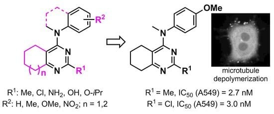
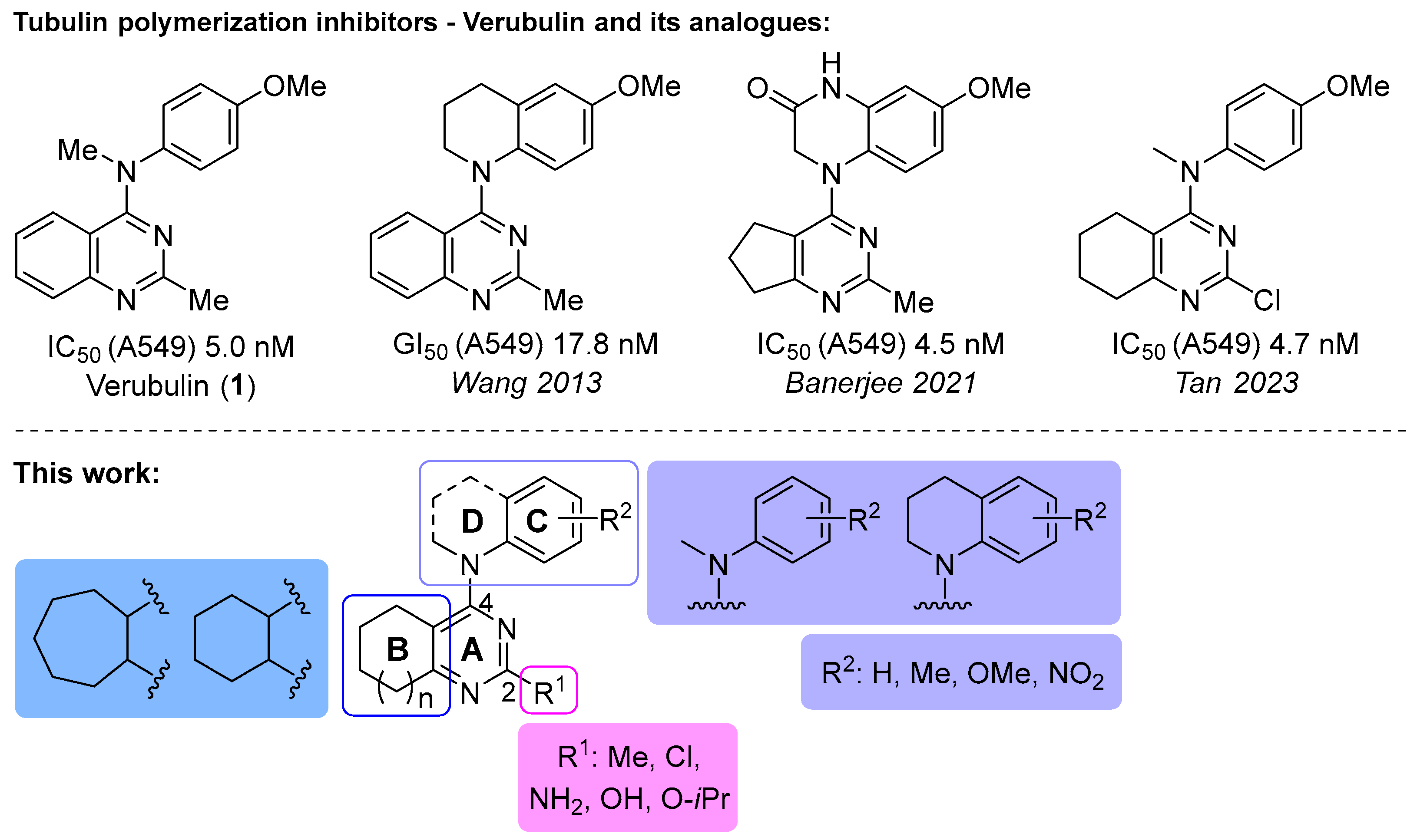
1. Introduction
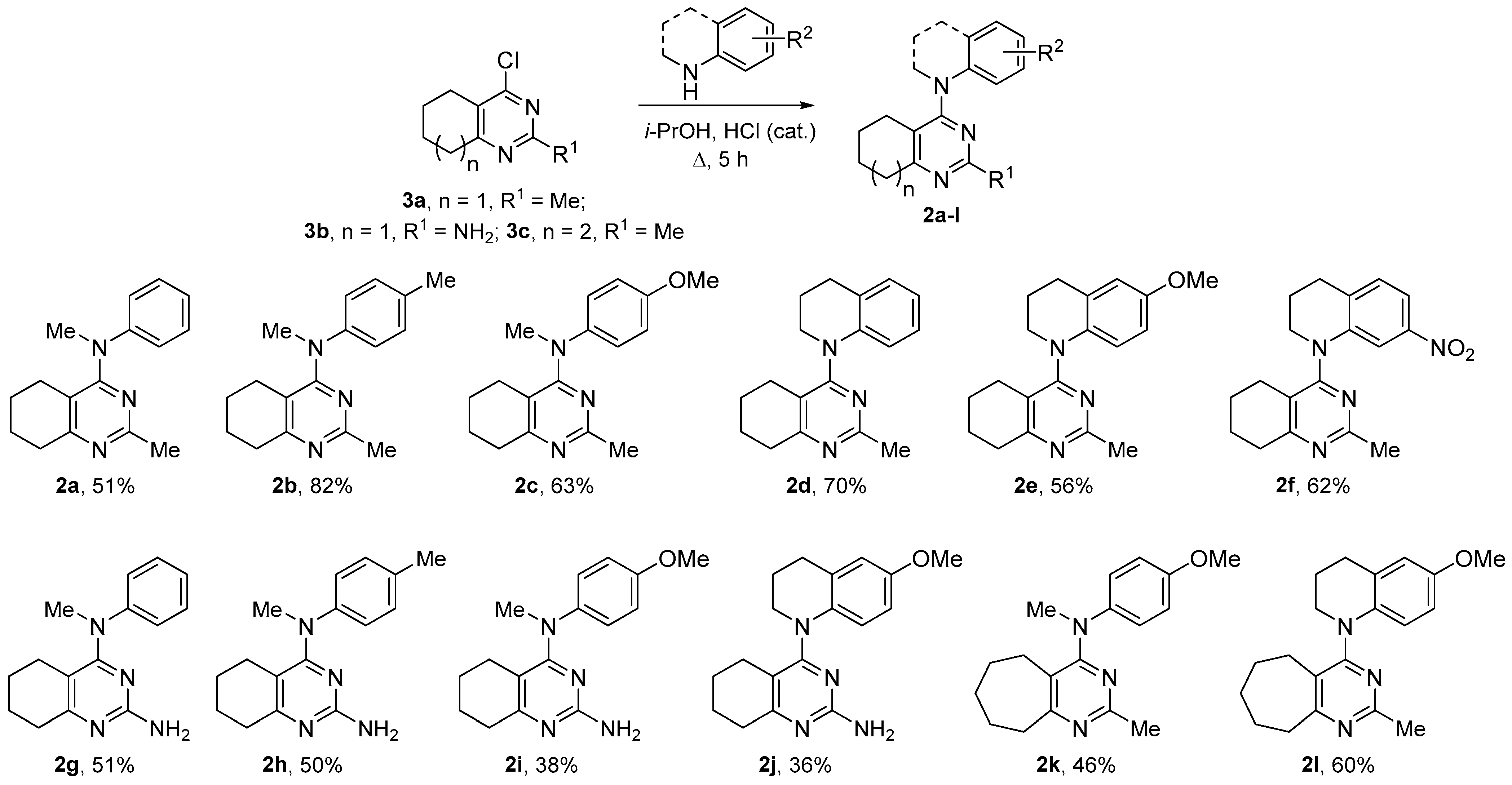
2. Results and Discussion
2.1. Chemistry
2.2. Bioactivity Testing
2.2.1. Cytotoxicity of Verubulin Analogues and SAR Analysis
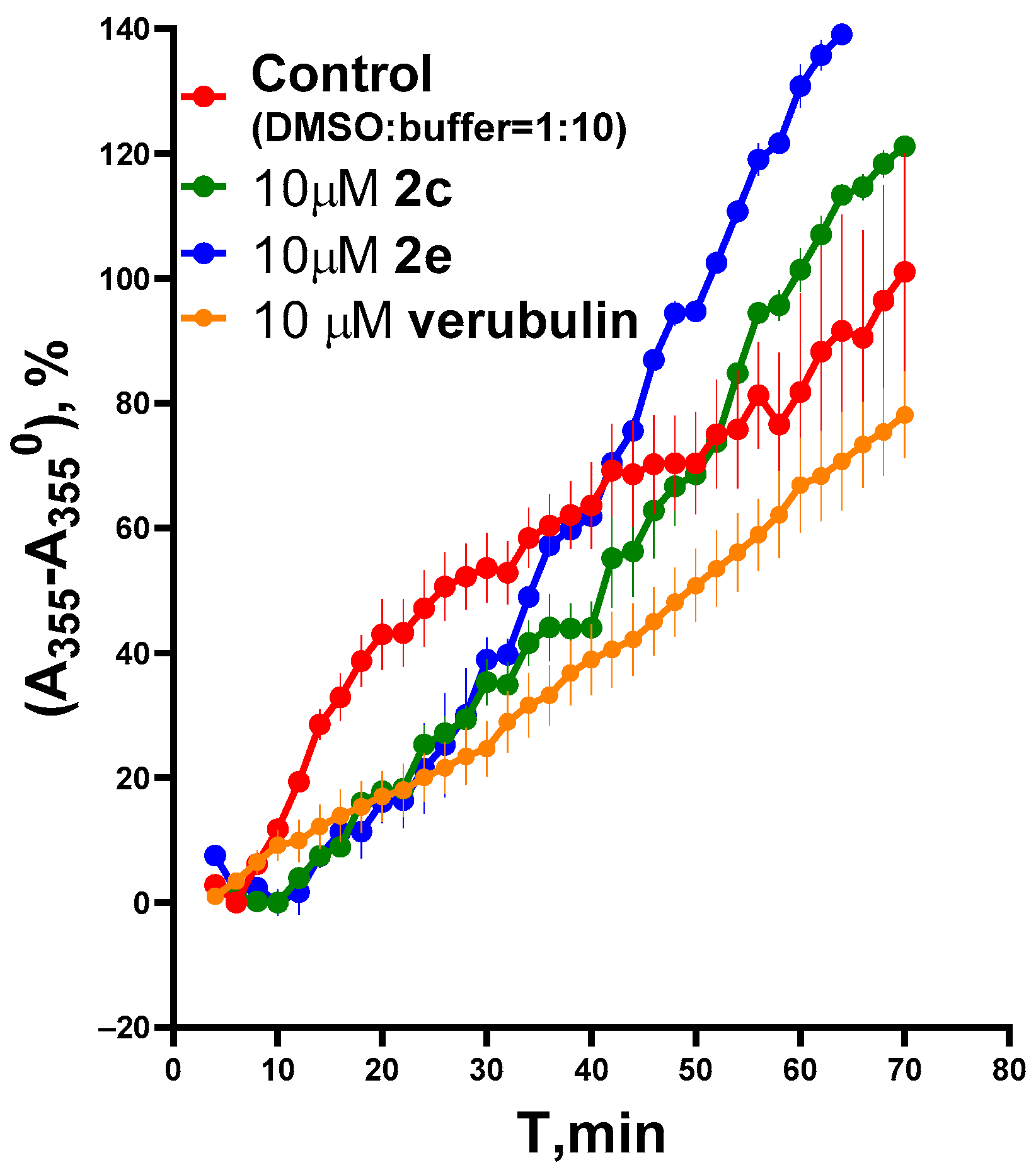
2.2.2. Effect of Verubulin Analogues on Microtubule Dynamics
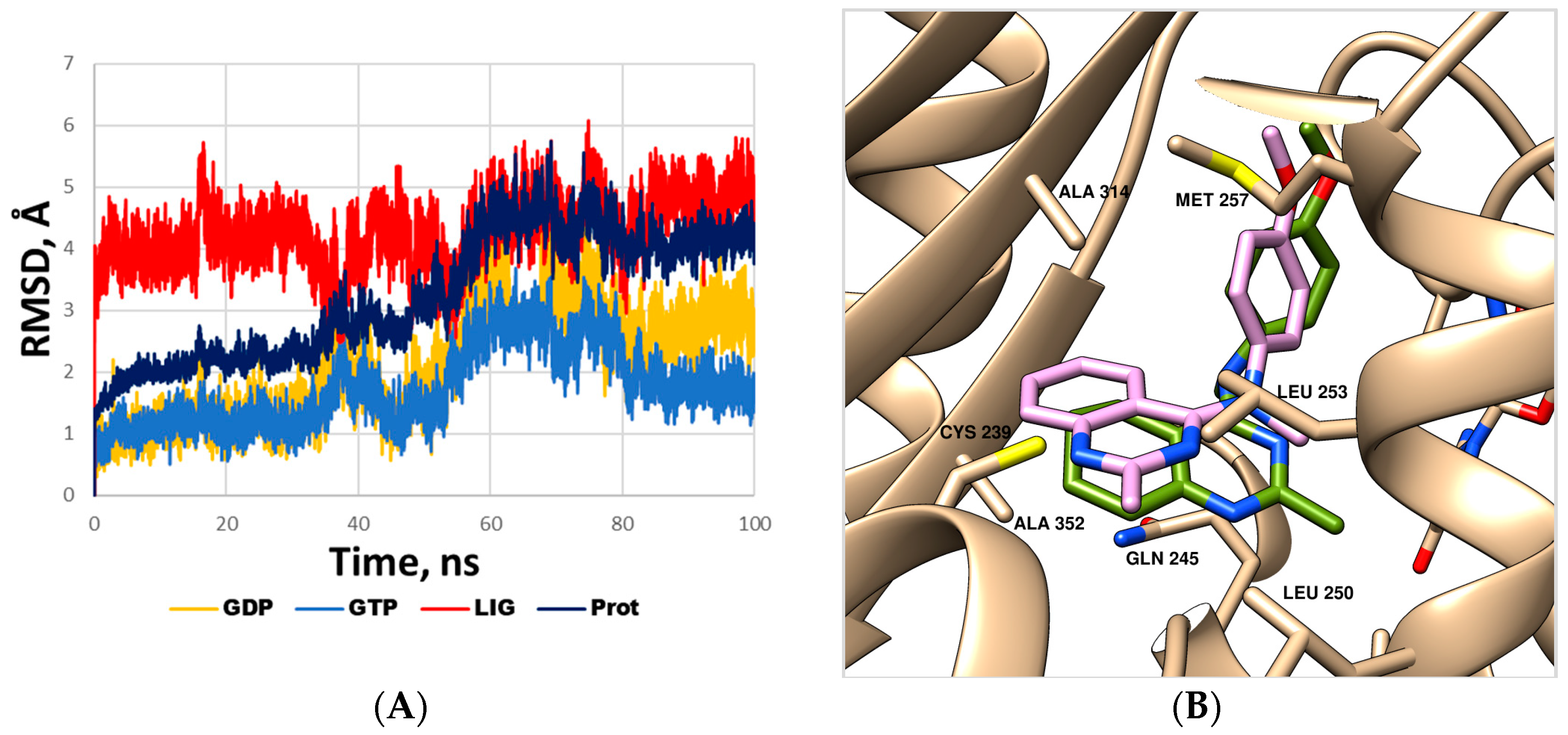
2.2.3. Molecular Dynamics Simulations for Compound 2c
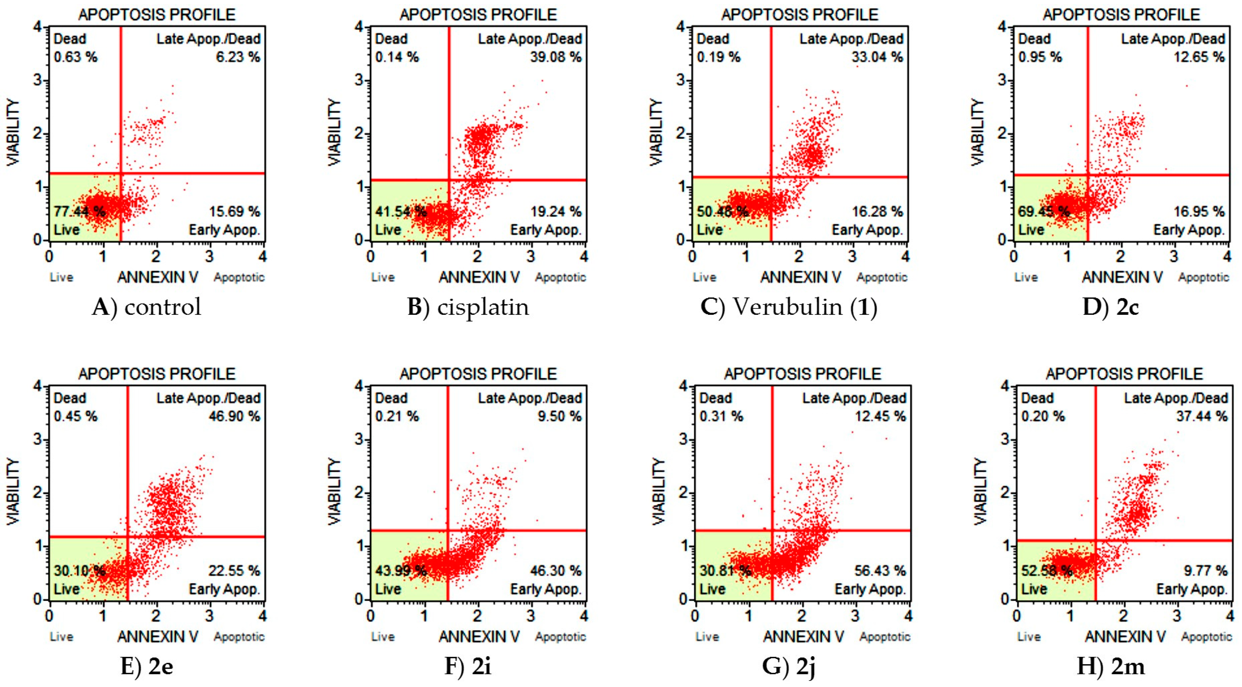
2.2.4. Apoptosis Induction by Verubulin Analogues
2.3. Encapsulation of Compound 2c in Biocompatible Nanocontainers Based on Ca2+/Mg2+ Cross-Linked Alginate and Investigation of Cytotoxicity of Formulations via MTT Assay
3. Materials and Methods
3.1. Chemistry
3.1.1. General Remarks
3.1.2. SNAr Reactions of 4-Chloropyrimidines 2a–d with Amines (General Method)
3.2. Biology
3.2.1. Cell Cultures
3.2.2. Cell Viability Assay (MTT Assay)
3.2.3. Immunofluorescent Microscopy
3.2.4. Tubulin + MAP Polymerization
3.2.5. Cell Death
3.3. MTT Study of Compound 2c, Encapsulated in Alginate-Based Nanocontainers
3.3.1. General Remarks
3.3.2. Preparation of Nanocontainers
3.3.3. Preparation of Nanocontainers Filled with 2c
3.3.4. Cytotoxicity Studies of Nanocontainers Filled with 2c
3.4. Molecular Dynamics Simulation
4. Conclusions
Supplementary Materials
Author Contributions
Funding
Institutional Review Board Statement
Informed Consent Statement
Data Availability Statement
Acknowledgments
Conflicts of Interest
References
- Pellegrini, F.; Budman, D.R. Review: Tubulin Function, Action of Antitubulin Drugs, and New Drug Development. Cancer Investig. 2005, 23, 264–273. [Google Scholar] [CrossRef]
- Borisy, G.; Heald, R.; Howard, J.; Janke, C. Microtubules: 50 years on from the discovery of tubulin. Nat. Rev. Mol. Cell Biol. 2016, 17, 322–328. [Google Scholar] [CrossRef]
- Janke, C.; Magiera, M.M. The tubulin code and its role in controlling microtubule properties and functions. Nat. Rev. Mol. Cell Biol. 2020, 21, 307–326. [Google Scholar] [CrossRef]
- Naaz, F.; Haidera, M.R.; Shafi, S.; Yar, M.S. Anti-tubulin agents of natural origin: Targeting taxol, vinca, and colchicine binding domains. Eur. J. Med. Chem. 2019, 171, 310–331. [Google Scholar] [CrossRef] [PubMed]
- Arnst, K.E.; Banerjee, S.; Chen, H.; Deng, S.; Hwang, D.-J.; Li, W.; Miller, D.D. Current advances of tubulin inhibitors as dual acting small molecules for cancer therapy. Med. Res. Rev. 2019, 39, 1398–1426. [Google Scholar] [CrossRef] [PubMed]
- Peng, X.; Ren, Y.; Pan, W.; Liu, J.; Chen, J. Discovery of Novel Acridane-Based Tubulin Polymerization Inhibitors with Anticancer and Potential Immunomodulatory Effects. J. Med. Chem. 2023, 66, 627–640. [Google Scholar] [CrossRef] [PubMed]
- Yuan, X.-Y.; Song, C.-H.; Liu, X.-J.; Wang, X.; Jia, M.-Q.; Wang, W.; Liu, W.-B.; Fu, X.-J.; Jin, C.-Y.; Song, J.; et al. Discovery of novel N-benzylarylamide-dithiocarbamate based derivatives as dual inhibitors of tubulin polymerization and LSD1 that inhibit gastric cancers. Eur. J. Med. Chem. 2023, 252, 115281. [Google Scholar] [CrossRef] [PubMed]
- Li, G.; Wu, J.-Q.; Cai, X.; Guan, W.; Zeng, Z.; Ou, Y.; Wu, X.; Li, J.; Fang, X.; Liu, J.; et al. Design, synthesis, and biological evaluation of diaryl heterocyclic derivatives targeting tubulin polymerization with potent anticancer activities. Eur. J. Med. Chem. 2023, 252, 115284. [Google Scholar] [CrossRef]
- Tang, M.; Liu, Y.-H.; Liu, H.; Mao, Q.; Yu, Q.; Kitagishi, H.; Zhang, Y.-M.; Xiao, L.; Liu, Y. Supramolecular Dual Polypeptides Induced Tubulin Aggregation for Synergistic Cancer Theranostics. J. Med. Chem. 2022, 65, 13473–13481. [Google Scholar] [CrossRef]
- Lin, S.; Du, T.; Zhang, J.; Wu, D.; Tian, H.; Zhang, K.; Jiang, L.; Lu, D.; Sheng, L.; Li, Y.; et al. Optimization of Benzamide Derivatives as Potent and Orally Active Tubulin Inhibitors Targeting the Colchicine Binding Site. J. Med. Chem. 2022, 65, 16372–16391. [Google Scholar] [CrossRef]
- Gallego-Jara, J.; Lozano-Terol, G.; Sola-Martínez, R.A.; Cánovas-Díaz, M.; de Diego Puente, T. A Compressive Review about Taxol®: History and Future Challenges. Molecules 2020, 25, 5986. [Google Scholar] [CrossRef]
- Sottomayor, M.; Ros Barceló, A. The vinca alkaloids: From biosynthesis and accumulation in plant cells, to uptake, activity and metabolism in animal cells. In Studies in Natural Products Chemistry (Bioactive Natural Products); Rahman, A., Ed.; Elsevier Science: Amsterdam, The Netherland, 2005; Volume 33, pp. 813–857. [Google Scholar]
- McLoughlin, E.C.; O’Boyle, N.M. Colchicine-Binding Site Inhibitors from Chemistry to Clinic: A Review. Pharmaceuticals 2020, 13, 8. [Google Scholar] [CrossRef]
- Li, W.; Sun, H.; Xu, S.; Zhu, Z.; Xu, J. Tubulin inhibitors targeting the colchicine binding site: A perspective of privileged structures. Future Med. Chem. 2017, 9, 1765–1794. [Google Scholar] [CrossRef] [PubMed]
- Wang, J.; Miller, D.D.; Li, W. Molecular interactions at the colchicines binding site in tubulin: An X-ray crystallography perspective. Drug Discov. Today 2022, 27, 759–776. [Google Scholar] [CrossRef] [PubMed]
- Kasibhatla, S.; Baichwal, V.; Cai, S.X.; Roth, B.; Skvortsova, I.; Skvortsov, S.; Lukas, P.; English, N.M.; Sirisoma, N.; Drewe, J.; et al. MPC-6827: A Small-Molecule Inhibitor of Microtubule Formation That Is Not a Substrate for Multidrug Resistance Pumps. Cancer Res. 2007, 67, 5865–5871. [Google Scholar] [CrossRef]
- Alvarez, R.; Aramburu, L.; Puebla, P.; Caballero, E.; Gonzalez, M.; Vicente, A.; Medarde, M.; Pelaez, R. Pyridine Based Antitumour Compounds Acting at the Colchicine Site. Curr. Med. Chem. 2016, 23, 1100–1130. [Google Scholar] [CrossRef]
- Bansal, R.; Malhotra, A. Therapeutic progression of quinazolines as targeted chemotherapeutic agents. Eur. J. Med. Chem. 2021, 211, 113016. [Google Scholar] [CrossRef]
- Chamberlain, M.C.; Grimm, S.; Recht, S.P.L.; Zhu, J.Z.; Kim, L.; Rosenfeld, S.; Fadul, C.E. Brain Tumor Investigational Consortium (BTIC). A phase 2 trial of verubulin for recurrent glioblastoma: A prospective study by the brain tumor investigational consortium (BTIC). J. Neurooncol. 2014, 118, 335–343. [Google Scholar] [CrossRef] [PubMed]
- Mahal, K.; Resch, M.; Ficner, R.; Schobert, R.; Biersack, B.; Mueller, T. Effects of the Tumor-Vasculature-Disrupting Agent Verubulin and Two Heteroaryl Analogues on Cancer Cells, Endothelial Cells, and Blood Vessels. ChemMedChem 2014, 9, 847–854. [Google Scholar] [CrossRef]
- Sirisoma, N.; Kasibhatla, S.; Pervin, A.; Zhang, H.; Jiang, S.; Willardsen, J.A.; Anderson, M.B.; Baichwal, V.; Mather, G.G.; Jessing, K.; et al. Discovery of 2-Chloro-N-(4-methoxyphenyl)-N-methylquinazolin-4-amine (EP128265, MPI-0441138) as a Potent Inducer of Apoptosis with High In Vivo Activity. J. Med. Chem. 2008, 51, 4771–4779. [Google Scholar] [CrossRef] [PubMed]
- Sirisoma, N.; Pervin, A.; Zhang, H.; Jiang, S.; Willardsen, J.A.; Anderson, M.B.; Mather, G.; Pleiman, C.M.; Kasibhatla, S.; Tseng, B.; et al. Discovery of N-(4-Methoxyphenyl)-N,2-dimethylquinazolin-4-amine, a Potent Apoptosis Inducer and Efficacious Anticancer Agent with High Blood Brain Barrier Penetration. J. Med. Chem. 2009, 52, 2341–2351. [Google Scholar] [CrossRef] [PubMed]
- Sirisoma, N.; Pervin, A.; Zhang, H.; Jiang, S.; Willardsen, J.A.; Anderson, M.B.; Mather, G.; Pleiman, C.M.; Kasibhatla, S.; Tseng, B.; et al. Discovery of N-methyl-4-(4-methoxyanilino)quinazolines as potent apoptosis inducers. Structure–activity relationship of the quinazoline ring. Bioorg. Med. Chem. Lett. 2010, 20, 2330–2334. [Google Scholar] [CrossRef]
- Gangjee, A.; Zhao, Y.; Lin, L.; Raghavan, S.; Roberts, E.G.; Risinger, A.L.; Hamel, E.; Mooberry, S.L. Synthesis and Discovery of Water-Soluble Microtubule Targeting Agents that Bind to the Colchicine Site on Tubulin and Circumvent Pgp Mediated Resistance. J. Med. Chem. 2010, 53, 8116–8128. [Google Scholar] [CrossRef] [PubMed]
- Wang, X.-F.; Wang, S.-B.; Ohkoshi, E.; Wang, L.-T.; Hamel, E.; Qian, K.; Morris-Natschke, S.L.; Lee, K.-H.; Xie, L. N-Aryl-6-methoxy-1,2,3,4-tetrahydroquinolines: A novel class of antitumor agents targeting the colchicine site on tubulin. Eur. J. Med. Chem. 2013, 67, 196–207. [Google Scholar] [CrossRef]
- Gangjee, A.; Zhao, Y.; Raghavan, S.; Rohena, C.C.; Mooberry, S.L.; Hamel, E. Structure−Activity Relationship and in Vitro and in Vivo Evaluation of the Potent Cytotoxic Anti-microtubule Agent N-(4-Methoxyphenyl)-N,2,6-trimethyl-6,7-dihydro-5H-cyclopenta[d]pyrimidin-4-aminium Chloride and Its Analogues As Antitumor Agents. J. Med. Chem. 2013, 56, 6829–6844. [Google Scholar] [CrossRef] [PubMed]
- Wang, X.-F.; Guan, F.; Ohkoshi, E.; Guo, W.; Wang, L.; Zhu, D.-Q.; Wang, S.-B.; Wang, L.-T.; Hamel, E.; Yang, D.; et al. Optimization of 4-(N-Cycloamino)phenylquinazolines as a Novel Class of Tubulin-Polymerization Inhibitors Targeting the Colchicine Site. J. Med. Chem. 2014, 57, 1390–1402. [Google Scholar] [CrossRef]
- Devambatla, R.K.V.; Namjoshi, O.A.; Choudhary, S.; Hamel, E.; Shaffer, C.V.; Rohena, C.C.; Mooberry, S.L.; Gangjee, A. Design, Synthesis, and Preclinical Evaluation of 4-Substituted-5-methyl-furo[2,3-d]pyrimidines as Microtubule Targeting Agents That Are Effective against Multidrug Resistant Cancer Cells. J. Med. Chem. 2016, 59, 5752–5765. [Google Scholar] [CrossRef] [PubMed]
- Banerjee, S.; Arnst, K.E.; Wang, Y.; Kumar, G.; Deng, S.; Yang, L.; Li, G.-B.; Yang, J.; White, S.W.; Li, W.; et al. Heterocyclic-Fused Pyrimidines as Novel Tubulin Polymerization Inhibitors Targeting the Colchicine Binding Site: Structural Basis and Antitumor Efficacy. J. Med. Chem. 2018, 61, 1704–1718. [Google Scholar] [CrossRef]
- Arnst, K.E.; Banerjee, S.; Wang, Y.; Chen, H.; Li, Y.; Yang, L.; Li, W.; Miller, D.D.; Li, W. X-ray Crystal Structure Guided Discovery and Antitumor Efficacy of Dihydroquinoxalinone as Potent Tubulin Polymerization Inhibitors. ACS Chem. Biol. 2019, 14, 2810–2821. [Google Scholar] [CrossRef]
- Lia, W.; Yina, Y.; Shuaia, W.; Xua, F.; Yaoa, H.; Liub, J.; Cheng, K.; Xua, J.; Zhud, Z.; Xu, S. Discovery of novel quinazolines as potential anti-tubulin agents occupying three zones of colchicine domain. Bioorg. Chem. 2019, 83, 380–390. [Google Scholar] [CrossRef]
- Loidreau, Y.; Nourrisson, M.-R.; Fruit, C.; Corbière, C.; Marchand, P.; Besson, T. Microwave-Assisted Synthesis of Potential Bioactive Benzo-, Pyrido- or Pyrazino-thieno[3,2-d]pyrimidin-4-amine Analogs of MPC-6827. Pharmaceuticals 2020, 13, 202. [Google Scholar] [CrossRef] [PubMed]
- Banerjee, S.; Mahmud, F.; Deng, S.; Ma, L.; Yun, M.-K.; Fakayode, S.O.; Arnst, K.E.; Yang, L.; Chen, H.; Wu, Z.; et al. X-ray Crystallography-Guided Design, Antitumor Efficacy, and QSAR Analysis of Metabolically Stable Cyclopenta-Pyrimidinyl Dihydroquinoxalinone as a Potent Tubulin Polymerization Inhibitor. J. Med. Chem. 2021, 64, 13072–13095. [Google Scholar] [CrossRef] [PubMed]
- Tan, L.; Wu, C.; Zhang, J.; Yu, Q.; Wang, X.; Zhang, L.; Ge, M.; Wang, Z.; Ouyang, L.; Wang, Y. Design, Synthesis, and Biological Evaluation of Heterocyclic-Fused Pyrimidine Chemotypes Guided by X-ray Crystal Structure with Potential Antitumor and Anti-multidrug Resistance Efficacy Targeting the Colchicine Binding Site. J. Med. Chem. 2023, 66, 3588–3620. [Google Scholar] [CrossRef] [PubMed]
- Evdokimova, A.V.; Alexeev, A.A.; Nurieva, E.V.; Milaeva, E.R.; Kuznetsov, S.A.; Zefirova, O.N. N-(4-Methoxyphenyl)-substituted bicyclic isothioureas: Effect on morphology of cancer cells. Mendeleev Commun. 2021, 31, 288–290. [Google Scholar] [CrossRef]
- Yang, H.; An, B.; Li, X.; Zeng, W. Evaluation of 4-phenylamino-substituted naphthalene-1,2-diones as tubulin polymerization inhibitors. Bioorg. Med. Chem. Lett. 2018, 28, 3057–3063. [Google Scholar] [CrossRef]
- Lovering, F.; Bikker, J.; Humblet, C. Escape from flatland: Increasing saturation as an approach to improving clinical success. J. Med. Chem. 2009, 52, 6752–6756. [Google Scholar] [CrossRef]
- Lovering, F. Escape from Flatland 2: Complexity and promiscuity. Med. Chem. Commun. 2013, 4, 515–519. [Google Scholar] [CrossRef]
- Cox, B.; Zdorichenko, V.; Cox, P.B.; Booker-Milburn, K.I.; Paumier, R.; Elliott, L.D.; Robertson-Ralph, M.; Bloomfield, G. Escaping from Flatland: Substituted Bridged Pyrrolidine Fragments with Inherent Three-Dimensional Character. ACS Med. Chem. Lett. 2020, 11, 1185–1190. [Google Scholar] [CrossRef]
- Klein, H.F.; Hamilton, D.J.; de Esch, I.J.P.; Wijtmans, M.; O’Brien, P. Escape from planarity in fragment-based drug discovery: A synthetic strategy analysis of synthetic 3D fragment libraries. Drug Discov. Today 2022, 27, 2484–2496. [Google Scholar] [CrossRef]
- Severino, P.; da Silva, C.F.; Andrade, L.N.; de Lima Oliveira, D.; Campos, J.; Souto, E.B. Alginate nanoparticles for drug delivery and targeting. Curr. Pharm. Des. 2019, 25, 1312–1334. [Google Scholar] [CrossRef]
- Lee, K.Y.; Mooney, D.J. Alginate: Properties and biomedical applications. Prog. Polym. Sci. 2012, 37, 106–126. [Google Scholar] [CrossRef]
- Shilpa, A.; Agrawal, S.; Ray, A.R. Controlled delivery of drugs from alginate matrix. J. Macromol. Sci. Part C Polym. Rev. 2003, 43, 187–221. [Google Scholar] [CrossRef]
- Spiridonov, V.V.; Sadovnikov, K.S.; Vasilenko, D.A.; Sedenkova, K.N.; Lukmanova, A.R.; Markova, A.A.; Shibaeva, A.V.; Bolshakova, A.V.; Karlov, S.S.; Averina, E.B.; et al. Synthesis and evaluation of the anticancer activity of the water-dispersible complexes of 4-acylaminoisoxazole derivative with biocompatible nanocontainers based on Ca2+ (Mg2+) cross-linked alginate. Mendeleev Commun. 2022, 32, 591–593. [Google Scholar] [CrossRef]
- Mosmann, T. Rapid Colorimetric Assay for Cellular Growth and Survival: Application to Proliferation and Cytotoxicity Assays. J. Immunol. Methods. 1983, 65, 55–63. [Google Scholar] [CrossRef] [PubMed]
- Yan, W.; Yang, T.; Yang, J.; Wang, T.; Yu, Y.; Wang, Y.; Chen, Q.; Bai, P.; Li, D.; Ye, H.; et al. SKLB060 Reversibly Binds to Colchicine Site of Tubulin and Possesses Efficacy in Multidrug-Resistant Cell Lines. Cell. Physiol. Biochem. 2018, 47, 489–504. [Google Scholar] [CrossRef] [PubMed]
- Tait, J.F.; Gibson, D.; Fujikawa, K. Phospholipid binding properties of human placental anticoagulant protein-I, a member of the lipocortin family. J. Biol. Chem. 1989, 264, 7944–7949. [Google Scholar] [CrossRef]
- Andree, H.A.; Reutelingsperger, C.P.; Hauptmann, R.; Hemker, H.C.; Hermens, W.T.; Willems, G.M. Binding of vascular anticoagulant alpha (VAC alpha) to planar phospholipid bilayers. J. Biol. Chem. 1990, 265, 4923–4928. [Google Scholar] [CrossRef]
- Spiridonov, V.V.; Lukmanova, A.R.; Pozdyshev, D.V.; Markova, A.A.; Volodina, Y.L.; Golovina, G.V.; Shakhmatov, V.V.; Kuzmin, V.A.; Muronetz, V.I.; Yaroslavov, A.A. Ionically cross-linked micro-sized hydrogels with encapsulated drug: Structure, cell uptake kinetics and cytotoxicity. Mendeleev Commun. 2023, 33, 553–555. [Google Scholar] [CrossRef]
- Farkas, N.; Kramar, J.A. Dynamic light scattering distributions by any means. J. Nanopart. Res. 2021, 23, 120. [Google Scholar] [CrossRef]
- Spiridonov, V.; Orlova, M.; Ivanov, I.; Panova, I.; Orlov, A.; Antonova, Y.; Yaroslavov, A. Synthesis of microgels based on carboxymethylcellulose cross-linked with zinc(II) ions and heterocyclic effectors of NO-synthase. Colloids Surf. A Physicochem. Eng. Asp. 2020, 585, 124104. [Google Scholar] [CrossRef]
- Grante, I.; Actins, A.; Orola, L. Protonation effects on the UV/Vis absorption spectra of imatinib: A theoretical and experimental study. Spectrochim. Acta A Mol. Biomol. Spectrosc. 2014, 129, 326–332. [Google Scholar] [CrossRef] [PubMed]
- Miller, G.W.; Rose, F.L. 1080. S-triazolopyrimidines. Part I. Synthesis as potential therapeutic agents. J. Chem. Soc. 1963, 5642–5659. [Google Scholar] [CrossRef]
- Ohno, S.; Mizukoshi, K.; Komatsu, O.; Kunoh, Y.; Nakamura, Y.; Katoh, E.; Nagasaka, M. Synthesis and Hypoglycemic Activity of 7,8-Dihydro-6H-thiopyrano[3,2-d]pyrimidine Derivatives and Related Compounds. Chem. Pharm. Bull. 1986, 34, 4150–4165. [Google Scholar] [CrossRef] [PubMed]
- Sedenkova, K.N.; Zverev, D.V.; Nazarova, A.A.; Lavrov, M.I.; Radchenko, E.V.; Grishin, Y.K.; Gabrel’yan, A.V.; Zamoyski, V.L.; Grigoriev, V.V.; Averina, E.B.; et al. Novel Nanomolar Allosteric Modulators of AMPA Receptor of Bis(pyrimidine) Series: Synthesis, Biotesting and SAR Analysis. Molecules 2022, 27, 8252. [Google Scholar] [CrossRef]
- Zhou, H.-J.; Wang, J.; Yao, B.; Wong, S.; Djakovic, S.; Kumar, B.; Rice, J.; Valle, E.; Soriano, F.; Menon, M.-K.; et al. Discovery of a First-in-Class, Potent, Selective, and Orally Bioavailable Inhibitor of the p97 AAA ATPase (CB-5083). J. Med. Chem. 2015, 58, 9480–9497. [Google Scholar] [CrossRef]
- Sadovnikov, K.S.; Vasilenko, D.A.; Gracheva, Y.A.; Zefirov, N.A.; Radchenko, E.V.; Palyulin, V.A.; Grishin, Y.K.; Vasilichin, V.A.; Shtil, A.A.; Shevtsov, P.N.; et al. Novel substituted 5-methyl-4-acylaminoisoxazoles as antimitotic agents: Evaluation of selectivity to LNCaP cancer cells. Arch. Pharm. 2022, 355, e2100425. [Google Scholar] [CrossRef] [PubMed]
- Brindisi, M.; Ulivieri, C.; Alfano, G.; Gemma, S.; de Asís Balaguer, F.; Khan, T.; Grillo, A.; Chemi, G.; Menchon, G.; Prota, A.E.; et al. Structure-activity relationships, biological evaluation and structural studies of novel pyrrolonaphthoxazepines as antitumor agents. Eur. J. Med. Chem. 2019, 162, 290–320. [Google Scholar] [CrossRef] [PubMed]
- Huang, J.; MacKerell, A.D. CHARMM36 all-atom additive protein force field: Validation based on comparison to NMR data. J. Comput. Chem. 2013, 34, 2135–2145. [Google Scholar] [CrossRef]
- Vanommeslaeghe, K.; Hatcher, E.; Acharya, C.; Kundu, S.; Zhong, S.; Shim, J.; Darian, E.; Guvench, O.; Lopes, P.; Vorobyov, I.; et al. CHARMM general force field: A force field for drug-like molecules compatible with the CHARMM all-atom additive biological force fields. J. Comput. Chem. 2010, 31, 671–690. [Google Scholar] [CrossRef]
- Abraham, M.J.; Murtola, T.; Schulz, R.; Páll, S.; Smith, J.C.; Hess, B.; Lindahl, E. GROMACS: High performance molecular simulations through multi-level parallelism from laptops to supercomputers. SoftwareX 2015, 1–2, 19–25. [Google Scholar] [CrossRef]
- Trott, O.A.; Olson, J. AutoDock Vina: Improving the speed and accuracy of docking with a new scoring function, efficient optimization, and multithreading. J. Comput. Chem. 2010, 31, 455–461. [Google Scholar] [CrossRef] [PubMed]
- Roe, D.R.; Cheatham, T.E. PTRAJ and CPPTRAJ: Software for Processing and Analysis of Molecular Dynamics Trajectory Data. J. Chem. Theory Comput. 2013, 9, 3084–3095. [Google Scholar] [CrossRef] [PubMed]
- Salomon-Ferrer, R.; Case, D.A.; Walker, R.C. An overview of the Amber biomolecular simulation package. WIREs Comput. Mol. Sci. 2013, 3, 198–210. [Google Scholar] [CrossRef]
- Pettersen, E.F.; Goddard, T.D.; Huang, C.C.; Couch, G.S.; Greenblatt, D.M.; Meng, E.C.; Ferrin, T.E. UCSF Chimera—A visualization system for exploratory research and analysis. J. Comput. Chem. 2004, 25, 1605–1612. [Google Scholar] [CrossRef] [PubMed]


| Compound | IC50, nM | Compound | IC50, nM | ||||||||
|---|---|---|---|---|---|---|---|---|---|---|---|
| MCF7′ | A549 | VA13 | HEK293T | MCF7′ | A549 | VA13 | HEK293T | ||||
| 1 | 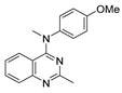 | 1.4 ± 0.05 | 1.1 ± 0.05 | 0.84 ± 0.06 | 1.0 ± 0.02 | 2j |  | 40.2 ± 7.8 | 50.5 ± 11.1 | 14.5 ± 3.9 | 28.9 ± 6.6 |
| 2a |  | 1840 ± 810 | 2200 ± 260 | 1400 ± 180 | 1750 ± 230 | 2k |  | 53 ± 7 | 79 ± 10 | 22 ± 6 | 39 ± 6 |
| 2b |  | 97 ± 7 | 110 ± 8 | 62 ± 8 | 89 ± 3 | 2l |  | 360 ± 70 | 790 ± 19 | 260 ± 80 | 450 ± 100 |
| 2c | 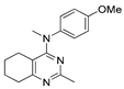 | 3.6 ± 0.4 | 2.7 ± 0.4 | 1.6 ± 0.4 | 2.9 ± 0.5 | 2m |  | 3.3 ± 0.14 | 3.0 ± 0.2 | 1.3 ± 0.18 | 2.7 ± 0.12 |
| 2d |  | 9100 ± 2200 | 12,600 ± 1600 | 3000 ± 700 | 7300 ± 1200 | 2n |  | 2500 ± 140 | 3270 ± 290 | 2290 ± 160 | 1720 ± 68 |
| 2e |  | 12.6 ± 2.1 | 13.5 ± 2.2 | 7.1 ± 2.0 | 8.8 ± 2.0 | 2o |  | >50,000 | >50,000 | >50,000 | >50,000 |
| 2f |  | >50,000 | >50,000 | >50,000 | >50,000 | 2p |  | 660 ± 150 | 640 ± 76 | 290 ± 74 | 510 ± 33 |
| 2g |  | 28,600 ± 4800 | 36,400 ± 7400 | 26,900 ± 9500 | 26,300 ± 5200 | 2q |  | 310 ± 22 | 360 ± 34 | 160 ± 20 | 250 ± 11 |
| 2h |  | 210 ± 15 | 310 ± 23 | 150 ± 14 | 170 ± 18 | 2r |  | 24,200 ± 4600 | 23,800 ± 5200 | 117,700 ± 40,100 | 53,500 ± 7200 |
| 2i |  | 10.2 ± 1.5 | 15.0 ± 2.0 | 6.0 ± 1.5 | 9.6 ± 2.2 | ||||||
| Entry | Compound | Hydrodynamic and Electrokinetic Properties | IC50, μM 1 | ||
|---|---|---|---|---|---|
| Dh, nm | EPM, μm/s/(V/cm) | A549 | hTERT-Immortalized Human Fibroblasts | ||
| 1 | 2c∙HCl–(Alg)–Ca2+ | 130 ± 7 | −3.2 ± 0.2 | 0.5 ± 0.04 | 1.1 ± 0.1 |
| 2 | (Alg)–Ca2+ | 150 ± 8 | −2.1 ± 0.1 | 18 ± 0.9 | >25 |
| 3 | 2c∙HCl–(Alg)–Mg2+ | 106 ± 4 | −4.5 ± 0.3 | 0.5 ± 0.05 | 0.5 ± 0.05 |
| 4 | (Alg)–Mg2+ | 205 ± 9 | −3.4 ± 0.2 | >25 | >25 |
| 5 | 2c | – | – | 0.6 ± 0.06 | 4.2 ± 0.3 |
Disclaimer/Publisher’s Note: The statements, opinions and data contained in all publications are solely those of the individual author(s) and contributor(s) and not of MDPI and/or the editor(s). MDPI and/or the editor(s) disclaim responsibility for any injury to people or property resulting from any ideas, methods, instructions or products referred to in the content. |
© 2023 by the authors. Licensee MDPI, Basel, Switzerland. This article is an open access article distributed under the terms and conditions of the Creative Commons Attribution (CC BY) license (https://creativecommons.org/licenses/by/4.0/).
Share and Cite
Sedenkova, K.N.; Leschukov, D.N.; Grishin, Y.K.; Zefirov, N.A.; Gracheva, Y.A.; Skvortsov, D.A.; Hrytseniuk, Y.S.; Vasilyeva, L.A.; Spirkova, E.A.; Shevtsov, P.N.; et al. Verubulin (Azixa) Analogues with Increased Saturation: Synthesis, SAR and Encapsulation in Biocompatible Nanocontainers Based on Ca2+ or Mg2+ Cross-Linked Alginate. Pharmaceuticals 2023, 16, 1499. https://doi.org/10.3390/ph16101499
Sedenkova KN, Leschukov DN, Grishin YK, Zefirov NA, Gracheva YA, Skvortsov DA, Hrytseniuk YS, Vasilyeva LA, Spirkova EA, Shevtsov PN, et al. Verubulin (Azixa) Analogues with Increased Saturation: Synthesis, SAR and Encapsulation in Biocompatible Nanocontainers Based on Ca2+ or Mg2+ Cross-Linked Alginate. Pharmaceuticals. 2023; 16(10):1499. https://doi.org/10.3390/ph16101499
Chicago/Turabian StyleSedenkova, Kseniya N., Denis N. Leschukov, Yuri K. Grishin, Nikolay A. Zefirov, Yulia A. Gracheva, Dmitry A. Skvortsov, Yanislav S. Hrytseniuk, Lilja A. Vasilyeva, Elena A. Spirkova, Pavel N. Shevtsov, and et al. 2023. "Verubulin (Azixa) Analogues with Increased Saturation: Synthesis, SAR and Encapsulation in Biocompatible Nanocontainers Based on Ca2+ or Mg2+ Cross-Linked Alginate" Pharmaceuticals 16, no. 10: 1499. https://doi.org/10.3390/ph16101499
APA StyleSedenkova, K. N., Leschukov, D. N., Grishin, Y. K., Zefirov, N. A., Gracheva, Y. A., Skvortsov, D. A., Hrytseniuk, Y. S., Vasilyeva, L. A., Spirkova, E. A., Shevtsov, P. N., Shevtsova, E. F., Lukmanova, A. R., Spiridonov, V. V., Markova, A. A., Nguyen, M. T., Shtil, A. A., Zefirova, O. N., Yaroslavov, A. A., Milaeva, E. R., & Averina, E. B. (2023). Verubulin (Azixa) Analogues with Increased Saturation: Synthesis, SAR and Encapsulation in Biocompatible Nanocontainers Based on Ca2+ or Mg2+ Cross-Linked Alginate. Pharmaceuticals, 16(10), 1499. https://doi.org/10.3390/ph16101499