
Heart Rate Variability Analysis on Electrocardiograms, Seismocardiograms and Gyrocardiograms of Healthy Volunteers and Patients with Valvular Heart Diseases


Abstract
:1. Introduction
2. Materials and Methods
2.1. Datasets
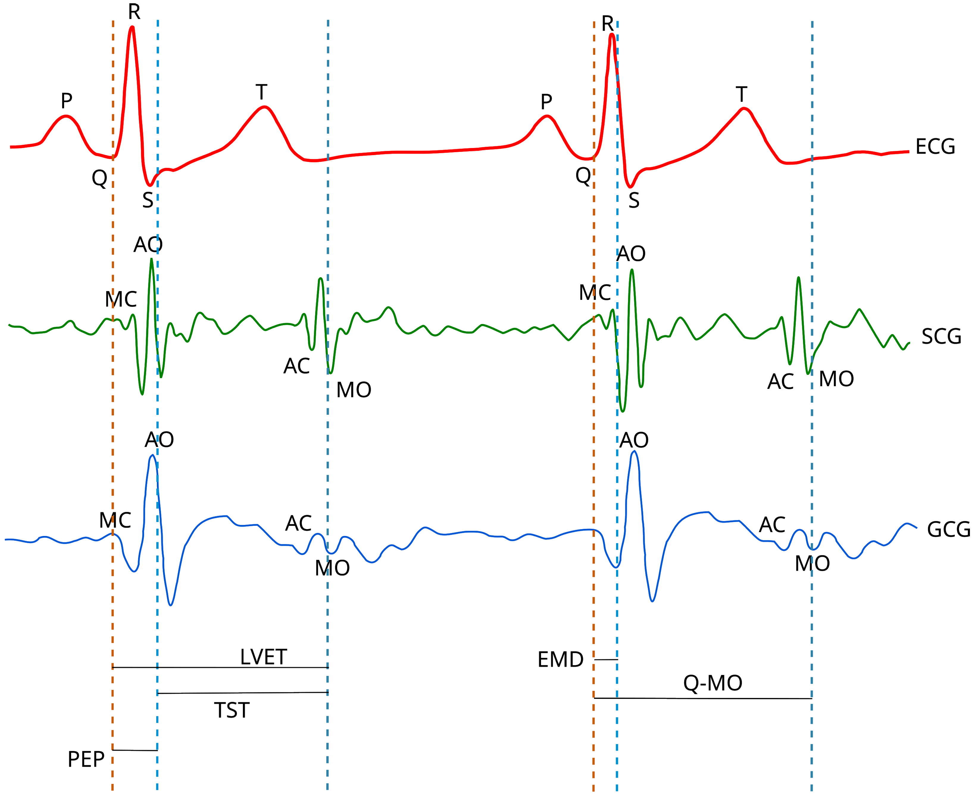
2.2. Signal Processing
2.3. HRV Analysis
3. Results
4. Discussion
5. Conclusions
Supplementary Materials
Author Contributions
Funding
Institutional Review Board Statement
Informed Consent Statement
Data Availability Statement
Conflicts of Interest
Abbreviations
| VHD | Valvular heart disease |
| HRV | Heart rate variability |
| ECG | Electrocardiography, electrocardiogram |
| SCG | Seismocardiography |
| GCG | Gyrocardiography |
| MEMS | Microelectromechanical systems |
| MRI | Magnetic resonance imaging |
| EMD | Empirical mode decomposition |
| CABG | Coronary artery bypass graft surgery |
| F | Female |
| MR | Mitral valve regurgitation |
| MS | Mitral valve stenosis |
| M | Male |
| MI | Myocardial infarction |
| NN | The interval between consecutive normal heartbeats |
| FIR | Fininte impulse response (filter) |
| AO | Aortic valve opening (wave) |
| RSA | Respiratory sinus arrhythmia |
| SNR | Signal-to-noise (ratio) |
| AVNN | Mean inter-beat interval |
| SDNN | Standard deviation of all interbeat intervals |
| RMSSD | Root mean square of differences (RMSSD) of successive inter-beat intervals |
| pNN50 | The proportion of the number of pairs of successive differences greater than 50 ms divided by total number of normal inter-beat intervals |
| VLF | The power of very low frequency band (0.0033–0.04 Hz) of HRV spectrum |
| LF | The power of low frequency band (0.04–0.15 Hz) of HRV spectrum |
| HF | The power of high frequency band (0.15–0.4 Hz) of HRV spectrum |
| LF/HF | LF/HF ratio |
| The width of the ellipse which containes the scatter points of Poincaré map | |
| The length of the ellipse which containes the scatter points of Poincaré map | |
| to ratio | |
| AVD | Aortic valve disease |
| AC | Aortic valve closure |
| AO | Aortic valve opening |
| MC | Mitral valve closure |
| MO | Mitral valve opening |
| PCI | Percutaneous coronary intervention |
| AS | Aortic valve stenosis |
| AR | Aortic valve regurgitation |
| TR | Tricupsid valve regurgitation |
| Pearson’s linear correlation coefficient |
Appendix A. Recording Descriptions in Datasets

| Subject | Length of Recording | Position | Breathing | Remarks |
|---|---|---|---|---|
| 1 | 3 min | Left or right side | 2 min normal, | |
| 30 s holding a breath, | ||||
| 30 s normal | ||||
| 2 | 3 min | Left or right side | 2 min normal, | |
| 30 s holding a breath, | ||||
| 30 s normal | ||||
| 3 | 3 min | Left or right side | 2 min normal, | |
| 30 s holding a breath, | ||||
| 30 s normal | ||||
| 4 | 3 min | Left or right side | 2 min normal, | |
| 30 s holding a breath, | ||||
| 30 s normal | ||||
| 5 | 3 min | Left or right side | 2 min normal, | |
| 30 s holding a breath, | ||||
| 30 s normal | ||||
| 6 | 3 min | Left or right side | 2 min normal, | Sensor not strictly secured |
| 30 s holding a breath, | on chest because of body hair. | |||
| 30 s normal | ||||
| 7 | 3 min | Left or right side | 2 min normal, | |
| 30 s holding a breath, | ||||
| 30 s normal | ||||
| 8 | 3 min | Supine | Normal | |
| 9 | 10 min | Supine | Normal | |
| 10 | 10 min | Supine | Normal | |
| 11 | 30 min | Supine | Normal | |
| 12 | 10 min | Supine | Normal | |
| 13 | 10 min | Supine | Normal | |
| 14 | 10 min | Supine | Normal | |
| 15 | 10 min | Supine | Normal | |
| 16 | 10 min | Supine | Normal | |
| 17 | 10 min | Supine | Normal | |
| 18 | 10 min | Supine | Normal | |
| 19 | 10 min | Supine | Normal | |
| 20 | 10 min | Supine | Normal | |
| 21 | 10 min | Supine | Normal | |
| 22 | 10 min | Supine | Normal | Sensor loose in the end. |
| 23 | 10 min | Left or right side | Normal | |
| 24 | 10 min | Supine | Normal | |
| 25 | 9 min | Supine | Normal | |
| 26 | 10 min | Supine | Normal | |
| 27 | 10 min | Left or right side | Normal | |
| 28 | 10 min | Supine | Normal | |
| 29 | 10 min | Supine | Normal |
| Subject Number | Length of Recording | Age (Years) | Gender | Height (cm) | Weight (kg) | History of | MS | MR | AR | AS | TR | ||
|---|---|---|---|---|---|---|---|---|---|---|---|---|---|
| MI | CABG | PCI | |||||||||||
| UP-01 | 6 min 8 s | 89 | M | 154.9 | 49.0 | 0 | 1 | 1 | 0 | 0 | 0 | 1 | 1 |
| UP-02 | 6 min 10 s | 89 | M | 170.2 | 82.0 | 1 | 1 | 1 | 0 | 0 | 0 | 1 | 0 |
| UP-03 | 6 min 1 s | 96 | M | 162.5 | 66.0 | 0 | 1 | 1 | 0 | 0 | 0 | 1 | 1 |
| UP-04 | 5 min 36 s | 84 | M | 152.4 | 65.0 | 0 | 1 | 1 | 0 | 0 | 0 | 1 | 0 |
| UP-05 | 6 min 35 s | 70 | F | 162.5 | 79.0 | 0 | 1 | 1 | 0 | 0 | 0 | 1 | 1 |
| UP-06 | 5 min 39 s | 90 | F | 160 | 48.0 | 1 | 1 | 1 | 0 | 0 | 0 | 1 | 1 |
| UP-07 | 6 min 3 s | 84 | M | 162.5 | 79.0 | 0 | 1 | 1 | 0 | 0 | 0 | 1 | 0 |
| UP-08 | 5 min 11 s | 95 | F | 152.4 | 44.0 | 0 | 1 | 1 | 1 | 0 | 0 | 1 | 1 |
| UP-09 | 5 min 15 s | 89 | M | 182.8 | 90.7 | 0 | 1 | 1 | 0 | 0 | 0 | 1 | 0 |
| UP-10 | 5 min 2 s | 80 | F | 157.4 | 74.0 | 0 | 1 | 1 | 1 | 0 | 0 | 1 | 0 |
| UP-11 | 5 min 6 s | 68 | M | 177.8 | 79.0 | 0 | 1 | 1 | 0 | 0 | 0 | 1 | 0 |
| UP-12 | 5 min 10 s | 79 | F | 154.9 | 78.0 | 0 | 1 | 1 | 1 | 0 | 0 | 1 | 0 |
| UP-13 | 5 min 18 s | 95 | F | 160 | 73.0 | 0 | 1 | 1 | 0 | 1 | 0 | 1 | 0 |
| UP-14 | 5 min 38 s | 85 | F | 152 | 82.0 | 0 | 1 | 1 | 1 | 0 | 0 | 1 | 0 |
| UP-15 | 5 min 34 s | 84 | F | 175 | 76.0 | 0 | 1 | 1 | 0 | 0 | 0 | 1 | 0 |
| UP-16 | 5 min 44 s | 97 | M | 157 | 77.0 | 0 | 1 | 1 | 0 | 0 | 0 | 1 | 0 |
| UP-17 | 5 min 54 s | 80 | M | 182.8 | 86.0 | 0 | 1 | 1 | 0 | 0 | 0 | 1 | 1 |
| UP-18 | 5 min 9 s | 90 | F | 152.4 | 92.0 | 0 | 1 | 1 | 0 | 0 | 0 | 1 | 0 |
| UP-19 | 5 min 17 s | 78 | M | 170.1 | 78.0 | 0 | 1 | 1 | 0 | 0 | 0 | 1 | 0 |
| UP-20 | 7 min 49 s | 92 | F | 139.7 | 53.0 | 0 | 1 | 1 | 0 | 1 | 0 | 1 | 0 |
| UP-21 | 5 min 10 s | 72 | M | 172.7 | 68.0 | 0 | 1 | 1 | 1 | 1 | 0 | 1 | 1 |
| UP-22 | 10 min 3 s | 77 | F | 165.1 | 51.3 | 0 | 0 | 0 | 0 | 0 | 0 | 1 | 1 |
| UP-23 | 9 min 59 s | 84 | F | 139.7 | 70.3 | 0 | 0 | 0 | 0 | 0 | 0 | 1 | 0 |
| UP-24 | 5 min | 80 | F | 155 | 67.6 | 0 | 0 | 0 | 0 | 0 | 0 | 1 | 1 |
| UP-25 | 9 min 5 s | 87 | F | 155.0 | 54.0 | 0 | 0 | 0 | 0 | 1 | 0 | 1 | 0 |
| UP-26 | 5 min 8 s | 80 | M | 175.3 | 85.7 | 0 | 0 | 1 | 0 | 0 | 0 | 1 | 0 |
| UP-27 | 5 min 5 s | 82 | M | 180.0 | 118.0 | 0 | 0 | 0 | 0 | 0 | 0 | 1 | 0 |
| UP-28 | 5 min 2 s | 71 | M | 175.0 | 117.0 | 0 | 0 | 0 | 0 | 0 | 0 | 1 | 0 |
| UP-29 | 4 min 58 s | 80 | M | 168.9 | 65.8 | 0 | 0 | 0 | 1 | 0 | 0 | 1 | 0 |
| UP-30 | 4 min 59 s | 71 | M | 177.8 | 81.6 | 0 | 0 | 1 | 1 | 0 | 0 | 1 | 0 |
References
- Mensah, G.A.; Roth, G.A.; Fuster, V. The Global Burden of Cardiovascular Diseases and Risk Factors: 2020 and Beyond. J. Am. Coll. Cardiol. 2019, 74, 2529–2532. [Google Scholar] [CrossRef]
- World Health Organization. Global Action Plan for the Prevention and Control of Noncommunicable Diseases 2013–2020; World Health Organization: Geneva, Switzerland, 2013. [Google Scholar]
- World Health Organization. The Top 10 Causes of Death. 2020. Available online: https://www.who.int/news-room/fact-sheets/detail/the-top-10-causes-of-death (accessed on 3 February 2023).
- World Health Organization. World Health Statistics 2020: Monitoring Health for the SDGs, Sustainable Development Goals; World Health Organization: Geneva, Switzerland, 2020; 77p. [Google Scholar]
- Virani, S.S.; Alonso, A.; Aparicio, H.J.; Benjamin, E.J.; Bittencourt, M.S.; Callaway, C.W.; Carson, A.P.; Chamberlain, A.M.; Cheng, S.; Delling, F.N.; et al. Heart Disease and Stroke Statistics—2021 Update. Circulation 2021, 143, e254–e743. [Google Scholar] [CrossRef]
- Coffey, S.; Roberts-Thomson, R.; Brown, A.; Carapetis, J.; Chen, M.; Enriquez-Sarano, M.; Zühlke, L.; Prendergast, B.D. Global epidemiology of valvular heart disease. Nat. Rev. Cardiol. 2021, 18, 853–864. [Google Scholar] [CrossRef]
- Nkomo, V.T.; Gardin, J.M.; Skelton, T.N.; Gottdiener, J.S.; Scott, C.G.; Enriquez-Sarano, M. Burden of valvular heart diseases: A population-based study. Lancet 2006, 368, 1005–1011. [Google Scholar] [CrossRef]
- Maganti, K.; Rigolin, V.H.; Sarano, M.E.; Bonow, R.O. Valvular Heart Disease: Diagnosis and Management. Mayo Clin. Proc. 2010, 85, 483–500. [Google Scholar] [CrossRef]
- Vahanian, A.; Beyersdorf, F.; Praz, F.; Milojevic, M.; Baldus, S.; Bauersachs, J.; Capodanno, D.; Conradi, L.; De Bonis, M.; De Paulis, R.; et al. 2021 ESC/EACTS Guidelines for the management of valvular heart disease: Developed by the Task Force for the management of valvular heart disease of the European Society of Cardiology (ESC) and the European Association for Cardio-Thoracic Surgery (EACTS). Eur. Heart J. 2022, 43, 561–632. [Google Scholar] [CrossRef]
- Yang, Y.; Wang, Z.; Chen, Z.; Wang, X.; Zhang, L.; Li, S.; Zheng, C.; Kang, Y.; Jiang, L.; Zhu, Z.; et al. Current status and etiology of valvular heart disease in China: A population-based survey. BMC Cardiovasc. Disord. 2021, 21, 339. [Google Scholar] [CrossRef]
- Yang, C.; Fan, F.; Aranoff, N.; Green, P.; Li, Y.; Liu, C.; Tavassolian, N. An Open-Access Database for the Evaluation of Cardio-Mechanical Signals From Patients with Valvular Heart Diseases. Front. Physiol. 2021, 12, 750221. [Google Scholar] [CrossRef]
- Alugubelli, N.; Abuissa, H.; Roka, A. Wearable Devices for Remote Monitoring of Heart Rate and Heart Rate Variability—What We Know and What Is Coming. Sensors 2022, 22, 8903. [Google Scholar] [CrossRef]
- Taoum, A.; Bisiaux, A.; Tilquin, F.; Le Guillou, Y.; Carrault, G. Validity of Ultra-Short-Term HRV Analysis Using PPG—A Preliminary Study. Sensors 2022, 22, 7995. [Google Scholar] [CrossRef]
- Glaveckaite, S.; Petrikonyte, D.; Latveniene, L.; Serpytis, P.; Laucevicius, A. Ocena choroby zastawkowej serca za pomocą elektrokardiografii wysiłkowej i echokardiografii obciążeniowej: Czy te badania są nadal potrzebne? Folia Cardiol. 2018, 13, 318–330. [Google Scholar] [CrossRef]
- Tadi, M.J.; Lehtonen, E.; Saraste, A.; Tuominen, J.; Koskinen, J.; Teräs, M.; Airaksinen, J.; Pänkäälä, M.; Koivisto, T. Gyrocardiography: A New Non-invasive Monitoring Method for the Assessment of Cardiac Mechanics and the Estimation of Hemodynamic Variables. Sci. Rep. 2017, 7, 6823. [Google Scholar] [CrossRef] [PubMed]
- Rai, D.; Thakkar, H.K.; Rajput, S.S.; Santamaria, J.; Bhatt, C.; Roca, F. A Comprehensive Review on Seismocardiogram: Current Advancements on Acquisition, Annotation, and Applications. Mathematics 2021, 9, 2243. [Google Scholar] [CrossRef]
- Tadi, M.J.; Lehtonen, E.; Pankäälä, M.; Saraste, A.; Vasankari, T.; Terás, M.; Koivisto, T. Gyrocardiography: A new non-invasive approach in the study of mechanical motions of the heart. Concept, method and initial observations. In Proceedings of the 2016 38th Annual International Conference of the IEEE Engineering in Medicine and Biology Society (EMBC), Orlando, FL, USA, 16–20 August 2016; pp. 2034–2037. [Google Scholar] [CrossRef]
- Sieciński, S.; Kostka, P.S.; Tkacz, E.J. Gyrocardiography: A Review of the Definition, History, Waveform Description, and Applications. Sensors 2020, 20, 6675. [Google Scholar] [CrossRef] [PubMed]
- Bozhenko, B. Seismocardiography—A new method in the study of the functional condition of the heart. Ter. Arkhiv 1961, 33, 55–64. [Google Scholar]
- Zanetti, J.M.; Salerno, D.M. Seismocardiography: A technique for recording precordial acceleration. In Proceedings of the [1991] Computer-Based Medical Systems, Proceedings of the Fourth Annual IEEE Symposium, Baltimore, MD, USA, 12–14 May 1991; pp. 4–9. [Google Scholar] [CrossRef]
- Meriheinä, U.; Juppo, M.; Koivisto, T.; Mikko, P.; Sairanen, K.; Grönholm, M. Heart Monitoring System. WIPO Patent WO 2015/036925 A1, 19 March 2015. [Google Scholar]
- Sieciński, S.; Kostka, P.S.; Tkacz, E.J. Heart Rate Variability Analysis on Electrocardiograms, Seismocardiograms and Gyrocardiograms on Healthy Volunteers. Sensors 2020, 20, 4522. [Google Scholar] [CrossRef]
- Zanetti, J.M.; Tavakolian, K. Seismocardiography: Past, present and future. In Proceedings of the 2013 35th Annual International Conference of the IEEE Engineering in Medicine and Biology Society (EMBC), Osaka, Japan, 3–7 July 2013; pp. 7004–7007. [Google Scholar] [CrossRef]
- Inan, O.T.; Migeotte, P.F.; Park, K.S.; Etemadi, M.; Tavakolian, K.; Casanella, R.; Zanetti, J.; Tank, J.; Funtova, I.; Prisk, G.K.; et al. Ballistocardiography and Seismocardiography: A Review of Recent Advances. IEEE J. Biomed. Health Inform. 2015, 19, 1414–1427. [Google Scholar] [CrossRef]
- Gurev, V.; Tavakolian, K.; Constantino, J.C.; Kaminska, B.; Blaber, A.P.; Trayanova, N. Mechanisms underlying isovolumic contraction and ejection peaks in seismocardiogram morphology. J. Med. Biol. Eng. 2012, 32, 103. [Google Scholar] [CrossRef]
- Dehkordi, P.; Khosrow-Khavar, F.; Di Rienzo, M.; Inan, O.T.; Schmidt, S.E.; Blaber, A.P.; Sørensen, K.; Struijk, J.J.; Zakeri, V.; Lombardi, P.; et al. Comparison of Different Methods for Estimating Cardiac Timings: A Comprehensive Multimodal Echocardiography Investigation. Front. Physiol. 2019, 10, 1057. [Google Scholar] [CrossRef]
- Santucci, F.; Lo Presti, D.; Massaroni, C.; Schena, E.; Setola, R. Precordial Vibrations: A Review of Wearable Systems, Signal Processing Techniques, and Main Applications. Sensors 2022, 22, 5805. [Google Scholar] [CrossRef]
- Wajdan, A.; Jahren, T.S.; Villegas-Martinez, M.; Khan, F.H.; Halvorsen, P.S.; Odland, H.H.; Elle, O.J.; Solberg, A.H.S.; Remme, E.W. Automatic Detection of Aortic Valve Events Using Deep Neural Networks on Cardiac Signals From Epicardially Placed Accelerometer. IEEE J. Biomed. Health Inform. 2022, 26, 4450–4461. [Google Scholar] [CrossRef]
- Taebi, A.; Solar, B.E.; Bomar, A.J.; Sandler, R.H.; Mansy, H.A. Recent Advances in Seismocardiography. Vibration 2019, 2, 64–86. [Google Scholar] [CrossRef]
- Yang, C.; Ojha, B.D.; Aranoff, N.D.; Green, P.; Tavassolian, N. Classification of aortic stenosis using conventional machine learning and deep learning methods based on multi-dimensional cardio-mechanical signals. Sci. Rep. 2020, 10, 17521. [Google Scholar] [CrossRef]
- Shokouhmand, A.; Aranoff, N.D.; Driggin, E.; Green, P.; Tavassolian, N. Efficient detection of aortic stenosis using morphological characteristics of cardiomechanical signals and heart rate variability parameters. Sci. Rep. 2021, 11, 23817. [Google Scholar] [CrossRef]
- Johnson, E.M.; Robinson, J.D.; Rigsby, C.K.; McCarthy, P.M.; Malaisrie, S.C.; Allen, B.D.; Barker, A.J.; Markl, M. Abstract 9903: Artificial Intelligence Driven Wearable 2-minute Seismocardiography Test for Detection of Aortic Valve Stenosis Severity. Circulation 2022, 146, A9903. [Google Scholar] [CrossRef]
- Johnson, E.M.; Barker, A.J.; Robinson, J.D.; Rigsby, C.K.; Markl, M. Abstract 11504: Elevated Seismocardiography-Derived Chest Energy is Associated With Aortic Flow Abnormalities in Patients With Aortic Valve Disease. Circulation 2022, 146, A11504. [Google Scholar] [CrossRef]
- Korzeniowska-Kubacka, I.; Bilinska, M.; Piotrowicz, R. Usefulness of Seismocardiography for the Diagnosis of Ischemia in Patients with Coronary Artery Disease. Ann. Noninvasive Electrocardiol. 2005, 10, 281–287. [Google Scholar] [CrossRef]
- Iftikhar, Z.; Lahdenoja, O.; Tadi, M.J.; Hurnanen, T.; Vasankari, T.; Kiviniemi, T.; Airaksinen, J.; Koivisto, T.; Pänkäälä, M. Multiclass Classifier based Cardiovascular Condition Detection Using Smartphone Mechanocardiography. Sci. Rep. 2018, 8, 9344. [Google Scholar] [CrossRef]
- Mehrang, S.; Jafari Tadi, M.; Kaisti, M.; Lahdenoja, O.; Vasankari, T.; Kiviniemi, T.; Airaksinen, J.; Koivisto, T.; Pänkäälä, M. Machine Learning Based Classification of Myocardial Infarction Conditions Using Smartphone-Derived Seismo- and Gyrocardiography. In Proceedings of the 2018 Computing in Cardiology Conference (CinC), Maastricht, The Netherlands, 23–26 September 2018; Volume 45, pp. 1–4. [Google Scholar] [CrossRef]
- Hurnanen, T.; Lehtonen, E.; Tadi, M.J.; Kuusela, T.; Kiviniemi, T.; Saraste, A.; Vasankari, T.; Airaksinen, J.; Koivisto, T.; Pankaala, M. Automated Detection of Atrial Fibrillation Based on Time–Frequency Analysis of Seismocardiograms. IEEE J. Biomed. Health Inform. 2017, 21, 1233–1241. [Google Scholar] [CrossRef]
- Jafari Tadi, M.; Mehrang, S.; Kaisti, M.; Lahdenoja, O.; Hurnanen, T.; Jaakkola, J.; Jaakkola, S.; Vasankari, T.; Kiviniemi, T.; Airaksinen, J.; et al. Comprehensive Analysis of Cardiogenic Vibrations for Automated Detection of Atrial Fibrillation Using Smartphone Mechanocardiograms. IEEE Sens. J. 2019, 19, 2230–2242. [Google Scholar] [CrossRef]
- Mehrang, S.; Tadi, M.J.; Hurnanen, T.; Knuutila, T.; Lahdenoja, O.; Jaakkola, J.; Jaakkola, S.; Vasankari, T.; Kiviniemi, T.; Airaksinen, J.; et al. Reliability of Self-Applied Smartphone Mechanocardiography for Atrial Fibrillation Detection. IEEE Access 2019, 7, 146801–146812. [Google Scholar] [CrossRef]
- Mehrang, S.; Tadi, M.J.; Knuutila, T.; Jaakkola, J.; Jaakkola, S.; Kiviniemi, T.; Vasankari, T.; Airaksinen, J.; Koivisto, T.; Pänkäälä, M. End-to-end sensor fusion and classification of atrial fibrillation using deep neural networks and smartphone mechanocardiography. Physiol. Meas. 2022, 43, 055004. [Google Scholar] [CrossRef] [PubMed]
- Sørensen, K.; Søgaard, P.; Emerek, K.; Jensen, A.S.; Struijk, J.J.; Schmidt, S.E. Seismocardiography as a tool for assessment of bi-ventricular pacing. Physiol. Meas. 2022, 43, 105007. [Google Scholar] [CrossRef] [PubMed]
- Koivisto, T.; Lahdenoja, O.; Hurnanen, T.; Koskinen, J.; Jafarian, K.; Vasankari, T.; Jaakkola, S.; Kiviniemi, T.O.; Airaksinen, K.E.J. Mechanocardiography-Based Measurement System Indicating Changes in Heart Failure Patients during Hospital Admission and Discharge. Sensors 2022, 22, 9781. [Google Scholar] [CrossRef]
- Ramos-Castro, J.; Moreno, J.; Miranda-Vidal, H.; García-González, M.A.; Fernández-Chimeno, M.; Rodas, G.; Capdevila, L. Heart rate variability analysis using a seismocardiogram signal. In Proceedings of the 2012 Annual International Conference of the IEEE Engineering in Medicine and Biology Society, San Diego, CA, USA, 28 August–1 September 2012; pp. 5642–5645. [Google Scholar] [CrossRef]
- Choudhary, T.; Das, M.; Sharma, L.; Bhuyan, M. Analyzing seismocardiographic approach for heart rate variability measurement. Biomed. Signal Process. Control 2021, 68, 102793. [Google Scholar] [CrossRef]
- Tadi, M.J.; Lehtonen, E.; Koivisto, T.; Pänkäälä, M.; Paasio, A.; Teräs, M. Seismocardiography: Toward heart rate variability (HRV) estimation. In Proceedings of the 2015 IEEE International Symposium on Medical Measurements and Applications (MeMeA) Proceedings, Turin, Italy, 7–9 May 2015; pp. 261–266. [Google Scholar] [CrossRef]
- Landreani, F.; Morri, M.; Martin-Yebra, A.; Casellato, C.; Pavan, E.; Frigo, C.; Caiani, E.G. Ultra-short-term heart rate variability analysis on accelerometric signals from mobile phone. In Proceedings of the 2017 E-Health and Bioengineering Conference (EHB), Sinaia, Romania, 22–24 June 2017; pp. 241–244. [Google Scholar] [CrossRef]
- Landreani, F.; Faini, A.; Martin-Yebra, A.; Morri, M.; Parati, G.; Caiani, E.G. Assessment of Ultra-Short Heart Variability Indices Derived by Smartphone Accelerometers for Stress Detection. Sensors 2019, 19, 3729. [Google Scholar] [CrossRef]
- Londhe, A.N.; Atulkar, M. Heart Rate Variability: A Methodological Survey. In Proceedings of the 2019 International Conference on Intelligent Sustainable Systems (ICISS), Palladam, India, 21–22 February 2019; pp. 57–63. [Google Scholar] [CrossRef]
- Shokouhmand, A.; Yang, C.; Aranoff, N.D.; Driggin, E.; Green, P.; Tavassolian, N. Mean Pressure Gradient Prediction Based on Chest Angular Movements and Heart Rate Variability Parameters. In Proceedings of the 2021 43rd Annual International Conference of the IEEE Engineering in Medicine Biology Society (EMBC), Guadalajara, Mexico, 1–5 November 2021; pp. 7170–7173. [Google Scholar] [CrossRef]
- Siecinski, S.; Kostka, P.S.; Tkacz, E.J. Heart Rate Variability Analysis on CEBS Database Signals. In Proceedings of the 2018 40th Annual International Conference of the IEEE Engineering in Medicine and Biology Society, Honolulu, HI, USA, 18–21 July 2018; pp. 5697–5700. [Google Scholar] [CrossRef]
- Siecinski, S.; Tkacz, E.J.; Kostka, P.S. Comparison of HRV indices obtained from ECG and SCG signals from CEBS database. BioMedical Eng. OnLine 2019, 18, 69. [Google Scholar] [CrossRef]
- Siecinski, S.; Kostka, P.S.; Tkacz, E.J. Time Domain Furthermore, Frequency Domain Heart Rate Variability Analysis on Gyrocardiograms. In Proceedings of the 2020 42nd Annual International Conference of the IEEE Engineering in Medicine and Biology Society (EMBC), Montreal, QC, Canada, 20–24 July 2020; pp. 2630–2633. [Google Scholar] [CrossRef]
- Siecinski, S.; Kostka, P.S.; Tkacz, E.J. Time Domain and Frequency Domain Heart Rate Variability Analysis on Electrocardiograms and Mechanocardiograms from Patients with Valvular Diseases. In Proceedings of the 2022 44th Annual International Conference of the IEEE Engineering in Medicine and Biology Society (EMBC), Glasgow, UK, 11–15 July 2022; pp. 653–656. [Google Scholar] [CrossRef]
- Siecinski, S.; Kostka, P.S.; Tkacz, E.J. Comparison of Heart Rate Variability Indices Based on Seismocardiograms from Healthy Volunteers and Patients with Valvular Heart Diseases. In Proceedings of the Computing in Cardiology Conference, Tampere, Finland, 4–7 September 2022. [Google Scholar]
- Milena, v.; Romano, C.; De Tommasi, F.; Carassiti, M.; Formica, D.; Schena, E.; Massaroni, C. Linear and Non-Linear Heart Rate Variability Indexes from Heart-Induced Mechanical Signals Recorded with a Skin-Interfaced IMU. Sensors 2023, 23, 1615. [Google Scholar] [CrossRef]
- Task Force of the European Society of Cardiology the North American Society of Pacing Electrophysiology. Heart Rate Variability. Standards of Measurement, Physiological Interpretation, and Clinical Use. Circulation 1996, 93, 1043–1065. [Google Scholar] [CrossRef]
- Saykrs, B. Analysis of Heart Rate Variability. Ergonomics 1973, 16, 17–32. [Google Scholar] [CrossRef]
- Montano, N.; Porta, A.; Cogliati, C.; Costantino, G.; Tobaldini, E.; Casali, K.R.; Iellamo, F. Heart rate variability explored in the frequency domain: A tool to investigate the link between heart and behavior. Neurosci. Biobehav. Rev. 2009, 33, 71–80. [Google Scholar] [CrossRef]
- Sassi, R.; Cerutti, S.; Lombardi, F.; Malik, M.; Huikuri, H.V.; Peng, C.K.; Schmidt, G.; Yamamoto, Y.; Gorenek, B.; Lip, G.Y.; et al. Advances in heart rate variability signal analysis: Joint position statement by the e-Cardiology ESC Working Group and the European Heart Rhythm Association co-endorsed by the Asia Pacific Heart Rhythm Society. Europace 2015, 17, 1341–1353. [Google Scholar] [CrossRef]
- Friedrich, D.; Aubert, X.L.; Führ, H.; Brauers, A. Heart rate estimation on a beat-to-beat basis via ballistocardiography—A hybrid approach. In Proceedings of the 2010 Annual International Conference of the IEEE Engineering in Medicine and Biology, Buenos Aires, Argentina, 31 August–4 September 2010; pp. 4048–4051. [Google Scholar] [CrossRef]
- Lahdenoja, O.; Hurnanen, T.; Tadi, M.J.; Pänkäälä, M.; Koivisto, T. Heart Rate Variability Estimation with Joint Accelerometer and Gyroscope Sensing. In Proceedings of the Computing in Cardiology, Vancouver, BC, Canada, 11–14 September 2016; Volume 43, pp. 717–720. [Google Scholar]
- Laurin, A.; Blaber, A.; Tavakolian, K. Seismocardiograms return valid heart rate variability indices. In Proceedings of the Computing in Cardiology 2013, Zaragoza, Spain, 22–25 September 2013; pp. 413–416. [Google Scholar]
- Rienzo, M.D.; Vaini, E.; Bruno, B.; Castiglioni, P.; Lombardi, P.; Parati, G.; Lombardi, C.; Meriggi, P.; Rizzo, F. Wearable Seismocardiography: Towards the beat-to-beat assessment of cardiac mechanics during sleep in microgravity. In Proceedings of the 2014 8th Conference of the European Study Group on Cardiovascular Oscillations (ESGCO), Trento, Italy, 25–28 May 2014; pp. 239–240. [Google Scholar] [CrossRef]
- Munck, K.; Sørensen, K.; Struijk, J.J.; Schmidt, S.E. Multichannel seismocardiography: An imaging modality for investigating heart vibrations. Physiol. Meas. 2020, 41, 115001. [Google Scholar] [CrossRef]
- Kaisti, M.; Tadi, M.J.; Lahdenoja, O.; Hurnanen, T.; Saraste, A.; Pankaala, M.; Koivisto, T. Stand-Alone Heartbeat Detection in Multidimensional Mechanocardiograms. IEEE Sens. J. 2019, 19, 234–242. [Google Scholar] [CrossRef]
- Kaisti, M.; Tadi, M.J.; Lahdenoja, O.; Hurnanen, T.; Pänkäälä, M.; Koivisto, T. Mechanocardiograms with ECG Reference. IEEE DataPort. 2018. Available online: https://ieee-dataport.org/documents/mechanocardiograms-ecg-reference (accessed on 3 February 2023).
- Yang, C.; Fan, F.; Aranoff, N.; Green, P.; Li, Y.; Liu, C.; Tavassolian, N. An Open-access Database for the Evaluation of Cardio-mechanical Signals from Patients with Valvular Heart Diseases (1.0). [Data set]. Zenodo. 2021. Available online: https://zenodo.org/record/5279448#.Y-tv3q1BxPY (accessed on 3 February 2023).
- Shimmer. Shimmer3 (IMU) Wireless Sensor Platform Specifications. 2022. Available online: https://shimmersensing.com/wp-content/uploads/2022/04/Shimmer3-IMU-Spec-Sheet.pdf (accessed on 3 February 2023).
- Yang, C.; Tavassolian, N. Combined Seismo- and Gyro-Cardiography: A More Comprehensive Evaluation of Heart-Induced Chest Vibrations. IEEE J. Biomed. Health Inform. 2018, 22, 1466–1475. [Google Scholar] [CrossRef]
- Pan, J.; Tompkins, W.J. A Real-Time QRS Detection Algorithm. IEEE Trans. Biomed. Eng. 1985, 32, 230–236. [Google Scholar] [CrossRef]
- Sørensen, K.; Schmidt, S.E.; Jensen, A.S.; Søgaard, P.; Struijk, J.J. Definition of Fiducial Points in the Normal Seismocardiogram. Sci. Rep. 2018, 8, 15455. [Google Scholar] [CrossRef]
- Vest, A.N.; Da Poian, G.; Li, Q.; Liu, C.; Nemati, S.; Shah, A.J.; Clifford, G.D. An open source benchmarked toolbox for cardiovascular waveform and interval analysis. Physiol. Meas. 2018, 39, 105004. [Google Scholar] [CrossRef]
- Poian, G.D.; Li, Q.; Schwabedal, J. embar. cliffordlab/PhysioNet-Cardiovascular-Signal-Toolbox: PhysioNet-Cardiovascular-Signal-Toolbox 1.0.2. GitHub. 2019. Available online: https://zenodo.org/record/3358559#.Y-tphq1BxPY (accessed on 3 February 2023).
- Lomb, N.R. Least-squares frequency analysis of unequally spaced data. Astrophys. Space Sci. 1976, 39, 447–462. [Google Scholar] [CrossRef]
- Scargle, J.D. Studies in astronomical time series analysis. II—Statistical aspects of spectral analysis of unevenly spaced data. Astrophys. J. 1982, 263, 835. [Google Scholar] [CrossRef]
- Ciccone, A.B.; Siedlik, J.A.; Wecht, J.M.; Deckert, J.A.; Nguyen, N.D.; Weir, J.P. Reminder: RMSSD and SD1 are identical heart rate variability metrics. Muscle Nerve 2017, 56, 674–678. [Google Scholar] [CrossRef] [PubMed]
- Shaffer, F.; Ginsberg, J.P. An Overview of Heart Rate Variability Metrics and Norms. Front. Public Health 2017, 5, 258. [Google Scholar] [CrossRef] [PubMed]
- Brennan, M.; Palaniswami, M.; Kamen, P. Do existing measures of Poincare plot geometry reflect nonlinear features of heart rate variability? IEEE Trans. Biomed. Eng. 2001, 48, 1342–1347. [Google Scholar] [CrossRef] [PubMed]
- İşler, Y.; Kuntalp, M. Combining classical HRV indices with wavelet entropy measures improves to performance in diagnosing congestive heart failure. Comput. Biol. Med. 2007, 37, 1502–1510. [Google Scholar] [CrossRef]
- Kitlas Golińska, A. Poincaré Plots in Analysis of Selected Biomedical Signals. Stud. Logic Gramm. Rhetor. 2013, 35, 117–127. [Google Scholar] [CrossRef]
- Ruan, X.; Liu, C.; Liu, C.; Wang, X.; Li, P. Automatic detection of atrial fibrillation using R-R interval signal. In Proceedings of the 2011 4th International Conference on Biomedical Engineering and Informatics (BMEI), Shanghai, China, 15–17 October 2011; Volume 2, pp. 644–647. [Google Scholar] [CrossRef]
- Biala, T.; Dodge, M.; Schlindwein, F.S.; Wailoo, M. Heart rate variability using Poincaré plots in 10 year old healthy and intrauterine growth restricted children with reference to maternal smoking habits during pregnancy. In Proceedings of the 2010 Computing in Cardiology, Belfast, UK, 26–29 September 2010; pp. 971–974. [Google Scholar]
- Kozak, M. What is Strong Correlation? Teach. Stat. 2009, 31, 85–86. [Google Scholar] [CrossRef]
- Arslan, U.; Özdemir, M.; Kocaman, S.A.; Balcıoğlu, S.; Cemri, M.; Çengel, A. Heart rate variability and heart rate turbulence in mild-to-moderate aortic stenosis. EP Eur. 2008, 10, 1434–1441. [Google Scholar] [CrossRef]
- Werner, B.; Piorecka-Makula, A.; Bobkowski, W. Heart rate variability in children with aortic valve stenosis—A pilot study. Arch. Med. Sci. 2013, 9, 535–539. [Google Scholar] [CrossRef]
- Charlier, P.; Cabon, M.; Herman, C.; Benouna, F.; Logier, R.; Houfflin-Debarge, V.; Jeanne, M.; Jonckheere, J.D. Comparison of multiple cardiac signal acquisition technologies for heart rate variability analysis. J. Clin. Monit. Comput. 2019, 34, 743–752. [Google Scholar] [CrossRef]
- NXP Semiconductors. MMA8451Q, 3-Axis, 14-bit/8-bit Digital Accelerometer Datasheet. 2017. Available online: https://www.nxp.com/docs/en/data-sheet/MMA8451Q.pdf (accessed on 3 February 2023).
- Maxim Integrated. MAX21000 Ultra-Accurate, Low Power, 3-Axis Digital Output Gyroscope Datasheet. 2013. Available online: https://www.analog.com/media/en/technical-documentation/data-sheets/MAX21000.pdf (accessed on 3 February 2023).
- Yang, C.; Ojha, B.; Aranoff, N.D.; Green, P.; Tavassolian, N. Classification of Aortic Stenosis Before and After Transcatheter Aortic Valve Replacement Using Cardio-mechanical Modalities. In Proceedings of the 2020 42nd Annual International Conference of the IEEE Engineering in Medicine Biology Society (EMBC), Montreal, QC, Canada, 20–24 July 2020; pp. 2820–2823. [Google Scholar] [CrossRef]


| Dataset | Number of | Age | Height | Weight | BMI | Recording |
|---|---|---|---|---|---|---|
| Subjects | (years) | (cm) | (kg) | (kg/m2) | Time (min) | |
| Healthy | 29 male | 23–41 | 170–190 | 60–98 | 18–30 | 253 |
| population | 29 ± 5 | 179 ± 5 | 76 ± 11 | 24 ± 3 | ||
| VHD | 14 female | 68–97 | 140–183 | 44–118 | 19–40 | 174.8 |
| patients | 16 male | 83 ± 8 | 163 ± 12 | 74 ± 17 | 28 ± 6 | |
| (30 in total) |
| HRV Index | Healthy | VHDs | ||
|---|---|---|---|---|
| Mean | SD | Mean | SD | |
| AVNN (ms) | 952.2551 | 112.1082 | 881.7178 | 155.9992 |
| SDNN (ms) | 93.7994 | 32.0249 | 94.7063 | 47.4722 |
| RMSSD (ms) | 84.7391 | 36.1640 | 121.6602 | 74.7506 |
| pNN50 | 0.3092 | 0.1924 | 0.3152 | 0.3178 |
| VLF (ms2) | 2108.3429 | 1555.0081 | 960.4883 | 828.3130 |
| LF (ms2) | 2947.2316 | 2468.9979 | 2190.3676 | 2270.7844 |
| HF (ms2) | 3493.6581 | 2550.7361 | 5687.0552 | 5676.3914 |
| LF/HF | 0.9345 | 0.5333 | 0.4307 | 0.1792 |
| SD1 (ms) | 59.9739 | 26.1703 | 86.1176 | 52.9350 |
| SD2 (ms) | 117.6626 | 39.0786 | 101.2172 | 44.6326 |
| SD1/SD2 | 0.5026 | 0.1258 | 0.8095 | 0.2275 |
| HRV Index | Healthy | VHDs | ||
|---|---|---|---|---|
| Mean | SD | Mean | SD | |
| AVNN (ms) | 952.2583 | 112.1185 | 881.5849 | 156.4511 |
| SDNN (ms) | 96.7361 | 31.9037 | 113.0716 | 40.8948 |
| RMSSD (ms) | 92.8507 | 37.1027 | 160.9644 | 63.2959 |
| pNN50 | 0.3590 | 0.1794 | 0.5499 | 0.2345 |
| VLF (ms2) | 2108.0188 | 1559.9375 | 1009.8038 | 849.8141 |
| LF (ms2) | 2967.0571 | 2477.1760 | 2413.8259 | 2320.6393 |
| HF (ms2) | 3898.4718 | 2926.5900 | 7275.5874 | 5670.2440 |
| LF/HF | 0.8986 | 0.5179 | 0.3177 | 0.1617 |
| SD1 (ms) | 65.7216 | 28.4287 | 113.9518 | 44.8231 |
| SD2 (ms) | 119.3105 | 39.8927 | 110.7745 | 40.7536 |
| SD1/SD2 | 0.5437 | 0.1265 | 1.0515 | 0.3080 |
| HRV Index | Healthy | VHDs | ||
|---|---|---|---|---|
| Mean | SD | Mean | SD | |
| AVNN (ms) | 952.2358 | 113.3623 | 929.9744 | 222.8833 |
| SDNN (ms) | 86.6979 | 31.6025 | 133.0636 | 66.8667 |
| RMSSD (ms) | 83.6785 | 36.3714 | 183.8181 | 79.3316 |
| pNN50 | 0.3712 | 0.1717 | 0.5551 | 0.2799 |
| VLF (ms2) | 2119.8767 | 1568.1258 | 3880.6816 | 13,313.9379 |
| LF (ms2) | 2978.3266 | 2484.0123 | 3251.1583 | 3594.0590 |
| HF (ms2) | 3663.0536 | 2657.3141 | 9481.6615 | 7681.8666 |
| LF/HF | 0.8997 | 0.5328 | 0.3217 | 0.1611 |
| SD1 (ms) | 63.7232 | 26.2834 | 130.1497 | 56.1906 |
| SD2 (ms) | 118.6764 | 39.0182 | 130.1935 | 80.8863 |
| SD1/SD2 | 0.5347 | 0.1369 | 1.0302 | 0.2766 |
| HRV Index | ECG | SCG | GCG | |||
|---|---|---|---|---|---|---|
| h * | p-Value | h * | p-Value | h * | p-Value | |
| AVNN | 0 | 0.0516 | 0 | 0.0516 | 0 | 0.6316 |
| SDNN | 0 | 0.9320 | 0 | 0.0748 | 1 | 0.0085 |
| RMSSD | 1 | 0.0201 | 1 | <0.0001 | 1 | <0.0001 |
| pNN50 | 0 | 0.8544 | 1 | <0.0001 | 1 | <0.0001 |
| VLF | 1 | <0.0001 | 1 | 0.0012 | 0 | 0.4823 |
| LF | 0 | 0.5215 | 0 | 0.3742 | 0 | 0.7365 |
| HF | 0 | 0.0621 | 1 | 0.0028 | 1 | <0.0001 |
| LF/HF | 1 | <0.0001 | 1 | <0.0001 | 1 | <0.0001 |
| SD1 | 1 | 0.0201 | 1 | <0.0001 | 1 | <0.0001 |
| SD2 | 0 | 0.4502 | 0 | 0.6924 | 0 | 0.3863 |
| SD1/SD2 | 1 | <0.0001 | 1 | <0.0001 | 1 | <0.0001 |
| HRV Index | (Healthy Subjects) | (VHD Subjects) |
|---|---|---|
| AVNN | 1.0000 | 0.9999 |
| SDNN | 0.9942 | 0.8767 |
| RMSSD | 0.9754 | 0.8164 |
| pNN50 | 0.6402 | 0.7026 |
| VLF | 0.9999 | 0.8867 |
| LF | 0.9996 | 0.9390 |
| HF | 0.9868 | 0.9493 |
| LF/HF | 0.9916 | 0.7296 |
| SD1 | 0.9754 | 0.8164 |
| SD2 | 0.9980 | 0.9364 |
| SD1/SD2 | 0.9375 | 0.4116 |
| HRV Index | (Healthy Subjects) | (VHD Subjects) |
|---|---|---|
| AVNN | 1.0000 | 0.5602 |
| SDNN | 0.9942 | 0.4830 |
| RMSSD | 0.9754 | 0.6134 |
| pNN50 | 0.6402 | 0.6497 |
| VLF | 0.9999 | −0.0663 |
| LF | 0.9996 | 0.5105 |
| HF | 0.9842 | 0.6818 |
| LF/HF | 0.9906 | 0.6531 |
| SD1 | 0.9976 | 0.6132 |
| SD2 | 0.9998 | 0.3829 |
| SD1/SD2 | 0.9841 | 0.3684 |
Disclaimer/Publisher’s Note: The statements, opinions and data contained in all publications are solely those of the individual author(s) and contributor(s) and not of MDPI and/or the editor(s). MDPI and/or the editor(s) disclaim responsibility for any injury to people or property resulting from any ideas, methods, instructions or products referred to in the content. |
© 2023 by the authors. Licensee MDPI, Basel, Switzerland. This article is an open access article distributed under the terms and conditions of the Creative Commons Attribution (CC BY) license (https://creativecommons.org/licenses/by/4.0/).
Share and Cite
Sieciński, S.; Tkacz, E.J.; Kostka, P.S. Heart Rate Variability Analysis on Electrocardiograms, Seismocardiograms and Gyrocardiograms of Healthy Volunteers and Patients with Valvular Heart Diseases. Sensors 2023, 23, 2152. https://doi.org/10.3390/s23042152
Sieciński S, Tkacz EJ, Kostka PS. Heart Rate Variability Analysis on Electrocardiograms, Seismocardiograms and Gyrocardiograms of Healthy Volunteers and Patients with Valvular Heart Diseases. Sensors. 2023; 23(4):2152. https://doi.org/10.3390/s23042152
Chicago/Turabian StyleSieciński, Szymon, Ewaryst Janusz Tkacz, and Paweł Stanisław Kostka. 2023. "Heart Rate Variability Analysis on Electrocardiograms, Seismocardiograms and Gyrocardiograms of Healthy Volunteers and Patients with Valvular Heart Diseases" Sensors 23, no. 4: 2152. https://doi.org/10.3390/s23042152
APA StyleSieciński, S., Tkacz, E. J., & Kostka, P. S. (2023). Heart Rate Variability Analysis on Electrocardiograms, Seismocardiograms and Gyrocardiograms of Healthy Volunteers and Patients with Valvular Heart Diseases. Sensors, 23(4), 2152. https://doi.org/10.3390/s23042152