
Investigating Cardiorespiratory Interaction Using Ballistocardiography and Seismocardiography—A Narrative Review


 ,
, 

 and
and
Abstract
1. Introduction
2. Search Strategy
3. The Cause of Respiratory Changes in BCG and SCG
3.1. BCG
3.2. SCG
3.3. Effect of the Modulation of Cardiac Output and Stroke Volume Caused by Breathing
4. Sensors and Preprocessing
4.1. BCG and SCG Sensors

| Sensor Type | Reference | Sensor Location |
|---|---|---|
| Suspended Bed | [3,4] | Bed |
| Load Cell | [85,87,89,93] | Under the legs of the bed |
| [94] | Weighing scale | |
| [95] | Force plate | |
| Force Sensor | [83,84] | Under mattress |
| Pressure Sensor | [70,82] | On mattress |
| Accelerometer | [74,75,96,97] | Wearable |
| Polyvinylidene Fluoride | [98] | Under mattress topper |
| Fiber Optic | [90] | Wearable |
| [71] | Back of a chair | |
| Optical Sensor | [72] | Under mattress |
| Fiber Bragg Grating | [99,100] | Wearable |
| Electro-Mechanical Films | [68,72,91] | On mattress |
| Static Charge Sensitive Bed | [78,79,80] | Bed |
| Radar | [76] | - |
4.2. Preprocessing
5. Studies That Combine Cardiac and Pulmonary Information
5.1. History of Diagnosis Based on Cardiorespiratory Variations of BCG
5.1.1. Sleep Apnea Screening
5.1.2. Respiratory Maneuvers
5.2. HRV Analysis
5.2.1. BCG
5.2.2. SCG
5.3. Respiratory-Related Features and Classification
5.3.1. BCG
5.3.2. SCG
6. Open Research Issues and Future Perspectives
7. Conclusions
Author Contributions
Funding
Institutional Review Board Statement
Informed Consent Statement
Data Availability Statement
Acknowledgments
Conflicts of Interest
Abbreviations
| A | Aortic cross section, cm2 |
| AC | Aortic valve closure |
| AO | Aortic valve opening |
| BCG | Ballistocardiography |
| C | Length of one heart cycle, s |
| ECG | Electrocardiography |
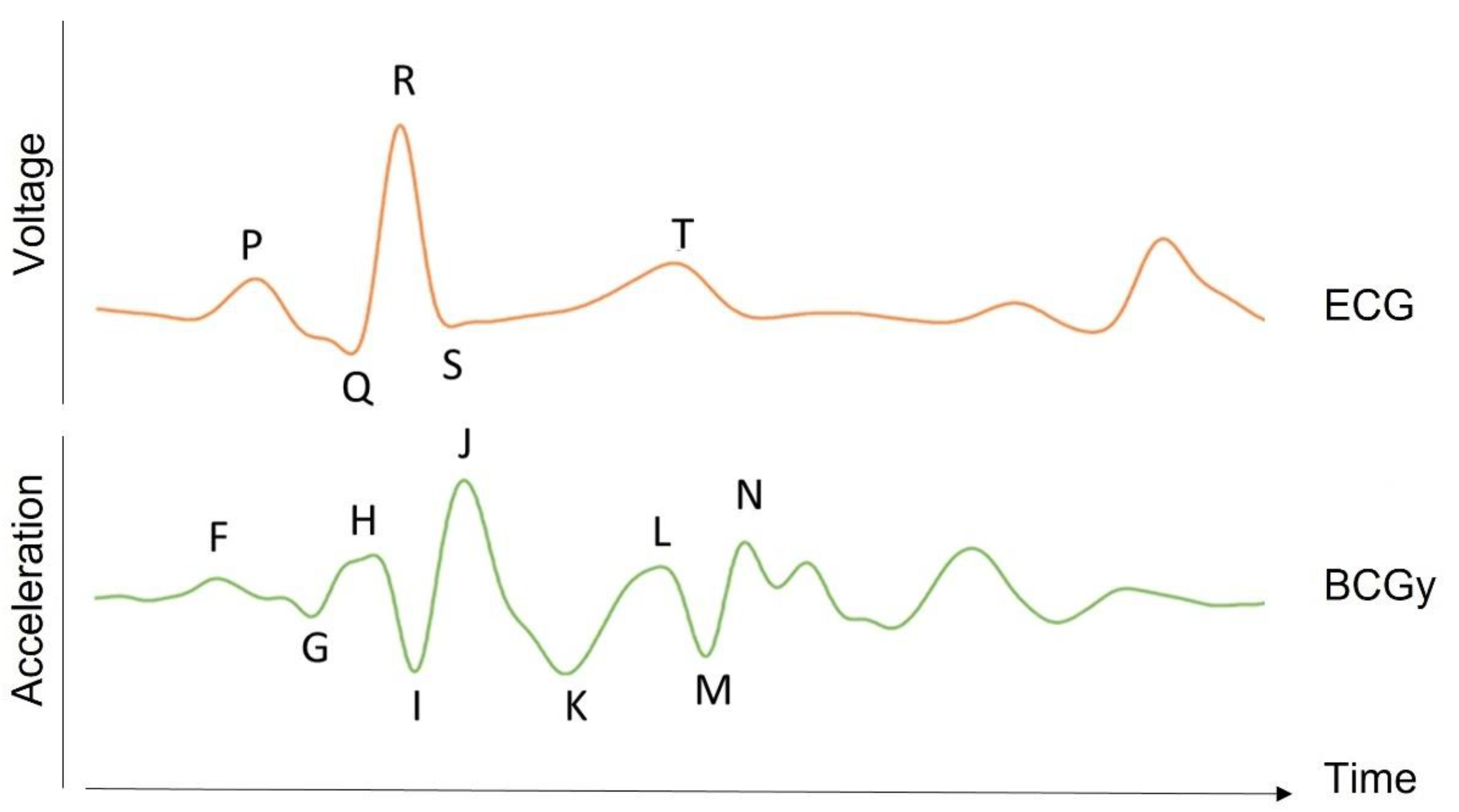
| F | Pre-systolic wave of the longitudinal acceleration BCG signal |
| G | Pre-systolic wave of the longitudinal acceleration BCG signal |
| H | Pre-ejection upward wave of the longitudinal acceleration BCG signal |
| HF | High frequency |
| HRV | Heart rate variability |
| I | First post-ejection downward wave of the longitudinal acceleration BCG signal |
| IVC | Isovolumetric contraction |
| J | First post-ejection upward wave of the longitudinal acceleration BCG signal |
| K | Second post-ejection downward wave of the longitudinal acceleration BCG signal |
| LF | Low frequency |
| MC | Mitral valve closure |
| MO | Mitral valve opening |
| MRI | Magnetic resonance imaging |
| NN | Normal-to-normal interval |
| P | ECG wave representing the electrical depolarization of the atria of the heart |
| PCG | Phonocardiography |
| pNN50 | Proportion of the successive pairs of NN intervals that differ more than 50 ms divided by the total number of NN intervals |
| Q | ECG wave representing the normal left-to-right depolarization of the interventricular septum |
| R | ECG wave corresponding to depolarization of the main mass of the ventricles |
| RE | Rapid ejection |
| RF | Rapid filling |
| RMSSD | Root mean square of successive differences between intervals |
| RR | Interval between two successive R waves, s |
| RSA | Respiratory sinus arrythmia |
| S | ECG wave representing the final depolarization of the ventricles |
| SCG | Seismocardiography |
| SDNN | Standard deviation of NN intervals |
| SV | Stroke volume, ml |
| S1 | First heart sound |
| S2 | Second heart sound |
| S3 | Third heart sound |
| S4 | Fourth heart sound |
| ULF | Ultra-low frequency |
| VLF | Very low frequency |
| Proportionality factor in SV formula, given by Starr |
References
- Ismail, S.; Siddiqi, I.; Akram, U. Localization and Classification of Heart Beats in Phonocardiography Signals—A Comprehensive Review. EURASIP J. Adv. Signal Process. 2018, 2018, 26. [Google Scholar] [CrossRef]
- Mirvis, D.; Goldberger, A. Electrocardiography. Heart Dis. 2001, 1, 82–128. [Google Scholar]
- Gordon, J.W. Certain Molar Movements of the Human Body Produced by the Circulation of the Blood. J. Anat. Physiol. 1877, 11, 533–536. [Google Scholar] [PubMed]
- Henderson, Y. The Mass-Movements of the Circulation as Shown by a Recoil Curve. Am. J. Physiol. Leg. Content 1905, 14, 287–298. [Google Scholar] [CrossRef]
- Heald, C.B.; Tucker, W.S. Recoil Curves as Shown by the Hot-Wire Microphone. Proc. R. Soc. Lond. Ser. B Contain. Pap. A Biol. Character 1922, 93, 281–298. [Google Scholar]
- Starr, I. The Ballistocardiograph-an Instrument for Clinical Research and for Routine Clinical Diagnosis. Harvey Lect. 1947, 42, 194–220. [Google Scholar]
- Rubenstein, E. A Review of Clinical Ballistocardiography. N. Engl. J. Med. 1952, 247, 166–173. [Google Scholar] [CrossRef] [PubMed]
- Inan, O.T.; Migeotte, P.-F.; Park, K.-S.; Etemadi, M.; Tavakolian, K.; Casanella, R.; Zanetti, J.; Tank, J.; Funtova, I.; Prisk, G.K.; et al. Ballistocardiography and Seismocardiography: A Review of Recent Advances. IEEE J. Biomed. Health Inform. 2015, 19, 1414–1427. [Google Scholar] [CrossRef]
- Pinheiro, E.; Postolache, O.; Girão, P. Theory and Developments in an Unobtrusive Cardiovascular System Representation: Ballistocardiography. Open Biomed. Eng. J. 2010, 4, 201–216. [Google Scholar] [CrossRef]
- Bozhenko, B.S. Seismocardiography—A New Method in the Study of Functional Conditions of the Heart. Ter. Arkh. 1961, 33, 55–64. [Google Scholar]
- Crow, R.S.; Hannan, P.; Jacobs, D.; Hedquist, L.; Salerno, D.M. Relationship between Seismocardiogram and Echocardiogram for Events in the Cardiac Cycle. Am. J. Noninvasive Cardiol. 1994, 8, 39–46. [Google Scholar] [CrossRef]
- Zanetti, J. Seismocardiography: A New Technique for Recording Cardiac Vibrations. Concept, Method, and Initial Observations. J. Cardiovasc. Technol. 1990, 9, 111–118. [Google Scholar]
- Andreozzi, E.; Fratini, A.; Esposito, D.; Naik, G.; Polley, C.; Gargiulo, G.D.; Bifulco, P. Forcecardiography: A Novel Technique to Measure Heart Mechanical Vibrations onto the Chest Wall. Sensors 2020, 20, 3885. [Google Scholar] [CrossRef] [PubMed]
- Centracchio, J.; Andreozzi, E.; Esposito, D.; Gargiulo, G.D.; Bifulco, P. Detection of Aortic Valve Opening and Estimation of Pre-Ejection Period in Forcecardiography Recordings. Bioengineering 2022, 9, 89. [Google Scholar] [CrossRef]
- Jafari Tadi, M.; Lehtonen, E.; Saraste, A.; Tuominen, J.; Koskinen, J.; Teräs, M.; Airaksinen, J.; Pänkäälä, M.; Koivisto, T. Gyrocardiography: A New Non-Invasive Monitoring Method for the Assessment of Cardiac Mechanics and the Estimation of Hemodynamic Variables. Sci. Rep. 2017, 7, 6823. [Google Scholar] [CrossRef] [PubMed]
- Clairmonte, N.; Skoric, J.; D’Mello, Y.; Hakim, S.; Aboulezz, E.; Lortie, M.; Plant, D. Neural Network-Based Classification of Static Lung Volume States Using Vibrational Cardiography. In Proceedings of the 2020 42nd Annual International Conference of the IEEE Engineering in Medicine & Biology Society (EMBC), Online, 20–24 July 2020; IEEE: Piscataway, NJ, USA, 2020; pp. 221–224. [Google Scholar]
- Morra, S.; Hossein, A.; Gorlier, D.; Rabineau, J.; Chaumont, M.; Migeotte, P.-F.; van de Borne, P. Modification of the Mechanical Cardiac Performance during End-Expiratory Voluntary Apnea Recorded with Ballistocardiography and Seismocardiography. Physiol. Meas. 2019, 40, 105005. [Google Scholar] [CrossRef]
- Kreit, J. Respiratory-Cardiovascular Interactions During Mechanical Ventilation: Physiology and Clinical Implications. In Comprehensive Physiology; Wiley: Hoboken, NJ, USA, 2022; pp. 3425–3448. [Google Scholar]
- Fisher, J.P.; Zera, T.; Paton, J.F.R. Respiratory–Cardiovascular Interactions. In Handbook of Clinical Neurology; Elsevier: Amsterdam, The Netherlands, 2022; pp. 279–308. [Google Scholar]
- Wise, R.A.; Robotham, J.L.; Summer, W.R. Effects of Spontaneous Ventilation on the Circulation. Lung 1981, 159, 175–186. [Google Scholar] [CrossRef] [PubMed]
- Magder, S. Heart-Lung Interaction in Spontaneous Breathing Subjects: The Basics. Ann. Transl. Med. 2018, 6, 348. [Google Scholar] [CrossRef]
- Feihl, F.; Broccard, A.F. Interactions between Respiration and Systemic Hemodynamics. Part I: Basic Concepts. Intensive Care Med. 2009, 35, 45–54. [Google Scholar] [CrossRef] [PubMed]
- Berntson, G.G.; Cacioppo, J.T.; Quigley, K.S. Respiratory Sinus Arrhythmia: Autonomic Origins, Physiological Mechanisms, and Psychophysiological Implications. Psychophysiology 1993, 30, 183–196. [Google Scholar] [CrossRef]
- Yasuma, F.; Hayano, J. Respiratory Sinus Arrhythmia. Chest 2004, 125, 683–690. [Google Scholar] [CrossRef] [PubMed]
- Elstad, M.; O’Callaghan, E.L.; Smith, A.J.; Ben-Tal, A.; Ramchandra, R. Cardiorespiratory Interactions in Humans and Animals: Rhythms for Life. Am. J. Physiol. Heart Circ. Physiol. 2018, 315, H6–H17. [Google Scholar] [CrossRef] [PubMed]
- Klum, M.; Urban, M.; Tigges, T.; Pielmus, A.-G.; Feldheiser, A.; Schmitt, T.; Orglmeister, R. Wearable Cardiorespiratory Monitoring Employing a Multimodal Digital Patch Stethoscope: Estimation of ECG, PEP, LVET and Respiration Using a 55 Mm Single-Lead ECG and Phonocardiogram. Sensors 2020, 20, 2033. [Google Scholar] [CrossRef]
- Han, X.; Wu, X.; Wang, J.; Li, H.; Cao, K.; Cao, H.; Zhong, K.; Yang, X. The Latest Progress and Development Trend in the Research of Ballistocardiography (BCG) and Seismocardiogram (SCG) in the Field of Health Care. Appl. Sci. 2021, 11, 8896. [Google Scholar] [CrossRef]
- De Ridder, S.; Migeotte, P.-F.; Neyt, X.; Pattyn, N.; Prisk, G.K. Three-Dimensional Ballistocardiography in Microgravity: A Review of Past Research. In Proceedings of the 2011 Annual International Conference of the IEEE Engineering in Medicine and Biology Society, Boston, MA, USA, 20 August–3 September 2011; IEEE: Piscataway, NJ, USA, 2011; pp. 4267–4270. [Google Scholar]
- Vogt, E.; MacQuarrie, D.; Neary, J.P. Using Ballistocardiography to Measure Cardiac Performance: A Brief Review of Its History and Future Significance. Clin. Physiol. Funct. Imaging 2012, 32, 415–420. [Google Scholar] [CrossRef] [PubMed]
- Inan, O.T. Recent Advances in Cardiovascular Monitoring Using Ballistocardiography. In Proceedings of the 2012 Annual International Conference of the IEEE Engineering in Medicine and Biology Society, San Diego, CA, USA, 28 August–1 September 2012; IEEE: Piscataway, NJ, USA, 2012; pp. 5038–5041. [Google Scholar]
- Zanetti, J.M.; Tavakolian, K. Seismocardiography: Past, Present and Future. In Proceedings of the 2013 35th Annual International Conference of the IEEE Engineering in Medicine and Biology Society (EMBC), Osaka, Japan, 3–7 July 2013; IEEE: Piscataway, NJ, USA, 2013; pp. 7004–7007. [Google Scholar]
- Bruser, C.; Antink, C.H.; Wartzek, T.; Walter, M.; Leonhardt, S. Ambient and Unobtrusive Cardiorespiratory Monitoring Techniques. IEEE Rev. Biomed. Eng. 2015, 8, 30–43. [Google Scholar] [CrossRef] [PubMed]
- Landreani, F.; Caiani, E.G. Smartphone Accelerometers for the Detection of Heart Rate. Expert Rev. Med. Devices 2017, 14, 935–948. [Google Scholar] [CrossRef] [PubMed]
- Leonhardt, S.; Leicht, L.; Teichmann, D. Unobtrusive Vital Sign Monitoring in Automotive Environments—A Review. Sensors 2018, 18, 3080. [Google Scholar] [CrossRef]
- Sadek, I.; Biswas, J.; Abdulrazak, B. Ballistocardiogram Signal Processing: A Review. Health Inf. Sci. Syst. 2019, 7, 10. [Google Scholar] [CrossRef] [PubMed]
- Taebi, A.; Solar, B.; Bomar, A.; Sandler, R.; Mansy, H. Recent Advances in Seismocardiography. Vibration 2019, 2, 64–86. [Google Scholar] [CrossRef]
- Sidikova, M.; Martinek, R.; Kawala-Sterniuk, A.; Ladrova, M.; Jaros, R.; Danys, L.; Simonik, P. Vital Sign Monitoring in Car Seats Based on Electrocardiography, Ballistocardiography and Seismocardiography: A Review. Sensors 2020, 20, 5699. [Google Scholar] [CrossRef] [PubMed]
- Jähne-Raden, N.; Gütschleg, H.; Marschollek, M. Trodden Lanes or New Paths: Ballisto- and Seismocardiography till Now. Stud. Health Technol. Inform. 2020, 270, 479–483. [Google Scholar] [CrossRef] [PubMed]
- Sieciński, S.; Kostka, P.S.; Tkacz, E.J. Gyrocardiography: A Review of the Definition, History, Waveform Description, and Applications. Sensors 2020, 20, 6675. [Google Scholar] [CrossRef]
- Jiang, F.; Zhou, Y.; Ling, T.; Zhang, Y.; Zhu, Z. Recent Research for Unobtrusive Atrial Fibrillation Detection Methods Based on Cardiac Dynamics Signals: A Survey. Sensors 2021, 21, 3814. [Google Scholar] [CrossRef]
- Sharma, M.; Rajput, J.S.; Tan, R.S.; Acharya, U.R. Automated Detection of Hypertension Using Physiological Signals: A Review. Int. J. Environ. Res. Public Health 2021, 18, 5838. [Google Scholar] [CrossRef]
- Rahmani, M.H.; Berkvens, R.; Weyn, M. Chest-Worn Inertial Sensors: A Survey of Applications and Methods. Sensors 2021, 21, 2875. [Google Scholar] [CrossRef]
- Santucci, F.; lo Presti, D.; Massaroni, C.; Schena, E.; Setola, R. Precordial Vibrations: A Review of Wearable Systems, Signal Processing Techniques, and Main Applications. Sensors 2022, 22, 5805. [Google Scholar] [CrossRef] [PubMed]
- Otis, A.B.; Rahn, H.; Brontman, M.; Mullins, L.J.; Fenn, W.O. Ballistocardiographic Study of Changes in Cardiac Output Due to Respiration 1. J. Clin. Investig. 1946, 25, 413–421. [Google Scholar] [CrossRef]
- Tavakolian, K.; Vaseghi, A.; Kaminska, B. Improvement of Ballistocardiogram Processing by Inclusion of Respiration Information. Physiol. Meas. 2008, 29, 771–781. [Google Scholar] [CrossRef] [PubMed]
- Josenhans, W.T.; Josenhans, W. Air Movements from the Chest and ULF Displacement BCG during Respiration and during Breathholding with Open and Closed Glottis. In Ballistocardiography and Cardiovascular Dynamics: 1st World Congress, Amsterdam, April 1965; S. Karger: Berlin, Germany, 1965; p. 327. [Google Scholar]
- Starr, I.; Friedland, C.K. On the Cause of the Respiratory Variation of the Ballistocardiogram, with a Note on Sinus Arrhythmia. J. Clin. Investig. 1946, 25, 53–64. [Google Scholar] [CrossRef]
- De Lalla, V.; Brown, H.R. Respiratory Variation of the Ballistocardiogram. Am. J. Med. 1950, 9, 728–733. [Google Scholar] [CrossRef]
- Brown, H.R.; de Lalla, V. Ballistocardiogram, Description and Clinical Use. Am. J. Med. 1950, 9, 718–727. [Google Scholar] [CrossRef]
- Dock, W. Effects of Respiration on Transmission of Ballistocardiographic Forces from the Heart to the Recording System. Am. Heart J. 1959, 58, 102–112. [Google Scholar] [CrossRef] [PubMed]
- NOORDERGRAAF, A. Further Studies on a Theory of the Ballistocardiogram. Circulation 1961, 23, 413–425. [Google Scholar] [CrossRef]
- Talbot, S.A.; Harrison, W.K., Jr.; Ginn, W.M., Jr. Features of ULF-BCG Pertinent to Coronary Heart Disease. In Ballistocardiography and Cardiovascular Dynamics; S. Karger: Berlin, Germany, 1966. [Google Scholar]
- Prisk, G.K.; Verhaeghe, S.; Padeken, D.; Hamacher, H.; Paiva, M. Three-Dimensional Ballistocardiography and Respiratory Motion in Sustained Microgravity. Aviat. Space Environ. Med. 2001, 72, 1067–1074. [Google Scholar] [PubMed]
- Pandia, K.; Inan, O.T.; Kovacs, G.T.A.; Giovangrandi, L. Extracting Respiratory Information from Seismocardiogram Signals Acquired on the Chest Using a Miniature Accelerometer. Physiol. Meas. 2012, 33, 1643–1660. [Google Scholar] [CrossRef] [PubMed]
- Zakeri, V.; Akhbardeh, A.; Alamdari, N.; Fazel-Rezai, R.; Paukkunen, M.; Tavakolian, K. Analyzing Seismocardiogram Cycles to Identify the Respiratory Phases. IEEE Trans Biomed. Eng. 2017, 64, 1786–1792. [Google Scholar] [CrossRef] [PubMed]
- Taghizadeh Alamdari, N. A Morphological Approach to Identify Respiratory Phases of Seismocardiogram; University of North Dakota: Grand Forks, ND, USA, 2016. [Google Scholar]
- Castiglioni, P.; Faini, A.; Parati, G.; di Rienzo, M. Wearable Seismocardiography. In Proceedings of the 2007 29th Annual International Conference of the IEEE Engineering in Medicine and Biology Society, Lyon, France, 22–26 August 2007; IEEE: Piscataway, NJ, USA, 2007; pp. 3954–3957. [Google Scholar]
- Dock, W. The Extravascular Basis for Respiratory Variation in the Ballistocardiogram with Notes on the Effects of Constrictive Pericarditis, Atrial Septal Defect, and the Valsalva Maneuver. Ann. Intern. Med. 1962, 57, 398–405. [Google Scholar] [CrossRef] [PubMed]
- Starr, I.; Rawson, A.J.; Schroeder, H.A.; Joseph, N.R. Studies on The Estimation of Cardiac Ouptut In Man, And Of Abnormalities In Cardiac Function, From The Heart’s Recoil And The Blood’s Impacts; The Ballistocardiogram. Am. J. Physiol. 1939, 127, 1–28. [Google Scholar] [CrossRef]
- Williams, A.H.; Gropper, A.L. Interrelationships of Cardiac Output, Blood Pressure, and Peripheral Resistance during Normal Respiration in Normotensive and Hypertensive Individuals. Circulation 1951, IV. [Google Scholar] [CrossRef]
- Knoop, A.A. Ballistocardiography and Cardiovascular Dynamics: 1st World Congress, Amsterdam, April 1965; Karger: Basel, Switzerland, 1966. [Google Scholar]
- Van Brummelen, A.G.W.; Scarborough, W.R.; Josenhans, W.T. On the Elimination of Pulse Wave Velocity in Stroke Volume Determination from the Ultralow-Frequency Displacement Ballistocardiography. Am. Heart J. 1964, 67, 374–378. [Google Scholar] [CrossRef] [PubMed][Green Version]
- Baevsky, R.M.; Funtova, I.I.; Luchitskaya, E.S. Role of the Right and Left Parts of the Heart in Mechanisms of Body Adaptation to the Conditions of Long Term Space Flight According to Longitudinal Ballistocardiography. Acta Astronaut. 2021, 178, 894–899. [Google Scholar] [CrossRef]
- Isaac Starr, A.N. Ballistocardiography in Cardiovascular Research: Physical Aspects of the Circulation in Health and Disease; Lippincott Williams & Wilkins: Philadelphia, PA, USA, 1967. [Google Scholar]
- Rabineau, J.; Nonclercq, A.; Leiner, T.; van de Borne, P.; Migeotte, P.-F.; Haut, B. Closed-Loop Multiscale Computational Model of Human Blood Circulation. Applications to Ballistocardiography. Front. Physiol. 2021, 12, 4311. [Google Scholar] [CrossRef] [PubMed]
- Semiz, B.; Carek, A.M.; Johnson, J.C.; Ahmad, S.; Heller, J.A.; Vicente, F.G.; Caron, S.; Hogue, C.W.; Etemadi, M.; Inan, O.T. Non-Invasive Wearable Patch Utilizing Seismocardiography for Peri-Operative Use in Surgical Patients. IEEE J. Biomed. Health Inform. 2021, 25, 1572–1582. [Google Scholar] [CrossRef]
- Hossein, A.; Mirica, D.C.; Rabineau, J.; del Rio, J.I.; Morra, S.; Gorlier, D.; Nonclercq, A.; van de Borne, P.; Migeotte, P.-F. Accurate Detection of Dobutamine-Induced Haemodynamic Changes by Kino-Cardiography: A Randomised Double-Blind Placebo-Controlled Validation Study. Sci. Rep. 2019, 9, 10479. [Google Scholar] [CrossRef]
- Hoog Antink, C.; Mai, Y.; Aalto, R.; Bruser, C.; Leonhardt, S.; Oksala, N.; Vehkaoja, A. Ballistocardiography Can Estimate Beat-to-Beat Heart Rate Accurately at Night in Patients After Vascular Intervention. IEEE J. Biomed. Health Inform. 2020, 24, 2230–2237. [Google Scholar] [CrossRef]
- Paalasmaa, J.; Waris, M.; Toivonen, H.; Leppakorpi, L.; Partinen, M. Unobtrusive Online Monitoring of Sleep at Home. In Proceedings of the 2012 Annual International Conference of the IEEE Engineering in Medicine and Biology Society, San Diego, CA, USA, 28 August–1 September 2012; IEEE: Piscataway, NJ, USA, 2012; pp. 3784–3788. [Google Scholar]
- Mack, D.C.; Patrie, J.T.; Suratt, P.M.; Felder, R.A.; Alwan, M. Development and Preliminary Validation of Heart Rate and Breathing Rate Detection Using a Passive, Ballistocardiography-Based Sleep Monitoring System. IEEE Trans. Inf. Technol. Biomed. 2009, 13, 111–120. [Google Scholar] [CrossRef]
- Ben Nasr, M.C.; ben Jebara, S.; Otis, S.; Abdulrazak, B.; Mezghani, N. A Spectral-Based Approach for BCG Signal Content Classification. Sensors 2021, 21, 1020. [Google Scholar] [CrossRef]
- Bruser, C.; Kerekes, A.; Winter, S.; Leonhardt, S. Multi-Channel Optical Sensor-Array for Measuring Ballistocardiograms and Respiratory Activity in Bed. In Proceedings of the 2012 Annual International Conference of the IEEE Engineering in Medicine and Biology Society, San Diego, CA, USA, 28 August–1 September 2012; IEEE: Piscataway, NJ, USA, 2012; pp. 5042–5045. [Google Scholar]
- Pino, E.J.; Arias, D.E.; Aqueveque, P.; Vilugron, L.; Hermosilla, D.; Curtis, D.W. Monitoring Technology for Wheelchair Users with Advanced Multiple Sclerosis. In Proceedings of the 2013 35th Annual International Conference of the IEEE Engineering in Medicine and Biology Society (EMBC), Osaka, Japan, 3–7 July 2013; IEEE: Piscataway, NJ, USA, 2013; pp. 961–964. [Google Scholar]
- Da He, D.; Winokur, E.S.; Heldt, T.; Sodini, C.G. The Ear as a Location for Wearable Vital Signs Monitoring. In Proceedings of the 2010 Annual International Conference of the IEEE Engineering in Medicine and Biology, Buenos Aires, Argentina, 31 August–4 September 2010; IEEE: Piscataway, NJ, USA, 2010; pp. 6389–6392. [Google Scholar]
- Morra, S.; Gauthey, A.; Hossein, A.; Rabineau, J.; Racape, J.; Gorlier, D.; Migeotte, P.-F.; le Polain de Waroux, J.B.; van de Borne, P. Influence of Sympathetic Activation on Myocardial Contractility Measured with Ballistocardiography and Seismocardiography during Sustained End-Expiratory Apnea. Am. J. Physiol. Regul. Integr. Comp. Physiol. 2020, 319, R497–R506. [Google Scholar] [CrossRef]
- Lu, G.; Wang, J.; Yue, Y.; Jing, X. Study of the Ballistocardiogram Signal in Life Detection System Based on Radar. In Proceedings of the 2007 29th Annual International Conference of the IEEE Engineering in Medicine and Biology Society, Lyon, France, 22–26 August 2007; IEEE: Piscataway, NJ, USA; pp. 2191–2194. [Google Scholar]
- Gurel, N.Z.; Wittbrodt, M.T.; Jung, H.; Ladd, S.L.; Shah, A.J.; Vaccarino, V.; Bremner, J.D.; Inan, O. Automatic Detection of Target Engagement in Transcutaneous Cervical Vagal Nerve Stimulation for Traumatic Stress Triggers. IEEE J. Biomed. Health Inform. 2020, 24, 1917–1925. [Google Scholar] [CrossRef]
- Alihanka, J.; Vaahtoranta, K.; Saarikivi, I. A New Method for Long-Term Monitoring of the Ballistocardiogram, Heart Rate, and Respiration. Am. J. Physiol. Regul. Integr. Comp. Physiol. 1981, 240, R384–R392. [Google Scholar] [CrossRef]
- Korhonen, I.; Iivainen, T.; Lappalainen, R.; Tuomisto, T.; Kööbi, T.; Pentikäinen, V.; Tuomisto, M.; Turjanmaa, V. TERVA: System for Long-Term Monitoring of Wellness at Home. Telemed. J. e-Health 2001, 7, 61–72. [Google Scholar] [CrossRef]
- Virtanen, I.; Polo, O.; Saaresranta, T.; Kuusela, T.; Polo-Kantola, P.; Ekholm, E. Medroxyprogesterone Improves Cardiac Autonomic Control in Postmenopausal Women with Respiratory Insufficiency. Respir. Med. 2004, 98, 126–133. [Google Scholar] [CrossRef][Green Version]
- Su, B.Y.; Ho, K.C.; Skubic, M.; Rosales, L. Pulse Rate Estimation Using Hydraulic Bed Sensor. In Proceedings of the 2012 Annual International Conference of the IEEE Engineering in Medicine and Biology Society, San Diego, CA, USA, 28 August–1 September 2012; IEEE: Piscataway, NJ, USA, 2012; pp. 2587–2590. [Google Scholar]
- Zhao, W.; Ni, H.; Zhou, X.; Song, Y.; Wang, T. Identifying Sleep Apnea Syndrome Using Heart Rate and Breathing Effort Variation Analysis Based on Ballistocardiography. In Proceedings of the 2015 37th Annual International Conference of the IEEE Engineering in Medicine and Biology Society (EMBC), Milan, Italy, 25–29 August 2015; pp. 4536–4539. [Google Scholar]
- Albukhari, A.; Lima, F.; Mescheder, U. Bed-Embedded Heart and Respiration Rates Detection by Longitudinal Ballistocardiography and Pattern Recognition. Sensors 2019, 19, 1451. [Google Scholar] [CrossRef]
- Cimr, D.; Studnicka, F.; Fujita, H.; Cimler, R.; Slegr, J. Application of Mechanical Trigger for Unobtrusive Detection of Respiratory Disorders from Body Recoil Micro-Movements. Comput. Methods Programs Biomed. 2021, 207, 106149. [Google Scholar] [CrossRef]
- Chung, G.S.; Choi, B.H.; Jeong, D.-U.; Park, K.S. Noninvasive Heart Rate Variability Analysis Using Loadcell-Installed Bed During Sleep. In Proceedings of the 2007 29th Annual International Conference of the IEEE Engineering in Medicine and Biology Society, Lyon, France, 22–26 August 2007; IEEE: Piscataway, NJ, USA, 2007; pp. 2357–2360. [Google Scholar]
- Lee, W.K.; Yoon, H.; Jung, D.W.; Hwang, S.H.; Park, K.S. Ballistocardiogram of Baby during Sleep. In Proceedings of the 2015 37th Annual International Conference of the IEEE Engineering in Medicine and Biology Society (EMBC), Milan, Italy, 25–29 August 2015; pp. 7167–7170. [Google Scholar]
- Mitsukura, Y.; Sumali, B.; Nagura, M.; Fukunaga, K.; Yasui, M. Sleep Stage Estimation from Bed Leg Ballistocardiogram Sensors. Sensors 2020, 20, 5688. [Google Scholar] [CrossRef]
- Shin, J.H.; Hwang, S.H.; Chang, M.H.; Park, K.S. Heart Rate Variability Analysis Using a Ballistocardiogram during Valsalva Manoeuvre and Post Exercise. Physiol. Meas. 2011, 32, 1239–1264. [Google Scholar] [CrossRef]
- Park, K.S.; Hwang, S.H.; Jung, D.; Yoon, H.N.; Lee, W.K. Ballistocardiography for Nonintrusive Sleep Structure Estimation. In Proceedings of the 2014 36th Annual International Conference of the IEEE Engineering in Medicine and Biology Society, Chicago, IL, USA, 26–30 August 2014; pp. 5184–5187. [Google Scholar]
- Nedoma, J.; Kepak, S.; Fajkus, M.; Cubik, J.; Siska, P.; Martinek, R.; Krupa, P. Magnetic Resonance Imaging Compatible Non-Invasive Fibre-Optic Sensors Based on the Bragg Gratings and Interferometers in the Application of Monitoring Heart and Respiration Rate of the Human Body: A Comparative Study. Sensors 2018, 18, 3713. [Google Scholar] [CrossRef]
- Huysmans, D.; Borzée, P.; Testelmans, D.; Buyse, B.; Willemen, T.; van Huffel, S.; Varon, C. Evaluation of a Commercial Ballistocardiography Sensor for Sleep Apnea Screening and Sleep Monitoring. Sensors 2019, 19, 2133. [Google Scholar] [CrossRef]
- Wusk, G.; Gabler, H. Non-Invasive Detection of Respiration and Heart Rate with a Vehicle Seat Sensor. Sensors 2018, 18, 1463. [Google Scholar] [CrossRef]
- Lee, W.K.; Yoon, H.; Han, C.; Joo, K.M.; Park, K.S. Physiological Signal Monitoring Bed for Infants Based on Load-Cell Sensors. Sensors 2016, 16, 409. [Google Scholar] [CrossRef]
- Shin, J.H.; Park, K.S. HRV Analysis and Blood Pressure Monitoring on Weighing Scale Using BCG. In Proceedings of the 2012 Annual International Conference of the IEEE Engineering in Medicine and Biology Society, San Diego, CA, USA, 28 August–1 September 2012; IEEE: Piscataway, NJ, USA, 2012; pp. 3789–3792. [Google Scholar]
- Martín-Yebra, A.; Landreani, F.; Casellato, C.; Pavan, E.; Migeotte, P.-F.; Frigo, C.; Martínez, J.P.; Caiani, E.G. Evaluation of Respiratory- and Postural-Induced Changes on the Ballistocardiogram Signal by Time Warping Averaging. Physiol. Meas. 2017, 38, 1426–1440. [Google Scholar] [CrossRef]
- Morra, S.; Hossein, A.; Gorlier, D.; Rabineau, J.; Chaumont, M.; Migeotte, P.-F.; van de Borne, P. Ballistocardiography and Seismocardiography Detection of Hemodynamic Changes during Simulated Obstructive Apnea. Physiol. Meas. 2020, 41, 065007. [Google Scholar] [CrossRef]
- Reinvuo, T.; Hannula, M.; Sorvoja, H.; Alasaarela, E.; Myllyla, R. Measurement of Respiratory Rate with High-Resolution Accelerometer and Emfit Pressure Sensor. In Proceedings of the Proceedings of the 2006 IEEE Sensors Applications Symposium, Houston, TX, USA, 7–9 February 2006; IEEE: Piscataway, NJ, USA, 2006; pp. 192–195. [Google Scholar]
- Vehkaoja, A.; Kontunen, A.; Lekkala, J. Effects of Sensor Type and Sensor Location on Signal Quality in Bed Mounted Ballistocardiographic Heart Rate and Respiration Monitoring. Annu. Int. Conf. IEEE Eng. Med. Biol. Soc. 2015, 2015, 4383–4386. [Google Scholar] [CrossRef]
- Chethana, K.; Guru Prasad, A.S.; Omkar, S.N.; Asokan, S. Fiber Bragg Grating Sensor Based Device for Simultaneous Measurement of Respiratory and Cardiac Activities. J. Biophotonics 2017, 10, 278–285. [Google Scholar] [CrossRef]
- Nedoma, J.; Fajkus, M.; Martinek, R.; Nazeran, H. Vital Sign Monitoring and Cardiac Triggering at 1.5 Tesla: A Practical Solution by an MR-Ballistocardiography Fiber-Optic Sensor. Sensors 2019, 19, 470. [Google Scholar] [CrossRef]
- Di Rienzo, M.; Meriggi, P.; Rizzo, F.; Vaini, E.; Faini, A.; Merati, G.; Parati, G.; Castiglioni, P. A Wearable System for the Seismocardiogram Assessment in Daily Life Conditions. In Proceedings of the 2011 Annual International Conference of the IEEE Engineering in Medicine and Biology Society, Boston, MA, USA, 30 August–3 September 2011; IEEE: Piscataway, NJ, USA, 2011; pp. 4263–4266. [Google Scholar]
- Jafari Tadi, M.; Koivisto, T.; Pänkäälä, M.; Paasio, A. Accelerometer-Based Method for Extracting Respiratory and Cardiac Gating Information for Dual Gating during Nuclear Medicine Imaging. Int. J. Biomed. Imaging 2014, 2014, 1–11. [Google Scholar] [CrossRef]
- Ramos-Castro, J.; Moreno, J.; Miranda-Vidal, H.; Garcia-Gonzalez, M.A.; Fernandez-Chimeno, M.; Rodas, G.; Capdevila, L. Heart Rate Variability Analysis Using a Seismocardiogram Signal. In Proceedings of the 2012 Annual International Conference of the IEEE Engineering in Medicine and Biology Society, San Diego, CA, USA, 28 August–1 September 2012; IEEE: Piscataway, NJ, USA, 2012; pp. 5642–5645. [Google Scholar]
- Siecinski, S.; Kostka, P.S.; Tkacz, E.J. Influence of Empirical Mode Decomposition on Heart Rate Variability Indices Obtained from Smartphone Seismocardiograms. In Proceedings of the 2019 41st Annual International Conference of the IEEE Engineering in Medicine and Biology Society (EMBC), Berlin, Germany, 23–27 July 2019; IEEE: Piscataway, NJ, USA, 2019; pp. 4913–4916. [Google Scholar]
- Xia, Z.; Shandhi, M.M.H.; Li, Y.; Inan, O.T.; Zhang, Y. The Delineation of Fiducial Points for Non-Contact Radar Seismocardiogram Signals Without Concurrent ECG. IEEE J. Biomed. Health Inform. 2021, 25, 1031–1040. [Google Scholar] [CrossRef]
- Tadi, M.J.; Lehtonen, E.; Koivisto, T.; Pänkäälä, M.; Paasio, A.; Teräs, M. Seismocardiography: Toward Heart Rate Variability (HRV) Estimation. In Proceedings of the IEEE International Symposium on Medical Measurements and Applications (MeMeA) Proceedings, Turin, Italy, 7–9 May 2015; pp. 261–266. [Google Scholar]
- Laurin, A.; Blaber, A.; Tavakolian, K. Seismocardiograms Return Valid Heart Rate Variability Indices. In Proceedings of the Computing in Cardiology 2013, Zaragoza, Spain, 22–25 September 2013; pp. 413–416. [Google Scholar]
- Hurnanen, T.; Lehtonen, E.; Tadi, M.J.; Kuusela, T.; Kiviniemi, T.; Saraste, A.; Vasankari, T.; Airaksinen, J.; Koivisto, T.; Pankaala, M. Automated Detection of Atrial Fibrillation Based on Time–Frequency Analysis of Seismocardiograms. IEEE J. Biomed. Health Inform. 2017, 21, 1233–1241. [Google Scholar] [CrossRef]
- Taebi, A.; Mansy, H.A. Grouping Similar Seismocardiographic Signals Using Respiratory Information. In Proceedings of the 2017 IEEE Signal Processing in Medicine and Biology Symposium (SPMB), Philadelphia, PA, USA, 2 December 2017; IEEE: Piscataway, NJ, USA, 2017; pp. 1–6. [Google Scholar]
- Pandia, K.; Inan, O.T.; Kovacs, G.T.A. A Frequency Domain Analysis of Respiratory Variations in the Seismocardiogram Signal. In Proceedings of the 2013 35th Annual International Conference of the IEEE Engineering in Medicine and Biology Society (EMBC), Osaka, Japan, 3–7 July 2013; IEEE: Piscataway, NJ, USA, 2013; pp. 6881–6884. [Google Scholar]
- Di Rienzo, M.; Vaini, E.; Castiglioni, P.; Merati, G.; Meriggi, P.; Parati, G.; Faini, A.; Rizzo, F. Wearable Seismocardiography: Towards a Beat-by-Beat Assessment of Cardiac Mechanics in Ambulant Subjects. Auton. Neurosci. 2013, 178, 50–59. [Google Scholar] [CrossRef]
- Jung, H.; Kimball, J.; Receveur, T.; Gazi, A.H.; Agdeppa, E.; Inan, O. Estimation of Tidal Volume Using Load Cells on a Hospital Bed. IEEE J. Biomed. Health Inform. 2022, 26, 3330–3341. [Google Scholar] [CrossRef] [PubMed]
- Gurel, N.Z.; Jeong, H.K.; Kloefkorn, H.; Hochman, S.; Inan, O.T. Unobtrusive Heartbeat Detection from Mice Using Sensors Embedded in the Nest. In Proceedings of the 2018 40th Annual International Conference of the IEEE Engineering in Medicine and Biology Society (EMBC), Honolulu, HI, USA, 17–21 July 2018; IEEE: Piscataway, NJ, USA, 2018; pp. 1604–1607. [Google Scholar]
- Cimr, D.; Studnicka, F.; Fujita, H.; Tomaskova, H.; Cimler, R.; Kuhnova, J.; Slegr, J. Computer Aided Detection of Breathing Disorder from Ballistocardiography Signal Using Convolutional Neural Network. Inf. Sci. 2020, 541, 207–217. [Google Scholar] [CrossRef]
- Yao, Y.; Bruser, C.; Pietrzyk, U.; Leonhardt, S.; van Waasen, S.; Schiek, M. Model-Based Verification of a Non-Linear Separation Scheme for Ballistocardiography. IEEE J. Biomed. Health Inform. 2014, 18, 174–182. [Google Scholar] [CrossRef] [PubMed]
- Yao, Y.; Brüser, C.; Vollmer, T.; Schiek, M. Signal Separation for Ballistocardiography via Locally Projective Noise Reduction. In Proceedings of the World Congress on Medical Physics and Biomedical Engineering, Beijing, China, 26–31 May 2012; pp. 501–504. [Google Scholar]
- Skoric, J.; D’Mello, Y.; Aboulezz, E.; Hakim, S.; Clairmonte, N.; Lortie, M.; Plant, D.V. Relationship of the Respiration Waveform to a Chest Worn Inertial Sensor. In Proceedings of the 2020 42nd Annual International Conference of the IEEE Engineering in Medicine & Biology Society (EMBC), Montreal, QC, Canada, 20–24 July 2020; IEEE: Piscataway, NJ, USA, 2020; pp. 2732–2735. [Google Scholar]
- Despins, L.A.; Guidoboni, G.; Skubic, M.; Sala, L.; Enayati, M.; Popescu, M.; Deroche, C.B. Using Sensor Signals in the Early Detection of Heart Failure: A Case Study. J. Gerontol. Nurs. 2020, 46, 41–46. [Google Scholar] [CrossRef]
- Brown, H.R.; Hoffman, M.J.; de Lalla, V. Ballistocardiographic Findings in Patients with Symptoms of Angina Pectoris. Circulation 1950, 1, 132–140. [Google Scholar] [CrossRef]
- Starr, I. Prognostic Value of Ballistocardiograms. JAMA 1964, 187, 511–517. [Google Scholar] [CrossRef]
- Anderson, W.H.; Urbach, K.; Doerner, A.A. Ballistic Respiratory Variation as Measured by the Electromagnetic Ballistocardiograph. Am. Heart J. 1954, 47, 15–29. [Google Scholar] [CrossRef]
- Fernandez-Garcia, B.C.G. Mitral Valvotomy. In Ballistocardiography and Cardiovascular Dynamics: 1st World Congress, Amsterdam, April 1965; S. Karger: Berlin, Germany, 1965; pp. 31–35. [Google Scholar]
- Davis, F.W.; Scarborough, W.R.; Mason, R.E.; Singewald, M.L.; Baker, B.M. The Effects of Exercise and Smoking on the Electrocardiograms and Ballistocardiograms of Normal Subjects and Patients with Coronary Artery Disease. Am. Heart J. 1953, 46, 529–542. [Google Scholar] [CrossRef]
- Buchanan, J. Ballistocardiographic Smoking Tests in Thyrotoxicosis. In Ballistocardiography and Cardiovascular Dynamics: 1st World Congress, Amsterdam, April 1965; S. Karger: Basel, Switzerland, 1966. [Google Scholar]
- Benjafield, A.V.; Ayas, N.T.; Eastwood, P.R.; Heinzer, R.; Ip, M.S.M.; Morrell, M.J.; Nunez, C.M.; Patel, S.R.; Penzel, T.; Pépin, J.-L.D.; et al. Estimation of the Global Prevalence and Burden of Obstructive Sleep Apnoea: A Literature-Based Analysis. Lancet Respir. Med. 2019, 7, 687. [Google Scholar] [CrossRef]
- Ho, M.L.; Brass, S.D. Obstructive Sleep Apnea. Neurol. Int. 2011, 3, e15. [Google Scholar] [CrossRef]
- Dempsey, J.A.; Smith, C.A.; Blain, G.M.; Xie, A.; Gong, Y.; Teodorescu, M. Role of Central/Peripheral Chemoreceptors and Their Interdependence in the Pathophysiology of Sleep Apnea. Adv. Exp. Med. Biol. 2012, 758, 343–349. [Google Scholar] [CrossRef]
- Linz, D.; Woehrle, H.; Bitter, T.; Fox, H.; Cowie, M.R.; Böhm, M.; Oldenburg, O. The Importance of Sleep-Disordered Breathing in Cardiovascular Disease. Clin. Res. Cardiol. 2015, 104, 705–718. [Google Scholar] [CrossRef]
- Polo, O.; Tafti, M.; Hämäläinen, M.; Vaahtoranta, K.; Alihanka, J. Respiratory Variation of the Ballistocardiogram during Increased Respiratory Load and Voluntary Central Apnoea. Eur. Respir. J. 1992, 5, 257–262. [Google Scholar] [CrossRef] [PubMed]
- Bronicki, R.A.; Anas, N.G. Cardiopulmonary Interaction. Pediatr. Crit. Care Med. 2009, 10, 313–322. [Google Scholar] [CrossRef]
- Qin, H.; Steenbergen, N.; Glos, M.; Wessel, N.; Kraemer, J.F.; Vaquerizo-Villar, F.; Penzel, T. The Different Facets of Heart Rate Variability in Obstructive Sleep Apnea. Front. Psychiatry 2021, 12, 2333. [Google Scholar] [CrossRef] [PubMed]
- Gao, W.; Xu, Y.; Li, S.; Fu, Y.; Zheng, D.; She, Y. Obstructive Sleep Apnea Syndrome Detection Based on Ballistocardiogram via Machine Learning Approach. Math. Biosci. Eng. 2019, 16, 5672–5686. [Google Scholar] [CrossRef] [PubMed]
- Salmi, T.; Partinen, M.; Hyyppä, M.; Kronholm, E. Automatic Analysis of Static Charge Sensitive Bed (SCSB) Recordings in the Evaluation of Sleep-Related Apneas. Acta Neurol. Scand. 2009, 74, 360–364. [Google Scholar] [CrossRef] [PubMed]
- Partinen, M.; Telakivi, T.; Salmi, T.; Alihanka, J.; Guilleminault, C. Screening for Obstructive Sleep Apnea with the SCSB Method. In Proceedings of the World Congress on Chronic Rhonchopathy, Barcelona, Spain, 22–24 May 1989; Volume 12. [Google Scholar]
- Lindqvist, A.; Halme, M.; Tukiainen, P.; Laitinen, L.A. Amplitude Variation in Static-Charge-Sensitive Bed Signal Increased in Obstructive Airways Disease. Clin. Physiol. 1998, 18, 369–376. [Google Scholar] [CrossRef] [PubMed]
- Migliorini, M.; Bianchi, A.M.; Nisticò, D.; Kortelainen, J.; Arce-Santana, E.; Cerutti, S.; Mendez, M.O. Automatic Sleep Staging Based on Ballistocardiographic Signals Recorded through Bed Sensors. In Proceedings of the 2010 Annual International Conference of the IEEE Engineering in Medicine and Biology, Buenos Aires, Argentina, 31 August–4 September 2010; IEEE: Piscataway, NJ, USA, 2010; pp. 3273–3276. [Google Scholar]
- Liu, F.; Zhou, X.; Wang, Z.; Wang, T.; Ni, H.; Yang, J. Identifying Obstructive Sleep Apnea by Exploiting Fine-Grained BCG Features Based on Event Phase Segmentation. In Proceedings of the 2016 IEEE 16th International Conference on Bioinformatics and Bioengineering (BIBE), Taichung, Taiwan, 32 October–2 November 2016; IEEE: Piscataway, NJ, USA, 2016; pp. 293–300. [Google Scholar]
- Sadek, I.; Seet, E.; Biswas, J.; Abdulrazak, B.; Mokhtari, M. Nonintrusive Vital Signs Monitoring for Sleep Apnea Patients: A Preliminary Study. IEEE Access 2018, 6, 2506–2514. [Google Scholar] [CrossRef]
- Wang, Z.; Zhou, X.; Zhao, W.; Liu, F.; Ni, H.; Yu, Z. Assessing the Severity of Sleep Apnea Syndrome Based on Ballistocardiogram. PLoS ONE 2017, 12, e0175351. [Google Scholar] [CrossRef] [PubMed]
- Guerrero, G.; Kortelainen, J.M.; Palacios, E.; Bianchi, A.M.; Tachino, G.; Tenhunen, M.; Méndez, M.O.; van Gils, M. Detection of Sleep-Disordered Breating with Pressure Bed Sensor. Annu. Int. Conf. IEEE Eng. Med. Biol. Soc. 2013, 2013, 1342–1345. [Google Scholar] [CrossRef] [PubMed]
- Zhao, T.; Fu, X.; Zhan, J.; Chen, K.; Li, Z. Vital Signs Monitoring Using the Macrobending Small-Core Fiber Sensor. Opt. Lett. 2021, 46, 4228–4231. [Google Scholar] [CrossRef]
- Siyahjani, F.; Garcia Molina, G.; Barr, S.; Mushtaq, F. Performance Evaluation of a Smart Bed Technology against Polysomnography. Sensors 2022, 22, 2605. [Google Scholar] [CrossRef] [PubMed]
- Alametsä, J.; Rauhala, E.; Huupponen, E.; Saastamoinen, A.; Värri, A.; Joutsen, A.; Hasan, J.; Himanen, S.-L. Automatic Detection of Spiking Events in EMFi Sheet during Sleep. Med. Eng. Phys. 2006, 28, 267–275. [Google Scholar] [CrossRef] [PubMed]
- Kirjavainen, T.; Polo, O.; McNamara, S.; Vaahtoranta, K.; Sullivan, C.E. Respiratory Challenge Induces High Frequency Spiking on the Static Charge Sensitive Bed (SCSB). Eur. Respir. J. 1996, 9, 1810–1815. [Google Scholar] [CrossRef]
- Bader, G.G.; Turesson, K.; Wallin, A. Sleep-Related Breathing and Movement Disorders in Healthy Elderly and Demented Subjects. Dementia 1996, 7, 279–287. [Google Scholar] [CrossRef]
- Sadek, I.; Mohktari, M. Nonintrusive Remote Monitoring of Sleep in Home-Based Situation. J. Med. Syst. 2018, 42, 64. [Google Scholar] [CrossRef] [PubMed]
- Zhou, Z.; Padgett, S.; Cai, Z.; Conta, G.; Wu, Y.; He, Q.; Zhang, S.; Sun, C.; Liu, J.; Fan, E.; et al. Single-Layered Ultra-Soft Washable Smart Textiles for All-around Ballistocardiograph, Respiration, and Posture Monitoring during Sleep. Biosens. Bioelectron. 2020, 155, 112064. [Google Scholar] [CrossRef]
- Brink, M.; Müller, C.H.; Schierz, C. Contact-Free Measurement of Heart Rate, Respiration Rate, and Body Movements during Sleep. Behav. Res. Methods 2006, 38, 511–521. [Google Scholar] [CrossRef] [PubMed]
- Wolber, P.; Meyer, M.F.; Knesic, K.; Rink, S.; Jansen, S.; Klussmann, J.P.; Grosheva, M. Prospective Study on the Eustachian Tube Function during Frenzel Maneuver in a Hypobaric/Hyperbaric Pressure Chamber. Eur. Arch. Oto-Rhino-Laryngol. 2022, 279, 1843–1850. [Google Scholar] [CrossRef]
- Ghazal, S.N. Valsalva Maneuver in Echocardiography. J. Echocardiogr. 2017, 15, 1–5. [Google Scholar] [CrossRef] [PubMed]
- Condos, W.R.; Latham, R.D.; Hoadley, S.D.; Pasipoularides, A. Hemodynamics of the Mueller Maneuver in Man: Right and Left Heart Micromanometry and Doppler Echocardiography. Circulation 1987, 76, 1020–1028. [Google Scholar] [CrossRef] [PubMed]
- Busch, S.A.; Bruce, C.D.; Skow, R.J.; Pfoh, J.R.; Day, T.A.; Davenport, M.H.; Steinback, C.D. Mechanisms of Sympathetic Regulation during Apnea. Physiol. Rep. 2019, 7, e13991. [Google Scholar] [CrossRef] [PubMed]
- Onodera, K. A Study on Ballistocardiogram Recorded during Valsalva Maneuver in Healthy Persons and Patients with Abnormal Blood Pressure. Jpn. Circ. J. 1964, 28, 493–504. [Google Scholar] [CrossRef] [PubMed]
- Fierro, G.; Armentano, R.; Silveira, F. Evaluation of Transit Time-Based Models in Wearable Central Aortic Blood Pressure Estimation. Biomed. Phys. Eng. Express 2020, 6, 035006. [Google Scholar] [CrossRef]
- Facci, M.; Poppi, A.; Cussini, G.; Chierici, F.; Lenzi, S. Valutazione Del Ballistocardiogramma Normale. Riv. Med. Bologna 1955, 1, 4. [Google Scholar]
- Kazmier, F.; Schild, W. Die Konstruktion Der Vektor-Ballistokardiogramme Aus 3 Ableitungen. Kreisl-Forsch 1955, 44, 537. [Google Scholar]
- Kazmier, F.; Schild, W. Derveinfluss Der Atmung Auf Des Ballistokardiogramm. Cardiologia 1955, 27, 97. [Google Scholar] [CrossRef]
- Audier; Rasmussen; Fructus; Auge. Contribution de La Ballistocardiographie à l’examen Des Plongeurs En Apnée. Maroc. Méd. 1961, 40, 628. [Google Scholar]
- Douma, J. The Influence of Lung Pressure upon the Ultra-Low Frequency Displacement Ballistocardiogram. In Ballistocardiography and Cardiovascular Dynamics: 1st World Congress, Amsterdam, April 1965; Karger: Basel, Switzerland, 1966; pp. 336–340. [Google Scholar]
- Skoric, J.; D’Mello, Y.; Lortie, M.; Gagnon, S.; Plant, D.V. Effect of Static Respiratory Volume on the Waveform of Cardiac-Induced Sternal Vibrations. In Proceedings of the 2019 41st Annual International Conference of the IEEE Engineering in Medicine and Biology Society (EMBC), Berlin, Germany, 23–27 July 2019; IEEE: Piscataway, NJ, USA, 2019; pp. 4917–4921. [Google Scholar]
- Shaffer, F.; Ginsberg, J.P. An Overview of Heart Rate Variability Metrics and Norms. Front. Public Health 2017, 5, 258. [Google Scholar] [CrossRef]
- De Godoy, M.F. Nonlinear Analysis of Heart Rate Variability: A Comprehensive Review. J. Cardiol. Ther. 2016, 3, 528–533. [Google Scholar] [CrossRef]
- Friedrich, D.; Aubert, X.L.; Führ, H.; Brauers, A. Heart Rate Estimation on a Beat-to-Beat Basis via Ballistocardiography—A Hybrid Approach. In Proceedings of the 2010 Annual International Conference of the IEEE Engineering in Medicine and Biology, Buenos Aires, Argentina, 31 August–4 September 2010; pp. 4048–4051. [Google Scholar]
- Parchani, G.; Kumar, G.; Rao, R.; Udupa, K.; Saran, V. Efficacy of Non-Contact BallistocardiographySystem to Determine Heart Rate Variability. Ann. Neurosci. 2022, 097275312110634. [Google Scholar] [CrossRef]
- Yu, B.; Zhang, B.; An, P.; Xu, L.; Xue, M.; Hu, J. An Unobtrusive Stress Recognition System for the Smart Office. In Proceedings of the 2019 41st Annual International Conference of the IEEE Engineering in Medicine and Biology Society (EMBC), Berlin, Germany, 23–27 July 2019; IEEE: Piscataway, NJ, USA, 2019; pp. 1326–1329. [Google Scholar]
- Narkiewicz, K.; Montano, N.; Cogliati, C.; van de Borne, P.J.H.; Dyken, M.E.; Somers, V.K. Altered Cardiovascular Variability in Obstructive Sleep Apnea. Circulation 1998, 98, 1071–1077. [Google Scholar] [CrossRef] [PubMed]
- Liu, F.; Zhou, X.; Wang, Z.; Cao, J.; Wang, H.; Zhang, Y. Unobtrusive Mattress-Based Identification of Hypertension by Integrating Classification and Association Rule Mining. Sensors 2019, 19, 1489. [Google Scholar] [CrossRef] [PubMed]
- Xu, Y.; Yang, Z.; Li, G.; Tian, J.; Jiang, Y. A Practical Application for Quantitative Brain Fatigue Evaluation Based on Machine Learning and Ballistocardiogram. Healthcare 2021, 9, 1453. [Google Scholar] [CrossRef] [PubMed]
- Landreani, F.; Morri, M.; Martin-Yebra, A.; Casellato, C.; Pavan, E.; Frigo, C.; Caiani, E.G. Ultra-Short-Term Heart Rate Variability Analysis on Accelerometric Signals from Mobile Phone. In Proceedings of the 2017 E-Health and Bioengineering Conference (EHB), Sinaia, Romania, 22–24 June 2017; pp. 241–244. [Google Scholar]
- Willemen, T.; van Deun, D.; Verhaert, V.; van Huffel, S.; Haex, B.; vander Sloten, J. Characterization of the Respiratory and Heart Beat Signal from an Air Pressure-Based Ballistocardiographic Setup. Annu. Int. Conf. IEEE Eng. Med. Biol. Soc. 2014, 2014, 6298–6301. [Google Scholar] [CrossRef]
- Sandler, R.H.; Hassan, T.; Azad, M.K.; Rahman, B.; Raval, N.; Mentz, R.; Mansy, H. Respiratory Phase Detection from Seismocardiographic Signals Using Machine Learning. J. Card. Fail. 2022, 28, S75–S76. [Google Scholar] [CrossRef]
- Gamage, P.T.; Khurshidul Azad, M.; Taebi, A.; Sandler, R.H.; Mansy, H.A. Clustering Seismocardiographic Events Using Unsupervised Machine Learning. In Proceedings of the 2018 IEEE Signal Processing in Medicine and Biology Symposium (SPMB), Philadelphia, PA, USA, 1 December 2018; IEEE: Piscataway, NJ, USA, 2019; pp. 1–5. [Google Scholar]
- Alamdari, N.; Tavakolian, K.; Zakeri, V.; Fazel-Rezai, R.; Akhbardeh, A. Fusion of Electrocardiogram and Accelerocardiogram Derived Respiration Methods for Estimation of Respiratory Phases. J. Med. Device 2016, 10, 020928. [Google Scholar] [CrossRef]
- Alamdari, N.; Tavakolian, K.; Zakeri, V.; Fazel-Rezai, R.; Paukkunen, M.; Sepponen, R.; Akhbardeh, A. Using Electromechanical Signals Recorded from the Body for Respiratory Phase Detection and Respiratory Time Estimation: A Comparative Study. In Proceedings of the 2015 Computing in Cardiology Conference (CinC), Nice, France, 6–9 September 2015; pp. 65–68. [Google Scholar]
- D’Mello, Y.; Skoric, J.; Xu, S.; Akhras, M.; Roche, P.J.R.; Lortie, M.A.; Gagnon, S.; Plant, D.V. Autocorrelated Differential Algorithm for Real-Time Seismocardiography Analysis. IEEE Sens. J. 2019, 19, 5127–5140. [Google Scholar] [CrossRef]
- Solar, B.E.; Taebi, A.; Mansy, H.A. Classification of Seismocardiographic Cycles into Lung Volume Phases. In Proceedings of the 2017 IEEE Signal Processing in Medicine and Biology Symposium (SPMB), Philadelphia, PA, USA, 2 December 2017; pp. 1–2. [Google Scholar]
- Taebi, A.; Solar, B.E.; Mansy, H.A. An Adaptive Feature Extraction Algorithm for Classification of Seismocardiographic Signals. In Proceedings of the SoutheastCon 2018, St. Petersburg, FL, USA, 19–22 April 2018; pp. 1–5. [Google Scholar]
- Kozia, C.; Herzallah, R. Advanced Fusion and Empirical Mode Decomposition-Based Filtering Methods for Breathing Rate Estimation from Seismocardiogram Signals. Information 2021, 12, 368. [Google Scholar] [CrossRef]
- Lin, Y.-D.; Jhou, Y.-F. Estimation of Heart Rate and Respiratory Rate from the Seismocardiogram under Resting State. Biomed. Signal. Process. Control 2020, 57, 101779. [Google Scholar] [CrossRef]
- Gilaberte, S.; Gómez-Clapers, J.; Casanella, R.; Pallas-Areny, R. Heart and Respiratory Rate Detection on a Bathroom Scale Based on the Ballistocardiogram and the Continuous Wavelet Transform. In Proceedings of the 2010 Annual International Conference of the IEEE Engineering in Medicine and Biology, Buenos Aires, Argentina, 31 August–4 September 2010; pp. 2557–2560. [Google Scholar]
- Ulrich, C.; Jensen, M.; Hansen, R.; Tavakolian, K.; Khosrow-khavar, F.; Blaber, A.; Sørensen, K.; Emil Schmidt, S. Determining the Respiratory State from a Seismocardiographic Signal—A Machine Learning Approach. In Proceedings of the Computing in Cardiology 2018, Maastricht, The Netherlands, 23–26 September 2018. [Google Scholar]
- Choudhary, T.; Sharma, L.N.; Bhuyan, M.K.; Bora, K. Identification of Human Breathing-States Using Cardiac-Vibrational Signal for m-Health Applications. IEEE Sens. J. 2021, 21, 3463–3470. [Google Scholar] [CrossRef]
- Azad, M.K.; Gamage, P.T.; Sandler, R.H.; Raval, N.; Mansy, H.A. Seismocardiographic Signal Variability During Regular Breathing and Breath Hold in Healthy Adults. In Proceedings of the 2019 IEEE Signal Processing in Medicine and Biology Symposium (SPMB), Philadelphia, PA, USA, 7 December 2019; pp. 1–7. [Google Scholar]
- Sandler, R.; Gamage, P.; Azad, M.K.; Dhar, R.; Raval, N.; Mentz, R.; Mansy, H. Potential SCG Predictors of Heart Failure Readmission. J. Card. Fail. 2020, 26, S87. [Google Scholar] [CrossRef]
- Sandler, R.H.; Hassan, T.; Rahman, B.; Raval, N.; Mentz, R.; Mansy, H.A. Seismocardiographic Signal Variability During Regular Breathing and Breath Holding. J. Card. Fail. 2022, 28, S75. [Google Scholar] [CrossRef]
- Paalasmaa, J. A Respiratory Latent Variable Model for Mechanically Measured Heartbeats. Physiol. Meas. 2010, 31, 1331–1344. [Google Scholar] [CrossRef] [PubMed]
- Wolf, G.A.; Wolff, H.G. Studies on the Nature of Certain Symptoms Associated with Cardiovascular Disorders. Psychosom. Med. 1946, 8, 293–319. [Google Scholar] [CrossRef]
- McGee, S. Hypovolemia. Evid.-Based Phys. Diagn. 2018, 77–80.e1. [Google Scholar] [CrossRef]
- Olsen, H.; Vernersson, E.; Länne, T.; Länne, L. Cardiovascular Response to Acute Hypovolemia in Relation to Age. Implications for Orthostasis and Hemorrhage. Am. J. Physiol. Heart Circ. Physiol. 2000, 278, H222–H232. [Google Scholar] [CrossRef]
- Diedrich, A.; Paranjape, S.Y.; Robertson, D. Plasma and Blood Volume in Space. Am. J. Med. Sci. 2007, 334, 80–86. [Google Scholar] [CrossRef]
- Baran, R.; Marchal, S.; Garcia Campos, S.; Rehnberg, E.; Tabury, K.; Baselet, B.; Wehland, M.; Grimm, D.; Baatout, S. The Cardiovascular System in Space: Focus on In Vivo and In Vitro Studies. Biomedicines 2021, 10, 59. [Google Scholar] [CrossRef] [PubMed]
- McGrath, J.L.; Bachmann, D.J. Vital Signs Measurement. In Roberts and Hedges’ Clinical Procedures in Emergency Medicine and Acute Care; Elsevier: Amsterdam, The Netherlands, 2019. [Google Scholar]
- Yadav, K.; Singh, A.; Jaryal, A.K.; Coshic, P.; Deepak, K.K. Temporal Analysis of Sequential Changes in Heart Rate Variability During Non-Hypotensive Hypovolemia. High Blood. Press. Cardiovasc. Prev. 2022, 29, 385–391. [Google Scholar] [CrossRef]
- Elstad, M.; Walløe, L. Heart Rate Variability and Stroke Volume Variability to Detect Central Hypovolemia during Spontaneous Breathing and Supported Ventilation in Young, Healthy Volunteers. Artic. Physiol. Meas. 2015, 36, 671. [Google Scholar] [CrossRef]
- Migeotte, P.-F.; Deliere, Q.; Tank, J.; Funtova, I.; Baevsky, R.; Neyt, X.; Pattyn, N. 3D-Ballistocardiography in Microgravity: Comparison with Ground Based Recordings. In Proceedings of the 2013 35th Annual International Conference of the IEEE Engineering in Medicine and Biology Society (EMBC), Osaka, Japan, 3–7 July 2013; pp. 7012–7016. [Google Scholar]
- Castiglioni, P.; Meriggi, P.; Rizzo, F.; Vaini, E.; Faini, A.; Parati, G.; di Rienzo, M. Seismocardiography While Sleeping at High Altitude. Annu. Int. Conf. IEEE Eng. Med. Biol. Soc. 2012, 2012, 3793–3806. [Google Scholar] [PubMed]
- Tadi, M.J.; Teuho, J.; Lehtonen, E.; Saraste, A.; Koivisto, T.; Pankaala, M.; Teras, M. MEMS Gating: A New Dual Gating Technique for Eliminating Motion-Related Inaccuracies in PET Imaging. In Proceedings of the 2016 IEEE Nuclear Science Symposium, Medical Imaging Conference and Room-Temperature Semiconductor Detector Workshop (NSS/MIC/RTSD), Strasbourg, France, 29 October–6 November 2016; pp. 1–5. [Google Scholar]
- Billman, G.E.; Huikuri, H.V.; Sacha, J.; Trimmel, K. An Introduction to Heart Rate Variability: Methodological Considerations and Clinical Applications. Front. Physiol. 2015, 6, 55. [Google Scholar] [CrossRef] [PubMed]
- Di Rienzo, M.; Vaini, E.; Lombardi, P. An Algorithm for the Beat-to-Beat Assessment of Cardiac Mechanics during Sleep on Earth and in Microgravity from the Seismocardiogram. Sci. Rep. 2017, 7, 15634. [Google Scholar] [CrossRef] [PubMed]


| Ref. | Year | ECG | PCG | SCG | BCG | Major Contributions |
|---|---|---|---|---|---|---|
| [28] | 2011 | ✓ | An overview of research on BCG in microgravity. | |||
| [29] | 2012 | ✓ | History of BCG, possible future advantages of measuring cardiac cycle events with BCG, and its use in clinical and applied research. | |||
| [30] | 2012 | ✓ | Review of BCG’s history, its technological advancements up to that point, and recommendations for future studies for home cardiovascular health monitoring. | |||
| [31] | 2013 | ✓ | Overview of SCG as a non-invasive cardiology method, its historical background, an assessment of the technology at the time, and an appraisal of the issues that should be addressed, such as the development and clarification of definitions, standards, and annotations. | |||
| [32] | 2015 | ✓ | ✓ | ✓ | ✓ | Review of recent advances in unobtrusive monitoring of cardiorespiratory rhythms, the sensors that have been employed for this purpose, as well as a discussion on identifying the underlying physiological mechanisms, which are observed by different methods. |
| [8] | 2015 | ✓ | ✓ | Overview of the instrumentation and signal processing advances of BCG and SCG and a summary of the key human subject studies that support the use of BCG and SCG in extra-clinical applications. | ||
| [33] | 2017 | ✓ | Review focused on extraction of the heart rate using accelerometric technology. | |||
| [34] | 2018 | ✓ | ✓ | ✓ | Overview of unobtrusive monitoring techniques that could be used to monitor some of the human vital signs in a car seat, such as heart rate, breathing rate, temperature, and oxygen saturation. | |
| [35] | 2019 | ✓ | Review of BCG sensors and the signal processing methods for analyzing the BCG signal and extracting physiological parameters, such as heart rate and breathing rate. | |||
| [36] | 2019 | ✓ | Review focused on the developments in the field of SCG, including advances in instrumentation and signal processing. | |||
| [37] | 2020 | ✓ | ✓ | ✓ | Summary of vital function measuring methods integrated into car seats, including ECG, BCG, SCG, signal processing methods and their potential application for the purpose of vital sign monitoring in cars. | |
| [38] | 2020 | ✓ | ✓ | Overview of the current relevance of BCG and SCG for the target medical diagnostics and current research content, such as subject properties or measuring system setups. | ||
| [39] | 2020 | ✓ | Summary of the history, definition, measurements, waveform description, and applications of gyrocardiography. | |||
| [40] | 2021 | ✓ | ✓ | Review on non-invasive atrial fibrillation detection methods based on cardiac dynamics. | ||
| [41] | 2021 | ✓ | ✓ | Systematic review of studies conducted on the automated detection of hypertension using ECG, PPG, and BCG signals. Details of the study methods, the physiological signal studied, feature extraction, and diagnostic performance parameters were discussed. | ||
| [27] | 2021 | ✓ | ✓ | Current status of research in BCG and SCG applied to the field of medical treatment, health care, and nursing. | ||
| [42] | 2021 | ✓ | ✓ | ✓ | Survey focused on the applications of chest-worn inertial sensors, as well as their placement on the body and the data processing methods associated with each application. | |
| [43] | 2022 | ✓ | Review on the currently available wearable devices for measuring precordial vibrations. The focus is on sensor technology and signal processing techniques for the extraction of the desired parameters, applications, and experimental protocols for each technique. |
Publisher’s Note: MDPI stays neutral with regard to jurisdictional claims in published maps and institutional affiliations. |
© 2022 by the authors. Licensee MDPI, Basel, Switzerland. This article is an open access article distributed under the terms and conditions of the Creative Commons Attribution (CC BY) license (https://creativecommons.org/licenses/by/4.0/).
Share and Cite
Balali, P.; Rabineau, J.; Hossein, A.; Tordeur, C.; Debeir, O.; van de Borne, P. Investigating Cardiorespiratory Interaction Using Ballistocardiography and Seismocardiography—A Narrative Review. Sensors 2022, 22, 9565. https://doi.org/10.3390/s22239565
Balali P, Rabineau J, Hossein A, Tordeur C, Debeir O, van de Borne P. Investigating Cardiorespiratory Interaction Using Ballistocardiography and Seismocardiography—A Narrative Review. Sensors. 2022; 22(23):9565. https://doi.org/10.3390/s22239565
Chicago/Turabian StyleBalali, Paniz, Jeremy Rabineau, Amin Hossein, Cyril Tordeur, Olivier Debeir, and Philippe van de Borne. 2022. "Investigating Cardiorespiratory Interaction Using Ballistocardiography and Seismocardiography—A Narrative Review" Sensors 22, no. 23: 9565. https://doi.org/10.3390/s22239565
APA StyleBalali, P., Rabineau, J., Hossein, A., Tordeur, C., Debeir, O., & van de Borne, P. (2022). Investigating Cardiorespiratory Interaction Using Ballistocardiography and Seismocardiography—A Narrative Review. Sensors, 22(23), 9565. https://doi.org/10.3390/s22239565