
Electrochemical Properties of Phytosynthesized Gold Nanoparticles for Electrosensing

 ,
,  ,
, 
Abstract
:1. Introduction
2. Materials and Methods
2.1. Chemicals and Reagents
2.2. Equipment and Electrodes
2.3. Procedures
2.3.1. Plant Extracts Preparation
2.3.2. AOA Determination
2.3.3. Synthesis of Gold Nanoparticles
2.3.4. Characterization of Phyto-AuNPs
2.3.5. Electrochemical Measurements
2.4. Data Treatment
3. Results
3.1. The Impact of Washing
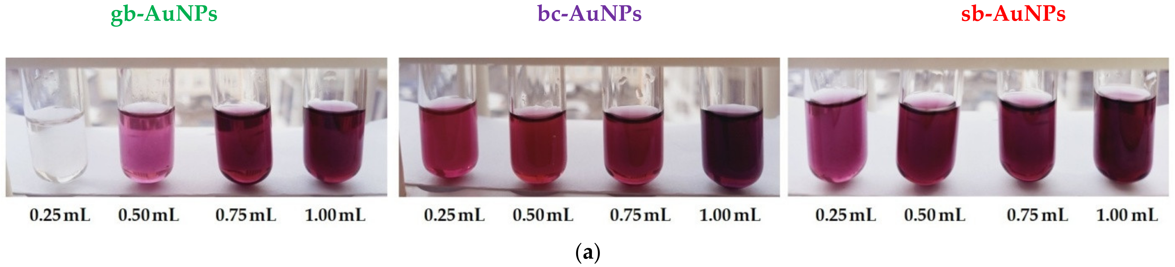
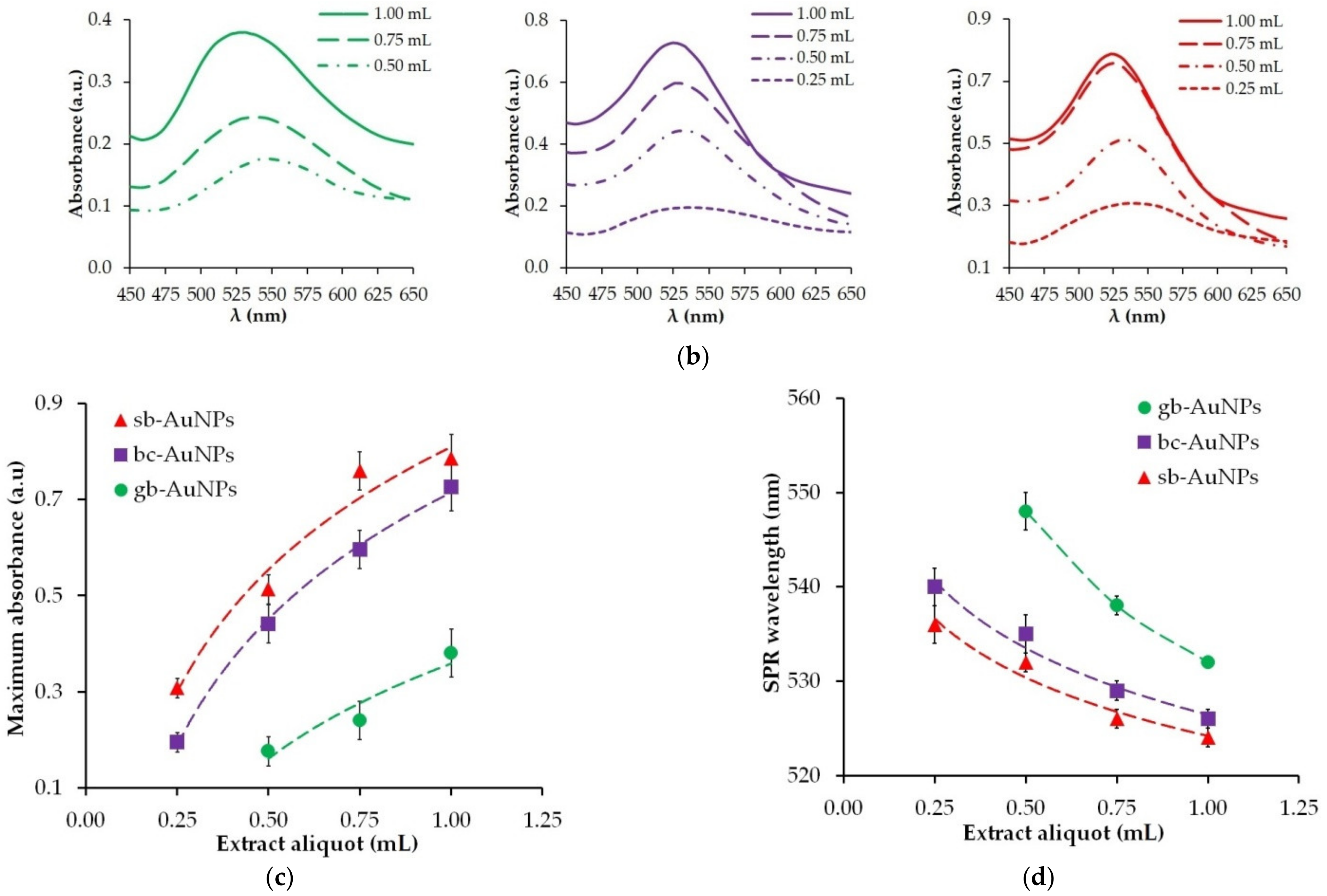
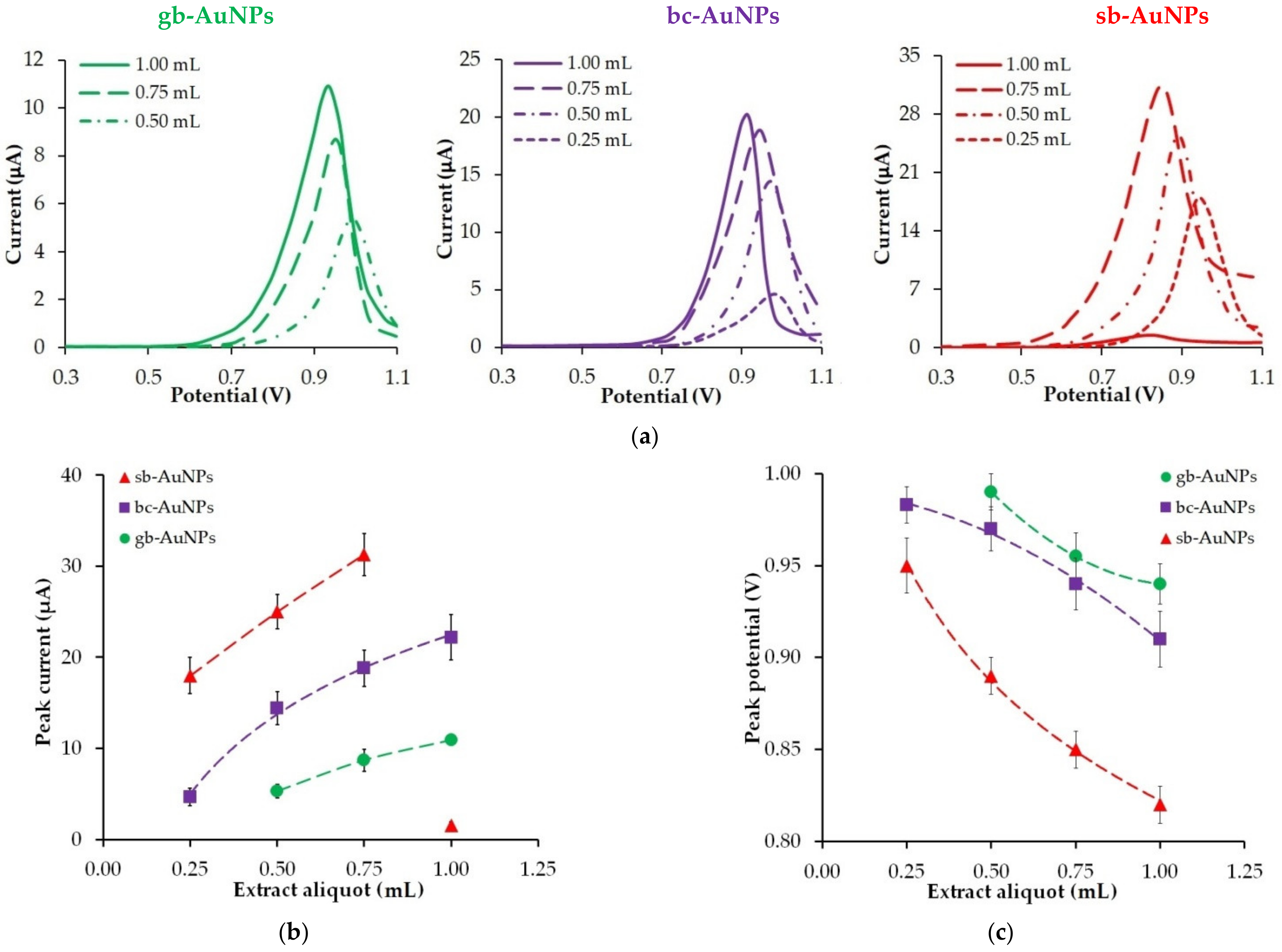
3.2. The Impact of Aliquots
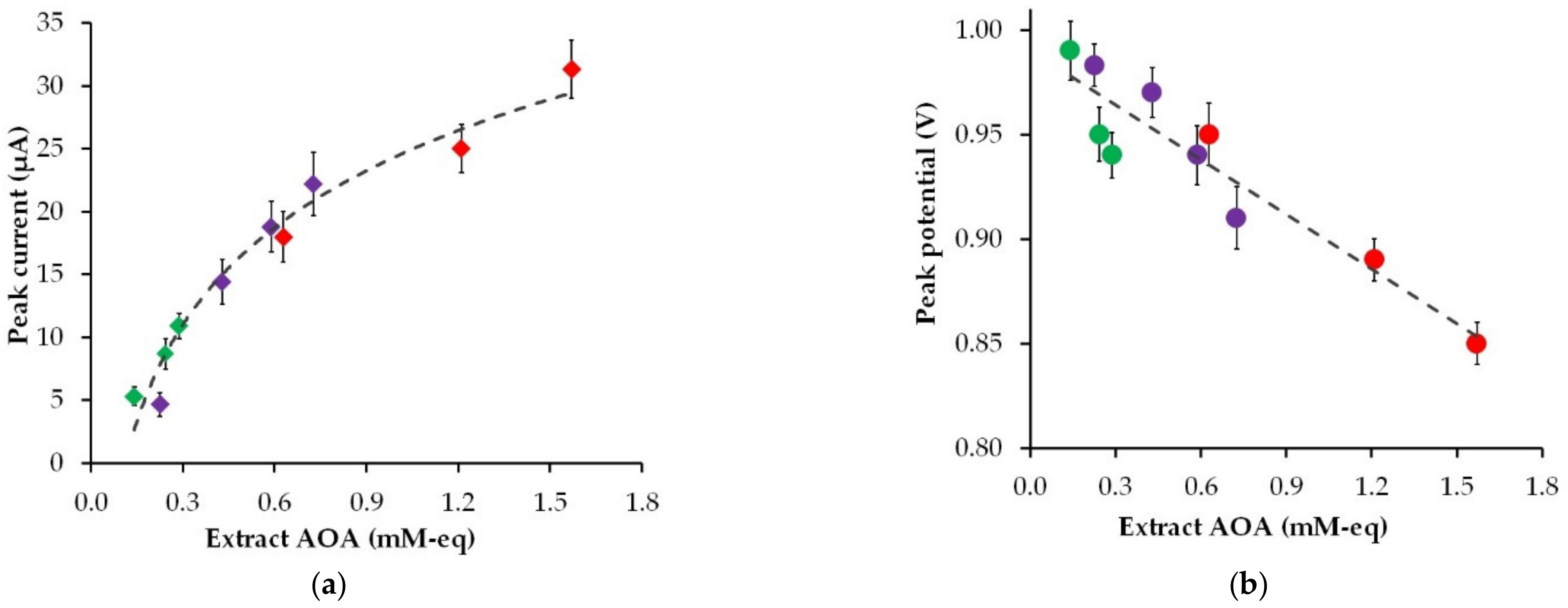
3.3. The Impact of Extract AOA
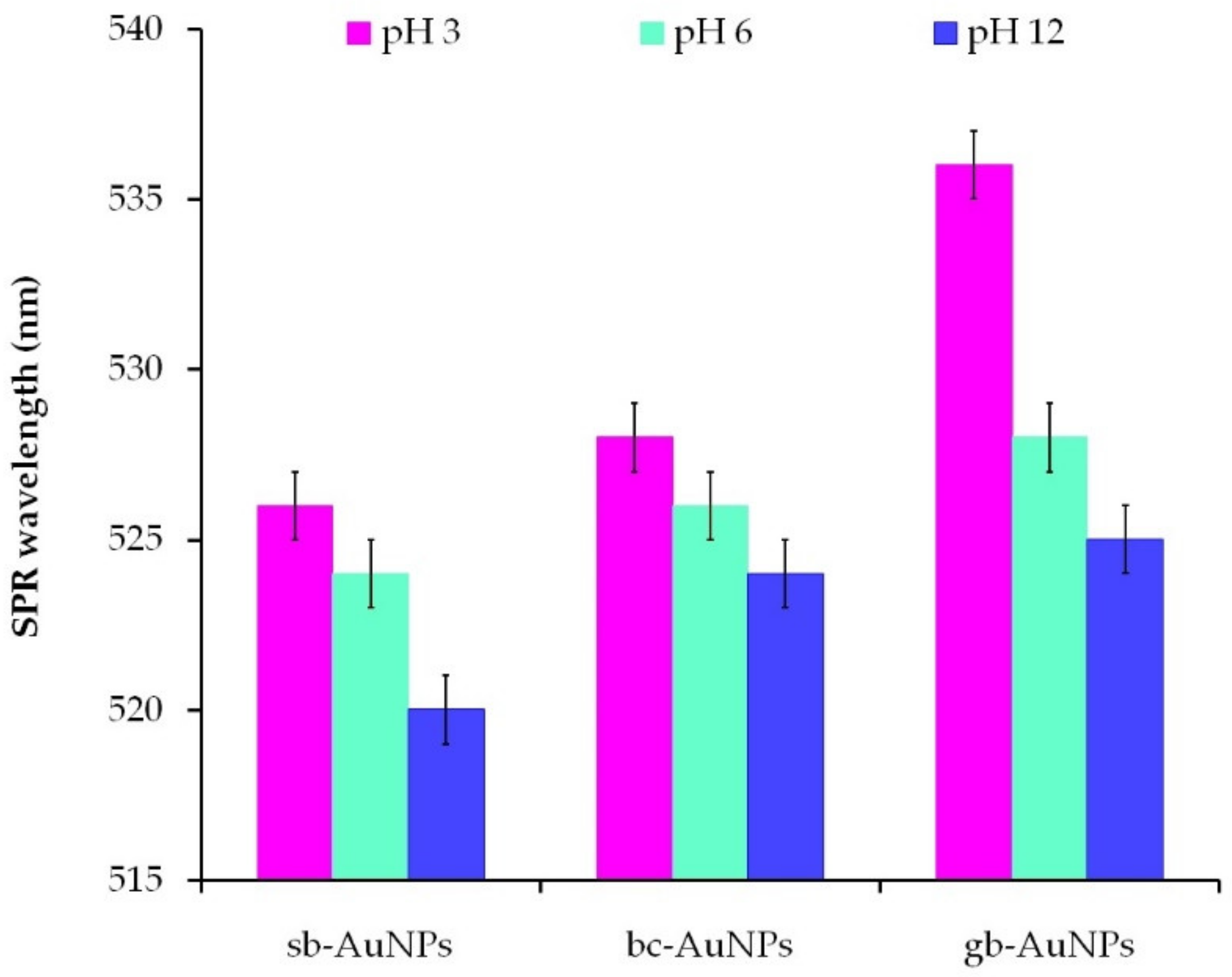
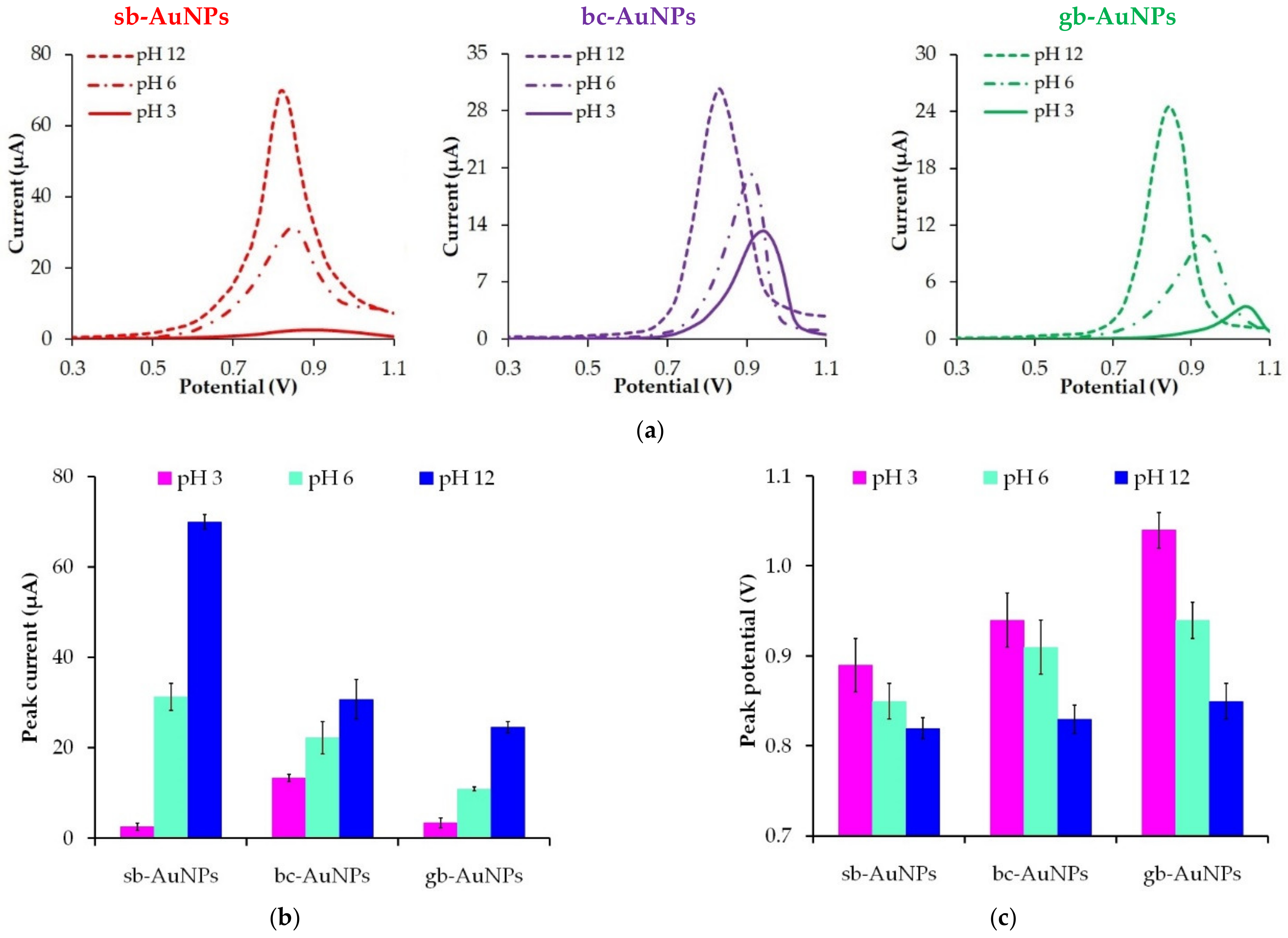
3.4. The Impact of Plant Extract pH
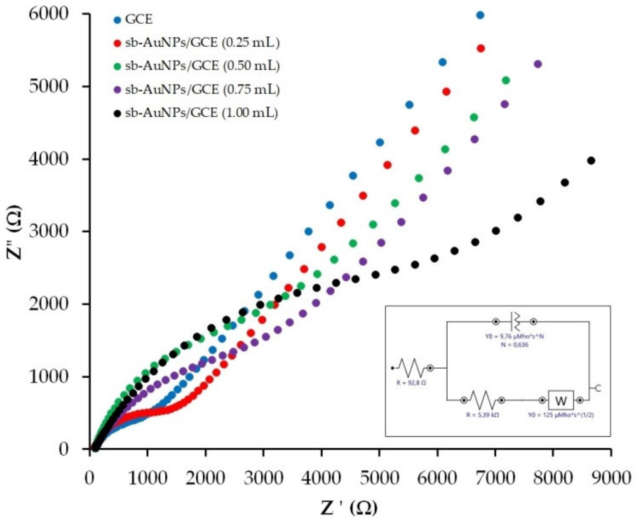
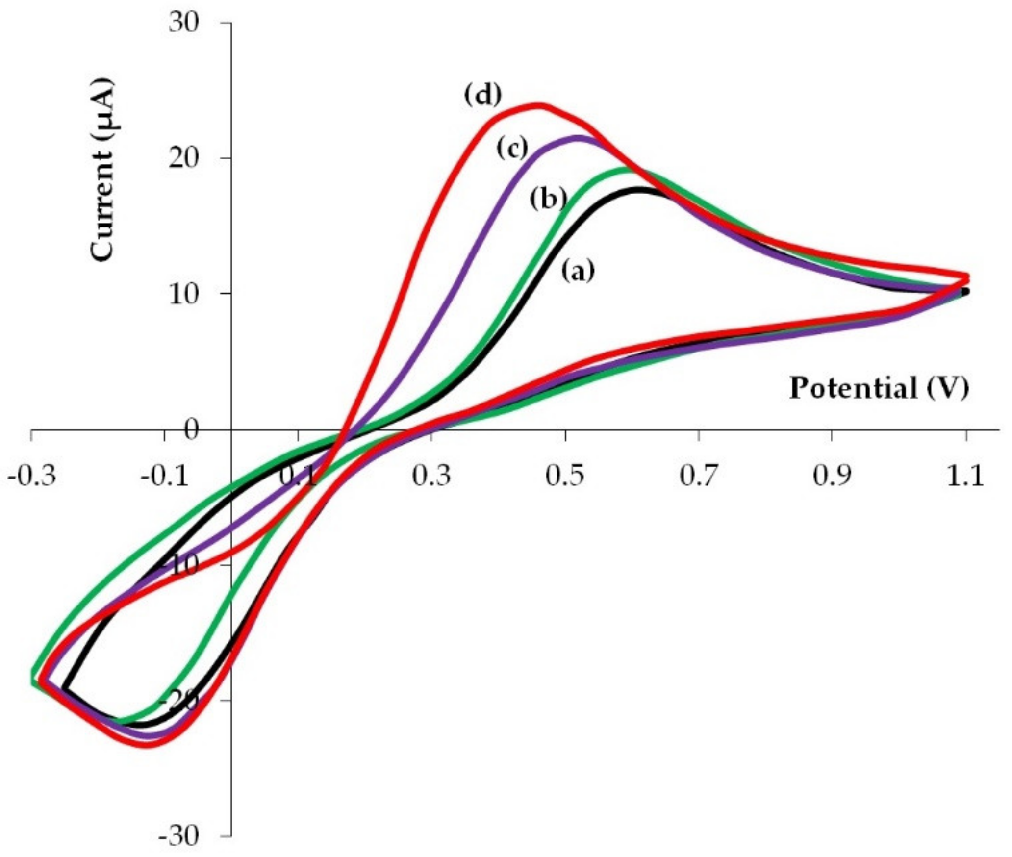
3.5. Electrochemical Behavior of K3[Fe(CN)6]/K4[Fe(CN)6] on Phyto-AuNPs
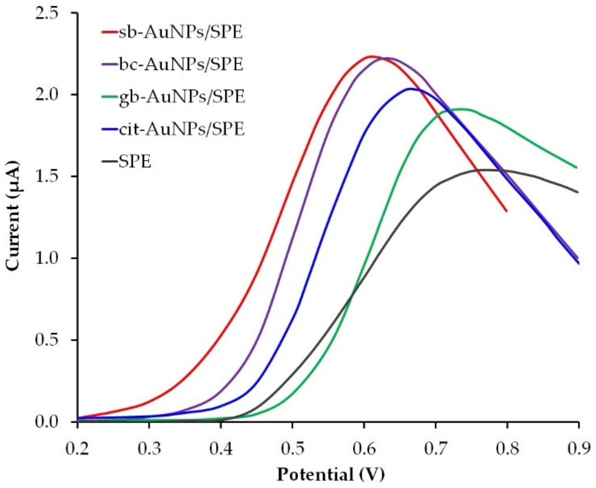
3.6. Analytical Application of sb-AuNPs
4. Conclusions
Supplementary Materials
Author Contributions
Funding
Institutional Review Board Statement
Informed Consent Statement
Data Availability Statement
Acknowledgments
Conflicts of Interest
References
- Brainina, K.; Stozhko, N.; Bukharinova, M.; Vikulova, E. Nanomaterials: Electrochemical properties and application in sensors. Phys. Sci. Rev. 2018, 3, 20188050. [Google Scholar] [CrossRef]
- Herrero-Calvillo, R.; Santovena-Uribe, A.; Esparza, R.; Rosas, G. A photocatalytic and electrochemical study of gold nanoparticles synthesized by a green approach. Mater. Res. Express 2020, 7, 015019. [Google Scholar] [CrossRef]
- Paiva-Santos, A.C.; Herdade, A.M.; Guerra, C.; Peixoto, D.; Pereira-Silva, M.; Zeinali, M.; Mascarenhas-Melo, F.; Paranhos, A.; Veiga, F. Plant-mediated green synthesis of metal-based nanoparticles for dermopharmaceutical and cosmetic applications. Int. J. Pharm. 2021, 597, 120311. [Google Scholar] [CrossRef] [PubMed]
- Jadoun, S.; Arif, R.; Jangid, N.K.; Meena, R.K. Green synthesis of nanoparticles using plant extracts: A review. Environ. Chem. Lett. 2021, 19, 355–374. [Google Scholar] [CrossRef]
- Lee, K.X.; Shameli, K.; Yew, Y.P.; Teow, S.-Y.; Jahangirian, H.; Rafiee-Moghaddam, R.; Webster, T.J. Recent developments in the facile bio-synthesis of gold nanoparticles (AuNPs) and their biomedical applications. Int. J. Nanomed. 2020, 15, 275–300. [Google Scholar] [CrossRef]
- Can, M. Green gold nanoparticles from plant-derived materials: An overview of the reaction synthesis types, conditions, and applications. Rev. Chem. Eng. 2020, 36, 859–877. [Google Scholar] [CrossRef]
- Hashmi, S.S.; Shah, M.; Muhammad, W.; Ahmad, A.; Ullah, M.A.; Nadeem, M.; Abbasi, B.H. Potentials of phyto-fabricated nanoparticles as ecofriendly agents for photocatalytic degradation of toxic dyes and waste water treatment, risk assessment and probable mechanism. J. Indian Chem. Soc. 2021, 98, 100019. [Google Scholar] [CrossRef]
- Boomi, P.; Ganesan, R.; Prabu Poorani, G.; Jegatheeswaran, S.; Balakumar, C.; Gurumallesh Prabu, H.; Anand, K.; Marimuthu Prabhu, N.; Jeyakanthan, J.; Saravanan, M. Phyto-engineered gold nanoparticles (AuNPs) with potential antibacterial, antioxidant, and wound healing activities under in vitro and in vivo conditions. Int. J. Nanomed. 2020, 15, 7553–7568. [Google Scholar] [CrossRef]
- El-Borady, O.M.; Fawzy, M.; Hosny, M. Antioxidant, anticancer and enhanced photocatalytic potentials of gold nanoparticles biosynthesized by common reed leaf extract. Appl. Nanosci. 2021; in press. [Google Scholar] [CrossRef]
- Dauthal, P.; Mukhopadhyay, M. Antioxidant activity of phytosynthesized biomatrix-loaded noble metallic nanoparticles. Chin. J. Chem. Eng. 2018, 26, 1200–1208. [Google Scholar] [CrossRef]
- Kalimuthu, K.; Cha, B.S.; Kim, S.; Park, K.S. Eco-friendly synthesis and biomedical applications of gold nanoparticles: A review. Microchem. J. 2020, 152, 104296. [Google Scholar] [CrossRef]
- Karthik, R.; Govindasamy, M.; Chen, S.-M.; Mani, V.; Lou, B.-S.; Devasenathipathy, R.; Hou, Y.-S.; Elangovan, A. Green synthesized gold nanoparticles decorated graphene oxide for sensitive determination of chloramphenicol in milk, powdered milk, honey and eye drops. J. Colloid Interface Sci. 2016, 475, 46–56. [Google Scholar] [CrossRef]
- Taib, S.H.M.; Shameli, K.; Nia, P.M.; Etesami, M.; Miyake, M.; Ali, R.R.; Abouzari-Lotf, E.; Izadiyan, Z. Electrooxidation of nitrite based on green synthesis of gold nanoparticles using Hibiscus sabdariffa leaves. J. Taiwan Inst. Chem. Eng. 2019, 95, 616–626. [Google Scholar] [CrossRef]
- Li, L.; Zhang, Z. Biosynthesis of gold nanoparticles using green alga Pithophora oedogonia with their electrochemical performance for determining carbendazim in soil. Int. J. Electrochem. Sci. 2016, 11, 4550–4559. [Google Scholar] [CrossRef]
- Karuppiah, C.; Palanisamy, S.; Chen, S.-M.; Emmanuel, R.; Muthupandi, K.; Prakash, P. Green synthesis of gold nanoparticles and its application for the trace level determination of painter’s colic. RSC Adv. 2015, 5, 16284–16291. [Google Scholar] [CrossRef]
- Devnani, H.; Satsangee, S.P. Green gold nanoparticle modified anthocyanin-based carbon paste electrode for voltammetric determination of heavy metals. Int. J. Environ. Sci. Technol. 2015, 12, 1269–1282. [Google Scholar] [CrossRef] [Green Version]
- Karthik, R.; Chen, S.-M.; Elangovan, A.; Muthukrishnan, P.; Shanmugam, R.; Lou, B.-S. Phyto mediated biogenic synthesis of gold nanoparticles using Cerasus serrulata and its utility in detecting hydrazine, microbial activity and DFT studies. J. Colloid Interface Sci. 2016, 468, 163–175. [Google Scholar] [CrossRef]
- Palanisamy, S.; Karuppiah, C.; Chen, S.-M.; Muthupandi, K.; Emmanuel, R.; Prakash, P.; Elshikh, M.S.; Ali, M.A.; Al-Hemaid, F.M.A. Selective and simultaneous determination of dihydroxybenzene isomers based on green synthesized gold nanoparticles decorated reduced graphene oxide. Electroanalysis 2015, 27, 1144–1151. [Google Scholar] [CrossRef]
- Brainina, K.Z.; Bukharinova, M.A.; Stozhko, N.Y.; Sokolkov, S.V.; Tarasov, A.V.; Vidrevich, M.B. Electrochemical sensor based on a carbon veil modified by phytosynthesized gold nanoparticles for determination of ascorbic acid. Sensors 2020, 20, 1800. [Google Scholar] [CrossRef] [Green Version]
- Liu, Y.; Pharr, M.; Salvatore, G.A. Lab-on-skin: A review of flexible and stretchable electronics for wearable health monitoring. ACS Nano 2017, 11, 9614–9635. [Google Scholar] [CrossRef]
- Son, D.; Bao, Z. Nanomaterials in skin-inspired electronics: Toward soft and robust skin-like electronic nanosystems. ACS Nano 2018, 12, 11731–11739. [Google Scholar] [CrossRef]
- Bariya, M.; Nyein, H.Y.Y.; Javey, A. Wearable sweat sensors. Nat. Electron. 2018, 1, 160–171. [Google Scholar] [CrossRef]
- Yang, J.C.; Mun, J.; Kwon, S.Y.; Park, S.; Bao, Z.; Park, S. Electronic skin: Recent progress and future prospects for skin-attachable devices for health monitoring, robotics, and prosthetics. Adv. Mater. 2019, 31, 1904765. [Google Scholar] [CrossRef] [Green Version]
- Brainina, K.Z.; Galperin, L.G.; Vikulova, E.V.; Stozhko, N.Y.; Murzakaev, A.M.; Timoshenkova, O.R.; Kotov, Y.A. Gold nanoparticles electrooxidation: Comparison of theory and experiment. J. Solid State Electrochem. 2011, 15, 1049–1056. [Google Scholar] [CrossRef]
- Brainina, K.Z.; Stozhko, N.Y.; Bukharinova, M.A.; Galperin, L.G.; Vidrevich, M.B.; Murzakaev, A.M. Mathematical modeling and experimental data of the oxidation of ascorbic acid on electrodes modified by nanoparticles. J. Solid State Electrochem. 2016, 20, 2323–2330. [Google Scholar] [CrossRef]
- Ahmad, T.; Iqbal, J.; Bustam, M.A.; Irfan, M.; Asghar, H.M.A. A critical review on phytosynthesis of gold nanoparticles: Issues, challenges and future perspectives. J. Clean. Prod. 2021, 309, 127460. [Google Scholar] [CrossRef]
- Ahmad, T.; Bustam, M.A.; Irfan, M.; Moniruzzaman, M.; Asghar, H.M.A.; Bhattacharjee, S. Effect of volume of gold chloroauric acid on size, shape and stability of biosynthesized AuNPs using aqueous Elaeis guineensis (Oil Palm) leaves extract. Int. J. Automot. Mech. Eng. 2018, 15, 5135–5145. [Google Scholar] [CrossRef]
- Salandari Rabori, M.; Noroozi Karimabad, M.; Reza Hajizadeh, M. Facile, low-cost and rapid phytosynthesis of stable and eco-friendly gold nanoparticles using green walnut shell and study of their anticancer potential. WCRJ World Cancer Res. J. 2021, 8, e2037. [Google Scholar] [CrossRef]
- Anbu, P.; Gopinath, S.C.B.; Jayanthi, S. Synthesis of gold nanoparticles using Platycodon grandiflorum extract and its antipathogenic activity under optimal conditions. Nanomater. Nanotechnol. 2020, 10, 1–9. [Google Scholar] [CrossRef]
- Stozhko, N.Y.; Bukharinova, M.A.; Khamzina, E.I.; Tarasov, A.V.; Vidrevich, M.B.; Brainina, K.Z. The effect of the antioxidant activity of plant extracts on the properties of gold nanoparticles. Nanomaterials 2019, 9, 1655. [Google Scholar] [CrossRef] [Green Version]
- Brainina, K.; Stozhko, N.; Bukharinova, M.; Khamzina, E.; Vidrevich, M. Potentiometric method of plant microsuspensions antioxidant activity determination. Food Chem. 2019, 278, 653–658. [Google Scholar] [CrossRef] [PubMed]
- Turkevich, J.; Stevenson, P.C.; Hillier, J. A Study of the nucleation and growth processes in the synthesis of colloidal gold. Discuss. Faraday Soc. 1951, 11, 55–75. [Google Scholar] [CrossRef]
- Marken, F.; Neudeck, A.; Bond, A.M. Cyclic Voltammetry. In Electroanalytical Methods, 2nd ed.; Scholz, F., Ed.; Springer: Berlin/Heidelberg, Germany, 2010; Chapter II.1; pp. 57–106. [Google Scholar] [CrossRef]
- Haiss, W.; Thanh, N.T.K.; Aveyard, J.; Fernig, D.G. Determination of size and concentration of gold nanoparticles from UV-Vis spectra. Anal. Chem. 2007, 79, 4215–4221. [Google Scholar] [CrossRef] [PubMed]
- Mainali, B.P.; Pattadar, D.K.; Zamborini, F.P. Reverse size-dependent electrooxidation of gold nanoparticles coated with alkanethiol self-assembled monolayers. J. Phys. Chem. C 2021, 125, 2719–2728. [Google Scholar] [CrossRef]
- Vijayaraghavan, K.; Ashokkumar, T. Plant-mediated biosynthesis of metallic nanoparticles: A review of literature, factors affecting synthesis, characterization techniques and applications. J. Environ. Chem. Eng. 2017, 5, 4866–4883. [Google Scholar] [CrossRef]
- Mittal, A.K.; Chisti, Y.; Banerjee, U.C. Synthesis of metallic nanoparticles using plant extracts. Biotechnol. Adv. 2013, 31, 346–356. [Google Scholar] [CrossRef]
- Guo, Q.; Guo, Q.; Yuan, J.; Zeng, J. Biosynthesis of gold nanoparticles using a kind of flavonol: Dihydromyricetin. Colloids Surf. A 2014, 441, 127–132. [Google Scholar] [CrossRef]
- Shafiqa, A.R.; Aziz, A.A.; Mehrdel, B. Nanoparticle optical properties: Size dependence of a single gold spherical nanoparticle. J. Phys. Conf. Ser. 2018, 1083, 012040. [Google Scholar] [CrossRef] [Green Version]
- Taleb, A.; Yanpeng, X.; Dubot, P. Self-organized gold nanoparticles modified HOPG electrodes: Electrochemical stability and its use for electrochemical nanosensing applications. Appl. Surf. Sci. 2017, 420, 110–117. [Google Scholar] [CrossRef] [Green Version]
- Taleb, A.; Silly, F.; Gusev, A.O.; Charra, F.; Pileni, M.-P. Electron transport properties of nanocrystals: Isolated, and “supra”-crystalline phases. Adv. Mater. 2000, 12, 633–637. [Google Scholar] [CrossRef]
- Yang, G.; Tan, L.; Yang, Y.; Chen, S.; Liu, G.-Y. Single electron tunneling and manipulation of nanoparticles on surfaces at room temperature. Surf. Sci. 2005, 589, 129–138. [Google Scholar] [CrossRef]
- Oueslati, M.H.; Tahar, L.B.; Harrath, A.H. Synthesis of ultra-small gold nanoparticles by polyphenol extracted from Salvia officinalis and efficiency for catalytic reduction of p-nitrophenol and methylene blue. Green Chem. Lett. Rev. 2020, 13, 18–26. [Google Scholar] [CrossRef] [Green Version]
- Dutta, P.P.; Bordoloi, M.; Gogoi, K.; Roy, S.; Narzary, B.; Bhattacharyya, D.R.; Mohapatra, P.K.; Mazumder, B. Antimalarial silver and gold nanoparticles: Green synthesis, characterization and in vitro study. Biomed. Pharmacother. 2017, 91, 567–580. [Google Scholar] [CrossRef]
- Aromal, A.S.; Philip, D. Green synthesis of gold nanoparticles using Trigonella foenum-graecum and its size-dependent catalytic activity. Spectrochim. Acta Part A 2012, 97, 1–5. [Google Scholar] [CrossRef]
- Pinto, R.J.B.; Lucas, J.M.F.; Morais, M.P.; Santos, S.A.O.; Silvestre, A.J.D.; Marques, P.A.A.P.; Freire, C.S.R. Demystifying the morphology and size control on the biosynthesis of gold nanoparticles using Eucalyptus globulus bark extract. Ind. Crops Prod. 2017, 105, 83–92. [Google Scholar] [CrossRef]
- Kalimuthu, P.; John, S.A. Size dependent electrocatalytic activity of gold nanoparticles immobilized onto three dimensional sol–gel network. J. Electroanal. Chem. 2008, 617, 164–170. [Google Scholar] [CrossRef]
- Wang, K.; Jiang, H.; Li, W.; Qiang, M.; Dong, T.; Li, H. Role of vitamin C in skin diseases. Front. Physiol. 2018, 9, 819. [Google Scholar] [CrossRef] [Green Version]
- Ridi, R.E.; Tallima, H. Physiological functions and pathogenic potential of uric acid: A review. J. Adv. Res. 2017, 8, 487–493. [Google Scholar] [CrossRef]
- Yang, Y.; Song, Y.; Bo, X.; Min, J.; Pak, O.S.; Zhu, L.; Wang, M.; Tu, J.; Kogan, A.; Zhang, H.; et al. A laser-engraved wearable sensor for sensitive detection of uric acid and tyrosine in sweat. Nat. Biotechnol. 2020, 38, 217–224. [Google Scholar] [CrossRef] [Green Version]
- Sempionatto, J.R.; Khorshed, A.A.; Ahmed, A.; Silva, A.N.D.L.E.; Barfidokht, A.; Yin, L.; Goud, K.Y.; Mohamed, M.A.; Bailey, E.; May, J.; et al. Epidermal enzymatic biosensors for sweat vitamin C: Toward personalized nutrition. ACS Sens. 2020, 5, 1804–1813. [Google Scholar] [CrossRef]


| Phyto-AuNPs | Peak Potential, V | Peak Current, μA | ||
|---|---|---|---|---|
| Without Purification | After Purification | Without Purification | After Purification | |
| sb-AuNPs | 0.85 ± 0.01 | 0.85 ± 0.02 | 7.3 ± 0.9 | 31.3 ± 3.0 |
| bc-AuNPs | 0.95 ± 0.03 | 0.94 ± 0.02 | 4.4 ± 0.6 | 18.8 ± 3.0 |
| gb-AuNPs | 1.00 ± 0.05 | 0.95 ± 0.03 | 2.0 ± 0.1 | 8.7 ± 0.7 |
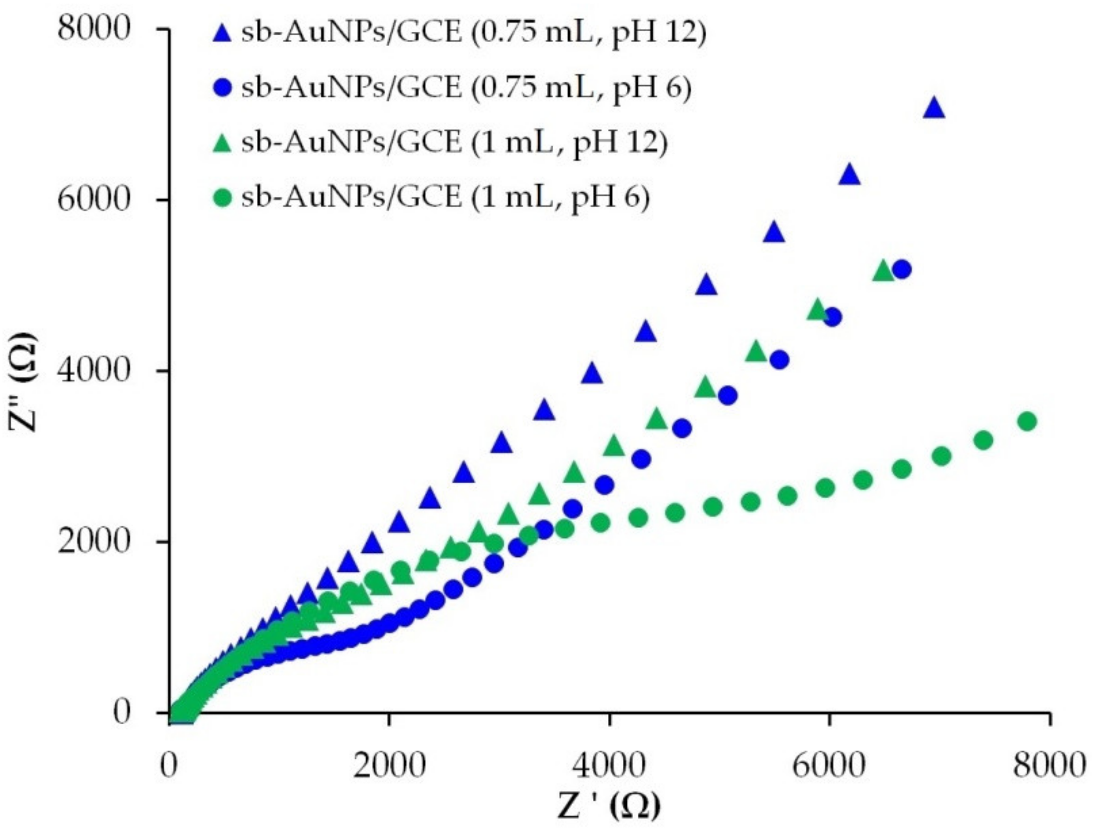
| Electrode | Vextr Used in the sb-AuNPs Synthesis, mL | Rs, Ω | Rct, kΩ | Q, μMho | n | W, μMho |
|---|---|---|---|---|---|---|
| GCE | 0 | 99.0 ± 4.6 | 0.87 ± 0.11 | 10.2 ± 1.1 | 0.65 ± 0.03 | 148 ± 9 |
| sb-AuNPs/GCE | 0.25 | 95.5 ± 4.5 | 1.60 ± 0.20 | 4.7 ± 0.6 | 0.71 ± 0.02 | 160 ± 13 |
| 0.50 | 111.0 ± 5.0 | 3.29 ± 0.21 | 21.2 ± 2.1 | 0.77 ± 0.05 | 176 ± 14 | |
| 0.75 | 105.7 ± 9.4 | 3.44 ± 0.18 | 18.7 ± 2.2 | 0.63 ± 0.04 | 158 ± 14 | |
| 1.0 | 95 ± 12 | 8.4 ± 1.2 | 7.2 ± 1.5 | 0.66 ± 0.10 | 182 ± 13 |
| pH of Strawberry Leaf Extract | DLS | UV–Vis | |
|---|---|---|---|
| ζ-Potential, mV | dhd, nm | d, nm | |
| 3 | −28 ± 1 | 38 ± 1 | 12 ± 1 |
| 6 | −27 ± 1 | 30 ± 1 | 10 ± 1 |
| 12 | −42 ± 4 | 23 ± 1 | 6 ± 1 |
| Electrode | d, nm | Ea, V | Ec, V | ∆E, V | Ia, μA | Ic, μA | Ia/Ic |
|---|---|---|---|---|---|---|---|
| sb-AuNPs/GCE | 6 ± 1 | 0.46 ± 0.04 | −0.13 ± 0.02 | 0.59 ± 0.02 | 23.9 ± 0.2 | −23.4 ± 0.2 | 1.0 ± 0.1 |
| bc-AuNPs/GCE | 6 ± 1 | 0.52 ± 0.05 | −0.12 ± 0.02 | 0.64 ± 0.02 | 21.6 ± 0.8 | −22.7 ± 0.7 | 1.0 ± 0.2 |
| gb-AuNPs/GCE | 9 ± 1 | 0.59 ± 0.01 | −0.18 ± 0.01 | 0.77 ± 0.03 | 19.2 ± 0.1 | −21.6 ± 0.4 | 0.9 ± 0.1 |
| GCE | - | 0.63 ± 0.13 | −0.16 ± 0.05 | 0.79 ± 0.17 | 18.4 ± 0.1 | −21.3 ± 0.4 | 0.8 ± 0.1 |
| Parameter | Uric Acid | Ascorbic Acid [19] | ||
|---|---|---|---|---|
| sb-AuNPs/SPE | cit-AuNPs/SPE | Phyto-AuNPs/CVE | cit-AuNPs/CVE | |
| Limit of detection, μM | 0.16 | 1.03 | 0.05 | 0.20 |
| Limit of quantification, μM | 0.49 | 3.13 | 0.15 | 0.60 |
| Linear range, μM | 0.1−0.98, 0.98−190 | 0.2–190 | 1–10, 10–5750 | 1–10, 10–11,700 |
| Sensitivity, μA/μM | 0.617, 0.169 | 0.128 | 0.130, 0.050 | 0.077, 0.025 |
| Sr of response of minimal concentration, % | 3 | 4 | 1.4 | 3.6 |
Publisher’s Note: MDPI stays neutral with regard to jurisdictional claims in published maps and institutional affiliations. |
© 2021 by the authors. Licensee MDPI, Basel, Switzerland. This article is an open access article distributed under the terms and conditions of the Creative Commons Attribution (CC BY) license (https://creativecommons.org/licenses/by/4.0/).
Share and Cite
Stozhko, N.Y.; Bukharinova, M.A.; Khamzina, E.I.; Tarasov, A.V. Electrochemical Properties of Phytosynthesized Gold Nanoparticles for Electrosensing. Sensors 2022, 22, 311. https://doi.org/10.3390/s22010311
Stozhko NY, Bukharinova MA, Khamzina EI, Tarasov AV. Electrochemical Properties of Phytosynthesized Gold Nanoparticles for Electrosensing. Sensors. 2022; 22(1):311. https://doi.org/10.3390/s22010311
Chicago/Turabian StyleStozhko, Natalia Yu., Maria A. Bukharinova, Ekaterina I. Khamzina, and Aleksey V. Tarasov. 2022. "Electrochemical Properties of Phytosynthesized Gold Nanoparticles for Electrosensing" Sensors 22, no. 1: 311. https://doi.org/10.3390/s22010311
APA StyleStozhko, N. Y., Bukharinova, M. A., Khamzina, E. I., & Tarasov, A. V. (2022). Electrochemical Properties of Phytosynthesized Gold Nanoparticles for Electrosensing. Sensors, 22(1), 311. https://doi.org/10.3390/s22010311