
Negative Index Metamaterial Lens for Subwavelength Microwave Detection

 ,
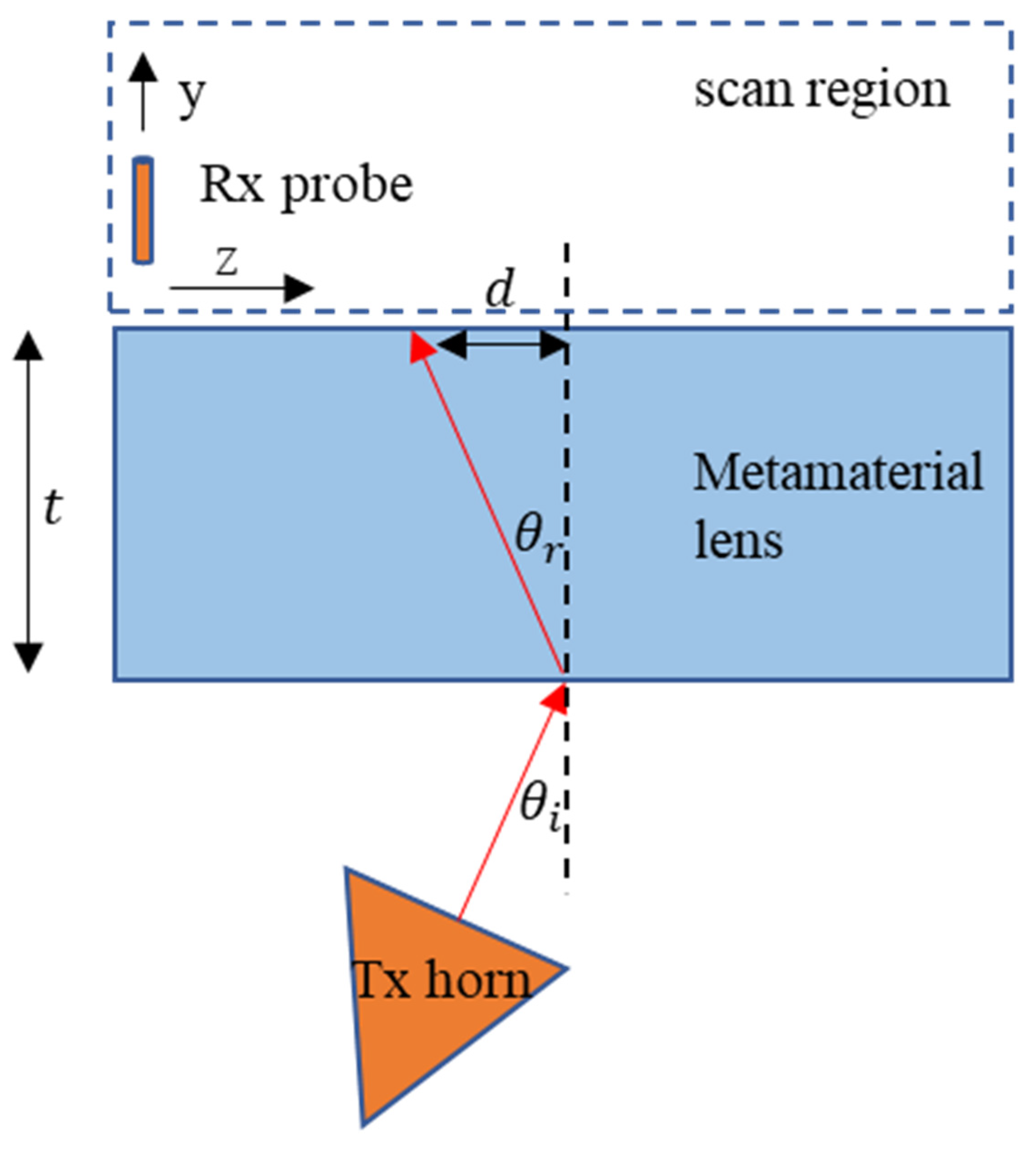
,  ,
, 
Abstract
1. Introduction
2. Theory
3. Simulation
3.1. Scattering Parameters
3.2. Electromagnetic Parameter Retrieval
4. Experiment
4.1. Transmission Characteristics
4.2. Left-Handed Characteristics
4.2.1. Negative Refraction
4.2.2. Subwavelength Focusing
4.2.3. Microwave NDE
5. Discussion
Author Contributions
Funding
Institutional Review Board Statement
Informed Consent Statement
Data Availability Statement
Acknowledgments
Conflicts of Interest
Appendix A
References
- Veselago, V.G. The electrodynamics of substances with simultaneously negative values of \epsilon and μ. Sov. Phys. Uspekhi 1968, 10, 509–514. [Google Scholar] [CrossRef]
- Smith, D.R.; Padilla, W.; Vier, D.C.; Nemat-Nasser, S.C.; Schultz, S. Composite Medium with Simultaneously Negative Permeability and Permittivity. Phys. Rev. Lett. 2000, 84, 4184–4187. [Google Scholar] [CrossRef]
- Shelby, R.A.; Smith, D.R.; Schultz, S. Experimental Verification of a Negative Index of Refraction. Science 2001, 292, 77–79. [Google Scholar] [CrossRef] [PubMed]
- Parazzoli, C.G.; Greegor, R.B.; Li, K.; Koltenbah, B.E.C.; Tanielian, M. Experimental Verification and Simulation of Negative Index of Refraction Using Snell’s Law. Phys. Rev. Lett. 2003, 90, 107401. [Google Scholar] [CrossRef]
- Aydin, K.; Guven, K.; Soukoulis, C.M.; Ozbay, E. Observation of negative refraction and negative phase velocity in left-handed metamaterials. Appl. Phys. Lett. 2005, 86, 124102. [Google Scholar] [CrossRef]
- Valanju, P.M.; Walser, R.M.; Valanju, A.P. Wave Refraction in Negative-Index Media: Always Positive and Very Inhomogeneous. Phys. Rev. Lett. 2002, 88, 187401. [Google Scholar] [CrossRef]
- Munk, B.A. Metamaterials: Critique and Alternatives; Wiley: Hoboken, NJ, USA, 2009. [Google Scholar]
- Houck, A.A.; Brock, J.B.; Chuang, I.L. Experimental Observations of a Left-Handed Material That Obeys Snell’s Law. Phys. Rev. Lett. 2003, 90, 137401. [Google Scholar] [CrossRef]
- Ziolkowski, R. Design, fabrication, and testing of double negative metamaterials. IEEE Trans. Antennas Propag. 2003, 51, 1516–1529. [Google Scholar] [CrossRef]
- Aydin, K.; Guven, K.; Kafesaki, M.; Zhang, L.; Soukoulis, C.M.; Ozbay, E. Experimental observation of true left-handed transmission peaks in metamaterials. Opt. Lett. 2004, 29, 2623–2625. [Google Scholar] [CrossRef]
- Alù, A. First-principles homogenization theory for periodic metamaterials. Phys. Rev. B Condens. Matter Mater. Phys. 2011, 84, 075153. [Google Scholar] [CrossRef]
- Pendry, J. Negative Refraction Makes a Perfect Lens. Phys. Rev. Lett. 2000, 85, 3966–3969. [Google Scholar] [CrossRef]
- Grbic, A.; Eleftheriades, G.V. Overcoming the Diffraction Limit with a Planar Left-Handed Transmission-Line Lens. Phys. Rev. Lett. 2004, 92, 117403. [Google Scholar] [CrossRef] [PubMed]
- Aydin, K.; Bulu, I.; Ozbay, E. Subwavelength resolution with a negative-index metamaterial superlens. Appl. Phys. Lett. 2007, 90, 254102. [Google Scholar] [CrossRef]
- Ozbay, E.; Li, Z.; Aydin, K. Super-resolution imaging by one-dimensional, microwave left-handed metamaterials with an effective negative index. J. Phys. Condens. Matter 2008, 20, 304216. [Google Scholar] [CrossRef]
- Roy, T.; Rogers, E.; Zheludev, N. Sub-wavelength focusing meta-lens. Opt. Express 2013, 21, 7577–7582. [Google Scholar] [CrossRef]
- Haxha, S.; Abdelmalek, F.; Ouerghi, F.; Charlton, M.D.B.; Aggoun, A.; Fang, X. Metamaterial Superlenses Operating at Visible Wavelength for Imaging Applications. Sci. Rep. 2018, 8, 16119. [Google Scholar] [CrossRef]
- Mukherjee, S.; Tamburrino, A.; Haq, M.; Udpa, S.; Udpa, L. Far field microwave NDE of composite structures using time reversal mirror. NDT E Int. 2018, 93, 7–17. [Google Scholar] [CrossRef]
- Savin, A.; Bruma, A.; Steigmann, R.; Iftimie, N.; Faktorova, D. Enhancement of Spatial Resolution Using a Metamaterial Sensor in Nondestructive Evaluation. Appl. Sci. 2015, 5, 1412–1430. [Google Scholar] [CrossRef]
- Mukherjee, S.; Shi, X.; Udpa, L.; Udpa, S.; Deng, Y.; Chahal, P.; Shi, X. Design of a Split-Ring Resonator Sensor for Near-Field Microwave Imaging. IEEE Sens. J. 2018, 18, 7066–7076. [Google Scholar] [CrossRef]
- Zhang, Y.; Zhao, J.; Cao, J.; Mao, B. Microwave Metamaterial Absorber for Non-Destructive Sensing Applications of Grain. Sensors 2018, 18, 1912. [Google Scholar] [CrossRef]
- O’Hara, J.F.; Singh, R.; Brener, I.; Smirnova, E.; Han, J.; Taylor, A.J.; Zhang, W. Thin-film sensing with planar terahertz metamaterials: Sensitivity and limitations. Opt. Express 2008, 16, 1786–1795. [Google Scholar] [CrossRef]
- Leggio, L.; Dadrasnia, E.; de Varona, O. Microwave Focusing within Arbitrary Refractive Index Media Using Left-Handed Metamaterial Lenses. Prog. Electromagn. Res. M 2016, 45, 51–58. [Google Scholar] [CrossRef]
- Chen, J.J.; Grzegorczyk, T.M.; Wu, B.-I.; Kong, J.A. Limitation of FDTD in simulation of a perfect lens imaging system. Opt. Express 2005, 13, 10840–10845. [Google Scholar] [CrossRef]
- Lu, J.; Grzegorczyk, T.M.; Wu, B.-I.; Pacheco, J.; Chen, M.; Kong, J.A. Effect of poles on subwavelength focusing by an LHM slab. Microw. Opt. Technol. Lett. 2005, 45, 49–53. [Google Scholar] [CrossRef]
- Tassin, P.; Veretennicoff, I.; Van Der Sande, G. Veselago’s lens consisting of left-handed materials with arbitrary index of refraction. Opt. Commun. 2006, 264, 130–134. [Google Scholar] [CrossRef]
- Shreiber, D.; Gupta, M.; Cravey, R. Microwave nondestructive evaluation of dielectric materials with a metamaterial lens. Sens. Actuators A Phys. 2008, 144, 48–55. [Google Scholar] [CrossRef]
- Tao, Y.; Yang, E.; Wang, G. Left-handed metamaterial lens applicator with built-in cooling feature for superficial tumor hyperthermia. Appl. Comput. Electromagn. Soc. J. 2017, 32, 1029–1034. [Google Scholar]
- Zhang, S.; Yin, L.; Fang, N. Focusing Ultrasound with an Acoustic Metamaterial Network. Phys. Rev. Lett. 2009, 102, 194301. [Google Scholar] [CrossRef]
- Amireddy, K.K.; Rajagopal, P.; Balasubramaniam, K.; Nadu, T. Holey-structured metalens for deep sub-wavelength resolution of delamination in layered materials using ultrasound. In Proceedings of the 15th Asia Pacific Conference for Non-Destructive Testing (APCNDT2017), Singapore, 13–17 November 2017; pp. 1–5. [Google Scholar]
- Walker, E.L.; Jin, Y.; Reyes, D.; Neogi, A. Sub-wavelength lateral detection of tissue-approximating masses using an ultrasonic metamaterial lens. Nat. Commun. 2020, 11, 5967. [Google Scholar] [CrossRef]
- Zaman, A.-U.-Z.; Song, K.; Lee, D.-G.; Hur, S. A novel approach to Fabry–Pérot-resonance-based lens and demonstrating deep-subwavelength imaging. Sci. Rep. 2020, 10, 10769. [Google Scholar] [CrossRef]
- Mukherjee, S.; Su, Z.; Udpa, L.; Udpa, S.; Tamburrino, A. Enhancement of Microwave Imaging Using a Metamaterial Lens. IEEE Sens. J. 2019, 19, 4962–4971. [Google Scholar] [CrossRef]
- Datta, S.; Shi, X.; Mukherjee, S.; Deng, Y.; Udpa, L. Model-Based Study of a Metamaterial Lens for Nondestructive Evaluation of Composites. J. Nondestruct. Eval. Diagn. Progn. Eng. Syst. 2020, 3, 041001. [Google Scholar] [CrossRef]
- Pendry, J.; Holden, A.; Robbins, D.; Stewart, W. Magnetism from conductors and enhanced nonlinear phenomena. IEEE Trans. Microw. Theory Tech. 1999, 47, 2075–2084. [Google Scholar] [CrossRef]
- Pendry, J.B.; Holden, A.J.; Stewart, W.J.; Youngs, I. Extremely Low Frequency Plasmons in Metallic Mesostructures. Phys. Rev. Lett. 1996, 76, 4773–4776. [Google Scholar] [CrossRef]
- Smith, D.R.; Kroll, N. Negative Refractive Index in Left-Handed Materials. Phys. Rev. Lett. 2000, 85, 2933–2936. [Google Scholar] [CrossRef]
- Smith, D.R.; Schurig, D.; Rosenbluth, M.; Schultz, S.; Ramakrishna, S.A.; Pendry, J.B. Limitations on subdiffraction imaging with a negative refractive index slab. Appl. Phys. Lett. 2003, 82, 1506–1508. [Google Scholar] [CrossRef]
- Aydin, K.; Bulu, I.; Ozbay, E. Focusing of electromagnetic waves by a left-handed metamaterial flat lens. Opt. Express 2005, 13, 8753–8759. [Google Scholar] [CrossRef] [PubMed]
- Wilson, P.; Ma, M.; Adams, J. Techniques for measuring the electromagnetic shielding effectiveness of materials. I. Far-field source simulation. IEEE Trans. Electromagn. Compat. 1988, 30, 239–250. [Google Scholar] [CrossRef]
- Nicolson, A.M.; Ross, G.F. Measurement of the Intrinsic Properties of Materials by Time-Domain Techniques. IEEE Trans. Instrum. Meas. 1970, 19, 377–382. [Google Scholar] [CrossRef]
- Weir, W. Automatic measurement of complex dielectric constant and permeability at microwave frequencies. Proc. IEEE 1974, 62, 33–36. [Google Scholar] [CrossRef]
- Chen, X.; Grzegorczyk, T.M.; Wu, B.-I.; Pacheco, J.J.; Kong, J.A. Robust method to retrieve the constitutive effective parameters of metamaterials. Phys. Rev. E Stat. Phys. Plasmas Fluids Relat. Interdiscip. Top. 2004, 70, 016608. [Google Scholar] [CrossRef]
- Arslanagic, S.; Hansen, T.V.; Mortensen, N.A.; Gregersen, A.H.; Sigmund, O.; Ziolkowski, R.; Breinbjerg, O. A Review of the Scattering-Parameter Extraction Method with Clarification of Ambiguity Issues in Relation to Metamaterial Homogenization. IEEE Antennas Propag. Mag. 2013, 55, 91–106. [Google Scholar] [CrossRef]
- Rothwell, E.J.; Frasch, J.L.; Ellison, S.M.; Chahal, P.; Ouedraogo, R.O. Analysis of the Nicolson-Ross-Weir Method for Characterizing the Electromagnetic Properties of Engineered Materials. Prog. Electromagn. Res. 2016, 157, 31–47. [Google Scholar] [CrossRef]


Publisher’s Note: MDPI stays neutral with regard to jurisdictional claims in published maps and institutional affiliations. |
© 2021 by the authors. Licensee MDPI, Basel, Switzerland. This article is an open access article distributed under the terms and conditions of the Creative Commons Attribution (CC BY) license (https://creativecommons.org/licenses/by/4.0/).
Share and Cite
Datta, S.; Mukherjee, S.; Shi, X.; Haq, M.; Deng, Y.; Udpa, L.; Rothwell, E. Negative Index Metamaterial Lens for Subwavelength Microwave Detection. Sensors 2021, 21, 4782. https://doi.org/10.3390/s21144782
Datta S, Mukherjee S, Shi X, Haq M, Deng Y, Udpa L, Rothwell E. Negative Index Metamaterial Lens for Subwavelength Microwave Detection. Sensors. 2021; 21(14):4782. https://doi.org/10.3390/s21144782
Chicago/Turabian StyleDatta, Srijan, Saptarshi Mukherjee, Xiaodong Shi, Mahmood Haq, Yiming Deng, Lalita Udpa, and Edward Rothwell. 2021. "Negative Index Metamaterial Lens for Subwavelength Microwave Detection" Sensors 21, no. 14: 4782. https://doi.org/10.3390/s21144782
APA StyleDatta, S., Mukherjee, S., Shi, X., Haq, M., Deng, Y., Udpa, L., & Rothwell, E. (2021). Negative Index Metamaterial Lens for Subwavelength Microwave Detection. Sensors, 21(14), 4782. https://doi.org/10.3390/s21144782