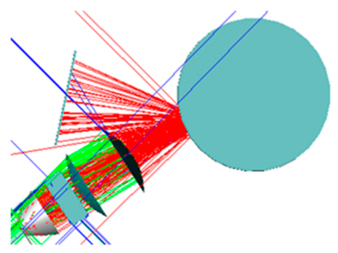
Smooth Surface Visual Imaging Method for Eliminating High Reflection Disturbance

Abstract
1. Introduction
2. The System for Visual Imaging
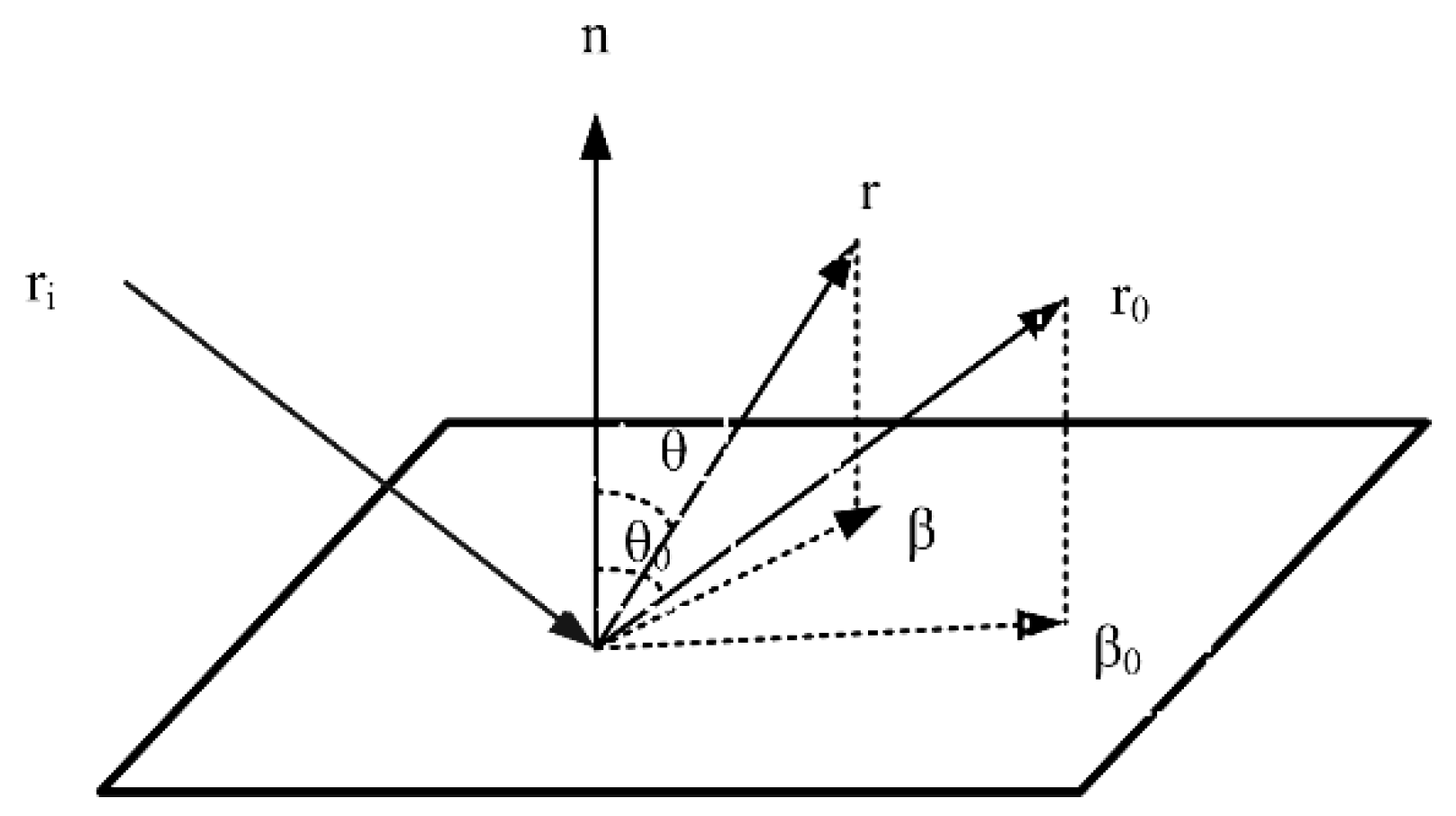
2.1. Visual Imaging Principle
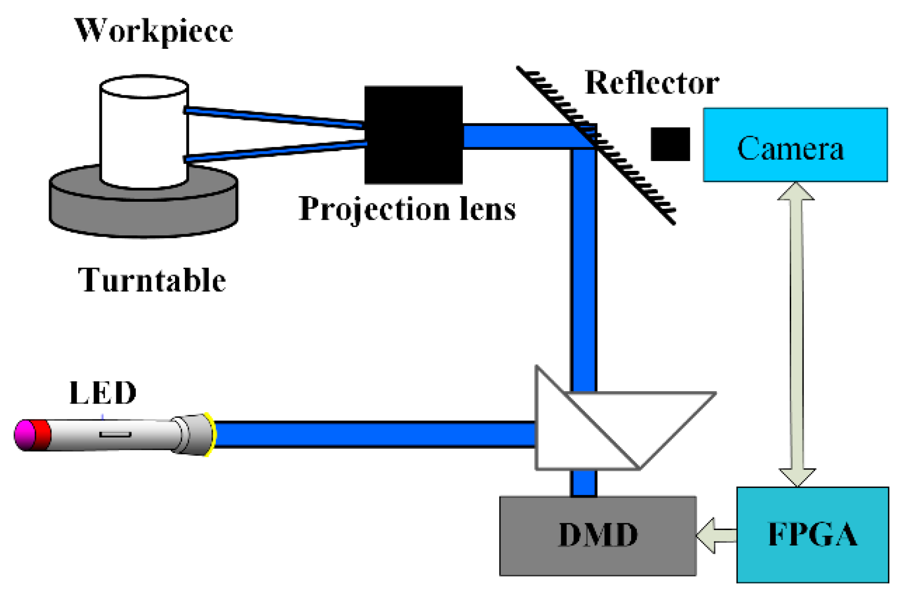
2.2. Composition of Adaptive Illumination System
2.2.1. LED Light Source
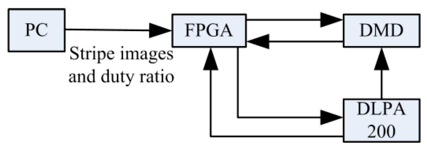
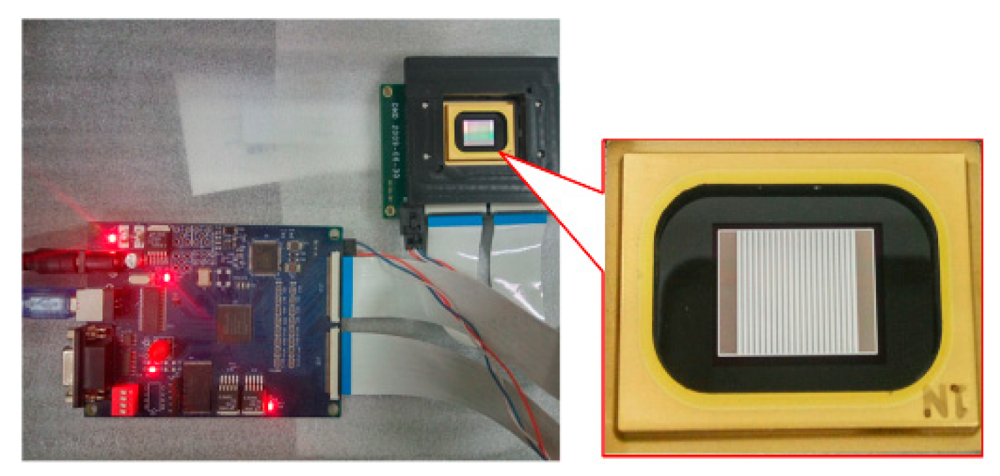
2.2.2. Spatiotemporal Light Modulation
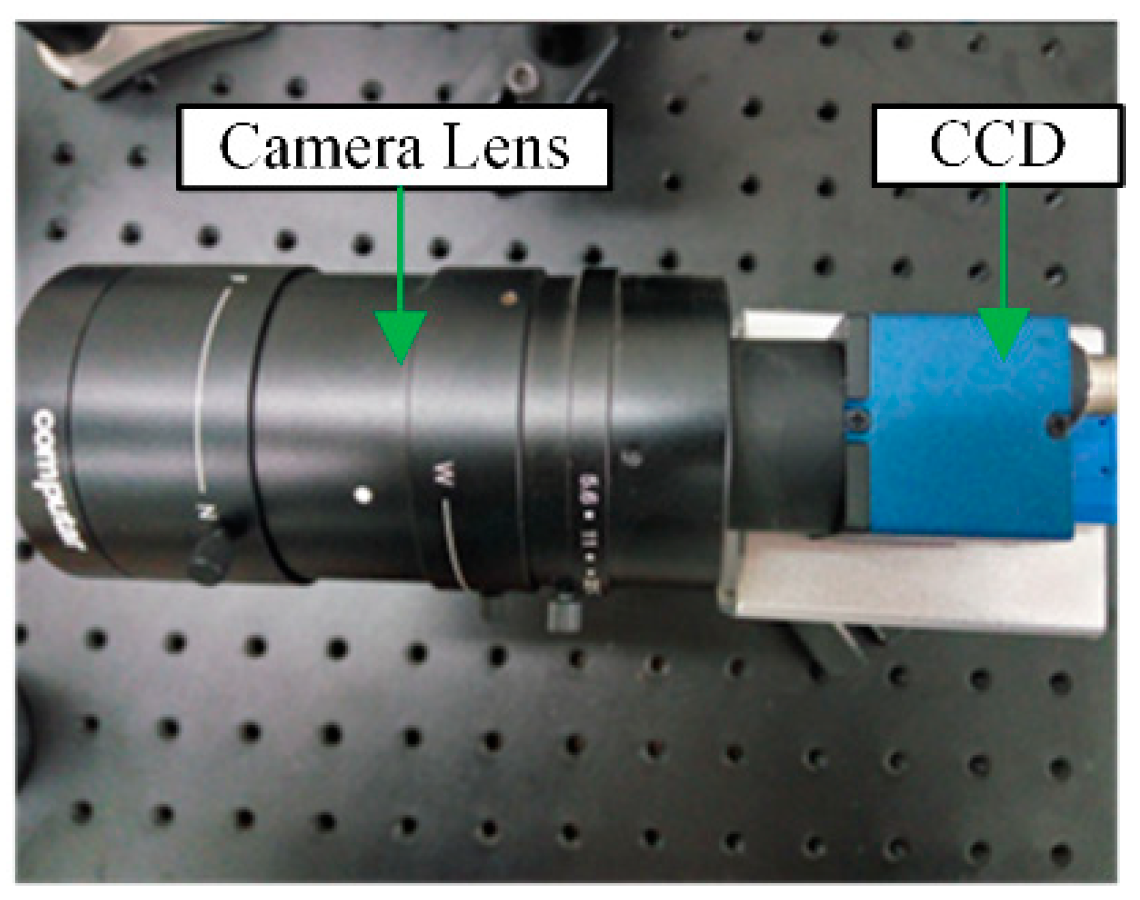
2.2.3. Image Acquisition
3. A Visual Imaging Method of Adaptive Illumination
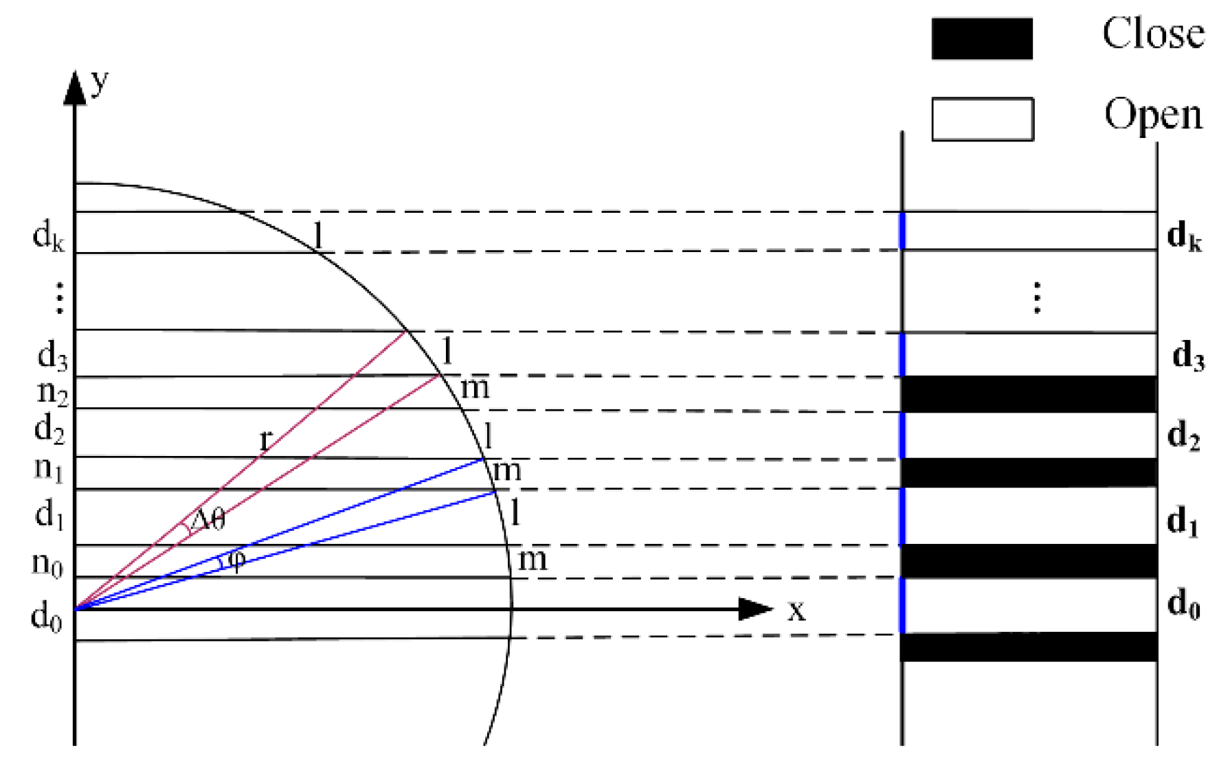
3.1. Uniform Straight Stripe Algorithm
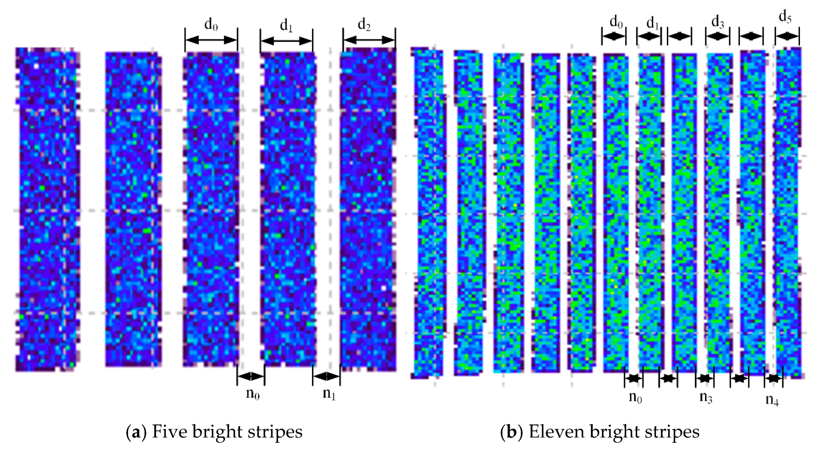
3.2. Simulation of Uniform Stripe Algorithm
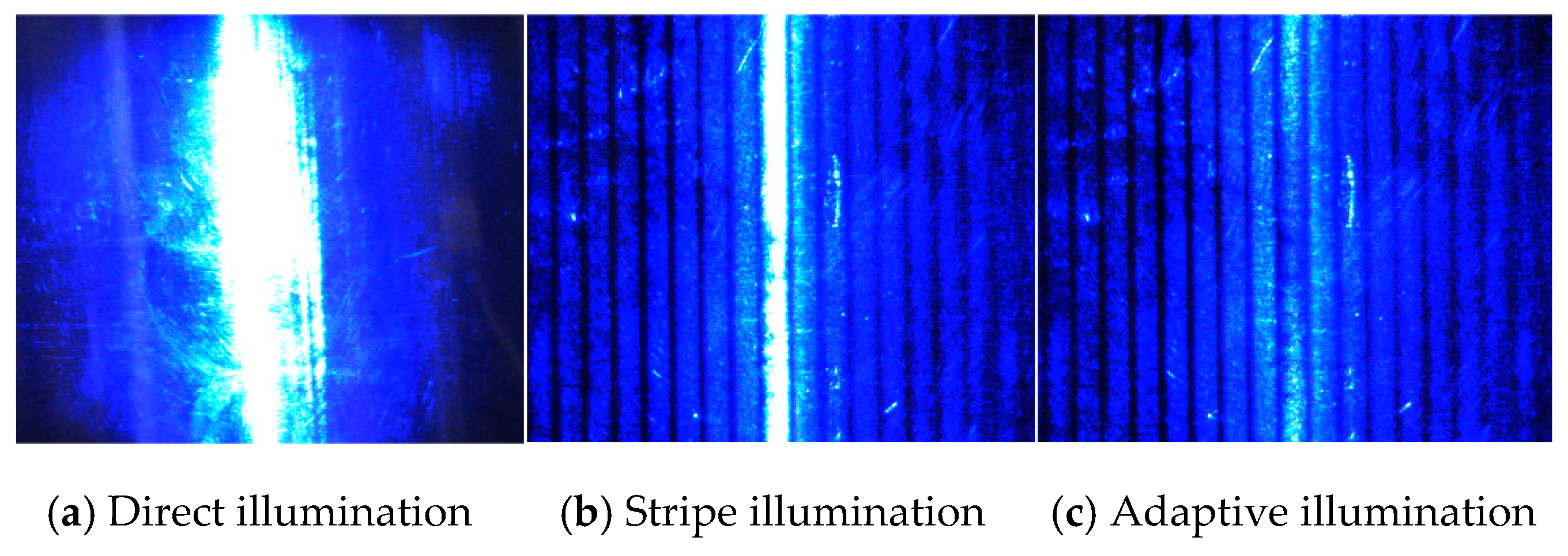
3.3. Visual Imaging of Adaptive Illumination of Light Intensity
- (1)
- Firstly, according to the uniform straight stripe algorithm, the illumination stripes P1–PN, as well as the distribution of the workpiece, are calculated, where N is the total number of stripes.
- (2)
- Suppose that the space-time duty ratio of each stripe is DRi = 50 %; then, i = 1–N.
- (3)
- Based on the space-time duty ratio of each stripe, the workpiece is illuminated by the time–space modulation stripe by the control of FPAG over DMD.
- (4)
- The image of the workpiece in the illumination area is captured by a digital camera. The grey value RGBi, standard deviation σ of each stripe, the deviation Erri = RGBi − Avg (average grey value) and the maximum (Max) and minimum (Min) grey value of each stripe are calculated.
- (5)
- The requirement of illumination uniformity can be determined according to the value of σ. If σ [σ], the space-time duty ratio DRi+1 = DRI + Erri/(Max − Min)∗K of each stripe should be revised, and the process should return to step (3); if σ , the adaptive modulation of light comes to an end.
4. Experiment Results
4.1. Experimental Platform
4.2. Experimental Analysis
5. Conclusions
Author Contributions
Funding
Conflicts of Interest
References
- Wang, Y.W.; Lin, Y.Z.; Jia, K.D.; Zhang Z., R.; Liu X., L. Comparative Analysis on Inspection of Steel Ball Surface Defect in Air and Oil Medium. Bearing 2010, 5, 37–39. [Google Scholar]
- Zhao, Y.L.; Wang, P.; Hao, H.R.; Liu, X. The Embedded Control System of Vision Inspecting Instrument for Steel Ball Surface Defect. In Proceedings of the 2008 IEEE Chinese Control & Decision Conference, Yantai, China, 2–4 July 2008; pp. 3469–3471. [Google Scholar]
- Do, Y.; Lee, S.; Kim, Y. Vision-Based Surface Defect Inspection of Metal Balls. Meas. Sci. Technol. 2011, 22, 107001. [Google Scholar] [CrossRef] [Scilit]
- Zhang, Z.; Yoshioka, H.; Imamura, T.; Miyake, T. Optimization of Light-Source Position in Appearance Inspection for Surface with Specular Reflection. In Proceedings of the IEEE International Conference on Information and Automation, Yinchuan, China, 26–28 August 2013; pp. 602–607. [Google Scholar]
- Ng, T.W. Optical Inspection of Ball Bearing Defects. Meas. Sci. Technol. 2007, 18, N73–N76. [Google Scholar] [CrossRef] [Scilit]
- Le, J.; Guo, J.J.; Zhu, H.; Fang, H.Y.; Shao, W. A fast defect-detecting method for smooth hemispherical shell surface. Opt. Electron. Eng. 2004, 10, 37–39. [Google Scholar]
- Wang, Z.; Xing, Q.; Fu, L.H.; Sun, H. Realtime Vision-Based Surface Defect Inspection of Steel Balls. Trans. Tianjin Univ. 2015, 21, 76–82. [Google Scholar] [CrossRef] [Scilit]
- Wang, H.; Miao, R.; Wu, X.S.; Ni, W.Y.; Tang, X.C. Polycrystalline Silicon Wafer Surface Color Defect Inspection Based on Machine Vision. Appl. Mech. Mater. 2014, 548–549, 48–52. [Google Scholar] [CrossRef] [Scilit]
- Guo, H.R.; Shao, W.; Zhou, A.W.; Yang, Y.; Liu, K. Novel defect recognition method based on adaptive global threshold for highlight metal surface. Chin. J. Sci. Instrum. 2017, 38, 2797–2804. [Google Scholar]
- Fu, R. Optimum Design of Fresnel Concentrator and Application Research in Concentrating Photovoltaic Technology. Thesis, North China Electric Power University, Beijing, China, 2017. [Google Scholar]
- Dan, D.; Lei, M.; Yao, B.; Wang, W.; Winterhalder, M.; Zumbusch, A.; Qi, Y.; Xia, L.; Yan, S.; Yang, Y.; et al. DMD-based LED-illumination super-resolution and optical sectioning microscopy. Sci. Rep. 2013, 3, 1116. [Google Scholar] [CrossRef] [Scilit] [PubMed]
- Schubert, F.; Wiggenhauser, H.; Lausch, R. On the accuracy of thickness measurements in impact-echo testing of finite concrete specimens—Numerical and experimental results. Ultrasonics 2004, 42, 897–901. [Google Scholar] [CrossRef] [Scilit] [PubMed]
- Juratli, T.A.; McCabe, D.; Nayyar, N.; Williams, E.A.; Silverman, I.M.; Tummala, S.S.; Fink, A.L.; Baig, A.; Martinez-Lage, M.; Selig, M.K.; et al. DMD genomic deletions characterize a subset of progressive/higher-grade meningiomas with poor outcome. Acta Neuropathol. 2018, 136, 779–792. [Google Scholar] [CrossRef] [Scilit] [PubMed]
- Zhu, Q.; Yang, X.B.; Li, M.S.; Li, H.; Wang, C.; Liu S., P.; Jian J., M.; Xiong D., X. Spot Array Optimization Parallel Confocal Imaging Based on Digital Micromirror Devices. Acta Opt. Sin. 2018, 38, 0118001. [Google Scholar]
- Wu, D.; Chen, T.; Li, A. A High Precision Approach to Calibrate a Structured Light Vision Sensor in a Robot-Based Three-Dimensional Measurement System. Sensors 2016, 16, 1388. [Google Scholar] [CrossRef] [Scilit] [PubMed]
- Feng, W.; Zhang, F.M.; Qu, X.H.; Zheng, S. Per-pixel coded exposure for high-speed and high-resolution imaging using a digital micromirror device camera. Sensors 2016, 16, 331. [Google Scholar] [CrossRef] [Scilit] [PubMed]
- Zhang, Y.; Strube, S.; Molnar, G.; Danzebrink, H.U.; Dai, G.; Bosse, H.; Hou, W. Parallel large-range scanning confocal microscope based on a digital micromirror device. Optik 2013, 124, 1585–1588. [Google Scholar] [CrossRef] [Scilit]
- Tang, W.Z.; Cao, Z.W.; Shi, J.H.; Zeng, G.H.; Fang, S.H. Back-Side Correlation Imaging with Digital Micro Mirror. Acta Opt. Sin. 2015, 35, 0511004. [Google Scholar] [CrossRef] [Scilit]
- Liu, Y.J. Design and Simulation of DLP Projection Lighting System Based on LED Light Source. Thesis, Shenzhen University, Shenzhen, China, 2015. [Google Scholar]


| Stripe No. | Theoretical Value/mm | Measured Value/mm | Err/mm |
|---|---|---|---|
| 1 | 0.698 | 0.702 | −0.004 |
| 2 | 0.349 | 0.321 | 0.028 |
| 3 | 0.697 | 0.684 | 0.013 |
| 4 | 0.348 | 0.336 | 0.012 |
| 5 | 0.694 | 0.704 | −0.010 |
| Stripe No. | Theoretical Value/mm | Measured Value/mm | Err/mm |
|---|---|---|---|
| 1 | 0.349 | 0.357 | −0.008 |
| 2 | 0.175 | 0.165 | 0.010 |
| 3 | 0.349 | 0.361 | −0.012 |
| 4 | 0.174 | 0.182 | −0.008 |
| 5 | 0.349 | 0.339 | 0.010 |
| 6 | 0.174 | 0.190 | −0.016 |
| 7 | 0.348 | 0.357 | −0.009 |
| 8 | 0.174 | 0.185 | −0.011 |
| 9 | 0.347 | 0.350 | −0.003 |
| 10 | 0.173 | 0.163 | 0.010 |
| 11 | 0.346 | 0.331 | 0.015 |
| Device | Device Type |
|---|---|
| Illuminant | LED (455 nm) |
| Prism | TIR |
| DMD | 0.7 Inch, 1024 × 768 |
| Projection lens | F = 150 mm |
| Stripe No. | Gray Value (106) | Stripe No | Gray Value (106) |
|---|---|---|---|
| 1 | 0.7 | 9 | 8.1 |
| 2 | 1.0 | 10 | 5.9 |
| 3 | 1.1 | 11 | 2.8 |
| 4 | 1.1 | 12 | 1.3 |
| 5 | 1.0 | 13 | 1.4 |
| 6 | 1.6 | 14 | 1.2 |
| 7 | 1.7 | 15 | 1.1 |
| 8 | 1.9 | 16 | 8.8 |
| Stripe No. | Gray Value (106) | Stripe No | Gray Value (106) |
|---|---|---|---|
| 1 | 0.7 | 9 | 3.5 |
| 2 | 1.0 | 10 | 3.2 |
| 3 | 1.0 | 11 | 2.7 |
| 4 | 1.0 | 12 | 1.3 |
| 5 | 1.1 | 13 | 1.4 |
| 6 | 1.7 | 14 | 1.2 |
| 7 | 1.8 | 15 | 1.1 |
| 8 | 1.9 | 16 | 0.8 |
© 2019 by the authors. Licensee MDPI, Basel, Switzerland. This article is an open access article distributed under the terms and conditions of the Creative Commons Attribution (CC BY) license (http://creativecommons.org/licenses/by/4.0/).
Share and Cite
Shao, W.; Liu, K.; Shao, Y.; Zhou, A. Smooth Surface Visual Imaging Method for Eliminating High Reflection Disturbance. Sensors 2019, 19, 4953. https://doi.org/10.3390/s19224953
Shao W, Liu K, Shao Y, Zhou A. Smooth Surface Visual Imaging Method for Eliminating High Reflection Disturbance. Sensors. 2019; 19(22):4953. https://doi.org/10.3390/s19224953
Chicago/Turabian StyleShao, Wei, Kaibin Liu, Yunqiu Shao, and Awei Zhou. 2019. "Smooth Surface Visual Imaging Method for Eliminating High Reflection Disturbance" Sensors 19, no. 22: 4953. https://doi.org/10.3390/s19224953
APA StyleShao, W., Liu, K., Shao, Y., & Zhou, A. (2019). Smooth Surface Visual Imaging Method for Eliminating High Reflection Disturbance. Sensors, 19(22), 4953. https://doi.org/10.3390/s19224953