Abstract
Aluminum is the most abundant metallic element in the Earth’s crust and acts as a non-essential element for biological species. The accumulation of excessive amounts of aluminum can be harmful to biological species. Thus, the development of convenient and selective tools for the aluminum detection is necessary. In this work, a highly selective aluminum ion fluorescent probe N’-(2,5-dihydroxybenzylidene)acetohydrazide (Al-II) has been successfully synthesized and systemically characterized. The fluorescence intensity of this probe shows a significant enhancement in the presence of Al3+, which is subject to the strong quench effects caused by Cu2+ and Fe3+. The binding ratio of probe-Al3+ was determined from the Job’s plot to be 1:1. Moreover, the probe was demonstrated to be effective for in vivo imaging of the intracellular aluminum ion in both living Drosophila S2 cells and Malpighian tubules.
1. Introduction
Aluminum is the most abundant metallic element in the Earth’s crust. It can be found in a wide range of plants, drinking water, and food. It is also commonly used as metallic materials in the industrial and medical fields [1,2,3]. However, aluminum is a non-essential element for the human body. The accumulation of excessive amounts of aluminum can be harmful to human bodies, causing many medical problems and illnesses, including neurodegenerative and neurological disorders, such as Alzheimer’s disease or Parkinson’s disease [3,4,5,6,7].
Compared with other transition metal ions, the detection of aluminum has always been challenged by its poor coordination ability, strong hydration tendency, and the lack of spectroscopic characteristics [8]. Therefore, it is urgent to develop the convenient and selective methods for the detection of aluminum. Fluorescent approach is the most attractive and highly sensitive method to detect low concentrations of analysts [9]. In recent years, there has been considerable progress for the detection of aluminum utilizing fluorescent chemo-sensors [10,11,12,13,14,15]. However, there are still many limitations during the application of most reported fluorescent probes, such as working well only in anhydrous systems, poor selectivity or sensitivity, and weak binding ability [13,16,17,18,19]. Especially, the chemo-sensors for biological system are rare, most of which were applied in in vitro cultured cells, but not the living organisms. We summarized the reported Al probes suitable for bio-imaging in Table 1.

Table 1.
Summary of the fluorescent probes for aluminum in biological systems.
In this work, we synthesized a highly selective turn-on fluorescent probe N’-(2,5-dihydroxybenzylidene)acetohydrazide (Al-II) for Al3+ detection, which worked well in aqueous systems. Moreover, we successfully applied this probe to monitor the in vivo distribution of Al3+ either in living cultured Drosophila cells or organs, which provides a useful tool to selectively detect aluminum not only in inorganic systems but also in biological systems.
2. Materials and Methods
2.1. HPLC Methods
Liquid chromatography-mass spectrometry (LC-MS) was performed using an Agilent 1200 Series apparatus with an LC/MSD trap and Daly conversion dynode detector with UV detection at 254 nm. The reverse phase high pressure liquid chromotragphy (RP-HPLC) method used in compound characterization is as follows: (A) Luna C18 column (100 × 2 mm); eluent A: H2O/0.1% formic acid, B: MeCN/0.1% formic acid; gradient: 5% B to 95% B over 3 min, 95% B for 1.5 min, 95% B to 5% B for 0.5 min, then 5% B for 2 min; flow rate 0.7 mL/min. (B) Luna C18 column (100 × 2 mm); eluent C: 10 mM ammonium acetate, D: 90% MeCN/10% 10 mM ammonium acetate; gradient 5% D for 0-1 min, 5 to 95% D from 1-11 min, 95% D from 11-12 min, 95% to 5% D from 12-13 min, 5% D from 13-15 min; flow rate 0.7 mL/min.
2.2. Synthesis of the Probe
N’-(2,5-dihydroxybenzylidene)acetohydrazide was prepared by the refluxing acetic hydrazide (0.37 g, 5.0 mmol) with salicylaldehyde (0.69 g, 5.0 mmol) in 50 mL methanol for 3 h. The solution was concentrated under reduced pressure, and the residue was further purified by recrystallization from methanol to afford 0.87 g (90%) as light green crystals.
LC-MS (method B): tR = 4.462 min, m/z = 195.0 [M+ H]+ (Figure S1).
1H NMR (major:minor isomer = 1:0.88) (500 MHz, (CD3)2SO) δ 11.49 (s, 1H, major), 11.16 (s, 1H, minor), 10.29 (s, 1H, major), 9.39 (s, 1H, minor), 8.94 (s, 1H, major), 8.88 (s, 1H, minor), 8.24 (s, 1H, major), 8.19 (s, 1H, minor), 7.03 (d, J = 3.0 Hz, 1H, minor), 6.91 (d, J = 2.6 Hz, 1H, major), 6.74–6.61 (m, 4H, 2 from major, 2 from minor), 2.16 (s, 3H, minor), 1.95 (s, 3H, major) (Figure S2).
13C NMR (126 MHz, (CD3)2SO) δ 171.45, 165.28, 150.00, 149.92, 149.80, 149.26, 145.55, 140.61, 120.29, 118.92, 118.61, 118.39, 116.93, 113.81, 111.39, 21.40, 20.30 (Figure S3).
2.3. Compound Stock and Storage
The Al-II probe powder was kept at 4 °C for long-term storage. The probe was dissolved into dimethyl sulfoxide (DMSO) to make a 10 mM stock solution, which was stored at 4 °C for temporary preservation.
2.4. Drosophila S2 Cell Culture and Experiments
Drosophila embryonic S2 cells were cultured with Gibco Schneider’s Drosophila Medium (Invitrogen) containing 10% fetal bovine serum and penicillin (50 IU/mL)/streptomycin (50 μg/mL) antibiotics at 25 °C.
For the aluminum detection experiments, the S2 cells were treated with Al3+-containing culture medium for 30 min, and then washed with PBS buffer three times, followed by the incubation with Al-II probe–containing medium for 30 min and additional PBS wash three times. Finally, the fluorescent signals indicating Al3+ ions in these cells were observed under the confocal fluorescence microscope. All steps were carried out at room temperature.
2.5. Al3+ Detection in Malpighian Tubules
We chose the Drosophila larval Malpighian tubules as the in vivo organ system to perform the Al3+ detection experiments using the Al-II probe.
Drosophila larvae at third instar stage were fed with fly food containing Al3+ for 30 min. Then the Malpighian tubules were dissected out from larval bodies, washed with PBS buffer, and further treated with Al-II probe for another 30 min followed by another round of PBS wash. Eventually, the Malpighian tubules were observed under the confocal fluorescence microscope. All steps were carried out at room temperature.
2.6. Confocal Fluorescence Imaging
Fluorescence imaging was acquired using a Nikon Ti laser scanning confocal fluorescence microscope with a laser (λex = 402 nm). The emission wavelength range was 500–600 nm. The images were captured under 60x oil lens (cells) or 20x lens (Malpighian tubules) with the pinhole size 71.5 μm and Si PMT HV 147.
The fluorescence intensity in cells was measured using the software ImageJ with the threshold (Triangle mode) adjustment. The fluorescence intensity in Drosophila Malpighian tubules was measured using the software ImageJ, then was normalized by the value of control group. The statistical significance was evaluated with Student’s t-test.
3. Results and Discussion
The desired fluorescent probe Al-II shown in Scheme 1 was synthesized as described above and characterized by LC-MS (Figure S1), 1H NMR (Figure S2) and 13C NMR (Figure S3).

Scheme 1.
Molecular structure of the Al-II probe.
3.1. Fluorescence Spectra of Detecting Al3+
The reaction time on the binding process of the probe with Al3+ was firstly studied. As shown in Figure 1a, there was obvious red-shift emission and dramatic intensity increase after addition of Al3+ in 5 min and the intensity reached to a stable value in 10 min. So, in the following studies, we pre-incubated probe-metal ions for 15 min before testing. The kinetic constant was estimated to be 0.0036 μM-1 min-1 based on the linear portion of increase on fluorescent intensity at 500 nm.
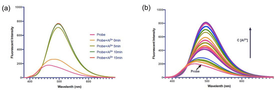
Figure 1.
Fluorescent spectra of Al-II probe with Al3+. (a) Fluorescent spectra of Al-II probe (10 μM) with Al3+ (50 μM) after incubation for 0 min, 5 min, 10 min and 15 min. (b) Fluorescent spectra of Al-II probe (10 μM) with titration of increased concentration of Al3+ (1 μM to 50 μM).
Then we performed a detailed investigation on the Al-II probe recognition of Al3+. As shown in Figure 1b, the fluorescence intensity of Al-II probe (10 μM) in aqueous solution at 465 nm was dramatically increased with the obvious emission red shift to 500 nm upon addition of Al3+. The changes of the emission intensities became constant when the amount of Al3+ added reached to 5.0 equiv. (50 μM).
As the emission of Al-II probe itself has spectra contribution to the spectra of probe-Al3+, so the spectra of Al-II probe were taken as the background signal and was deducted from probe-Al3+ spectra. The relative fluorescent intensity was used for future analysis. The same trend was seen after background deduction (Figure S4). After one-site specific binding analysis, the binding constant of Al-II probe with Al3+ was 10.8 μM (Figure S5).
3.2. Selectivity Over Metal Ions
The selectivity of Al-II probe as a fluorescent chemo-sensor for the detection of Al3+ among a wide range of environmentally and physiologically active metal ions was investigated by examining the fluorescence of solutions containing Al-II probe and the metal ions in distilled water. As shown in Figure 2a, when 10 equiv. of various metal ions (Al3+, Li+, Na+, Mg2+, K+, Ca2+, Cr3+, Mn2+, Fe2+, Fe3+, Ni2+, Cu2+, Zn2+, Ga3+, Ba2+, Gd2+, and Er3+) were added to the Al-II probe solution (10 μM), Al3+ produced a strong green fluorescence under excitation at 365 nm, whereas other metal ions made no obvious fluorescence.
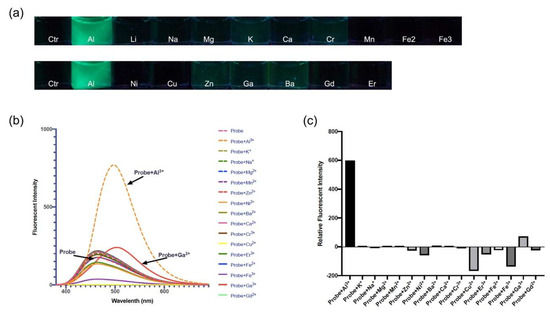
Figure 2.
Fluorescence response of the probe Al-II to different metal ions. (a) Changes in the fluorescence of probe Al-II (10 μM) upon addition of various metal ions (10 equiv.) under excitation at 365 nm. (b) Fluorescent spectra of Al-II probe (10 μM) in the presence of Al3+ (50 μM) and various metal ions (50 μM). (c) Relative fluorescent intensity of Al-II probe in the presence of Al3+ and various metal ions (λex 350 nm, λem 500 nm).
To more accurately explore the selectivity of probe Al-II over various metal ions, the fluorescence spectra of probe was measured (Figure 2b,c). In the presence of Al3+, the fluorescence intensity of Al-II probe showed a large enhancement along with a red-shifted emission. In the cases of other metal ions, such as K+, Na+, Mg2+, Mn2+, Zn2+, Ba2+, Ca2+, Cr3+, Fe2+ and Gd2+, there were no apparent changes in the fluorescence spectrum of Al-II probe (Figure 2b). Among these various metal ions, Ga3+ also showed an emission red shift, but its intensity was much lower in comparison with Al3+ (Figure 2b). Several metal ions such as Ni2+, Er3+, Cu2+ and Fe3+, quenched the emission intensity (Figure 2c), which might be attributed to their intrinsic quenching nature. These results indicated a highly selective ability for probe Al-II to detect Al3+ ions.
To further investigate the binding affinity of Al-II probe with Al3+ in the presence of various competing metal ions, Al-II probe was incubated with 5 equiv. of Al3+ in the presence of 5 equiv. of other metal ions. There was little interference for the detection of Al3+ in the presence of K+, Na+, Mg2+, Mn2+, Zn2+, Ni2+, Ba2+, Ca2+, Cr3+, Er3+, Fe2+ and Gd2+ (Figure 3, Figure S6). The Ga3+ ion did have weak competition effect for the Al-II-Al3+ binding, while Cu2+ and Fe3+ ions have strong quench effects for the fluorescence of probe Al-II with Al3+.
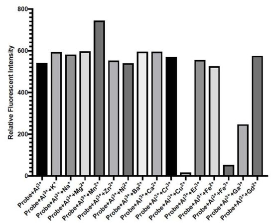
Figure 3.
Relative fluorescent intensity of Al-II probe (10 μM) upon addition of Al3+ (50 μM) in the presence of various metal ions (50 μM) in aqueous solution (λex 350 nm, λem 500 nm).
3.3. Detection Range
The detection limit was calculated based on the fluorescence titration of the probe Al-II. A good linear relationship between the Al-II fluorescence intensity and Al3+ concentration could be obtained in the range from 1 to 5 μM (R = 0.9947). The detection limit was calculated with the equation: Detection limit = 3*y-intercept/slope (as shown in Figure 4), and it was measured to be 0.66 μm.
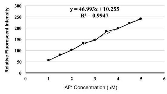
Figure 4.
Relative fluorescent intensity of Al-II probe (10 μM) upon addition of Al3+ (1 to 5 μM) (λex 350 nm, λem 500 nm).
3.4. Job’s Plot
The stoichiometry between the Al-II probe and Al3+ was validated by Job’s plot. The fluorescence emission intensity was measured by changing the molar fraction of Al3+ (the total concentration was 10 μM) (Figure S7). As shown in Figure 5, the fluorescence emission intensity indicated a maximum at a molar fraction of about 0.5, exhibiting a 1:1 stoichiometry of the probe-Al3+ complex.
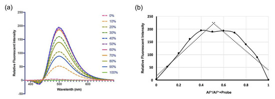
Figure 5.
The stoichiometry between the Al-II probe and Al3+. (a) Relative fluorescent spectra of different combination system of Al3+ and Al-II probe (with Al3+ from 0% to 100%, the total concentration is 10 μM). (b) Job’s plot for determining the stoichiometric complexation of Al-II probe with Al3+ in aqueous solution. The fluorescence at 500 nm emission was plotted.
3.5. UV-vis Spectra
To elucidate the binding mode of the probe and Al3+, we have performed the UV-vis spectra test. As shown in Figure 6, probe Al-II (25 μM) has two absorbance peaks at 280 nm and 340 nm. As the Al3+ concentration was increasing, the absorption peaks at 280 nm and 340 nm were gradually red-shifted to 290 nm and 370 nm. The intensity of new absorption peaks at 290 nm and 370 nm were increased upon addition of increased concentration of Al3+ and the intensity reached to stable value when the concentration of Al3+ added was 150 μM (6 equiv.). The large spectral bathochromic shift indicated the deprotonation, as a consequence of Al3+ binding to phenol.
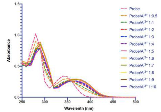
Figure 6.
Absorbance spectra of Probe Al-II (25 μM) with titration of increased concentration of Al3+ (1.5 μM to 250 μM).
We also measured the absorbance spectra of probe in the presence of Cu2+ and Fe3+ (Figure S8). Compared with Al3+, Cu2+ gave a larger red-shifted peak around 410 nm and Fe3+ has a very broad spectrum, which is consistent with the presence of ligand-to-metal charge-transfer (LMCT) in case of Cu2+ and Fe3+.
3.6. Application of Probe Al-II for Aluminum Detection in Living Cells
We examined the ability of Al-II probe for the detection of intracellular Al3+ in living biological systems under a confocal laser scanning microscopy. Drosophila S2 cells were incubated in the culture medium containing Al3+ (100 mM) for 30 min at room temperature. Then the Al3+ medium was removed and cells were washed with PBS buffer solution three times. The cells were further treated with Al-II probe (500 μM) for 30 min at room temperature, followed by PBS washing, three times, and imaging under confocal fluorescence microscope (Figure 7). As a negative control, the cells without Al3+ treatment showed no fluorescence (Figure 7a,b). The S2 cells with Al3+ treatment exhibited a strong green fluorescence (Figure 7c). Moreover, the cell morphology remained in a good condition after the addition of Al-II probe, indicating low toxicity of the probe. The Al-II probe can also be applied to the fixed cells, giving the similar results. These data demonstrated that the Al-II probe is cell membrane permeable and is useful for the imaging of intracellular Al3+ ions in living cells and fixed cells as well.
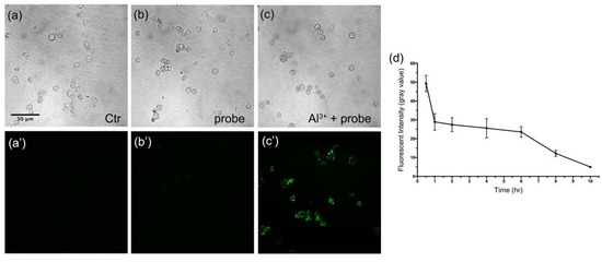
Figure 7.
Detection of Al3+ in Drosophila S2 cells using the Al-II probe upon excitation at 402 nm. (a,a’) The cells without any treatment. (b,b’) The cells incubated with the probe (500 μM, 30 min). (c,c’) The cells sequentially incubated with Al3+ (100 mM, 30 min) and the probe (500 μM, 30 min). The fluorescence images (a’,c’) are corresponding to the bright field images (a,c), respectively. The horizontal bar in panel a represents 50 μm and applies for all images of this figure. (d) The measurement of the fluorescent intensity in Al-probe-treated cells.
In addition, we measured the retention time of the Al-probe fluorescence inside cells (Figure 7d). As shown, the fluorescence intensity of Al-probe was quickly decreased after 30 min and maintained at a half level from 1 h to 6 h, then gradually decreased until nearly invisible at 10 h time point.
3.7. Imaging of Al3+ in Drosophila Malpighian Tubules Using Al-II Probe
The most attractive application for Al-II probe is sensing Al3+ in living organisms [31]. We chose the Drosophila Malpighian tubules as the model to detect Al3+ ions using the Al-II probe in living biological systems. The Malpighian tubule is the main organ to process metal ions in Drosophila, functionally analogous to mammalian kidney, and has recently become an important model organism for the study of metal ion transportation [32]. We first fed the 3rd instar larvae with Al3+ (100 mM) for 30 min and dissected the Malpighian tubules from larval bodies and washed them with PBS buffer. Then, we treated the Malpighian tubules with the Al-II probe (1 mM) for 30 min at room temperature, followed by image capture using confocal fluorescence microscope (Figure 8). As shown, compared with control sample without treatments, the Malpighian tubules with Al3+ and probe treatments showed strong fluorescence. These evidence indicated that the Al-II probe can be used for monitoring the distribution of Al3+ in living bodies.
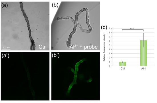
Figure 8.
Detection of Al3+ in Drosophila Malpighian tubules using the Al-II probe upon excitation at 402 nm. (a) A bright field image of Malpighian tubules without treatments. (a’) The corresponding fluorescence image of Malpighian tubules in panel a. (b) A bright field image of Malpighian tubules treated with Al3+ (100 mM, 30 min) and probe (1 mM, 30 min). (b’) The corresponding fluorescence image of Malpighian tubules in panel b. The horizontal bar in panel a represents 200 μm and applies for all four images of this figure. (c) The quantification of relative fluorescence intensity of Al-II probe in Malpighian tubules. n >= 3. * represents p < 0.05, ** represents p < 0.005, *** represents p < 0.0005.
4. Conclusions
In summary, we have designed and successfully synthesized a fluorescent probe Al-II to detect Al3+ ions. The probe revealed high selectivity and strong fluorescence to Al3+. Moreover, we successfully detected the intracellular Al3+ in the living cells as well as Drosophila Malpighian tubules utilizing the Al-II probe. Therefore, the Al-II probe not only is able to detect Al3+ ions in aqueous solution, but also serves as a potentially useful tool for monitoring the intracellular distribution of Al3+ in biological systems.
Supplementary Materials
The following are available online at https://www.mdpi.com/1424-8220/19/11/2423/s1: Figure S1: HPLC spectrum of compound Al-II. Figure S2: 1H NMR spectrum of compound Al-II. Figure S3: 13C NMR spectrum of compound Al-II. Figure S4: Fluorescent spectra of Al-II probe (10 μM) with titration of increased concentration of Al3+ (1 μM to 50 μM) after spectra subtraction of Al-II probe. Figure S5: Nonlinear one-site specific binding curve with relative fluorescent intensity after Al3+ (1 μM to 50 μM) titration (R2 = 0.9816, Kd = 10.8). Figure S6: Fluorescence response of the probe to Al3+ in the presence of higher concentration of Na+ and K+. Figure S7: Original fluorescent spectra of different combination system of Al3+ and probe (with Al3+ from 0% to 100%, the total concentration is 10 μM). Figure S8: Absorbance spectra of Probe Al-II (25 μM) with Al3+, Cu2+ or Fe3+ (250 μM).
Author Contributions
Conceptualization, L.Z.; data curation, L.Z.; formal analysis, J.Y. and H.W.; funding acquisition, Y.S.; investigation, L.W. and J.Y.; methodology, L.W. and J.Y.; project administration, L.Z.; supervision, L.Z.; validation, C.R. and Y.S.; writing—original draft, L.W., J.Y. and H.W.; writing—review & editing, Y.S.
Funding
This work was supported by the National Natural Science Foundation of China (31701274), the Fundamental Research Funds for the Central Universities (201612010 and 201822016).
Conflicts of Interest
The authors declare no conflict of interest.
References
- Krewski, D.; Yokel, R.A.; Nieboer, E.; Borchelt, D.; Cohen, J.; Harry, J.; Kacew, S.; Lindsay, J.; Mahfouz, A.M.; Rondeau, V. Human health risk assessment for aluminium, aluminium oxide, and aluminium hydroxide. J. Toxicol. Environ. Health B Crit. Rev. 2007, 10, 1–269. [Google Scholar] [CrossRef] [PubMed]
- Inostroza-Blancheteau, C.; Rengel, Z.; Alberdi, M.; de la Luz Mora, M.; Aquea, F.; Arce-Johnson, P.; Reyes-Diaz, M. Molecular and physiological strategies to increase aluminum resistance in plants. Mol. Biol. Rep. 2012, 39, 2069–2079. [Google Scholar] [CrossRef] [PubMed]
- Mohseni, H.K.; Chettle, D.R. A History of In Vivo Neutron Activation Analysis in Measurement of Aluminum in Human Subjects. J. Alzheimers. Dis. 2016, 50, 913–926. [Google Scholar] [CrossRef] [PubMed]
- Perl, D.P.; Brody, A.R. Alzheimer’s disease: X-ray spectrometric evidence of aluminum accumulation in neurofibrillary tangle-bearing neurons. Science 1980, 208, 297–299. [Google Scholar] [CrossRef]
- Tavakoli, O.; Yoshida, H. Effective recovery of harmful metal ions from squid wastes using subcritical and supercritical water treatments. Environ. Sci. Technol. 2005, 39, 2357–2363. [Google Scholar] [CrossRef]
- Lou, Z.; Li, P.; Song, P.; Han, K. Ratiometric fluorescence imaging of cellular hypochlorous acid based on heptamethine cyanine dyes. Analyst 2013, 138, 6291–6295. [Google Scholar] [CrossRef]
- Blackburn, A.C.; Doe, W.F.; Buffinton, G.D. Protein carbonyl formation on mucosal proteins in vitro and in dextran sulfate-induced colitis. Free Radic. Biol. Med. 1999, 27, 262–270. [Google Scholar] [CrossRef]
- Soroka, K.; Vithanage, R.S.; Phillips, D.A.; Walker, B.; Dasgupta, P.K. Fluorescence properties of metal complexes of 8-Hydroxyquinoline-5-sulfonic acid and chromatographic applications. Anal. Chem. 1987, 59, 629–636. [Google Scholar] [CrossRef]
- Valeur, B.; Leray, I. Design principles of fluorescent molecular sensors for cation recognition. Coord. Chem. Rev. 2000, 205, 3–40. [Google Scholar] [CrossRef]
- Wang, Y.; Hou, L.J.; Wu, Y.B.; Shi, L.L.; Shang, Z.B.; Jin, W.J. Alizarin Complexone as a highly selective ratiometric fluorescent probe for Al3+ detection in semi-aqueous solution. J. Photoch. Photobio. A 2014, 281, 40–46. [Google Scholar] [CrossRef]
- Li, M.X.; Zhang, X.; Fan, Y.H.; Bi, C.F. A novel fluorescent probe based on rhodamine hydrazone derivatives bearing a thiophene group for Al3+. Luminescence 2016, 31, 851–855. [Google Scholar] [CrossRef]
- Jiang, Y.; Sun, L.L.; Ren, G.Z.; Niu, X.; Hu, W.Z.; Hu, Z.Q. A New Fluorescence Turn-On Probe for Aluminum(III) with High Selectivity and Sensitivity, and its Application to Bioimaging. ChemistryOpen 2015, 4, 378–382. [Google Scholar] [CrossRef] [PubMed]
- Xiao, H.; Chen, K.; Jiang, N.; Cui, D.; Yin, G.; Wang, J.; Wang, R. A highly selective turn-on fluorescent probe for Al(III) based on coumarin and its application in vivo. Analyst 2014, 139, 1980–1986. [Google Scholar] [CrossRef]
- Peng, H.; Shen, K.; Mao, S.; Shi, X.; Xu, Y.; Aderinto, S.O.; Wu, H. A Highly Selective and Sensitive Fluorescent Turn-on Probe for Al3+ Based on Naphthalimide Schiff Base. J. Fluoresc. 2017, 27, 1191–1200. [Google Scholar] [CrossRef]
- Ahmed, M.; Faisal, M.; Ihsan, A.; Naseer, M.M. Fluorescent organic nanoparticles (FONs) as convenient probes for metal ion detection in aqueous medium. Analyst 2019, 144, 2480–2497. [Google Scholar] [CrossRef] [PubMed]
- Hsieh, W.H.; Wan, C.-F.; Liao, D.-J.; Wu, A.-T. A turn-on Schiff base fluorescence sensor for zinc ion. Tetrahedron Lett. 2012, 53, 5848–5851. [Google Scholar] [CrossRef]
- Jia, T.-J.; Cao, W.; Zheng, X.-J.; Jin, L.-P. A turn-on chemosensor based on naphthol–triazole for Al(III) and its application in bioimaging. Tetrahedron Lett. 2013, 54, 3471–3474. [Google Scholar] [CrossRef]
- Chen, Y.; Jiang, J. Decoding the phosphorylation code in Hedgehog signal transduction. Cell Res. 2013, 23, 186–200. [Google Scholar] [CrossRef] [PubMed]
- In, B.; Hwang, G.W.; Lee, K.H. Highly sensitive and selective detection of Al(III) ions in aqueous buffered solution with fluorescent peptide-based sensor. Bioorg. Med. Chem. Lett. 2016, 26, 4477–4482. [Google Scholar] [CrossRef]
- Li, C.-Y.; Zhou, Y.; Li, Y.-F.; Zou, C.-X.; Kong, X.-F. Efficient FRET-based colorimetric and ratiometric fluorescent chemosensor for Al3+ in living cells. Sens. Actuators, B 2013, 186, 360–366. [Google Scholar] [CrossRef]
- Ezhumalai, D.; Mathivanan, I.; Chinnadurai, A. Turn on macrocyclic chemosensor for Al(3+) ion with facile synthesis and application in live cell imaging. Spectrochim. Acta A Mol. Biomol. Spectrosc. 2018, 199, 209–219. [Google Scholar] [CrossRef]
- Li, Z.; Liu, C.; Wang, J.; Wang, S.; Xiao, L.; Jing, X. A selective diaminomaleonitrile-based dual channel emissive probe for Al3+ and its application in living cell imaging. Spectrochim. Acta A Mol. Biomol. Spectrosc. 2019, 212, 349–355. [Google Scholar] [CrossRef]
- Zhou, F.; Wang, H.; Liu, P.; Hu, Q.; Wang, Y.; Liu, C.; Hu, J. A highly selective and sensitive turn-on probe for aluminum(III) based on quinoline Schiff’s base and its cell imaging. Spectrochim. Acta A Mol. Biomol. Spectrosc. 2018, 190, 104–110. [Google Scholar] [CrossRef]
- Gupta, S.R.; Singh, P.; Koch, B.; Singh, V.P. A water soluble, highly sensitive and selective fluorescent probe for Al3+ ions and its application in live cell imaging. J. Photochem. Photobiol. A 2017, 348, 246–254. [Google Scholar] [CrossRef]
- Balamurugan, G.; Velmathi, S.; Thirumalaivasan, N.; Wu, S.P. New phenazine based AIE probes for selective detection of aluminium(iii) ions in presence of other trivalent metal ions in living cells. Analyst 2017, 142, 4721–4726. [Google Scholar] [CrossRef]
- Ding, W.-H.; Wang, D.; Zheng, X.-J.; Ding, W.-J.; Zheng, J.-Q.; Mu, W.-H.; Cao, W.; Jin, L.-P. A turn-on fluorescence chemosensor for Al3+, F− and CN− ions, and its application in cell imaging. Sens. Actuators, B 2015, 209, 359–367. [Google Scholar] [CrossRef]
- Saini, A.K.; Sharma, V.; Mathur, P.; Shaikh, M.M. The development of fluorescence turn-on probe for Al(III) sensing and live cell nucleus-nucleoli staining. Sci. Rep. 2016, 6, 34807. [Google Scholar] [CrossRef]
- Liu, Y.; Bi, A.; Gao, T.; Cao, X.; Gao, F.; Rong, P.; Wang, W.; Zeng, W. A novel self-assembled nanoprobe for the detection of aluminum ions in real water samples and living cells. Talanta 2019, 194, 38–45. [Google Scholar] [CrossRef]
- Li, C.L.; Lu, P.H.; Fu, S.F.; Wu, A.T. A Highly Selective and Sensitive Fluorescent Chemosensor for Detecting Al3+ Ion in Aqueous Solution and Plant Systems. Sensors 2019, 19. [Google Scholar] [CrossRef]
- Tian, H.; Qiao, X.; Zhang, Z.L.; Xie, C.Z.; Li, Q.Z.; Xu, J.Y. A high performance 2-hydroxynaphthalene Schiff base fluorescent chemosensor for Al3+ and its applications in imaging of living cells and zebrafish in vivo. Spectrochim. Acta A Mol. Biomol. Spectrosc. 2019, 207, 31–38. [Google Scholar] [CrossRef]
- Devirgiliis, C.; Zalewski, P.D.; Perozzi, G.; Murgia, C. Zinc fluxes and zinc transporter genes in chronic diseases. Mutat. Res. 2007, 622, 84–93. [Google Scholar] [CrossRef]
- Xiao, G.; Zhou, B. What can flies tell us about zinc homeostasis? Arch. Biochem. Biophys. 2016, 611, 134–141. [Google Scholar] [CrossRef]
© 2019 by the authors. Licensee MDPI, Basel, Switzerland. This article is an open access article distributed under the terms and conditions of the Creative Commons Attribution (CC BY) license (http://creativecommons.org/licenses/by/4.0/).