Article

Taxonomic Revision of Pasiphaea (Pasiphaeidae: Crustacea) of the Southwest Tropical Pacific with a Description of Eight New Species †

by
Anton M. Tikhomirov
1,
Dmitrii N. Kulagin
1,
Anastasiia A. Lunina
1,
Elodie Vourey
2 and
Alexander L. Vereshchaka
1,*
1
Shirshov Institute of Oceanology, Russian Academy of Sciences, 117218 Moscow, Russia
2
Pacific Community—SPC, Taxonomy Laboratory of FAME-OFPEMA, B.P. D5, Noumea 98848, New Caledonia
*
Author to whom correspondence should be addressed.
Zoobank link: urn:lsid:zoobank.org:pub:5010C09B-0418-4D6C-B4CC-50DF8797A80C.
Diversity 2025, 17(9), 656; https://doi.org/10.3390/d17090656
Submission received: 7 August 2025 / Revised: 10 September 2025 / Accepted: 11 September 2025 / Published: 19 September 2025
(This article belongs to the Special Issue 2025 Feature Papers by Diversity’s Editorial Board Members)

Abstract

The genus Pasiphaea Savigny, 1816, comprises a diverse assemblage of meso- to bathypelagic shrimps, currently including 69 valid species. Taxonomic investigations have long been hampered by numerous synonymies and by species known only from their original, overly brief descriptions. Here, we address these deficiencies by examining Pasiphaea specimens collected in the Western and Central Pacific during multiple research cruises using mid-water trawls. Integrating traditional morphological characters (carapace carination, pleonal relief, telson end morphology, and pereopodal dentition) with a novel morphometric framework, we standardized terminology and defined a suite of quantitative measurements across the carapace, pleon, telson, and chelae. In order to corroborate species boundaries, we sequenced three mitochondrial gene markers (COI, 16S rDNA, and 12S rDNA) and calculated genetic distances under the Kimura two-parameter model. This combined approach extended or confirmed the geographic ranges of five described taxa, facilitated the formal description of eight new species, and demonstrated the synonymy of P. gracilis Hayashi, 1999, and P. fragilis Hayashi, 1999. Phylogenetic reconstructions further highlighted cases of pseudocryptic speciation in neighboring bathyal habitats. Our results expose substantial lacunae in the Southwest Pacific Pasiphaea fauna and substantiate the necessity for a comprehensive, genus-level revision. This study thereby contributes to both refining species diagnoses and advancing our understanding of deep-sea crustacean biodiversity.

1. Introduction

The genus Pasiphaea Savigny, 1816, is a diverse group of meso- to bathypelagic shrimps which encompasses 69 currently accepted species [1]. Pasiphaea is morphologically distinguished from the remaining genera of the family Pasiphaeidae by a reduced branchial formula, absence of a mandibular palp, pectinate chelae, and the absence of an orbital tooth of the carapace, varying in rostrum shape and length, shape of the branchiostegal sinus, pleonic relief, shape of the posterior part of telson, and armament of the first and second pereopods. Recent molecular studies suggest the monophyly of the genus [2].
The genus Pasiphaea was established by Savigny [3] (type species P. sivado Risso, 1816). Early works [4,5,6,7,8,9] added many species in the late 19th and early 20th centuries. Professor R.N. Burukovsky [10,11] added revisions, but the taxonomy remained fragmented. Hayashi [12,13,14] revised a few Pasiphaea species groups in tropical waters. Notably, Hayashi [13] redescribed the P. cristata species group (22 species), synonymizing P. nishiei Iwasaki, 1990, with P. merriami Schmitt, 1931, and P. vereschhaka Burukovsky, 1993, with P. amplidens Spence Bate, 1888. He also recognized that P. australis Hanamura, 1989 (lacking branchiae), actually belongs to a separate genus Alainopasiphaea, and he subsumed the old genus Phye [15] into Pasiphaea. In summary, the genus concept has been narrowed and clarified: all species of Pasiphaea now share a complete or only mildly reduced branchial formula, and the name Phye is no longer valid.
Hayashi recognized three informal species-groups within Pasiphaea, each defined by suites of morphological traits:
  • P. cristata” group: Characterized by a complete branchia formula and unarmed posterior margins on the first two pereopods (no spines on the merus of the first pereopod, and no spines on the ischium/basis of the second pereopod). Some representatives of the group are P. cristata Spence Bate, 1888, P. sirenkoi Burukovsky, 1987, and P. crosnieri Hayashi, 2004.
  • P. alcocki” group: Characterized by a deeply concave posterior margin of the telson and a strongly carinate dorsal margin on the carapace and pleon. In this group, the merus of the first and second pereopods bears numerous spiniform setae, often with spines also on the ischium or the basis of the second pereopod. This group (17 species) generally includes the large species like P. alcocki (Wood-Mason in Wood-Mason & Alcock, 1891), P. ledoyeri Hayashi, 2006, and P. major Hayashi, 2006.
  • P. sivado” group: Basically characterized by a reduced branchial formula (fewer pleurobranchial plates than typical, i.e., lost branchiae). Members of this group tend to have dorsally rounded pleonic somites 1–5 and usually possess a posterodorsal spine on the sixth somite. (Hayashi [14] noted P. sivado-group shrimps as having a “reduced branchial formula”.) Pasiphaea sivado (Risso, 1816), P. japonica Omori, 1976, P. propinqua De Man, 1916, etc., fall here.
Aside from these groups, Hayashi [13,14] recognized 14 species that were not correctly placed in any group due to a different combination of the characters above and other characters (e.g., rostrum shape, armament of pereopods, carination on carapace and pleon, proportions and shape of the apex of the telson) that are also used in species identification.
Taxonomic work on Pasiphaea has revealed many issues, including numerous synonymies (e.g., P. nishiei = P. merriami, P. vereschhaka = P. amplidens), a number of species known only from their original, overly brief description (e.g., P. dofleini Schmitt, 1932; P. emarginata Rathbun, 1902; P. notosivado Yaldwyn, 1971; P. truncata Rathbun, 1906) and subtle differences (e.g., pleomere shape, number of teeth), making field identification difficult [14].
Komai et al. [16] additionally expanded Pasiphaea taxonomy in the West Pacific, specifically in Taiwanese waters, describing four new species yet also revealing substantial gaps in existing records. The tropical Southwest Pacific, encompassing New Caledonia, Fiji, Vanuatu, Tonga, Indonesia, and adjacent regions, covers a far larger and less-studied area, where Pasiphaea diversity remains poorly documented. Recorded species in this region include P. debitusae Hayashi, 1999, and P. gracilis [12], recently also reported from Japanese waters [17], indicating a broad tropical-Pacific distribution. Some other congeners known from nearby localities (but not New Caledonia) include P. alcocki, P. falx Komai, Lin & Chan, 2012, P. levicarinata Hanamura, 1994, P. mclaughlinae Hayashi, 2006, P. orientalis Schmitt, 1931, P. taiwanica Komai, Lin & Chan, 2012, P. japonica [16], P. crosnieri [13], P. laevis Hayashi, 1999, P. philippinensis Hayashi, 1999, P. propinqua [12], and P. pseudacantha Hayashi, 2004 [13]. Despite these scattered records, the bathyal Pasiphaea fauna of the Southwest Pacific remains understudied.
In order to address this deficit, we examined Pasiphaea specimens collected off New Caledonia and kept at the Taxonomy Laboratory of the Fisheries, Aquaculture, and Marine Ecosystems Division (The Pacific Community, Noumea) and the Muséum National d’Histoire Naturelle (Paris).
We morphologically analyzed and additionally sequenced three molecular markers of each species, combined in a three-gene molecular tree, to test two hypotheses:
  • Even moderately abundant new collections will yield a significant number of undescribed Pasiphaea species in the Southwest Pacific;
  • The discovery of additional species will further challenge Hayashi’s morphological species-group concept and will not resolve the incongruence between morphology- and DNA-based evidence.

2. Materials and Methods

2.1. Sampling

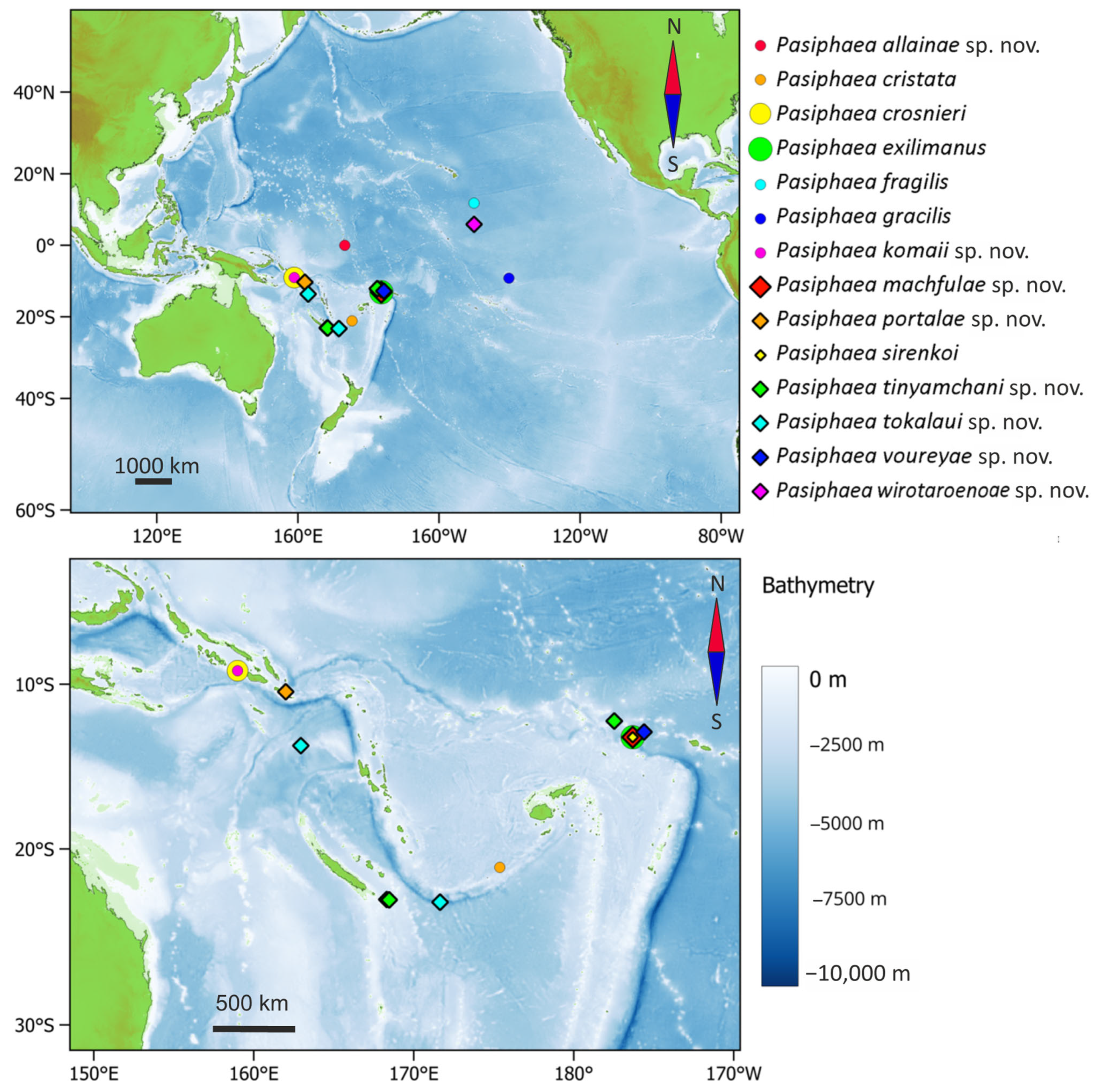
Material was collected during NECTALIS 5 [18], WALLALIS [19], MARACAS 7D [20], WARMALIS 2 [21] and 3 [22] cruises (Figure 1) with a pelagic fishing trawl, 10 m2 opening, 10 mm mesh size of the cod end, and between 2 and 3 knots trawling speed. All specimens were sorted immediately onboard, frozen at −20 °C in sea water, and later examined in the laboratory.

2.2. Measurements

In addition to traditional characters (e.g., carination of the carapace, pleonic relief, number of telson spines, number of pereopodal teeth), we applied a morphometric approach. All available specimens were measured across the following anatomical regions: carapace, pleon, telson, and chelae. The most important measurements are as follows.
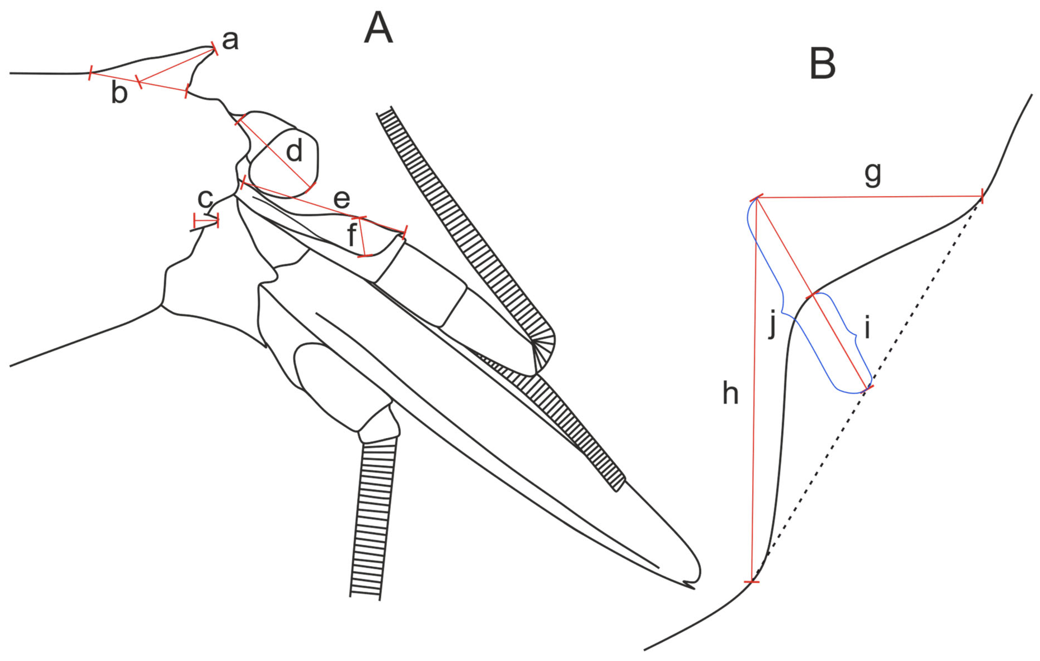
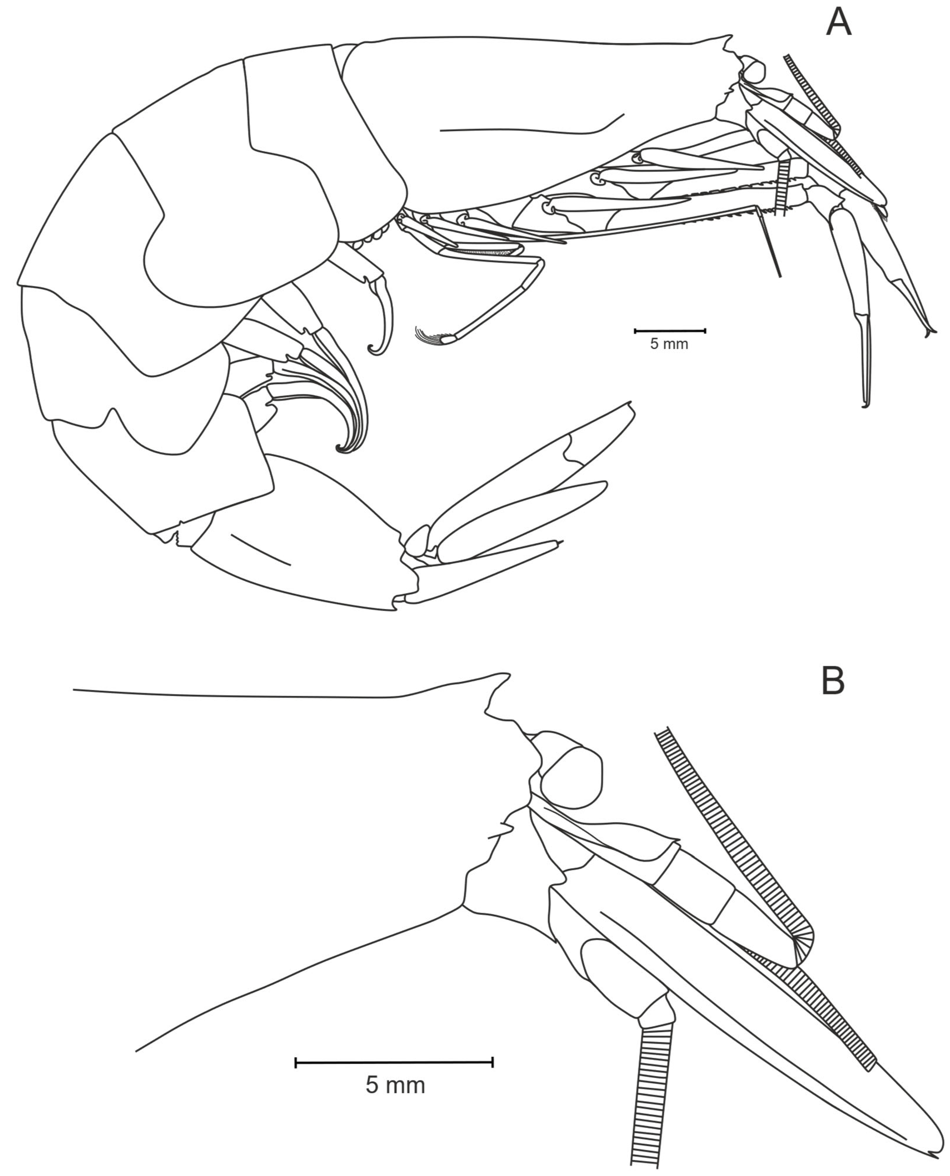
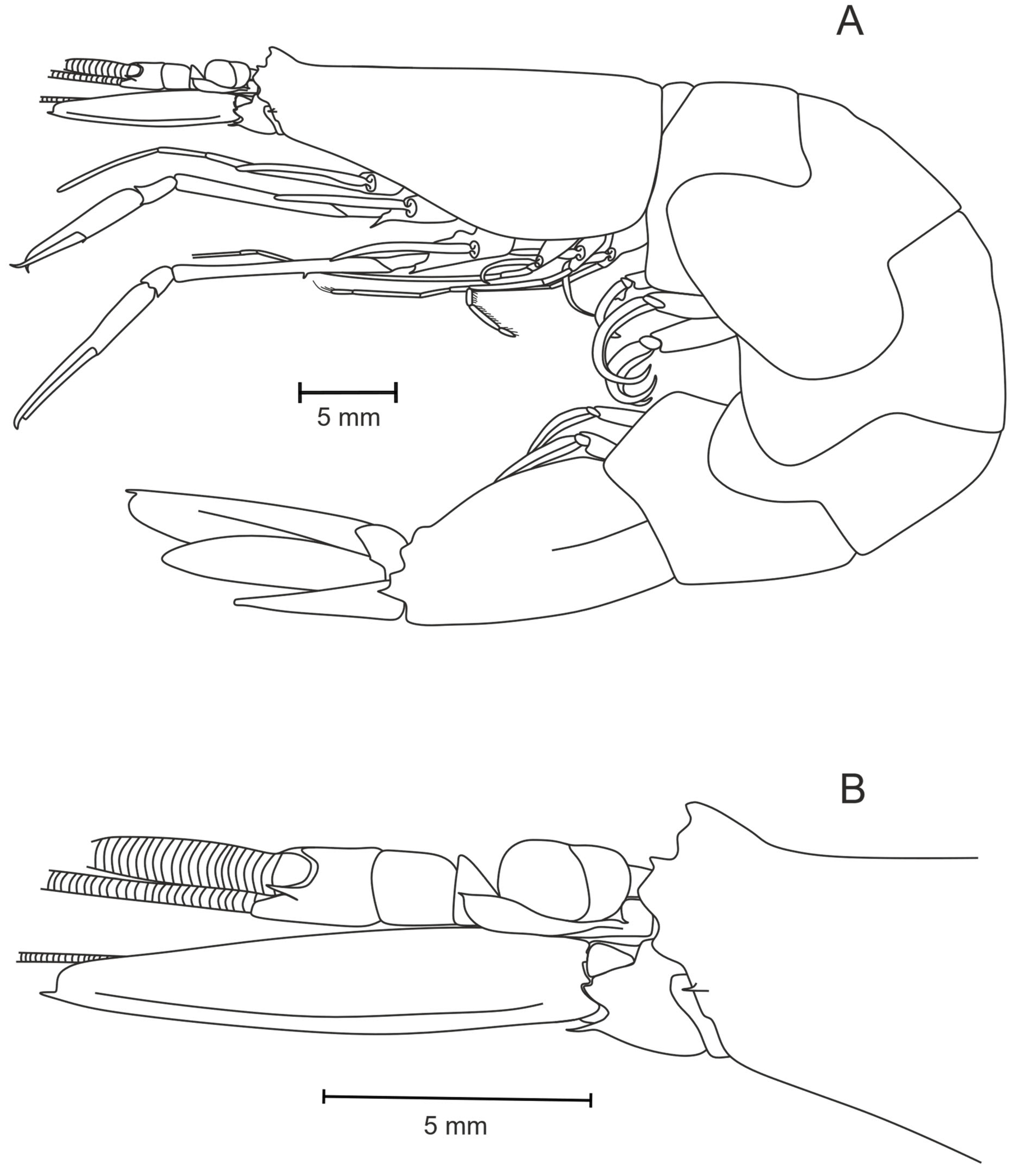
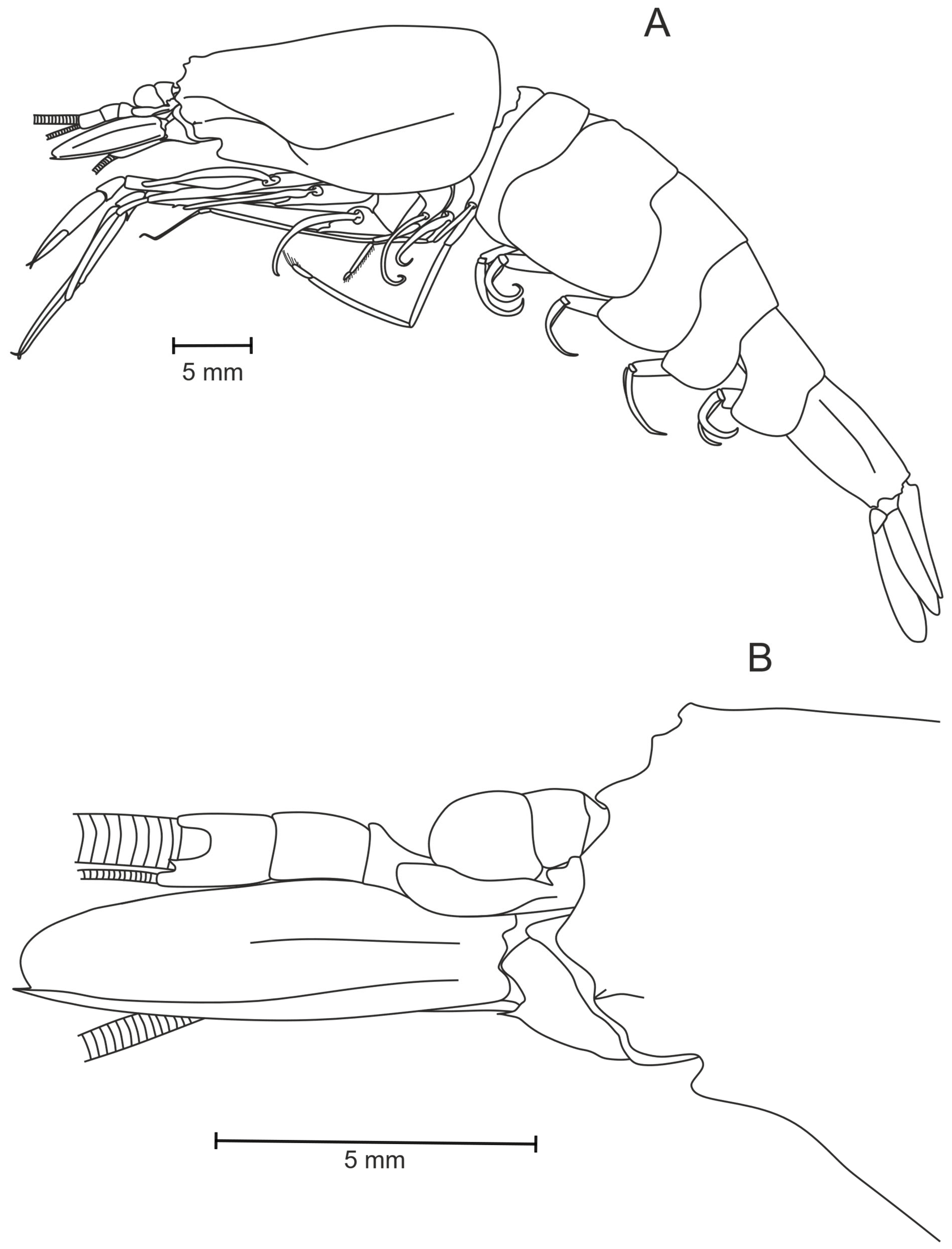
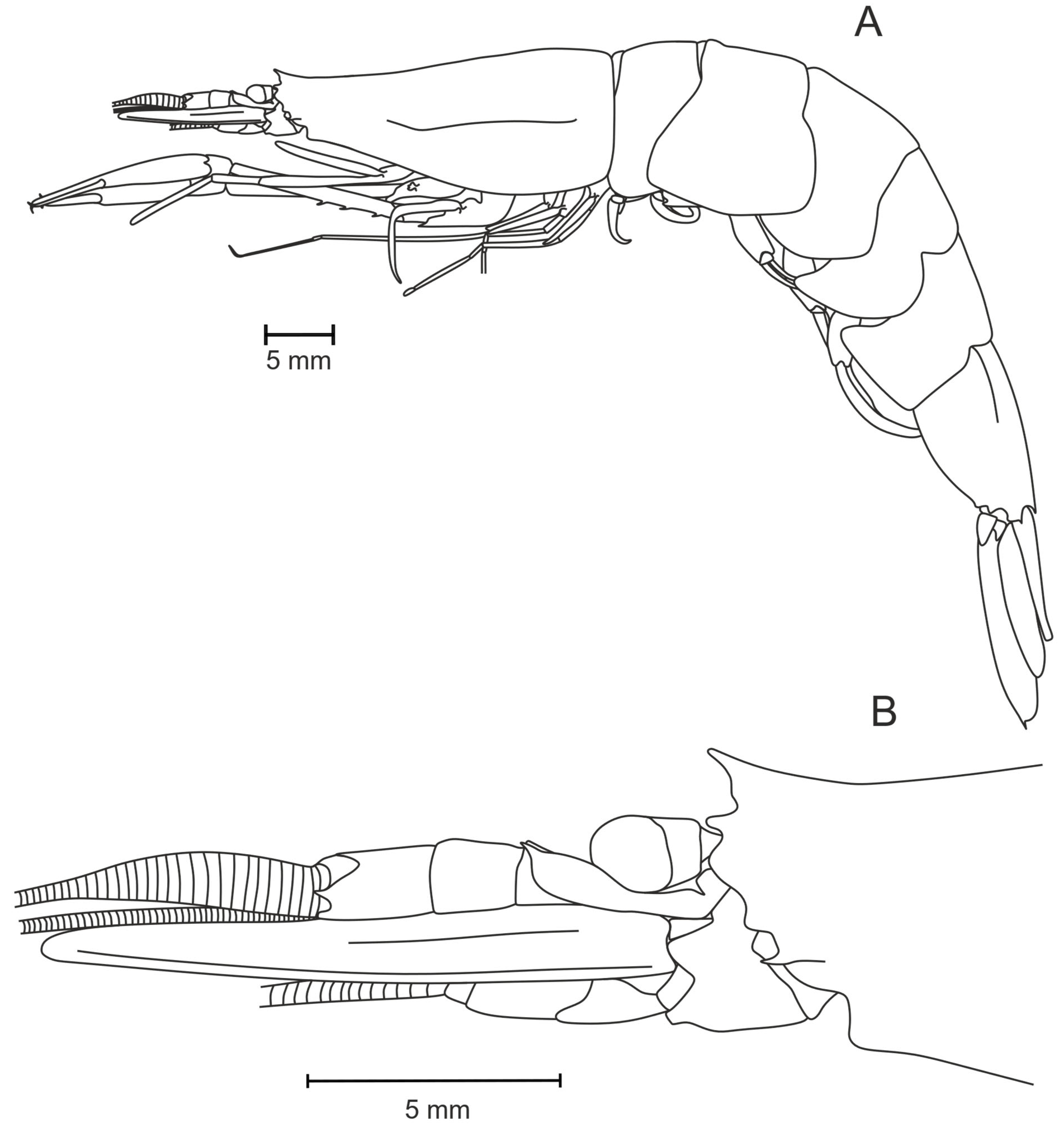
  • Carapace (Figure 2A and Figure 3A,B): Postsinusial height, maximum height, carapace length, length of the rostrum, rostrum base length, length of the branchiostegal tooth, length of the branchiostegal sinus, height of the branchiostegal sinus, sinus median, median sinus depth, length of the eye cornea and the eyestalk, length of the stylocerite, height of the stylocerite at its highest part;
  • Pleon (Figure 2A): Length of somites IV–VI, maximum height of the sixth somite, distal height of the sixth somite;
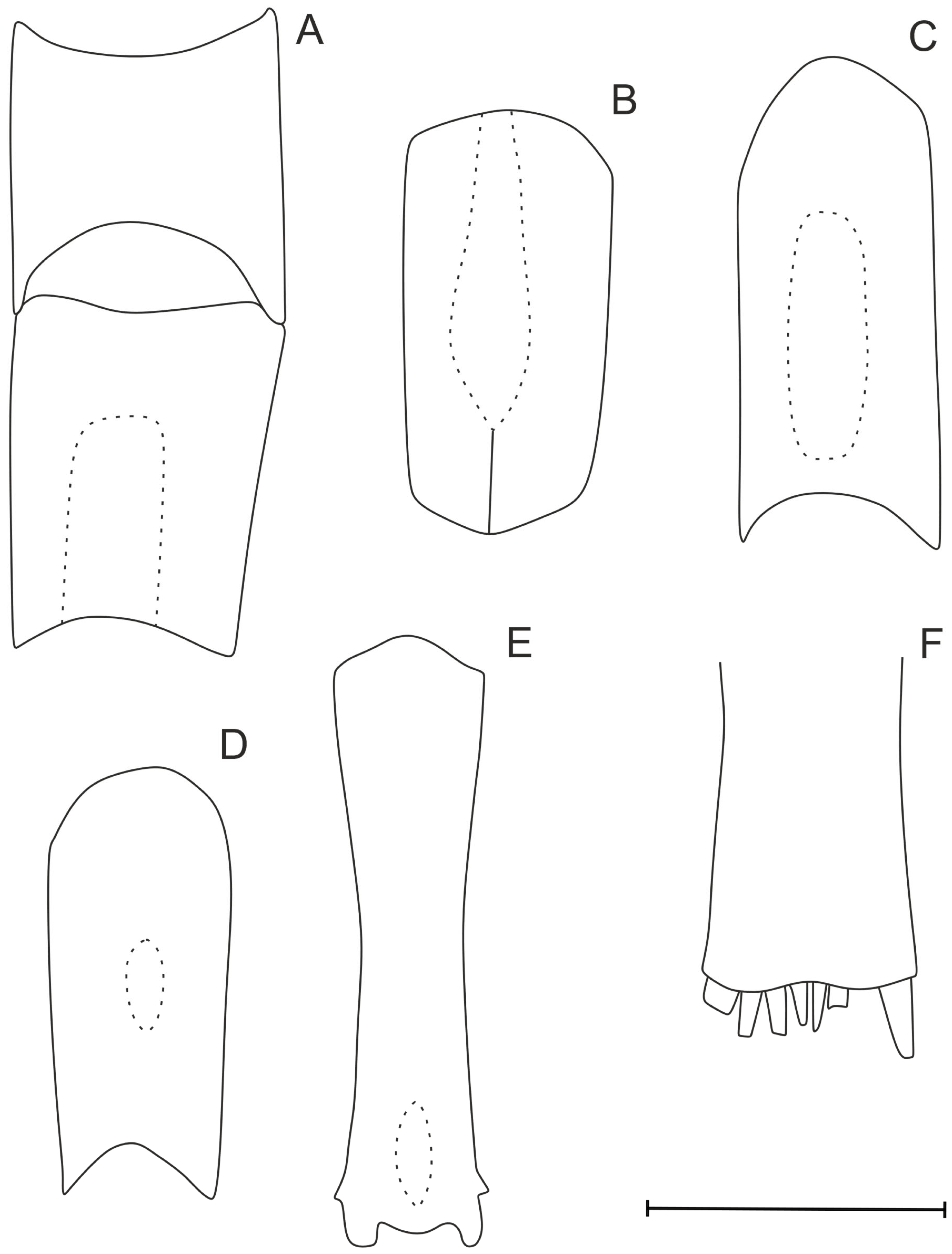
  • Telson (Figure 2A): Length of the telson, anterior width of the telson, posterior width of the telson end, depth of the telson concavity/convex curvature (Figure 2B);
  • Chelae (of pereopods I and II) (Figure 2C,D): Length of the fingers, length of the palm, maximum height of the palm.
The postsinusial height of the carapace (Figure 2A[a]) was measured from the posterior border of the branchiostegal sinus to the upper margin of the carapace, excluding the rostral elevation. The carapace length (Figure 2A[c]) was measured from the posterior orbital border to the posterior margin of the carapace in the dorsal midline.
The rostrum base length (Figure 3A[b]) was measured from the anterior margin of the rostrum (i.e., the point at which the carapace transitions into the rostral elevation) to the point where the rostrum smoothly merges with the carapace. Rostrum length (Figure 3A[a]) was measured from the midpoint of the rostrum base to the rostrum apex.
Measurements of the branchiostegal sinus are the most complex. The length of the branchiostegal sinus (Figure 3B[g]) was measured parallel to the dorsal margin of the carapace. The height of the branchiostegal sinus (Figure 3B[h]) was measured orthogonally relative to the sinus length. The branchiostegal sinus median (Figure 3B[j]) was defined as the median line of the right-angled triangle formed by the sinus length and height. The branchiostegal sinus median depth (Figure 3B[i]) was calculated as the distance from the carapace margin in the deepest part to the intersection of the sinus median and the hypotenuse of this triangle (as shown by the dotted line in Figure 3B).
The depth of the telson concavity was measured from the most protruding point at the telson’s posterior end to the level of the most concave point (Figure 2B, dotted line). If the telson was convex, the curvature was measured from the apex of the curvature to the straight line connecting the bases of the paired basal spines.
The length of the fingers should be understood as the line extending from the proximal articulation of the movable finger with the palm to the line connecting the tips of both fingers.

2.3. Morphological Approach

Throughout nearly two hundred years of studying Pasiphaea, different scientists have used various terms describing species of the genus. Here we suggest a standardized terminology regardless of the observer to describe all species in the same manner. Also, we suggest some terminology revision for a common understanding of all important characteristics:
  • The spine-like or triangle-like structure in the anterior part of carapace is often named as an epigastral tooth. However, this structure may be considered as a rostrum, albeit usually short and poorly developed. For consistency with other decapods, we call this structure a rostrum (Figure 3A[a,b]).
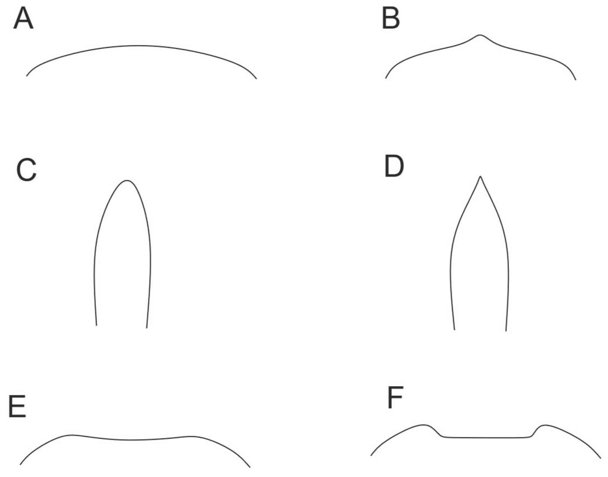
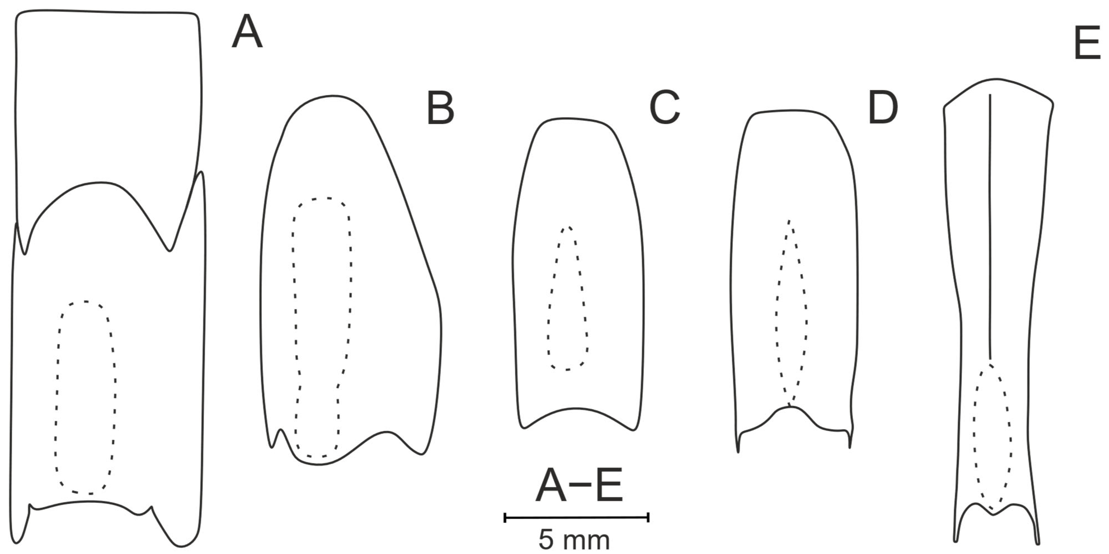
  • Pleonic relief is a big stumbling block in species describing. First, misinterpretation often comes in carinae identification in the case of a greatly flattened somite. We consider the somite to be rounded if the rib is absent (Figure 4A) and carinate if it has a distinct dorsal rib (Figure 4B); an evenly rounded dorsal margin may look rib-like due to a lateral compression but is considered here as rounded. This especially concerns the sixth pleonic somite, which is greatly flattened in a lateral direction and, although being rounded, may ostensibly look carinate (Figure 4C,D).
  • Another point at issue in the pleonic relief is the presence of a flattened (Figure 4E) or grooved (Figure 4F) zone on the pleonic somites. Almost any flattened zone is grooved to some extent in the middle and any grooved zone is flattened in the anterior or posterior part, so we synonymize these terms, assuming that flattenings and grooves may represent the same underlying structure, expressed to varying degrees, but the assessment of their prominence is subjective. As our material shows, differences in groove depth in the dorsal part of the somites are not significant in species differentiation. We suggest the term “depressions” for this kind of dorsal ornamentation as an intermediate between the two mentioned above.
  • Some species have a median protrusion or a proper tooth on the posterodorsal parts of their sixth pleonic somites (Figure 5). We consider both structures as two different characters: (1) a posterodorsal tooth which often has an acute apex, narrow base, and is well distinguished from other parts on the posterior somite (Figure 5C,F); (2) a posterodorsal protrusion which has a smooth apex, a base (visible from the dorsal view, posteriorly it appears as a blunt projection) at least as wide as the posterior margin, and smoothly continues into the posterior part of the somite (Figure 5B,E).

2.4. Genetic Approach

We used three mitochondrial gene markers to assess the genetic diversity of the collected shrimp specimens: cytochrome c oxidase subunit I (COI), 16S rDNA, and 12S rDNA. These markers offer comparable taxonomic resolution and are widely applied in DNA barcoding studies [23,24].
Total genomic DNA was extracted from pleonal muscle tissue using a modified direct lysis method [25]. Each 1.5 mL Eppendorf tube contained 150 µL of WLB buffer comprising: 1× Encyclo PCR buffer without MgCl2 (Evrogen™), 5% Chelex solution (v/v), and 5% Proteinase K (Qiagen™). Target regions of the selected gene markers were amplified using published primers: COI (LCO1490/HCO2198 [26]), 16S (16L2/16H3 [27,28]), and 12S (12S-psF1/R1 [29]).
Polymerase chain reactions were performed using a commercial master mix (ScreenMix-HS, Evrogen), containing 1× ScreenMix-HS, 0.4 μM of each primer, 1 μL of DNA template, and Milli-Q H2O to a final volume of 20 μL. Primer sequences, annealing temperatures, and PCR conditions are provided in Supplementary Table S1. PCR products were purified via ethanol precipitation and sequenced bidirectionally using BigDye Terminator v3.1 (Applied Biosystems, Foster City, CA, USA) on an ABI Prism 3500 Genetic Analyzer (Applied Biosystems, Foster City, CA, USA), following standard protocols at the Joint Centre for Molecular Diagnostics, Severtsov Institute of Ecology and Evolution, Russian Academy of Sciences. Chromatograms were processed and assembled using CodonCode Aligner v9.0.1 (CodonCode Corporation).
All newly obtained sequences were compared with existing GenBank entries using BLAST v.2.16.0 (NCBI) to identify potential contamination and were subsequently deposited in the GenBank database (accession numbers PV984862–PV984875, PV987548–PV987579).
To confirm the validity of newly described species, pairwise genetic distances were calculated between newly obtained sequences and those available in GenBank for Pasiphaea spp., using the Kimura 2-parameter (K2P) model [30] as implemented in MEGA X [31]. All available Pasiphaea sequences for the three target genes were retrieved from GenBank. For each gene fragment, sequences were aligned using MAFFT v7.526 [32]. Alignments of the protein-coding COI gene were additionally verified by translation into amino acid sequences using TranslatorX [33], to ensure the absence of premature stop codons.
The final datasets (Supplementary Table S2) were trimmed to the length of the shortest high-quality sequence in each alignment—532 bp for COI, 440 bp for 16S, and 388 bp for 12S—and were used for the calculation of K2P distances.
The concatenated dataset was used to assess phylogenetic relationships between species under Bayesian inference (BI) and ML analyses. Representatives of other genera of the family Pasiphaeidae were added to the dataset as an outgroup (Supplementary Table S2). The partitioning scheme and best-fitting substitution models for each partition were determined using PartitionFinder2, according to the corrected Akaike information criterion (AICc). The final dataset was divided into 4 partitions with corresponding best-fit models of nucleotide substitutions (Supplementary Table S3). The BI was run in MrBayes 3.2; two independent runs, each consisting of 4 chains, were executed for 10,000,000 generations with model parameters estimated during the analysis. Chains were sampled every 1000 generations, and the first 25% trees were discarded as “burn-in”. A 1% average standard deviation of split frequencies was reached after 950,000 generations, and a 75% majority rule consensus tree was constructed from the remaining trees to evaluate clade confidence from posterior probabilities estimation. In the ML analysis, we used the GTR + G model for each partition in the concatenated dataset, with all free parameters estimated by RAxML with 10 independent runs. Confidence values of branches in the resulting trees were evaluated by a thorough bootstrap procedure with 1000 replicates. We considered the clades statistically supported if they had a support of posterior probabilities ≥ 0.9 in the BI analysis and a bootstrap value of ≥70% in the ML analysis.

3. Results

3.1. Taxonomic Part

Pasiphaeidae Dana, 1852
Pasiphaea Savigny, 1816
Pasiphaea allainae sp. nov.
urn:lsid:zoobank.org:act:CC80DE30-2F55-4798-B313-19C6313807E2
Material Examined: Holotype (Collection number WAL09-M195-200)—ovigerous female CL 25.6 mm. Off NW coast of Wallis and Futuna Islands, R/V Alis. Campagne WALLALIS: st. WAL09, 13°12′50.4″ S, 176°16′51.6″ W, horizontal trawl: 260 m, 14 July 2018 (night).
Paratypes: Female (WA3-17-M334-03) CL 23.2 mm. Off W coast of Gilbert Islands, R/V Antea. Campagne WARMALIS3: st. WA3-17, 0°0′2.94″ N, 173°19′22.98″ E, oblique trawl: 24.4–245 m, 29 October 2023 (night); female (WAL07-M191-49) CL 23.7 mm. Off NW coast of Wallis and Futuna Islands, R/V Alis. Campagne WALLALIS: st. WAL07, 13°15′46.8″ S, 176°18′54″ W, horizontal trawl: 500 m, 12 July 2018 (night); male (MNHN-IU-2018-1617) CL 20.4 mm. Solomon Islands, SW Russell Island, R/V Alis. Campagne SALOMON 2: CP 2176, 09°09.4′ S, 158°59.2′E, 600–875 m, 21 October 2004.
Diagnosis: Rostrum reaching anterodorsal margin of carapace, triangular, apex slightly curved forward. Carapace dorsally rounded, branchiostegal tooth set behind carapace margin and overlapping this margin. Branchiostegal sinus prominent, deepest part extended in vertical direction. Pleonic somites second to fifth depressed dorsally and lacking posterodorsal tooth. Sixth pleonic somite dorsally carinate in anterior part, depressed and dorsally protruded in posterior part. Telson shorter than sixth pleonic somite, dorsally flattened, posterior end straight to slightly concave at middle, with four pairs of spines. First pereopod: basis with single distal tooth, ischium unarmed, merus with 10–16 teeth, fingers 0.71–0.75 times as long as palm of chela. Second pereopod: basis with single distal tooth, ischium unarmed, merus with 19–23 teeth, fingers 1.08–1.16 times as long as palm of chela. Five pleurobranchs and three arthrobranchs on each side.
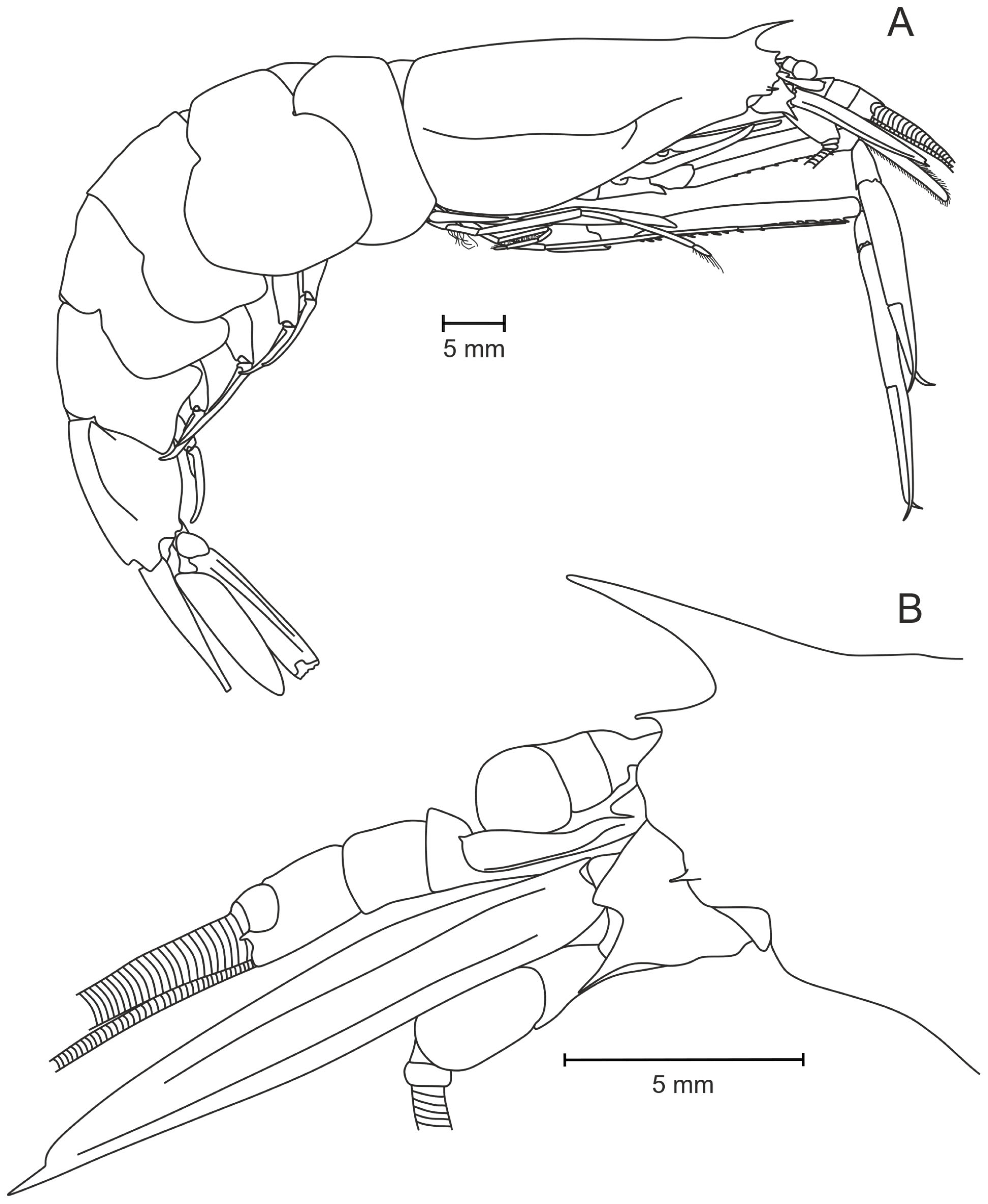
Description (of Holotype): Rostrum (Figure 7A,B) triangular, frontal margin slightly convex basally, set near anterior margin of carapace, apex straight or slightly curved and reaching anterior margin of carapace. Branchiostegal tooth overlapping anterior margin of carapace by 0.62 of its length in lateral view. Carapace (Figure 7A) without dorsal carina, suprabranchial carina distinct. Branchiostegal sinus 0.64 times as long as wide.
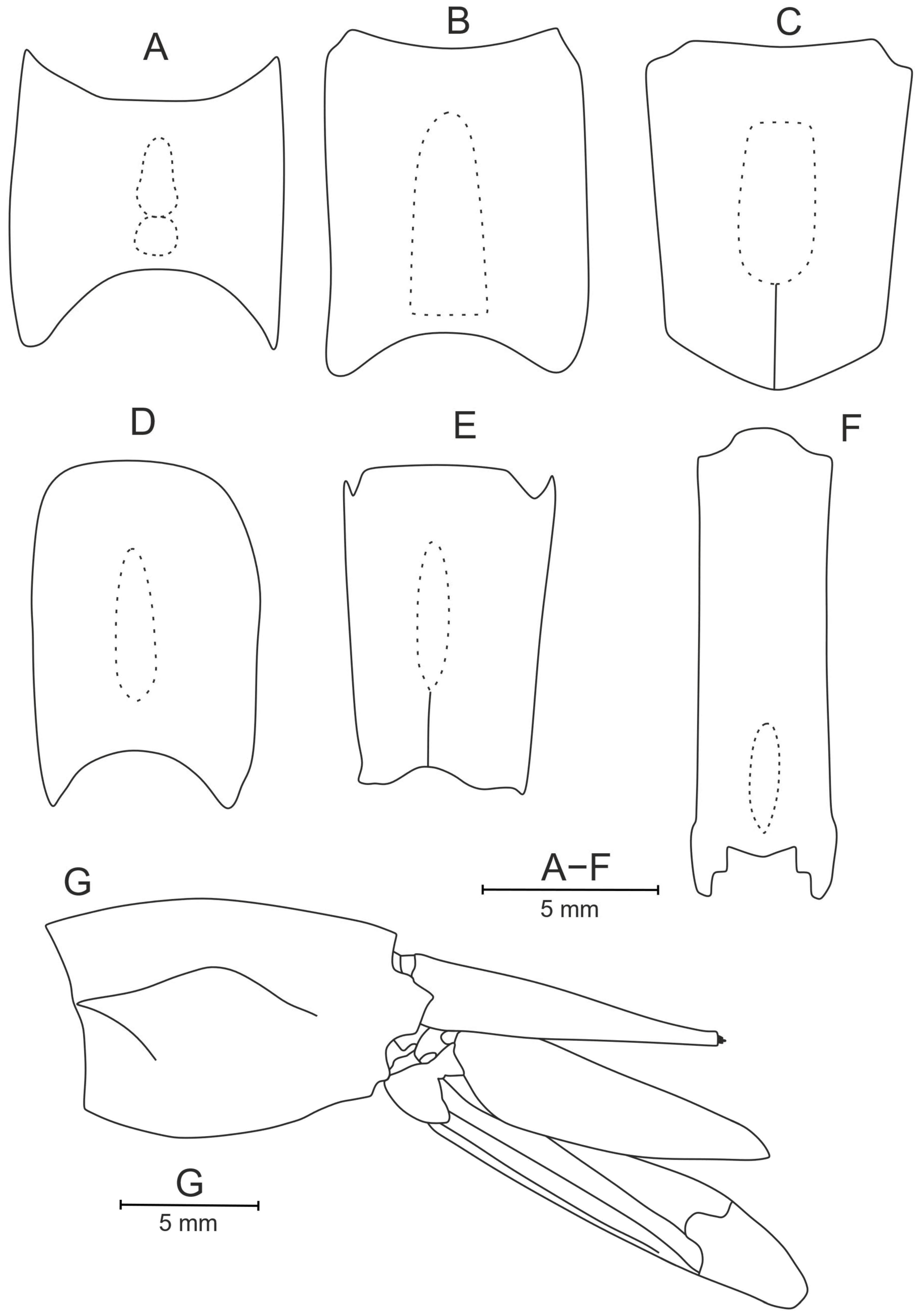
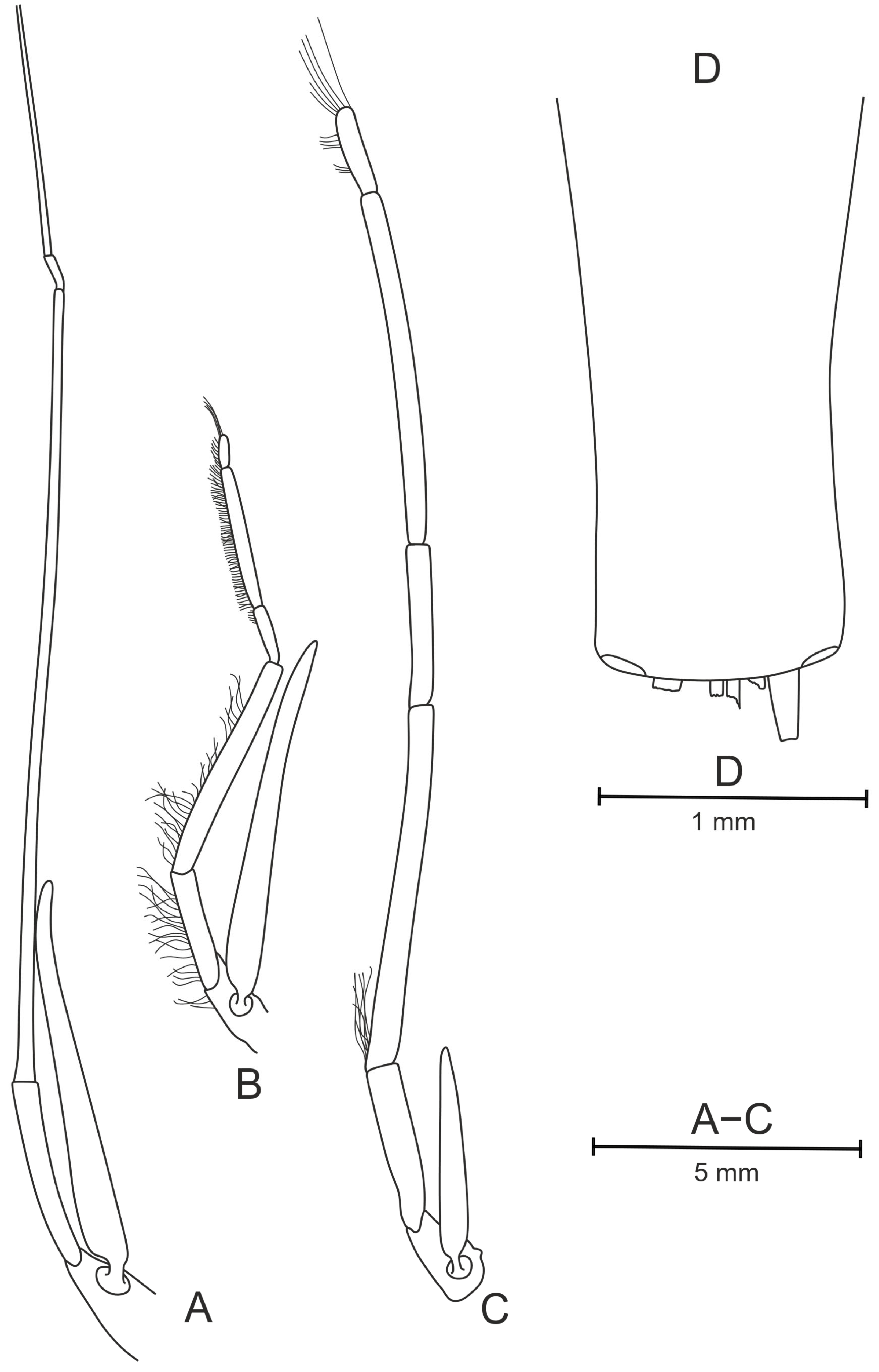
All pleonic somites (Figure 8) without posterodorsal teeth, sixth somite posterodorsal protrusion 0.31 times as long as wide and resembling posterodorsal tooth from lateral view. First somite dorsally rounded along entire length; second to fifth somites dorsally depressed in posterior 0.6–1.0, 0.75–1.0, 0.35–0.80, and 0.5–1.0 parts of their length, respectively; sixth somite dorsally carinate in anterior 2/3 part, depressed in posterior 1/3 part and laterally carinate in anterior 0–0.5 part. Telson 0.76–0.78 times as long as sixth pleonic somite, with well-marked dorsal groove along its entire length. Telson end almost straight laterally and slight medial depression 0.10 times as deep as wide, armed with four pairs of terminal robust setae, and single pair of subterminal lateral dorsal setae (broken—Figure 11D).
Eye cornea well pigmented, dark-brown. Stylocerite (Figure 7B) elongate, curved, widened at end, narrowing in sharp end, not reaching distal end of first segment of antennule. Scaphocerite distinctly overlapping antennular peduncle, 3.53 times as long as wide, distolateral tooth distinct. Basicerite with long robust tooth.
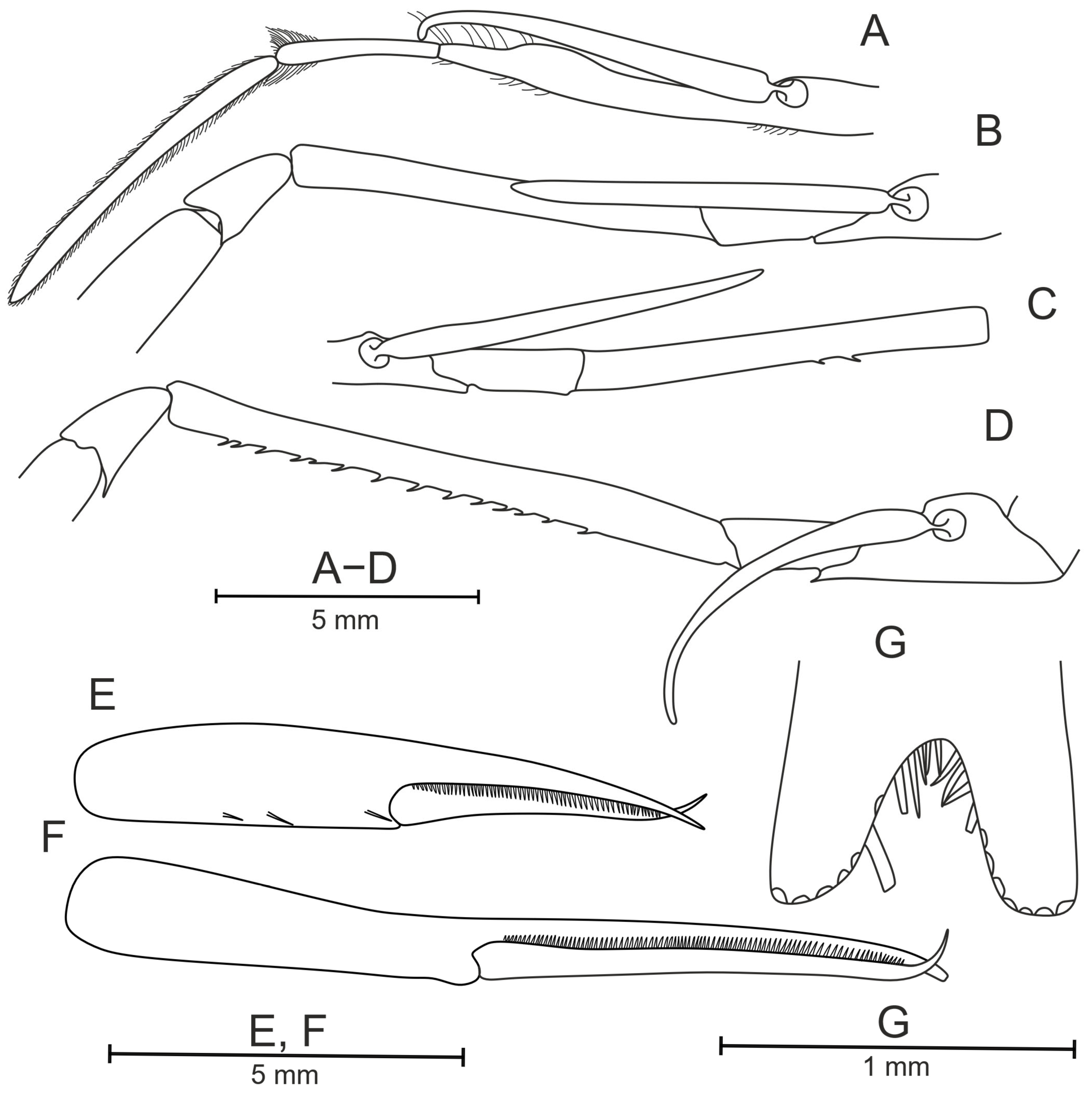
Mandible (Figure 9A) with 10 teeth on cutting part; maxillae I without distinct subdistal process on endopodite and long bristle, basal endite armed with row of large teeth and small teeth in between, coxal endite armed distally with small spines (Figure 9B); maxilliped I with visible reduced endopodite (Figure 9C). Maxilliped III (Figure 10A) slightly overlapping scaphocerite; terminal segment (dactylus + propodus) 1.7 times longer than penultimate (carpus); exopod well developed.
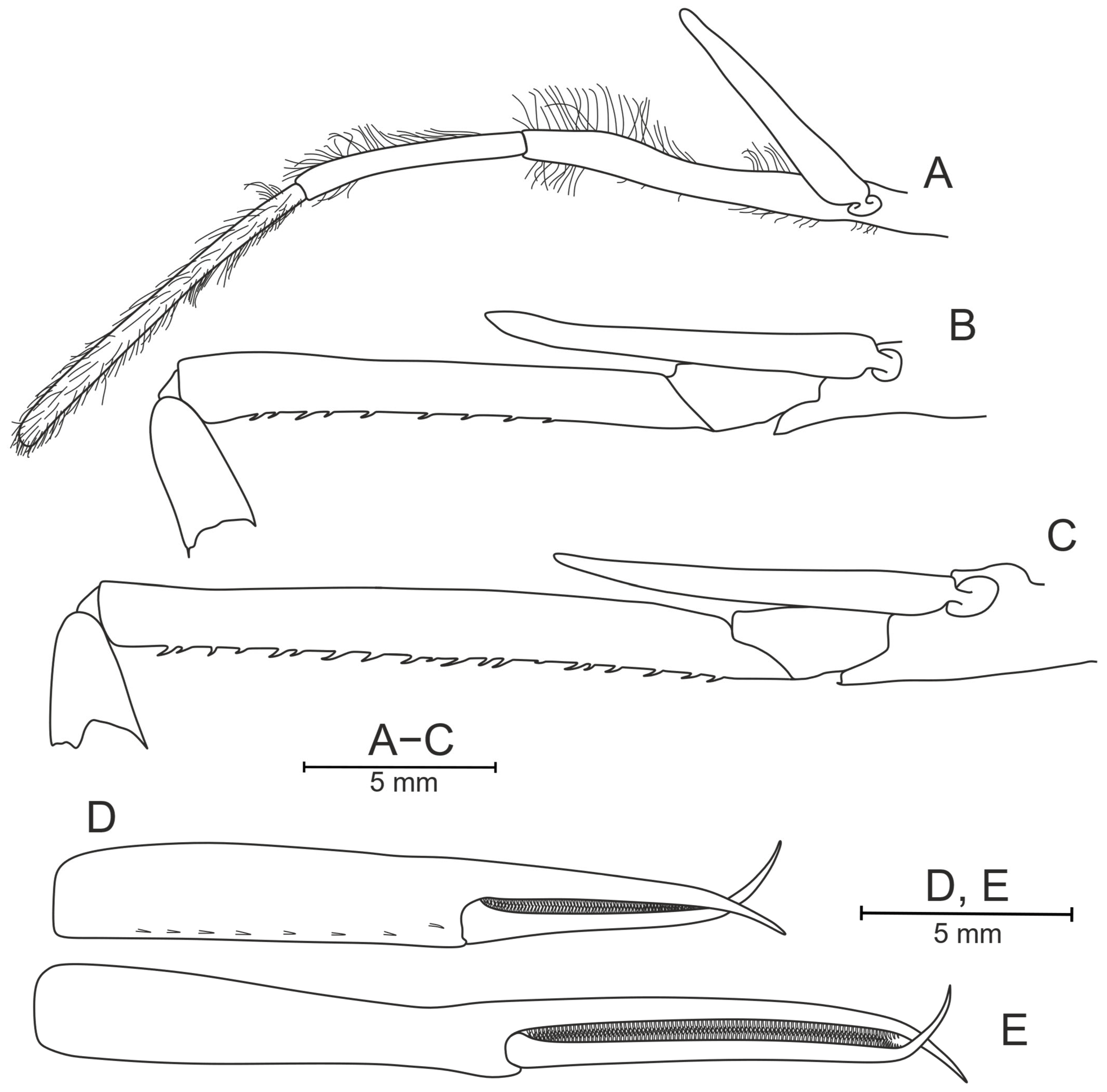
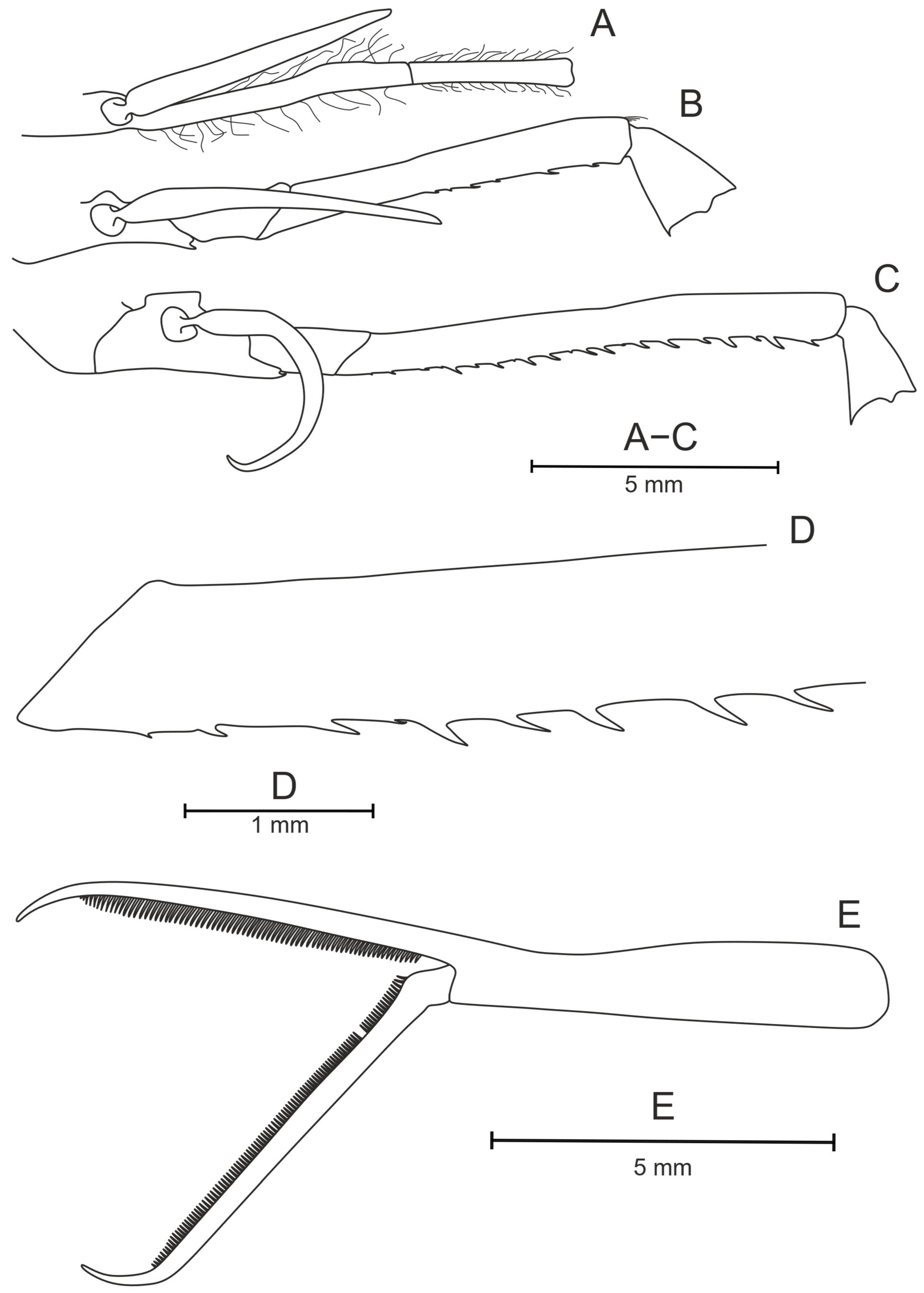
First pereopod (Figure 10B) extending beyond scaphocerite by about 0.4 times length of palm. Basis with single very long ventrally directed distal tooth. Merus with 15–16 teeth evenly distributed along entire length of segment. Carpus short, with large teeth on dorsal and ventral parts of distal margins. Chela with two ventral bristles on propodus, fingers 0.71–0.75 times as long as palm (Figure 10D).
Second pereopod (Figure 10C) overlapping first pereopod by 0.21 times length of fingers. Basis with single long ventrally directed distal tooth that is smaller than that on first pereopod. Merus with 23 teeth on lower margin evenly distributed along entire length of segment. Carpus short, with large teeth on lower and upper regions of distal margin. Fingers 1.08–1.16 times as long as palm (Figure 10E).
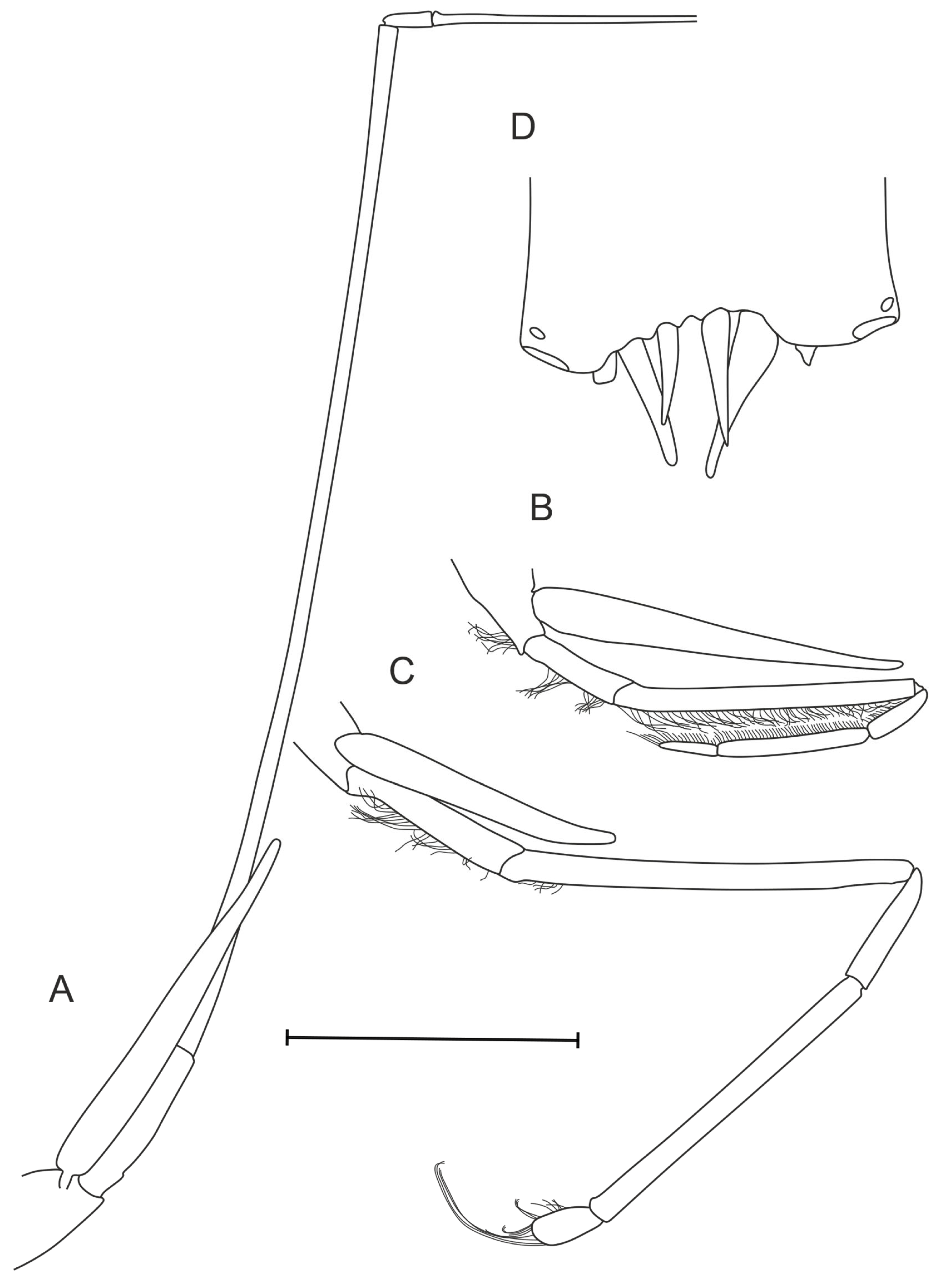
Third pereopod (Figure 11A) slender, extending beyond anterior margin of carapace by last two segments; not bearing teeth on any segment. Fourth pereopod (Figure 11B) shortest, bearing dense brush of short bristles on each segment including distal part of basis; teeth absent on all segments. Fifth pereopod (Figure 11C) longer than fourth, bearing bristles only on basis and dactylus, latter rounded and bearing longer bristles; teeth absent on all segments.
Branchial Formula: Five pleurobranchs, three arthrobranchs.
Eggs small, 1.6 × 1.0 mm. Size—CL 23.2–25.6 mm.
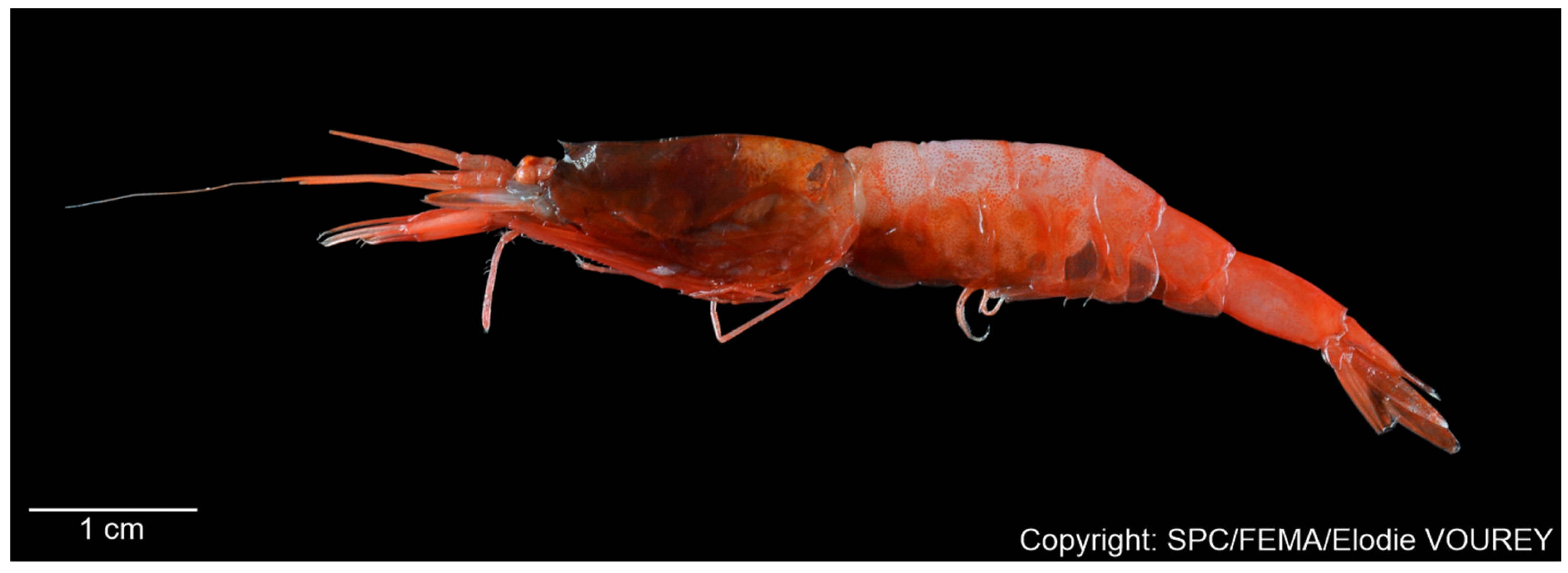
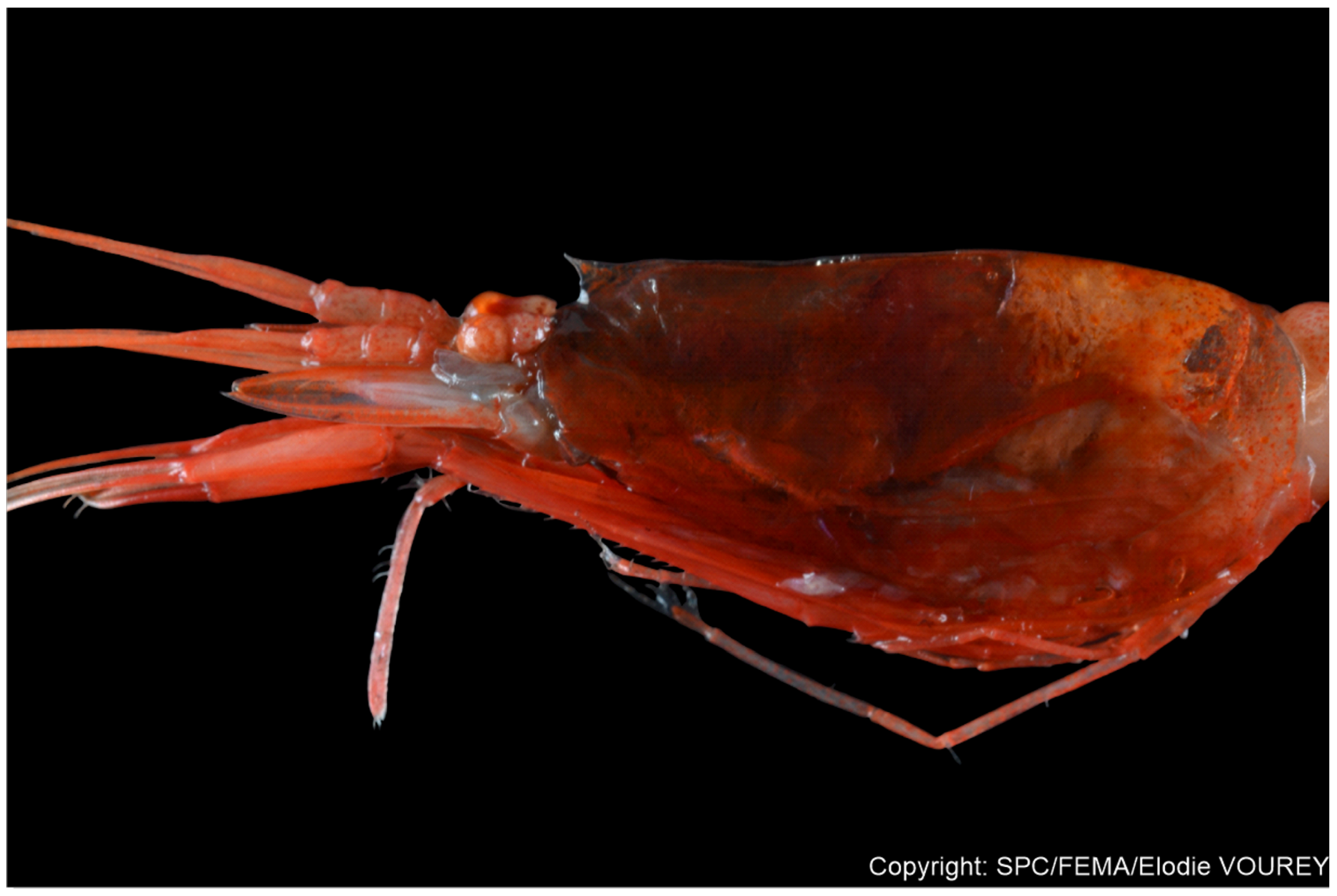
Color: Semitransparent with orange-red dots.
Range: From the NW coast of Wallis Island to Solomon Islands and Gilbert Islands on the north (Figure 1); mesopelagic above bottom depths 25–875 m.
Etymology: The species name allainae is given in honor of Dr Valérie Allain, whose leadership of the Taxonomy Laboratory of the Fisheries, Aquaculture, and Marine Ecosystems Division (The Pacific Community, Noumea) has been instrumental in establishing a rigorous taxonomic framework in the central Pacific. Her efforts have underpinned numerous discoveries in Pacific marine biodiversity.
Authorship of Species Name: The taxonomic basis for the erection of this species was prepared by Tikhomirov & Vereshchaka, who are thus solely responsible for making the new species name Pasiphaea allainae sp. nov. available.
Remarks: The specimen of P. allainae sp. nov. from MNHN (MNHN-IU-2018-1617) was erroneously identified by Hayashi as P. truncata. Unfortunately, the P. truncata description is rather incomplete and superficial, being based mainly on comparison with the species P. kaiwiensis Rathbun, 1906; however, it is still possible to identify reliable distinctions from the described species. Both species are easily distinguished by (1) no dorsal carina of carapace in P. allainae sp. nov. vs. carapace carinated right after rostrum in P. truncata; (2) pleonic somites second to sixth are depressed in P. allainae sp. nov. vs. rounded in P. truncata; (3) sixth somite dorsally carinated in anterior 2/3 with distinct depression after it in P. allainae sp. nov. vs. fully dorsally carinated merging smoothly into a posterodorsal spine in P. truncata; (4) the posterior shape of the sixth pleonic somite: posterodorsal protrusion in P. allainae sp. nov. vs. posterodorsal spine in P. truncata.
Morphologically, the new species belongs to a complex defined here as the “P. planidorsalis” complex. This complex shares vertically extended branchiostegal sinus, depressions of the pleonic somites (second to sixth somites depressed in any part), and elongated distal tooth on the basis of the first pereopod. (Table 1).
Variations: All specimens are similar to each other possessing only minor variations in pleonic ornamentation, telson, and number of teeth on pereopods.
(1) Some pleonic somites deepen to visible groove in flattened area (in WA3-17-M334-03);
(2) Fifth somite depressed zone covers distal 0.6–1.0 of all somite length (in WA3-17-M334-03 and WAL07-M191-49);
(3) Posterodorsal protrusion of sixth somite varies from 0.20 (in MNHN-IU-2018-1617) and 0.27 (in WAL07-M191-49) to 0.4 times as long as wide (in WA3-17-M334-03);
(4) All paratypes have straight or slightly convex telson end, telson end curvature varies from 0.04 (in WA3-17-M334-03) and 0.06 (in WAL07-M191-49) to 0.10 times as long as wide (in MNHN-IU-2018-1617).
(5) Pereopod I merus has 10 teeth on ventral side, pereopod II merus has 19 (in MNHN-IU-2018-1617); 11–14 on pereopod I, 21 on pereopod II (in WAL07-M191-49); 14–15 on pereopod I, 22–23 on pereopod II (in WA3-17-M334-03).
Molecular distances suggest significant divergence between the new species and the rest of Pasiphaea. The new species is close to P. planidorsalis, P. aequus, and P. portalae sp. nov., but recorded differences show species-level genetic distances among them (Table 2).
Pasiphaea exilimanus Komai, Lin & Chan, 2012
Material Examined: Male (WAL07-M191-64) CL 21.7 mm. Off NW coast of Wallis and Futuna Islands, R/V Alis. Campagne WALLALIS: st. WAL07, 13°15′46.8″ S, 176°18′54″ W, horizontal trawl: 500 m, 12 July 2018 (night).
Diagnosis: Rostrum directed slightly dorsally, terminating in sharp tooth, apex just reaching anterodorsal margin of carapace (according to original description, since tip of our specimen’s rostrum is broken) [16]. Carapace dorsally with carina only in anterior part behind rostrum, medial and posterior parts rounded, branchiostegal tooth set just behind carapace margin and overlapping this margin. Branchiostegal sinus slightly extended more in its longitudinal then in vertical direction. All pleonic somites rounded dorsally and lacking posterodorsal tooth. Telson shorter than sixth pleonic somite, dorsally flattened, posteriorly slightly concave, armed with five pairs of spines. First pereopod: basis with single distal tooth, ischium unarmed, merus with 6–8 ventral teeth, fingers 0.96–1 times as long as palm of chela. Second pereopod: basis with single distal tooth, ischium unarmed, merus with 22–23 ventral teeth, fingers 1.27 times as long as palm of chela. Five pleurobranchs and three arthrobranchs on each side.
Size: Largest male CL 22.4 mm and largest female CL 22.3 mm, smallest ovigerous female CL 18.9 mm [16].
Color: Orange-red.
Distribution: Known only from the northeastern and southwestern coasts of Taiwan [16] and the northwestern coast of Wallis and Futuna Islands (this study), mesopelagic above bottom depths of 451 to 2556 m [16].
Pasiphaea fragilis Hayashi, 1999
Material Examined: Female (WA2-04-M284-51) CL 13.8 mm. Central Pacific, R/V Alis. Campagne WARMALIS2: st. WA2-04, 11°59′13.2″ S, 150°2′16.8″ W, horizontal trawl: 496.2–506.6 m, 18 September 2022 (night); ovigerous female (MNHN-IU-2018-1599) CL 15.1 mm and male (MNHN-IU-2018-1599) CL 13.4 mm. Central Pacific, Marquesas Islands. Campagne MUSORSTOM 9: st. CP 1262, 09°20′ S, 140°08′ W, 805–905 m, 7 September 1997.
Diagnosis (based on both P. fragilis and P. gracilis): Rostrum very small, barely reaching midlength between anterior margin of carapace and rostrum base. Carapace dorsally rounded, branchiostegal tooth set behind carapace margin and overlapping it. Branchiostegal sinus extended more in its longitudinal than in its vertical direction. All pleonic somites rounded dorsally and lacking posterodorsal tooth, except sixth somite. Posterodorsal tooth on sixth somite directed slightly upwards. Telson shorter than sixth pleonic somite, dorsally grooved, posteriorly truncated, with four pairs of spines (telson tip modified from Hayashi) [12]. First pereopod: basis with single distal tooth, ischium unarmed, merus with 6–9 ventral teeth (merged data of two species), fingers 0.79–0.81 times as long as palm of chela. Second pereopod: basis with single distal tooth, ischium unarmed, merus with 9–14 ventral teeth (merged data of two species), fingers 1.10 times as long as palm of chela. Four pleurobranchs and three arthrobranchs on each side.
Size: CL 13.8 mm.
Distribution: Known only from the New Caledonia (Loyalty Islands) [12], Wallis and Futuna Islands (as P. gracilis [12]) and the Marquesas Islands, Central Pacific (this study); mesopelagic to bottom depths of 496–905 m.
Remarks: The rostrum in the examined specimen is either underdeveloped or injured, bud-like; however, it is apparent that its length extends to approximately the midpoint between the rostral base and the anterior dorsal margin of the carapace. The telson tip is damaged. Excluding discussed defects, the present specimen differs from the original description solely in having a slightly greater number of teeth on the meri of the first (7–8 vs. 6–9) and second pereopods (13–14 vs. 9–12 teeth in P. fragilis). In all other morphological attributes, the specimen conforms almost entirely to the descriptions of P. gracilis and P. fragilis.
Hayashi [12] delineated four primary distinctions between P. gracilis and P. fragilis:
  • Integumental consistency, described as “fragile” in P. fragilis versus “firmer” in P. gracilis.
  • Orientation of the posterodorsal tooth on the sixth pleonic somite, which is inclined slightly upward in P. fragilis but aligned flush with the dorsal surface of the somite in P. gracilis.
  • Armament of the first pereopod merus, bearing 6–9 ventral teeth in P. fragilis compared to 7–9 in P. gracilis.
  • Armament of the second pereopod merus, bearing 9–12 ventral teeth in P. fragilis versus 11–14 in P. gracilis.
These minor distinctions are highly likely attributable to differences in specimen maturity. Indeed, the observed series of P. fragilis comprised individuals with carapace lengths of 10.8–10.9 mm, whereas those of P. gracilis measured 12.3–15.1 mm [12]. This disparity in size readily accounts for the observed variation in integumental firmness (character 1) and the ranges of ventral dentition on pereopods I and II (characters 3 and 4), particularly given substantial overlap in spine counts and the limited number of specimens examined. Moreover, the purported divergence in posterodorsal tooth orientation (character 2) is subjective, as acknowledged in the original methods.
Our newly analyzed specimen (WA2-04-M284-51) exhibits intermediate characters, combining aspects of both P. fragilis and P. gracilis, thereby bridging the morphological gap between the two nominal taxa. This intermediate morphology substantiates the hypothesis that the previously described differences are ontogenetically based rather than indicative of distinct species.
Finally, the molecular data in the Supplementary Materials Table S2 corroborate the morphological evidence, confirming that P. fragilis and P. gracilis represent a single species. Consequently, we herein synonymize both species and consider P. fragilis as a senior synonym of P. gracilis.
Pasiphaea komaii sp. nov.
urn:lsid:zoobank.org:act:F1B29EF0-8C01-4871-9C29-680B1842C357
Material Examined: Holotype—ovigerous female (MNHN-IU-2018-1619) CL 20.2 mm. Solomon Islands. SALOMON 2 CP2176, 600–875 m, 09°09.4′ S, 158°59.2′E, 21 October 2004.
Diagnosis: Rostrum apex not overlapping rostrum base, not reaching anterodorsal margin of carapace. Carapace dorsally rounded, branchiostegal tooth set behind carapace margin and overlapping this margin. Branchiostegal sinus slightly extended more in its vertical then in longitudinal direction, barely visible. All pleonic somites rounded dorsally and lacking posterodorsal tooth. Telson shorter than sixth pleonic somite, dorsally rounded, posteriorly slightly concave, with four pairs of spines. First pereopod: basis with single distal tooth, ischium and merus unarmed, fingers 0.77 times as long as palm of chela. Second pereopod: basis with single distal tooth, ischium unarmed, merus with single ventral tooth, fingers 1.36 times as long as palm of chela. Five pleurobranchs and three arthrobranchs on each side.
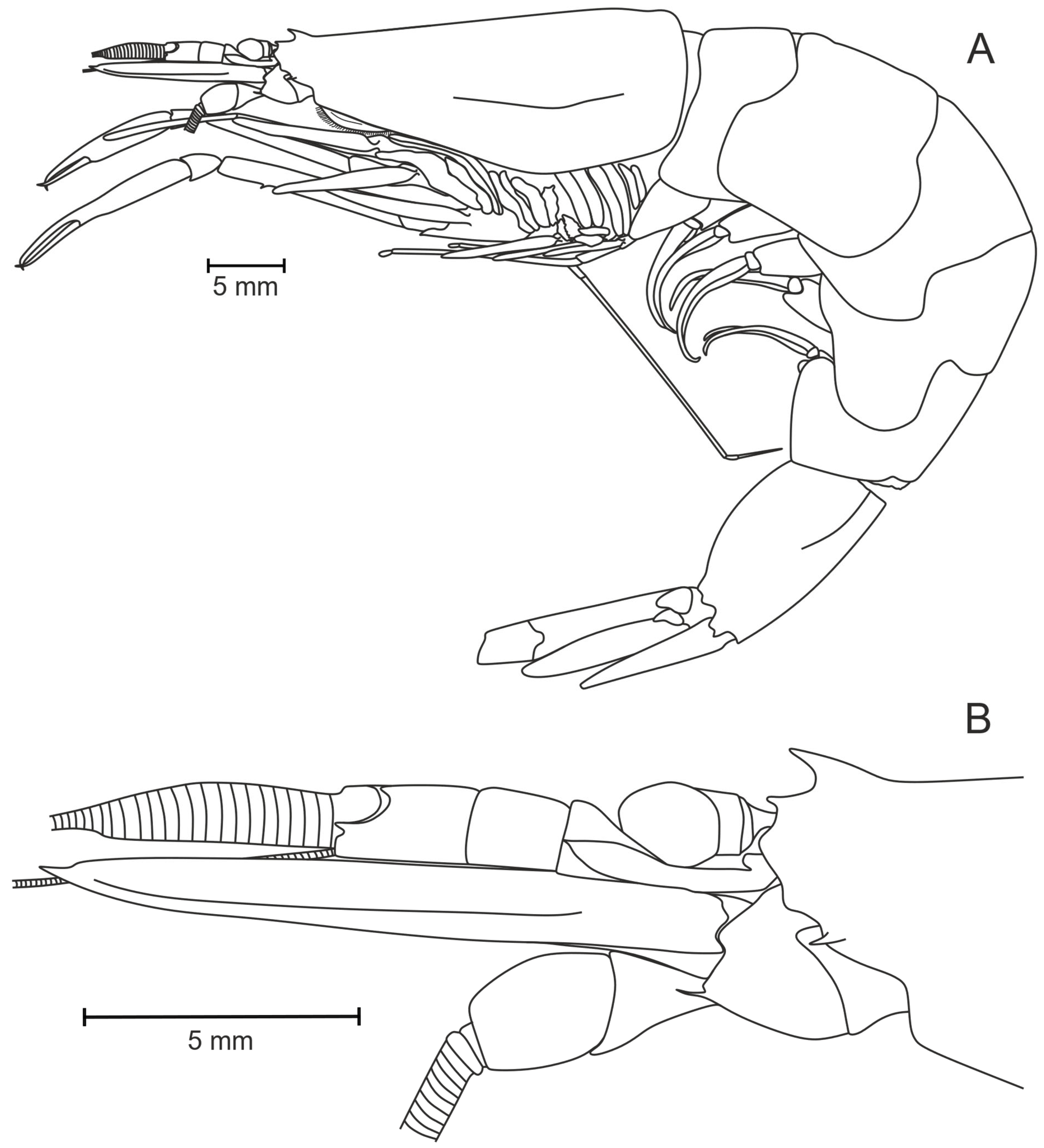
Description (of Holotype): Rostrum (Figure 13A,B) as wide triangle, basally extended in anterior direction, set near anterior margin of carapace. Carapace (Figure 13A) dorsally rounded along entire length, no lateral carinae. Branchiostegal tooth overlapping anterior margin of carapace by 0.50 of its length in lateral view. Branchiostegal sinus 0.76 times as long as wide.
All pleonic somites without posterodorsal teeth. Sixth somite with lateral carina in anterior 0–0.43 part of somite (Figure 13A). Telson 0.64 times as long as sixth pleonic somite, rounded along its entire dorsal side. Telson terminally concave in middle, incision 0.10 times as deep as wide, armed with four pairs and one nonpaired terminal robust spines (Figure 14F).
Eye cornea well pigmented, dark-brown. Stylocerite (Figure 13B) elongate, curved, widened at end, narrowing in sharp end, slightly not reaching distal end of first segment of antennule. Scaphocerite distinctly overlapping antennular peduncle, 4.00 times as long as wide, distolateral tooth distinct. Basicerite with thin tooth.
Maxilliped III (Figure 14A) not reaching scaphocerite distal edge, scaphocerite overlapping maxilliped III by 0.11 of scaphocerite length; terminal segment (dactylus + propodus) 1.79 times longer than penultimate (carpus); exopod well developed.
First pereopod (Figure 14B) extending beyond scaphocerite by 0.63 times length of fingers. Basis armed with long ventrally directed distal tooth. Carpus with small tooth on distal external lateral margin. Chela with two ventral bristles on propodus, fingers 0.76 times as long as palm on both chelae (Figure 14D).
Second pereopod (Figure 14C) overlapping first pereopod by about 0.18 times length of fingers. Basis with single distal tooth subparallel to margin of basis, apex slightly curved dorsally. Merus with one tooth on lower margin in proximal 0.39–0.43 times length of segment. Carpus short, with long tooth on distal part of ventral margin.
Third pereopod slender, not bearing teeth on any segment. Fourth pereopod shortest, bearing dense brush of short bristles on dactylus, propodus, distal part of carpus and ischium; teeth absent on all segments. Fifth pereopod longer than fourth, bearing bristles only on dactylus which is rounded; teeth absent on all segments.
Branchial Formula: Five pleurobranchs, three arthrobranchs.
Size: CL 20.2 mm.
Distribution: Waters of Solomon Islands.
Etymology: Named in tribute to Dr Tomoyuki Komai, a distinguished carcinologist whose pioneering research has greatly advanced modern decapod taxonomy.
Authorship of Species Name: The taxonomic basis for the erection of this species was prepared by Tikhomirov & Vereshchaka, who are thus solely responsible for making the new species name Pasiphaea komaii sp. nov. available.
Remarks: The holotype was identified by Hayashi as Pasiphaea planidorsalis Hayashi, 2004 in 29 August 2005. However, this specimen does not agree with the original description of P. planidorsalis for the following reasons: (1) the rostral tip does not extend beyond the rostral base, and the rostrum bears a broad ventral projection in P. komaii sp. nov. vs. nearly reaching the anterior margin of the carapace, without ventral projection in P. planidorsalis; (2) there are no dorsal depressions on pleonic somites in P. komaii sp. nov. vs. 2–5 somites being dorsally depressed in P. planidorsalis; (3) the telson is terminally concave in P. komaii sp. nov. vs. truncated in P. planidorsalis.
Morphologically, the new species is most similar to three species—P. cristata, P. americana Faxon, 1893, and P. poeyi Chace, 1939—in (1) carapace relief, (2) pleonic relief, and (3) pereopod armament.
Pasiphaea komaii sp. nov. is also similar to P. cristata in (1) rostrum shape and (2) position of the meral tooth on pereopod II (at 0.39–0.43 of its length in P. komaii sp. nov. vs. 0.40–0.46 of its length in P. cristata) and differs in the following ways: (1) the telson apex (slightly concave in P. komaii sp. nov. vs. slightly convex in P. cristata); (2) armament of basis in pereopod I (long distal ventrally directed tooth in P. komaii sp. nov. vs. no teeth on basis in P. cristata).
The new species differs from P. americana in four ways: (1) branchiostegal sinus shape (barely distinguishable and shallow in P. komaii sp. nov. vs. deep and noticeable in P. americana), (2) pleonic relief (fully rounded in P. komaii sp. nov. vs. uncertainly plain in P. americana according to Hayashi [12]), (3) telson apex (slightly concave in P. komaii sp. nov. vs. deeply concave in P. americana), (4) number of terminal spines on the telson (4 pairs in P. komaii sp. nov. vs. 7–10 pairs in P. americana).
The new species differs from P. poeyi in five ways: (1) rostrum shape (wide and short in P. komaii sp. nov. vs. curved directed more forwardly triangular rostrum in P. poeyi), (2) distal tooth of the basis on pereopod II (curved upwards in P. komaii sp. nov. vs. parallel or directed ventrally to basis margin in P. poeyi), (3) shorter pereopod I (overlapping scaphocerite by 0.63 of finger length in P. komaii sp. nov. vs. 0.73 in P. poeyi), (4) pereopod II meral tooth position (at 0.39–0.43 of its length in P. komaii sp. nov. vs. 0.21–0.26 of its length in P. poeyi), (5) telson dorsal surface (fully rounded in P. komaii sp. nov. vs. flattened in P. poeyi).
Other species with similar armament of the meri of pereopod I (unarmed) and of pereopod II (a single tooth) have either a dorsally different carapace (carinate in Pasiphaea merriami, Pasiphaea sirenkoi, Pasiphaea unispinosa Wood-Mason, 1892) or pleon (some somites have carinate or variably depressed parts in Pasiphaea merriami, Pasiphaea sirenkoi, Pasiphaea telacantha Hayashi, 2004, and Pasiphaea tinyamchani sp. nov.).
Molecular distances suggest significant divergence between the new species and the rest of Pasiphaea. The new species is close to P. cristata, P. telacantha, and P. allainae sp. nov. (10.9, 16.3% and 16.9% difference in COI gene markers), to P. cristata, P. aequus, and P. tinyamchani sp. nov. (6.9, 13.0, and 13.9% difference in 16S gene markers) and to P. cristata, P. aequus, and P. tinyamchani sp. nov. (11.8, 14.4, and 14.8% difference in 12S gene markers). The closest species in both morphological and molecular ways—P. cristata—significantly differs from the discussed P. komaii sp. nov.
Pasiphaea machfulae sp. nov.
urn:lsid:zoobank.org:act:236A60F6-FE5D-4CDD-86C5-B0A13A41E7E2
Material Examined: Holotype—ovigerous female (WAL07-M191-63) CL 20.1 mm. Off NW coast of Wallis and Futuna Islands, R/V Alis. Campagne WALLALIS: st. WAL07, 13°15′46.8″ S, 176°18′54″ W, horizontal trawl: 500 m, 12 July 2018 (night).
Diagnosis: Rostrum not reaching anterodorsal margin of carapace, formed as continuation of carapace carina. Carapace dorsally carinate along entire length, branchiostegal tooth set behind carapace margin, reaching this margin. Branchiostegal sinus equal in its vertical and longitudinal directions, subrectangular. All pleonic somites rounded dorsally and lacking posterodorsal tooth. Telson subequal to sixth pleonic somite, dorsally grooved, posteriorly deeply concave, with 11 pairs of spines. First pereopod: basis and ischium unarmed, merus with 0–2 teeth, fingers 0.94–0.95 times as long as palm of chela. Second pereopod: basis with single distal tooth, ischium unarmed, merus with 14–15 teeth, fingers 1.14–1.18 times as long as palm of chela. Five pleurobranchs and three arthrobranchs on each side.
Description (of Holotype): Rostrum (Figure 17A,B) set near anterior margin of carapace. Carapace (Figure 17A) dorsally carinate along entire length, but weak in posterior 0.25 carina; lateral carinae are suprabranchial, short oblique, postorbital, and branchiostegal, although suprabranchial carina is most defined. Branchiostegal tooth barely reaching carapace margin and overlapping it by 0.07 of tooth length. Branchiostegal sinus 1.03 times as long as wide.
All pleonic somites without posterodorsal teeth. Sixth somite with lateral carina on 0.11–0.82 part of somite (Figure 17A). Telson 0.94 times as long as sixth pleonic somite, grooved along its entire dorsal side. Telson terminally concave, incision 0.58 times as deep as wide, armed with 11 pairs of terminal robust spines (many of them are missing—Figure 18G).
Eye cornea well pigmented, orange. Stylocerite (Figure 17B) elongate, curved ventrally, nearly straight dorsally, narrowing in sharp wide end, not reaching distal end of first segment of antennule. Scaphocerite distinctly overlapping antennular peduncle, 3.75 times as long as wide, distolateral tooth distinct. Basicerite with short robust tooth.
Maxilliped III (Figure 18A) overlapping scaphocerite distal edge by 0.25 of its terminal segment; terminal segment (dactylus + propodus) 1.97 times longer than penultimate (carpus); exopod well developed.
First pereopod extending beyond scaphocerite by about 0.28 times length of palm. Basis unarmed, ventrodistal margin with blunt angle. Merus armed with two teeth on right pereopod I set in distal 0.42 and 0.35 parts (Figure 18B), unarmed on left pereopod I on ventral side (Figure 18C). Carpus short, with tooth on distal external lateral margin. Chela slender, with row of ventral bristles on propodus, fingers 0.94–0.95 times as long as palm (Figure 18E).
Second pereopod (Figure 18D) overlapping first pereopod by about 0.56 times length of fingers. Basis with single distal tooth subtransversal to margin of basis. Merus with 14–15 teeth on lower margin evenly distributed along entire length of segment. Carpus short, with long tooth on distal part of ventral margin.
Third pereopod slender, not bearing teeth on any segment. Fourth pereopod shortest, bearing dense brush of short bristles on dactylus, propodus and distal part of carpus; teeth absent on all segments. Fifth pereopod longer than fourth, bearing bristles only on dactylus which is rounded; teeth absent on all segments.
Branchial Formula: Five pleurobranchs, three arthrobranchs.
Eggs, albeit damaged, are big, 2.6 × 1.8 mm.
Size: CL 20.1 mm.
Color: Orange to dark red.
Distribution: Known only from the NW coast of Wallis and Futuna Islands; mesopelagic above bottom depths of about 500 m.
Etymology: Named after Ms Pauline Machful, a valued member of the Taxonomy Laboratory of the Fisheries, Aquaculture, and Marine Ecosystems Division (The Pacific Community, Noumea), whose dedicated assistance in the laboratory greatly advanced our research on this species.
Authorship of Species Name: The taxonomic basis for the erection of this species was prepared by Tikhomirov & Vereshchaka, who are thus solely responsible for making the new species name P. machfulae sp. nov. available.
Remarks: Rostrum, albeit broken in the midlength of the holotype, leaves no doubt that it does not reach anterodorsal margin of carapace.
Morphologically, the new species is most similar to Pasiphaea crosnieri; both species significantly differ only in a ratio telson to sixth pleonic somite: subequal in the P. machfulae sp. nov. and shorter (0.75–0.79) in P. crosnieri (Figure 19). Also, in P. crosnieri, according to the original description, sixth somite should bear a carina [13], but the specimen available to us lacks it. Instead, the segment is strongly compressed laterally, as discussed in the terminology section (see Section 2.3 and particularly Figure 4C). Due to the lack of additional material available for assessment, we have provisionally retained this difference between the species; however, the issue requires additional material to be collected in the future. Pending a comprehensive revision, this character may for now be treated as a distinguishing character between the species.
Molecular distances suggest significant divergence between the new species and the rest of Pasiphaea. The closest species is P. crosnieri but the Kimura’s distanced between both species (6.4, 2.7, and 2.2% difference in COI, 16S, and 12S gene markers) confirm significant molecular divergence at a species level.
Pasiphaea portalae sp. nov.
urn:lsid:zoobank.org:act:3AF6F68C-CA02-413F-91E3-0AE392F7C3Awiro
Material Examined: Holotype—male (MNHN-IU-2018-1614) CL 24.1 mm. Solomon Islands. Campagne Salomon 1, Stn. CP 1851, 10°27.6′ S, 162°00′ E, 297–350 m, 6 October 2001.
Diagnosis: Rostrum reaching anterodorsal margin of carapace, subtriangular, apex slightly curved forward. Carapace dorsally rounded, branchiostegal tooth set just on carapace margin and overlapping this margin. Branchiostegal sinus slightly extended more in its vertical than in longitudinal direction, resembling obtuse angle. All pleonic somites lacking posterodorsal tooth, second to sixth somites dorsally depressed, sixth somite anteriorly carinate. Sixth pleonic somite dorsally carinate in anterior part, depressed and dorsally protruded in posterior part. Telson shorter than sixth pleonic somite, dorsally grooved, posteriorly barely concave or truncated, armed with four pairs of spines. First pereopod: basis with single long distal tooth, ischium and merus unarmed, fingers 0.71–0.73 times as long as palm of chela. Second pereopod: basis with single distal tooth, ischium unarmed, merus with three ventral teeth, fingers 1.10–1.11 times as long as palm of chela. Five pleurobranchs and three arthrobranchs on each side.
Description (of Holotype): Rostrum (Figure 20A,B) subtriangular, basally widened anteriorly, set near anterior margin of carapace, apex slightly curved forward. Carapace dorsally rounded along entire length, with suprabranchial lateral carina. Branchiostegal tooth overlapping anterior margin of carapace by 0.77 of its length in lateral view. Branchiostegal sinus 0.86 times as long as wide.
All pleonic somites (Figure 21A–D) without posterodorsal teeth, sixth somite with posterodorsal protrusion 0.82 times as long as wide and resembling posterodorsal tooth from dorsal and lateral views. First somite dorsally rounded along entire length. Second to sixth somites dorsally depressed in their posterior: 0.25–1, 0.19–0.92, 0.22–0.92, 0.24–0.98 and 0.76–0.92 parts, respectively. Sixth somite dorsally carinate in 0–0.68 and laterally carinate in anterior 0–0.46 part of somite. Telson 0.75 times as long as sixth pleonic somite, grooved along its entire dorsal side. Telson terminally barely concave at middle, depression 0.03 times as deep as wide, armed with four pairs of terminal robust spines and single pair of subterminal lateral dorsal setae (Figure 21E).
Eye cornea well pigmented, dark-brown. Stylocerite (Figure 20B) elongate, curved, widened at end, narrowing in sharp end which is overlapping distal end of first segment of antennule by 0.4 of length of sharp protrusion. Scaphocerite distinctly overlapping antennular peduncle, 4.17 times as long as wide, distolateral tooth distinct. Basicerite with long robust tooth.
Maxilliped III (Figure 22A) not reaching scaphocerite distal edge, scaphocerite overlapping maxilliped III by 0.10 of scaphocerite length; terminal segment (dactylus + propodus) 1.69 times longer than penultimate (carpus); exopod well developed.
First pereopod (Figure 22B) extending beyond scaphocerite by 0.15 times length of palm. Basis armed with single, long, slightly curved, ventrally directed distal tooth. Carpus with small tooth on distal external lateral margin. Chela with two ventral bristles on propodus, fingers 0.71–0.73 times as long as palm (Figure 22D).
Second pereopod (Figure 22C) overlapping first pereopod by about 0.33 times length of fingers. Basis with single distal tooth subparallel to margin of basis, apex slightly curved dorsally. Merus with three teeth on lower margin in proximal 0.15–0.18, 0.35–0.42 and 0.53–0.56 part of segment, respectively. Carpus short, with long tooth on distal part of ventral margin.
Third pereopod slender, extending beyond anterior margin of carapace by last two segments; not bearing teeth on any segment. Fourth pereopod shortest, bearing dense brush of short bristles on each segment including distal part of basis; teeth absent on all segments. Fifth pereopod longer than fourth, bearing bristles only on dactylus; teeth absent on all segments.
Branchial Formula: Five pleurobranchs, three arthrobranchs.
Size: CL 24.1 mm.
Distribution: off Solomon Islands on 297–350 m depth.
Etymology: The name honors Mrs Annie Portal, whose contributions as part of the Taxonomy Laboratory of the Fisheries, Aquaculture, and Marine Ecosystems Division (The Pacific Community, Noumea) were vital to the successful collection, identification, and analysis of this taxon.
Authorship of Species Name: The taxonomic basis for the erection of this species was prepared by Tikhomirov & Vereshchaka, who are thus solely responsible for making the new species name P. portalae sp. nov. available.
Remarks: The holotype was identified by Hayashi as Pasiphaea pseudacantha Hayashi, 2004 in 28 August 2003. However, this specimen does not agree with the original description of P. pseudacantha because the new species has second to sixth somites dorsally depressed and sixth somite additionally carinate in anterior part vs. all somites rounded in P. pseudacantha.
Morphologically, the new species belongs to a complex that we mentioned here as “P. planidorsalis” complex. This complex shares vertically extended branchiostegal sinus, depressions of the pleonic somites, and elongated distal tooth of the basis of the first pereopod (see in Table 1).
Other species with similar armament of the meri of pereopod I (unarmed) and of pereopod II (a single tooth) have dorsally different carapace (carinate in P. merriami, P. sirenkoi, P. unispinosa) or other pleonic relief including carinate parts (e.g., P. merriami).
Molecular differences confirming the erection of new species (Table 3).
Pasiphaea sirenkoi Burukovsky, 1987
Material Examined: Male (WAL07-M191-89) CL 20.2 mm. Off NW coast of Wallis and Futuna Islands, R/V Alis. Campagne WALLALIS: st. WAL07, 13°15′46.8″ S, 176°18′54″ W, horizontal trawl: 500 m, 12 July 2018 (night).
Diagnosis: Rostrum apex reaching frontal side of anterodorsal margin of carapace, formed as continuation of carapace carina, slightly widened anteriorly. Carapace dorsally sharply carinate, except posterior 0.19 part, branchiostegal tooth set near carapace margin and overlapping this margin. Branchiostegal sinus absent. Second to sixth pleonic somites deeply depressed dorsally and lacking posterodorsal tooth. Telson shorter than sixth pleonic somite, dorsally rounded, posteriorly slightly concave, armed with four pairs of spines. First pereopod: basis with single distal tooth, ischium and merus unarmed, fingers 0.85–0.86 times as long as palm of chela. Second pereopod: basis with single distal tooth, ischium unarmed, merus with single ventral tooth set in posterior 0.61–0.66, fingers 1.14 times as long as palm of chela. Five pleurobranchs and three arthrobranchs on each side.
Size: CL 31.7 mm.
Distribution: South China Sea [32], Japan and New Caledonia [13], Taiwan [16]) and the northwestern coast of Wallis Island (this study); mesopelagic above bottom depths of 500–772 m.
Pasiphaea tinyamchani sp. nov.
urn:lsid:zoobank.org:act:B340E0EF-BD82-475B-9A95-C1783184B128
Material Examined: Holotype—male (M7D07-M233-88) CL 24.5 mm. Off SE coast of New Caledonia, R/V Alis. Campagne MARACAS7: st. M7D07, 22°58′26.4″ S, 168°28′4.8″ E, horizontal trawl: 320–337 m, 28 September 2019 (day).
Paratypes: Female (M7C05-M222-150) CL 25.2 mm. Off SE coast of New Caledonia, R/V Alis. Campagne MARACAS7: st. M7C05, 22°57′7.2″ S, 168°18′36″ E, horizontal trawl: 330.4–331.4 m, 17 August 2019 (night); male (M7D07-M233-90) CL 21.2 mm. Off SE coast of New Caledonia, R/V Alis. Campagne MARACAS7: st. M7D07, 22°58′26.4″ S, 168°28′4.8″ E, horizontal trawl: 320–337 m, 28 September 2019 (day); female (MNHN-IU-2008-14045) CL 14.4 mm. South West Pacific, Banc Combe. Campagne Musorstrom-7: stn CP 551, 12°15′ S, 177°28′ W, 791–795 m, 18 May 1992; female (MNHN-IU-2008-14046) CL 12.5 mm. South West Pacific, Banc Combe. Campagne Musorstrom-7: stn CP 551, 12°16′ S, 177°28′ W, 786–800 m, 18 May 1992.
Diagnosis: Rostrum reaching midlength between anterior part of rostrum base and anterior margin of carapace, subtriangular, from thin to stout, slightly curved forward. Carapace dorsally rounded, branchiostegal tooth set just on carapace margin and overlapping this margin. Branchiostegal sinus subequal in its vertical and longitudinal directions, resembling obtuse angle. All pleonic somites lacking posterodorsal tooth, second to fifth somites dorsally depressed. Telson shorter than sixth pleonic somite, dorsally grooved or flattened, posteriorly slightly concave, armed with four pairs of spines. First pereopod: basis, ischium and merus unarmed, fingers 0.70–0.77 times as long as palm of chela. Second pereopod: basis with single distal tooth, ischium unarmed, merus with one ventral tooth, fingers 1.34–1.40 times as long as palm of chela. Five pleurobranchs and three arthrobranchs on each side.
Description (of Holotype): Rostrum (Figure 23A,B) as curved triangle, set near anterior margin of carapace, apex slightly overlapping midlength between rostrum base and anterior margin of carapace. Carapace (Figure 23A) dorsally rounded along entire length, with indistinct suprabranchial lateral carina. Branchiostegal tooth overlapping anterior margin of carapace by 0.40 of its length in lateral view. Branchiostegal sinus 0.98 times as long as wide.
All pleonic somites (Figure 24A–E) without posterodorsal teeth. First somite dorsally rounded along entire length. Second to fifth somites dorsally depressed in their posterior: 0.26–0.79, 0.17–0.90, 0.38–1.0, and 0.37–1.0 parts, respectively. Sixth somite dorsally rounded, and laterally carinate in anterior 0–0.45 part of somite. Telson 0.76 times as long as sixth pleonic somite, grooved along its entire dorsal side, terminally barely concave at middle, depression 0.10 times as deep as wide, armed with four pairs of terminal robust spines and one pair of subterminal setae (all missing except one—Figure 24F).
Eye cornea well pigmented, black. Stylocerite (Figure 23B) elongate, curved, slightly widened at end, narrowing in sharp end which is not reaching distal end of first segment of antennule. Scaphocerite distinctly overlapping antennular peduncle, 4.17 times as long as wide, distolateral tooth distinct. Basicerite with thin tooth.
Maxilliped III (Figure 25A) not reaching scaphocerite distal edge, scaphocerite overlapping maxilliped III by 0.08 of scaphocerite length; terminal segment (dactylus + propodus) 1.79 times longer than penultimate (carpus); exopod well developed.
First pereopod (Figure 25B) extending beyond scaphocerite by 0.14 times length of palm. Basis unarmed, ventrodistal margin with blunt angle. Merus with row of setae on dorsodistal margin, unarmed on ventral side. Carpus with small tooth on distal ventral margin. Chela with two ventral bristles on propodus, fingers 0.72–0.75 times as long as palm (Figure 25D).
Second pereopod (Figure 25C) overlapping first pereopod by about 0.24 times length of fingers. Coxa without ventrodistal tooth. Basis with single distal tooth subparallel to margin of basis and slightly curved ventrally. Merus with one tooth on lower margin in distal 0.73–0.75 part of segment. Carpus short, with robust tooth on external distolateral margin and long tooth on distal part of ventral margin.
Third pereopod slender, extending beyond anterior margin of carapace by last three segments and about 0.11 of merus; not bearing teeth on any segment. Fourth pereopod shortest, bearing dense brush of short bristles on each segment including distal part of basis; teeth absent on all segments. Fifth pereopod longer than fourth, bearing bristles only on dactylus; teeth absent on all segments.
Branchial Formula: Five pleurobranchs, three arthrobranchs.
Size: CL 12.5–25.2 mm.
Distribution: Known only from the southeast coast of New Caledonia and Banc Combe on 320–800 m depth.
Etymology: The species epithet tinyamchani sp. nov. recognizes Dr Tin-Yam Chan, a preeminent carcinologist whose scholarly contributions have substantially promoted progress in decapod systematics.
Authorship of Species Name: The taxonomic basis for the erection of this species was prepared by Tikhomirov & Vereshchaka, who are thus solely responsible for making the new species name P. tinyamchani sp. nov. available.
Remarks: The paratypes (MNHN-IU-2008-14045 and MNHN-IU-2008-14046) were identified by Hayashi as Pasiphaea telacantha Hayashi, 2004, in august 2000. However, these specimens do not fully agree with the original description of P. telacantha because they have (like the holotype of the new species) a telson that is slightly concave and an acute tooth on the ventrodistal side of coxa and the basis of pereopod II is absent.
Variations:
Three specimens (M7D07-M233-88, M7D07-M233-90, M7C05-M222-150) are more similar to each other possessing only variations in pleonic relief and its distinguishability, telson apex and meral teeth positions:
(1) Second somite flattened zone hard to distinguish in M7D07-M233-90 and located in the posterior 0.55–0.83 of dorsal side of somite. In M7C05-M222-150, flattening of second somite located in 0.34–0.78 of dorsal side.
(2) Third somite flattened zone covers 0.19–0.97 in M7D07-M233-90 and 0.18–0.91 in M7C05-M222-150.
(3) Fourth somite flattened zone covers 0.47–0.84 in M7D07-M233-90 and 0.5–1 in M7C05-M222-150.
(4) Fifth somite flattened zone indistinguishable in M7D07-M233-90 and 0.27–1 in M7C05-M222-150.
(5) Specimen M7D07-M233-90 has damaged telson apex; M7C05-M222-150 has faintly concave telson apex about 0.01 times as deep as wide.
(6) Ventral meral tooth of second pereopod set in the posterior 0.72–0.73 in M7D07-M233-90 and 0.76–0.77 in M7C05-M222-150.
(7) No teeth on ventrodistal side of coxa of pereopod II in both specimens.
Two specimens identified by Hayashi as P. telacantha are juvenile (their CLs are 12.5 and 14.4 mm and they are both 1.5–2 times smaller than adults, and therefore regarded as juveniles) and do not possess some characters that characterize this new species:
(1) Second somite is rounded; flattened zone indistinguishable in both species (MNHN-IU-2008-14045, MNHN-IU-2008-14046).
(2) Third somite flattened zone covers 0.26–0.69 in MNHN-IU-2008-14045; in MNHN-IU-2008-14046, flattened zone is indistinguishable (somite dorsally fully rounded).
(3) Fourth somite flattened zone barely visible and covers 0.53–0.91 in MNHN-IU-2008-14045; in MNHN-IU-2008-14046, flattened zone is indistinguishable (somite dorsally fully rounded).
(4) Fifth somite is rounded; flattened zone indistinguishable in both species (MNHN-IU-2008-14045, MNHN-IU-2008-14046).
(5) Telson apex is 0.04 times as deep as wide in MNHN-IU-2008-14045 and 0.14 times in MNHN-IU-2008-14046.
(6) Ventral meral tooth of second pereopod is set in the posterior 0.70–0.75 in MNHN-IU-2008-14045 and 0.72 in MNHN-IU-2008-14046.
(7) No teeth on ventrodistal side of coxa of pereopod II in both specimens.
Morphologically, the new species is most similar to P. telacantha. P. tinyamchani sp. nov. differs from P. telacantha in six ways: (1) pleonic relief (second to fifth somites dorsally flattened in P. tinyamchani sp. nov. vs. third and rarely fourth depressed, also second to fifth somites rounded or indistinctly flat dorsally according to Hayashi, 2004, in P. telacantha), (2) telson of new species, compared with sixth somite is much longer (0.71–0.80 in P. tinyamchani sp. nov. vs. 0.63–0.67 in P. telacantha), (3) telson apex (slightly concave in P. tinyamchani sp. nov. vs. truncated in P. telacantha), (4) ventrodistal tooth on basis of pereopod I (bluntly pointed in all mentioned specimens of P. tinyamchani sp. nov. vs. real ventrodistal tooth in some specimens of P. telacantha according to Hayashi [13]), (5) position of meral tooth of pereopod II (in posterior 0.70–0.77 part in P. tinyamchani sp. nov. vs. in posterior 0.89 part in P. telacantha), (6) armament of coxa of pereopod II (blunt ventrodistal edge of coxa in P. tinyamchani sp. nov. (Figure 25C) vs. acute ventrodistal tooth in P. telacantha).
Other species with similar armament of the meri of pereopod I (unarmed) and of pereopod II (a single tooth) have either dorsally different carapace (carinate in P. merriami, P. sirenkoi, and P. unispinosa) or pleon (some somites carinate in P. merriami). Also, a meral tooth of pereopod II set in posterior 0.70 or further is a unique distinguishing character for new species shared only with P. telacantha.
Molecular distances suggest significant divergence between the new species and the rest of Pasiphaea. The new species is close to P. telacantha (12.4 and 10.9% difference in COI and 16S gene markers) and to P. allainae sp. nov. (13.3% difference in 12S gene marker). The given Kimura’s distances are significant evidence for new species erection.
Pasiphaea tokalaui sp. nov.
urn:lsid:zoobank.org:act:32ED0CD8-B94D-4677-85A3-AA0D089E6A47
Material Examined: Holotype—female (NEC5011-M130-51) CL 29.1 mm. Off SE coast of New Caledonia, R/V Alis. Campagne NECTALIS5: st. NEC5011, 23°6′28.8″ S, 171°38′45.6″ E, horizontal trawl: 306.4–333.6 m, 1 December 2016 (night).
Paratypes: Female (WA1-04-M260-34) CL 25.8 mm. Northwest of Vanuatu, R/V Alis. Campagne WARMALIS1: st. WA1-04, 13°46′33.6″ S, 162°56′34.8″ E, horizontal trawl: 483–523 m, 12 September 2021 (night); female (NEC5011-M130-53) CL 23.4 mm. Off SE coast of New Caledonia, R/V Alis. Campagne NECTALIS5: st. NEC5011, 23°6′28.8″ S, 171°38′45.6″ E, horizontal trawl: 306.4–333.6 m, 1 December 2016 (night).
Diagnosis: Rostrum overlapping anterodorsal margin of carapace, not reaching proximal part of eye cornea, spine-like, all rostrum slightly curved forward. Carapace dorsally with carina only in anterior part, medial and posterior parts rounded, branchiostegal spine set behind carapace margin and overlapping this margin. Branchiostegal sinus from equal in its vertical and longitudinal directions to extended in longitudinal direction, subrectangular. Pleonic somites all dorsally depressed and lacking posterodorsal tooth, third and fifth somites posterodorsally carinate. Telson equal or subequal to sixth pleonic somite, dorsally grooved, posterior end straight to slightly convex, armed with four pairs of spines. First pereopod: basis with single distal tooth, ischium unarmed, merus with 8–12 teeth, fingers 0.71–0.76 times as long as palm of chela. Second pereopod: basis with single distal tooth, ischium unarmed, merus with 17–24 teeth, fingers 0.95–1.05 times as long as palm of chela. Five pleurobranchs and three arthrobranchs on each side.
Description (of Holotype): Rostrum (Figure 26A,B) set near anterior margin of carapace, overlapping anterodorsal margin of carapace, not reaching proximal edge of eye cornea, shape of slightly curved spine. Carapace (Figure 26A) dorsally carinate in first 0.3 of carapace length, suprabranchial carina distinct, short oblique carina faint. Branchiostegal tooth overlapping carapace margin by 0.5 of its length in lateral view. Branchiostegal sinus 1.14 times as long as wide.
All pleonic somites (Figure 27A–F) without posterodorsal teeth. All somites depressed dorsally in their posterior 0.22–0.92, 0.26–0.94, 0.22–0.68, 0.30–0.82, 0.25–0.75, and 0.69–0.95 parts, respectively. Third and fifth somites dorsally carinate in their posterior 0.68–1.0 and 0.75–1.0 parts, respectively, sixth somite laterally carinate in anterior 0–0.72 part (Figure 26A and Figure 27G). Telson equal to sixth pleonic somite, distinctly grooved along its entire dorsal side. Telson terminally slightly convex, convex curvature 0.11 times as long as wide, armed with four pairs of terminal robust spines (Figure 29D).
Eye cornea well pigmented, dark-brown. Stylocerite (Figure 26B) elongate, curved, with nearly constant height along entire length, narrowing in sharp short end, not reaching distal end of first segment of antennule. Scaphocerite distinctly overlapping antennular peduncle, 4.04 times as long as wide, distolateral tooth distinct and long. Basicerite with long robust tooth.
Maxilliped III (Figure 28A) overlapping scaphocerite distal edge by 0.22 of terminal segment; terminal segment (dactylus + propodus) 1.64 times longer than penultimate (carpus); exopod well developed.
First pereopod (Figure 28B) extending beyond scaphocerite by about 0.45 times length of palm. Basis armed with single wide distal tooth transversal to margin of basis. Merus with 9–12 teeth on ventral side, equally spaced, but starting from 0.28 anterior part of segment, dorsodistal part of segment with row of setae. Carpus short, with tooth on distal part of ventral margin. Chela with row of ventral bristles on propodus (Figure 28D).
Second pereopod (Figure 28C) overlapping first pereopod by about 0.81 times length of fingers. Basis with single small distal tooth subparallel to margin of basis with apex slightly curving dorsally. Merus with 20–24 teeth on lower margin, equally spaced along all ventral length. Carpus short, with robust tooth on distal part of ventral margin.
Third pereopod (Figure 29A) slender, extending beyond anterior margin of carapace by at least 0.74 of posterior part of penultimate segment, not bearing teeth on any segment. Fourth pereopod (Figure 29B) shortest, bearing dense brush of short bristles on dactylus, propodus, carpus and posterior half of ischium; teeth absent on all segments. Fifth pereopod (Figure 29C) longer than fourth, bearing bristles on basis, anterior part of ischium and dactylus which is rounded and bearing longer bristles; teeth absent on all segments.
Branchial Formula: Five pleurobranchs, three arthrobranchs.
Size: CL 23.4–29.1 mm.
Distribution: Known only near the SE coast of New Caledonia and Northwest of Vanuatu; mesopelagic above bottom depths of 306.4–523 m.
Etymology: The epithet tokalaui sp. nov. honors Mr Wame Tokalau, whose hands-on collaboration as a member of the Taxonomy Laboratory of the Fisheries, Aquaculture, and Marine Ecosystems Division (The Pacific Community, Noumea) was crucial to the discovery and description of this species.
Authorship of Species Name: The taxonomic basis for the erection of this species was prepared by Tikhomirov & Vereshchaka, who are thus solely responsible for making the new species name P. tokalaui sp. nov. available.
Remarks: The sixth pleonic somite is compressed laterally greatly more than any other segments; therefore, although being rounded, may ostensibly look carinate.
Variation:
(1) Both paratypes have more elongated branchiostegal sinus than the holotype: 1.40 (in NEC5011-M130-53) and 1.59 times as long as high (in WA1-04-M260-34).
(2) NEC5011-M130-53 has nearly straight rostrum compared with slightly curved ones in the holotype and another paratype.
(3) The dorsal carina of the carapace is as long as 0.23 of CL (in WA1-04-M260-34).
(4) The difference in size of the depressed zones in pleonic relief: 0.26–0.66 (in NEC5011-M130-53) or 0.13–0.88 (in WA1-04-M260-34) in the first somite; 0.3–0.75 (in NEC5011-M130-53) or 0.26–0.85 (in WA1-04-M260-34) in the fourth somite; 0.31–0.63 (in NEC5011-M130-53) or 0.26–0.79 (in WA1-04-M260-34) in the fifth somite.
(5) The paratypes differ in the size of carinate parts: 0.81–1.0 (in NEC5011-M130-53) or 0.79–1 (in WA1-04-M260-34) on the fifth somite.
(6) The depressed part and the carina of the fifth somite are faint for both paratypes.
(7) Both paratypes show a smaller number of the pereopodal teeth: pereopod I has 8 teeth in WA1-04-M260-34 and 9–10 teeth in NEC5011-M130-53; pereopod II has 17 teeth in WA1-04-M260-34 and 18–19 teeth in NEC5011-M130-53.
Morphologically, the new species belongs to a complex that we define here as “P. diaphana” complex. (Table 4).
Molecular Distances: Suggest significant divergence between the new species and the rest of Pasiphaea. The new species is close to P. diaphana, P. wirotaroenoae sp. nov., and P. falx but recorded differences show species-level genetic distances among them (Table 5).
Pasiphaea voureyae sp. nov.
urn:lsid:zoobank.org:act:D862C9E9-81C0-42E4-9274-305511F06CED
Material Examined: Holotype—male (WAL08-M193-54) CL 21.4 mm. Off NE coast of Wallis and Futuna Islands, R/V Alis. Campagne WALLALIS: st. WAL08, 12°56′20.4″ S, 175°36′10.8″ W, horizontal trawl: 500 m, 13 July 2018 (night).
Diagnosis: Rostrum not reaching anterodorsal margin of carapace, subtriangular, apex slightly curved forward. Carapace dorsally rounded, branchiostegal tooth set behind carapace margin, reaching this margin. Branchiostegal sinus equal in its vertical and longitudinal directions, subrectangular. Pleonic somites second to fourth depressed dorsally, all lacking posterodorsal tooth. Telson shorter than sixth pleonic somite, dorsally flattened, posteriorly deeply concave, armed with 6–8 pairs of spines. First pereopod: basis, ischium, and merus unarmed, fingers 0.90–0.93 times as long as palm of chela. Second pereopod: basis with single distal tooth, ischium unarmed, merus with two teeth, fingers 1.23–1.29 times as long as palm of chela. Five pleurobranchs and three arthrobranchs on each side.
Description (of Holotype): Rostrum (Figure 31A,B) frontal margin slightly convex basally, set near anterior margin of carapace, apex slightly curved and overlapping middle of distance between rostrum base and anterior margin of carapace, not reaching anterior margin of carapace. Branchiostegal tooth barely reaching carapace margin. No dorsal or lateral carinae (Figure 31A). Branchiostegal sinus 1.36 times as long as wide.
All pleonic somites (Figure 32A–F) without posterodorsal teeth. First somite dorsally rounded along entire length; second to fourth somites dorsally depressed in their anterior 0–0.75, 0.16–0.60, and 0.24–0.77 parts, respectively. Fifth and sixth somites dorsally rounded along entire length, sixth somite laterally carinate in anterior 0–0.5 part. Telson (Figure 32F,G) 0.84 times as long as sixth pleonic somite, distinctly flattened along its entire dorsal side. Telson terminally concave, incision 0.30 times as deep as wide, armed with eight pairs of terminal robust (two pairs broken—Figure 32G).
Eye cornea well pigmented, dark-brown. Stylocerite (Figure 31B) elongate, curved, widened at end, narrowing in sharp end, not reaching distal end of first segment of antennule. Scaphocerite distinctly overlapping antennular peduncle, 3.89 times as long as wide, distolateral tooth distinct. Basicerite with long robust tooth.
Maxilliped III (Figure 33A) not reaching scaphocerite distal edge; terminal segment (dactylus + propodus) 1.63 times longer than penultimate (carpus); exopod well developed.
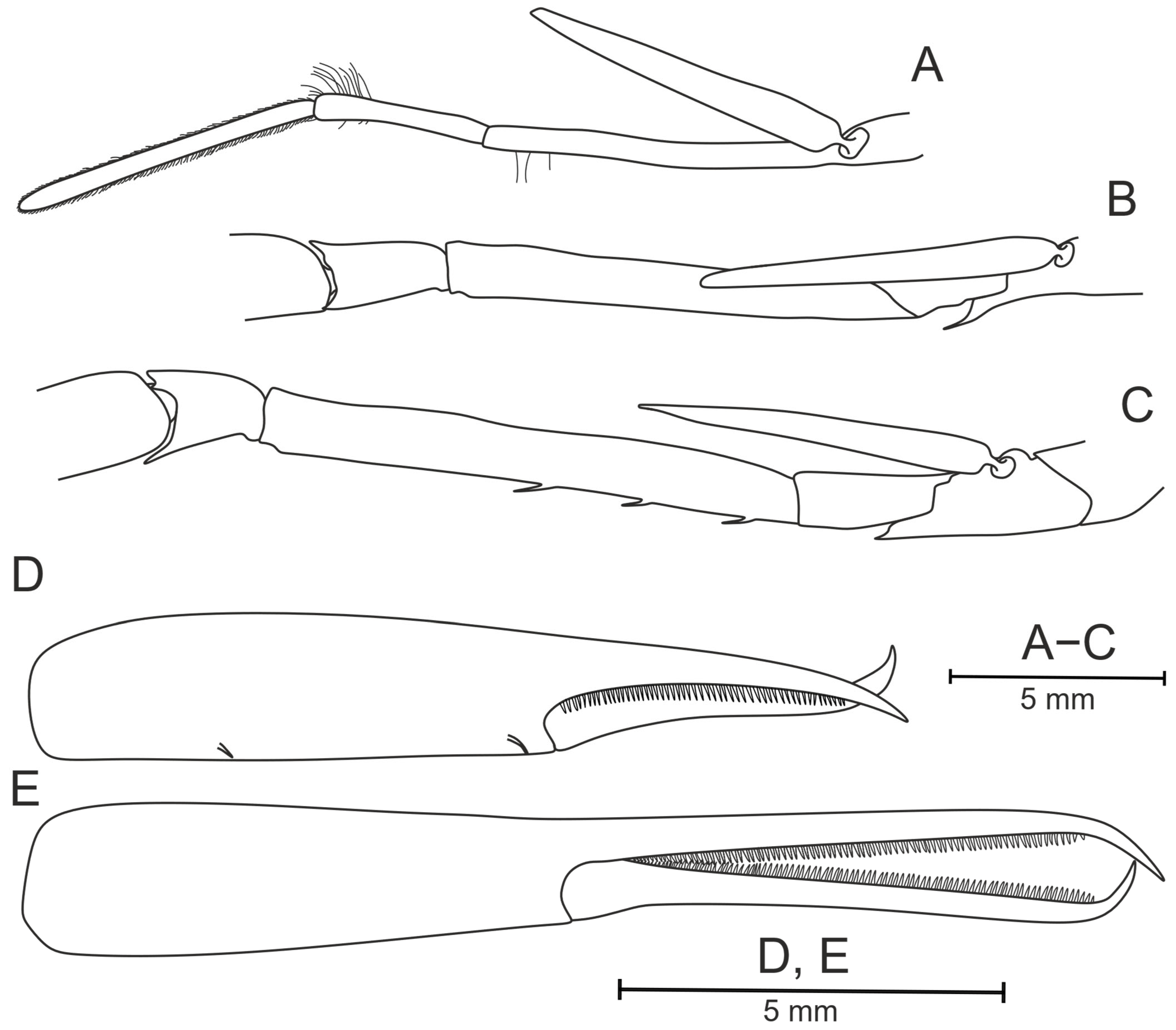
First pereopod (Figure 33B) extending beyond scaphocerite by about 0.25 times length of palm. Basis unarmed, ventrodistal margin with blunt angle. Merus unarmed on ventral side, with row of setae on dorsodistal part of segment. Carpus short, without teeth on any margin. Chela slender, with two ventral bristles on propodus, fingers 0.91–0.93 times as long as palm (Figure 33D).
Second pereopod (Figure 33C) overlapping first pereopod by about 0.52 times length of fingers. Basis with single distal tooth subparallel to margin of basis. Merus with two teeth on lower margin, set in anterior 0.16–0.17 and posterior 0.76–0.77 of ventral length. Carpus short, with small tooth on distal part of ventral margin. Fingers 1.23–1.29 times as long as palm (Figure 33E).
Third pereopod (Figure 34A) slender, not bearing teeth on any segment. Fourth pereopod (Figure 34B) shortest, bearing dense brush of short bristles on dactylus, propodus and carpus; teeth absent on all segments. Fifth pereopod (Figure 34C) longer than fourth, bearing bristles only on dactylus which is rounded; teeth absent on all segments.
Branchial Formula: Five pleurobranchs, three arthrobranchs.
Size: CL 21.4 mm.
Color: Semitransparent with orange-red dots.
Distribution: Known only from the NE coast of Wallis and Futuna Islands from the depth of about 500 m.
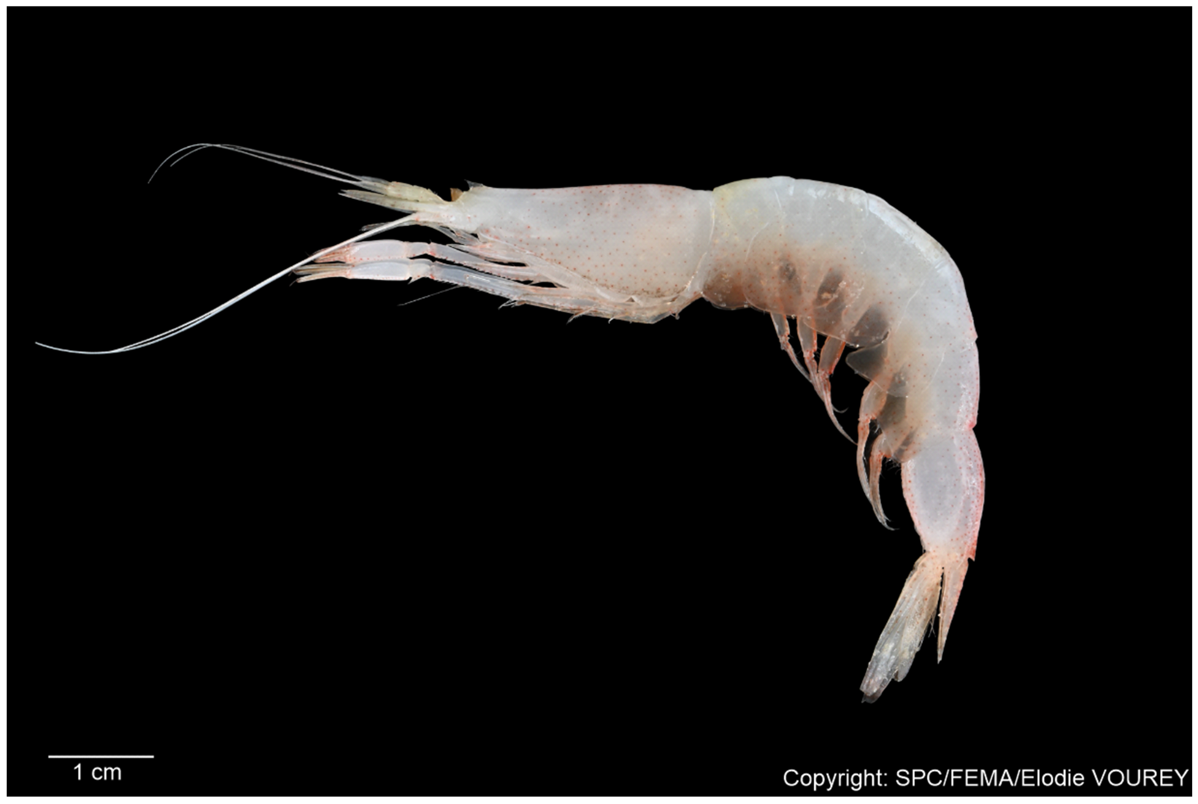
Etymology: The species is named after Mrs Elodie Vourey of Taxonomy Laboratory of the Fisheries, Aquaculture, and Marine Ecosystems Division (The Pacific Community, Noumea). She personally collected the type material of this species and provided exceptional support throughout its study.
Authorship of Species Name: The taxonomic basis for the erection of this species was prepared by Tikhomirov & Vereshchaka, who are thus solely responsible for making the new species name P. voureyae sp. nov. available.
Remarks: The fifth pleonic somite, albeit rounded dorsally, differs from the first and sixth somites in more plain shape. This character may likely be variable in this species and other specimens may bear flattening on this somite.
Combination of pleonic relief and unique armament of second pereopod clearly differentiates new species from other known ones.
Morphologically, the new species is most similar to two species: P. taiwanica and P. americana.
Pasiphaea voureyae sp. nov. is similar to P. taiwanica in pleonic relief and differs in three ways: (1) fifth somite of P. voureyae sp. nov. is rounded vs. flattened in P. taiwanica, (2) peropod I has no teeth in P. voureyae sp. nov. vs. 8 teeth in P. taiwanica, (3) peropod II in P. voureyae sp. nov. has only 2 teeth vs. 18–19 teeth in P. taiwanica.
The new species is similar to P. americana in armament of pereopods: both P. voureyae sp. nov. and P. americana have unarmed pereopod I and two teeth on pereopod II (but P. americana more often have a single tooth). Also, P. voureyae sp. nov. differs from P. americana in pleonic relief: P. voureyae sp. nov. has obviously flattened somites 2–4, whereas P. americana has obscure flattenings only on somites 1 and 2 (according to Hayashi [13]).
Molecular distances suggest significant distances among the new species and the rest of Pasiphaea. The new species is close to P. exilimanus, P. tarda and P. multidentata (13.6, 14.5 and 15.4% difference in COI gene markers), to P. sinensis, P. multidentata and P. romenskyi (6.0, 6.3 and 6.6% difference in 16S gene markers) and to P. scotiae, P. romenskyi and P. acutifrons (7.4, 7.6 and 7.7% difference in 12S gene markers).
Pasiphaea wirotaroenoae sp. nov.
urn:lsid:zoobank.org:act:F2404105-4988-4F00-ACF4-651EABA70BF3
Material Examined: Holotype—female (WA2-13-M300-104) CL 19.5 mm. NE of Line Islands, R/V Alis. Campagne WARMALIS2: st. WA2-13, 06°0′10.8″ N, 150°0′36″ W, oblique trawl: 3.4–288.2 m, 27 September 2022 (night).
Diagnosis: Rostrum overlapping anterodorsal margin of carapace, barely overreaching proximal part of eye cornea, spine-like, straight. Carapace dorsally with carina only in anterior part, medial and posterior parts rounded, branchiostegal spine set behind carapace margin and overlapping this margin. Branchiostegal sinus equal in its vertical and longitudinal directions, subrectangular. Pleonic somites: second and third dorsally depressed, third somite posterodorsally faintly carinate, fourth to sixth dorsally depressed, all lacking posterodorsal tooth. Sixth pleonic somite dorsally weakly protruded in posterior part. Telson shorter than sixth pleonic somite, dorsally grooved, posterior end slightly concave to straight, armed with four pairs of spines. First pereopod: basis with single distal tooth, ischium unarmed, merus with 6–7 teeth. Second pereopod: basis with single distal tooth, ischium unarmed, merus with 19–21 teeth, fingers 1.06 times as long as palm of chela. Five pleurobranchs and three arthrobranchs on each side.
Description (of Holotype): Rostrum (Figure 35A,B) set very close to anterior margin of carapace, barely overreaching proximal part of eye cornea for about 0.06 of rostrum length, spine-like, straight, subparallel to carapace dorsal margin. Carapace (Figure 35A) dorsally carinate in first 0.26 of carapace length, suprabranchial carina distinct, postorbital carina quite noticeable, short oblique carina and subhepatical carina faint. Branchiostegal tooth overlapping carapace margin by 0.25 of its length in lateral view. Branchiostegal sinus 1.15 times as long as wide.
All pleonic somites (Figure 36A–E) without posterodorsal teeth. Somites second and third dorsally depressed in 0.34–1.0 and 0–0.75 parts, respectively. Third somite dorsally carinate in posterior 0.75–1.0. Fourth to sixth somites dorsally depressed in their 0.36–0.92, 0.46–0.71 and 0.78–0.96 parts, respectively, sixth somite laterally carinate in 0.15–0.79 part. Telson 0.78 times as long as sixth pleonic somite, distinctly grooved along its entire dorsal side. Telson terminally slightly concave in middle, incision 0.03 times as long as wide, armed with four pairs of terminal robust spines (Figure 36F).
Eye cornea well pigmented, dark-brown. Stylocerite (Figure 35B) elongate, curved, with nearly constant height along entire length, narrowing in sharp end, not reaching distal end of first segment of antennule. Scaphocerite distinctly overlapping antennular peduncle, 4.05 times as long as wide, distolateral tooth distinct and long. Basicerite with long robust tooth.
First pereopod (Figure 37B) with basis armed with single distal tooth transversal to margin of basis. Merus with 6–7 unequal teeth on ventral side, equally spaced, but starting from 0.34 to 0.40 anterior part of segment. Carpus short, with tooth on distal part of ventral margin.
Second pereopod (Figure 37C) with basis bearing single small distal tooth subparallel to margin of basis with apex curved dorsally. Merus with 19–21 unequal teeth on lower margin, equally spaced along all ventral length. Carpus short, with robust tooth on distal part of ventral margin.
Third pereopod (Figure 38A) slender, not bearing teeth on any segment. Fourth pereopod (Figure 38B) shortest, bearing dense brush of short bristles only on dactylus and propodus; teeth absent on all segments. Fifth pereopod (Figure 38C) longer than fourth, bearing bristles only on dactylus which is rounded; teeth absent on all segments.
Branchial Formula: Five pleurobranchs, three arthrobranchs.
Size: CL 19.5 mm.
Distribution: Known only from the NE of Line Islands; depths are 3.4–288.2 m.
Etymology: This species is named wirotaroenoae sp. nov. in recognition of Mrs. Lysa Wirotaroeno, whose diligent work within the Taxonomy Laboratory of the Fisheries, Aquaculture, and Marine Ecosystems Division (The Pacific Community, Noumea) significantly facilitated our New Caledonian field and laboratory studies.
Authorship of Species Name: The taxonomic basis for the erection of this species was prepared by Tikhomirov & Vereshchaka, who are thus solely responsible for making the new species name P. wirotaroenoae sp. nov. available.
Remarks: The second and third pleonic are slightly smashed, yet being depressed with no doubts. Carina on the third pleonic somite and depressions on the fifth and sixth somites are faint and may ostensibly look absent. The sixth pleonic somite is compressed laterally greatly more than any other segments; therefore, although being rounded, they may mistakenly look carinate.
The new species belongs to the “P. falx” complex; see differences in Table 4.
Molecular Distances: Suggest significant distances among the new species and the rest of Pasiphaea. The new species is close to P. diaphana, P. tokalaui sp. nov., and P. falx but recorded differences show species-level genetic distances among them (Table 6).
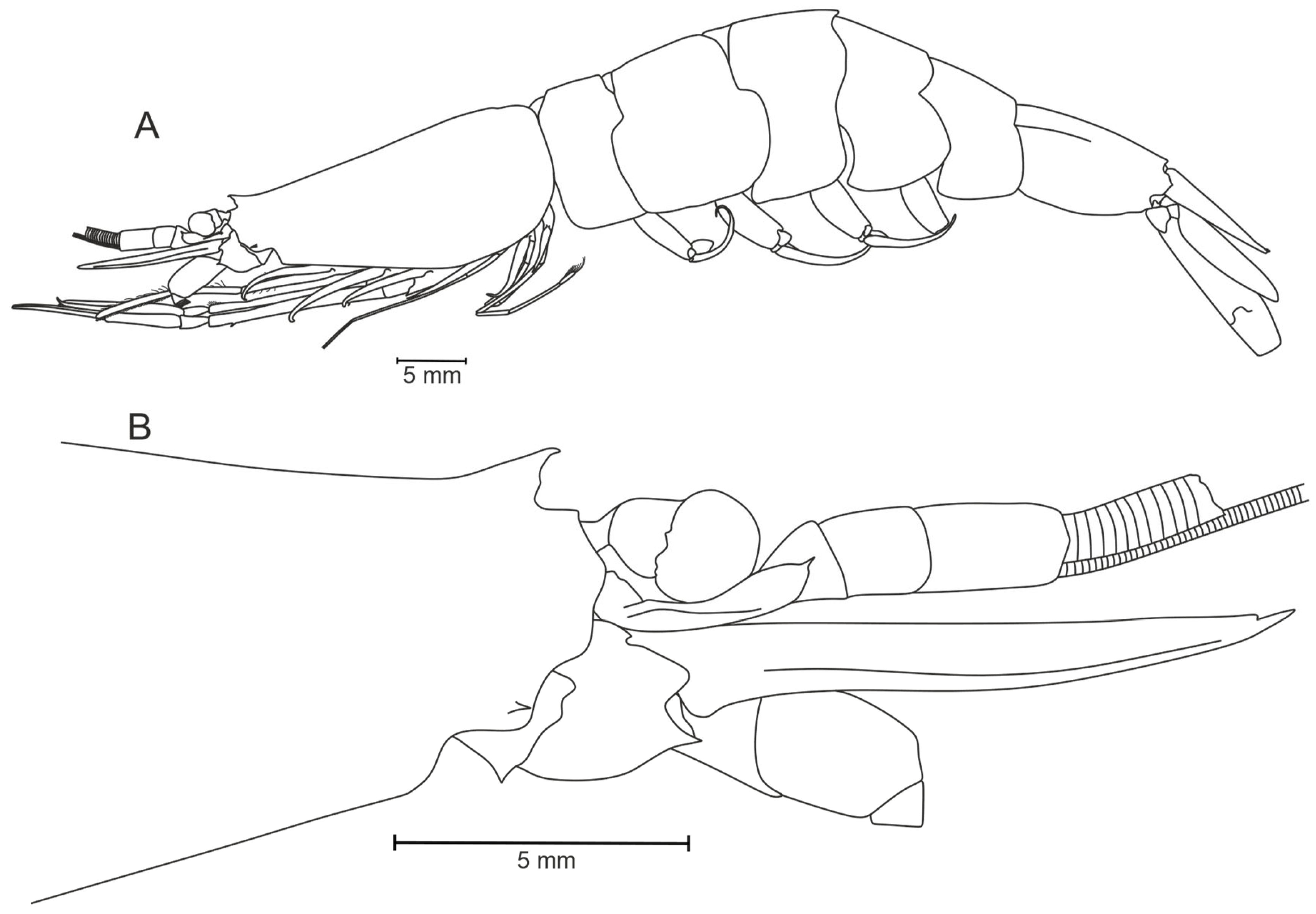
3.2. Phylogenetic Part

According to Hayashi’s definitions [13,14], P. allainae sp. nov. and P. machfulae sp. nov. belong to the P. alcocki species group, whereas the “P. cristata” group encompasses five species: P. komaii sp. nov., P. portalae sp. nov., P. tokalaui sp. nov., P. wirotaroenoae sp. nov., and P. voureyae sp. nov. Pasiphaea tinyamchani sp. nov. cannot be attributed to any of Hayashi’s groups.
The phylogenetic tree retrieved for these and other available Pasiphaea species shows that the genus comprises two robust major clades (A and B), each including four new species (Figure 39, Supplementary Figures S1 and S2). Clade A is further divided into three well-supported subclades (A1–A3) including eight, four, and four species, respectively, and one partly supported (on the BI tree only) subclade (A4) with three species. Clade B is divided into three supported subclades (B1–B3) including two, five, and eight species, respectively, plus an isolated clade containing P. americana. The clades B2, B3, and P. americana also formed a robust clade.
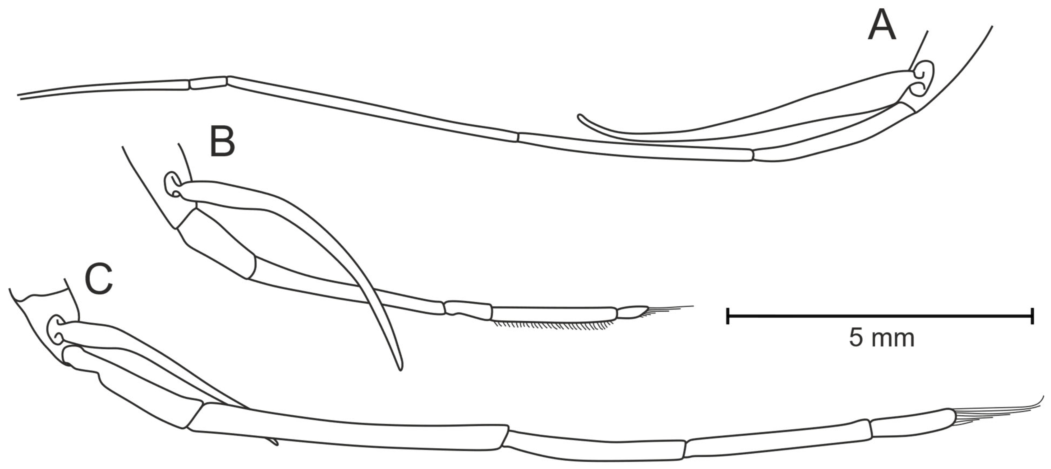
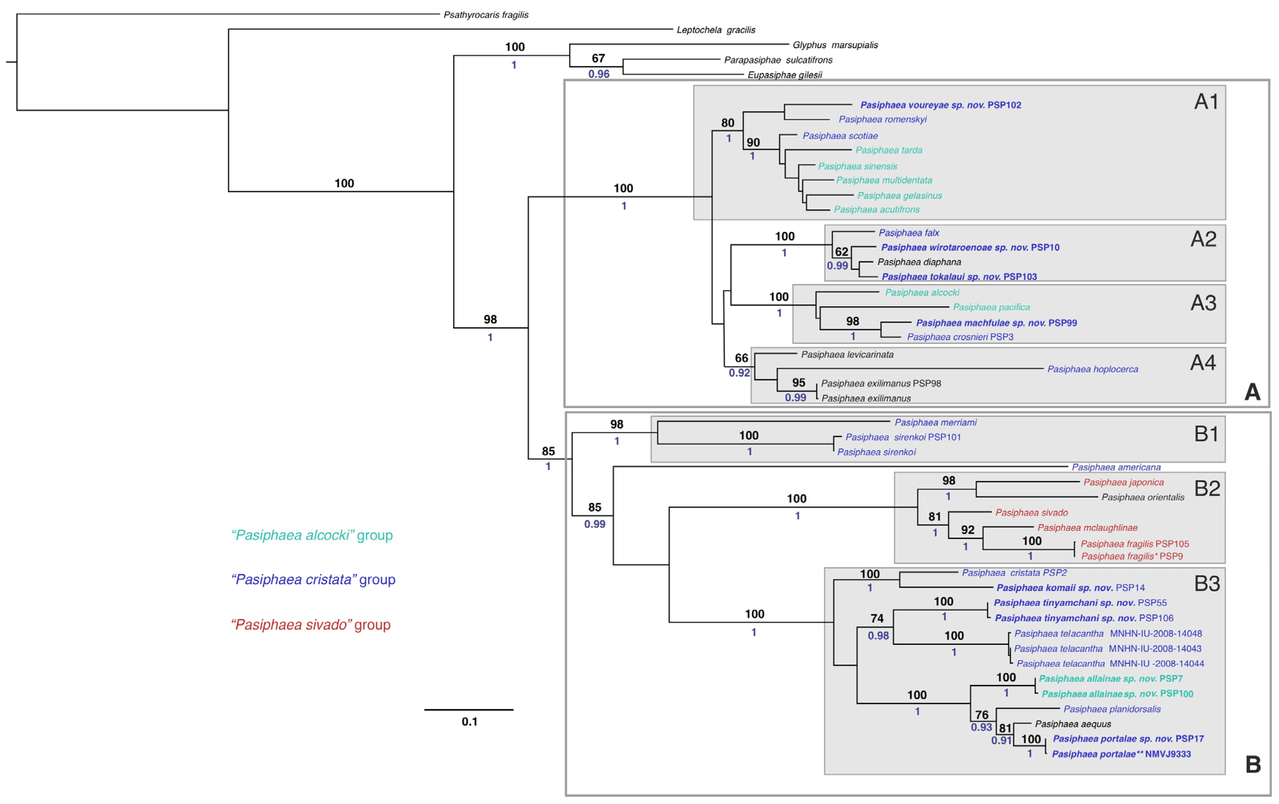
Hayashi’s species groups are distributed almost randomly across molecular clades. Only the “P. sivado” group partly corresponds to a monophyletic clade B2 which, at the same time, includes three other species. The “P. alcocki” group is scattered across A1, A3, and B3, while the “P. cristata” group is distributed across A1, A2, B1, B3, and the isolated P. americana. Notably, species of the “P. alcocki” and “P. cristata” groups occur in both major clades, further underscoring the discrepancies between Hayashi’s classification and molecular data.

4. Discussion

Our results confirm the first hypothesis: even moderately abundant new collections can yield a significant number of undescribed Pasiphaea species in the Southwest Pacific. Several new species are represented by a single specimen, highlighting large gaps in our knowledge of their habitats and true diversity and emphasizing the need for intensified sampling in the region.
The increasing accumulation of morphological data for Pasiphaea observed in the Southwest Tropical Pacific highlights the necessity of a standardized framework for species recognition. To address this, we propose a key (see Appendix A) that allows specimen identification and contributes to a clearer understanding of the regional diversity.
Secondly, examination of the available specimens revealed a pronounced discrepancy between the anticipated and observed morphological variation. Previous authors have generally reported substantial intraspecific variability within Pasiphaea [14,16,34,35,36,37], a phenomenon that has contributed to both frequent misidentifications and the proliferation of junior synonyms. Juvenile individuals, in particular, often lack diagnostic characters essential for reliable identification. In contrast, our analyses demonstrate that mature specimens exhibit a consistently elaborate external morphology, such that even ostensibly minor characters can serve as robust taxonomic markers. For instance, P. tinyamchani sp. nov. closely resembles P. telacantha, yet careful examination of telson morphology, the telson-to-sixth-pleon somite ratio, and the presence and form of the ventrodistal tooth on the basis of the first pereopod provides clear differentiation. These characters remain virtually invariant across ontogenetic stages, sexes, and individual specimens.
Moreover, we document the co-occurrence of pseudocryptic species that are morphologically nearly indistinguishable but genetically distinct at the species level. Such patterns imply rapid speciation driven by the heterogeneous ecological niches surrounding New Caledonia, including seamounts, banks, and island slopes. Subtle differences, such as variation in spination of the second pereopod (e.g., between P. tinyamchani sp. nov. and P. telacantha) or in the telson-to-sixth-pleon somite ratio (e.g., P. crosnieri versus P. machfulae sp. nov.), may reflect divergent feeding strategies or escape behaviors, the latter involving characteristic backward flips (see refs. [38,39,40]). A more extensive quantitative comparison among these pseudocryptic taxa, using larger sample sizes, is required to test this hypothesis rigorously.
A particularly illustrative case involves the sympatric occurrence of P. telacantha and P. tinyamchani sp. nov. off New Caledonia (Figure 1). All type specimens of P. telacantha originate solely from this region, whereas P. tinyamchani sp. nov. has also been recorded at Banc Combe (see remarks on P. tinyamchani sp. nov.). Historical collections from the 1980s and early 1990s yielded exclusively P. telacantha, whereas surveys in the late 2010s and early 2020s recovered only P. tinyamchani sp. nov. This pattern raises the question of whether P. tinyamchani sp. nov. represents an exceptionally rapid speciation event from P. telacantha, or whether the latter has been locally supplanted—potentially via immigration from Banc Combe.
Overall, the integration of refined morphological criteria with molecular evidence has extended or confirmed the geographic ranges of five described species and facilitated the description of eight new species. In addition, we synonymize P. gracilis and P. fragilis. When considered alongside fragmentary Indo-Pacific revisions over the past three decades [12,13,16,41], our findings underscore both the substantial gaps in our knowledge of regional biodiversity and the urgent need for a comprehensive, genus-level revision of Pasiphaea.
Our results also confirm the second hypothesis: the discovery of additional species further challenges Hayashi’s morphological species-group concept and does not resolve the incongruence between morphology- and DNA-based evidence. Although preliminary, our three-gene phylogenetic tree clearly demonstrates the incongruence between morphological and molecular datasets—a pattern unusual for eucarids but known in other crustacean groups. Hayashi’s [12,13,14] species groups are almost randomly distributed across molecular clades, and species that could be attributed to the same morphological group often occur in different molecular clades. While such incongruence is not unprecedented [42], and morphological–molecular partitions may yield markedly different trees [43], previous phylogenetic analyses of pelagic eucarids generally recovered similar topologies from both datasets [29,44,45,46,47,48,49,50]. This pronounced discrepancy is therefore a novel finding for pelagic eucarids.

Supplementary Materials

The following supporting information can be downloaded at https://www.mdpi.com/article/10.3390/d17090656/s1, Figure S1: Three-gene (16S + 12S + COI, 1373 bp) ML molecular tree of Pasiphaea; Figure S2: Three-gene (16S + 12S + COI, 1373 bp) BI molecular tree of Pasiphaea; Table S1: Primer information and PCR scheme used for amplification; Table S2: Details of the analyzed species and sequences used in the study. Newly generated sequences are highlighted in bold; Table S3: Partitioning scheme and best models selected by PartitionFinder2.

Author Contributions

Conceptualization, A.M.T. and A.L.V.; molecular analysis, D.N.K.; morphological analysis, A.M.T. and A.A.L.; visualization, A.M.T. and A.A.L.; writing—original draft preparation, A.M.T. and A.L.V.; investigating—A.M.T., D.N.K., A.A.L., E.V. and A.L.V.; writing—review and editing, A.M.T., D.N.K., A.A.L., E.V. and A.L.V. All authors have read and agreed to the published version of the manuscript.

Funding

This research was carried out in the framework of the state assignment [FMWE-2024–0023—morphological and molecular analyses] and supported by the Russian Science Foundation [Project No. 25-77-30002—plankton biodiversity research]. The collection of these samples (https://doi.org/10.18142/250, https://doi.org/10.17600/18000892, https://doi.org/10.18142/243, https://doi.org/10.17600/18000523, https://doi.org/10.17600/18003922) has received the financial support of the French ministry of the armed forces, French Pacific Funds, BEST 2.0 Programme, funded by the European Union, IRD, in association with Opération Cétacés, Ministere de la Transition Ecologique et Solidaire, Gouvernement de Nouvelle-Calédonie, WWF, FOF.

Institutional Review Board Statement

Not applicable.

Acknowledgments

We are grateful to the WCPFC Pacific Marine Specimen Bank (https://www.spc.int/ofp/PacificSpecimenBank) and the Pacific Community (SPC), Fisheries, Aquaculture, and Marine Ecosystems (FAME) division for collating and providing Pasiphaea samples. We thank the R/V Alis and R/V Antea crew and the science parties who participated in these cruises. We are also grateful to the Muséum National d’Histoire Naturelle (MNHN, Paris) for providing access to comparative material and making specimens available for study.

Conflicts of Interest

The authors declare no conflict of interest.

Abbreviations

BIBayesian inference (molecular analysis)
CLCarapace length
ML (analysis)Maximum likelihood (molecular analysis)
MNHNMuséum National d’Histoire Naturelle (Paris)

Appendix A. Key to Pasiphaea of the Southwest Tropical Pacific

The increasing accumulation of morphological data for Pasiphaea observed in the Southwest Tropical Pacific highlights the necessity of a standardized framework for species recognition. To address this, we propose a phylogenetic key that allows specimen identification and contributes to a clearer understanding of the regional diversity.
  • Branchial formula rudimentary (fifth pleurobranch absent or rudimentary) ...................2
    -
    Branchial formula full (all five pleurobranchs are fully developed)................................9
  • Fifth pleurobranch absent.........................................................................................................3
    -
    Fifth pleurobranch rudimentary...........................................................................................6
  • Basis of second pereopod with 2–7 teeth (excluding the posterior one)...............................
    ....................................................................................................P. philippinensis Hayashi, 1999
    -
    Basis of second pereopod with a single posterior tooth.....................................................4
  • Rostrum nearly reaching frontal margin of rostrum base and subparallel to dorsal
    carapace margin. Ischium of second pereiopod with tooth, P. debitusae Hayashi, 1999
    -
    Rostrum distinctly reaching midlength between anterior margin of carapace and
    rostrum base and directed dorsally. Ischium of second pereopod unarmed....................
    ..................................................................................................................................................5
  • Branchiostegal sinus absent or obscure. Height of branchiostegal sinus (Figure 3B[h])
    0.10 or less times as high as postsinusial height of carapace (Figure 2A[a])........................
    .................................................................................................................P. laevis Hayashi, 1999
    -
    Branchial sinus distinct. Height of branchiostegal sinus (Figure 3B[h]) 0.13 or more
    times as high as postsinusial height of carapace (Figure 2A[a])..........................................
    ...........................................................................................................P. gracilis Hayashi, 1999
  • Posterodorsal teeth on third and sixth pleonic somite.................P. orientalis Schmitt, 1931
    -
    Posterodorsal tooth only on sixth pleonic somite...............................................................7
  • At least one pleonic somite carinated or having depressions...............................................8
    -
    All pleonic somites rounded dorsally.............................................P. japonica Omori, 1976
  • Sixth pleonic somite sharply carinated. Posterior margin of telson convex.........................
    ..........................................................................................................P. propinqua De Man, 1916
    -
    Second to sixth pleonic somites having depressions. Posterior margin of telson
    truncated.................................................................................P. mclaughlinae Hayashi, 2006
  • Branchiostegal sinus absent.......................................................P. sirenkoi Burukovsky, 1987
    -
    Branchiostegal sinus present...............................................................................................10
  • None of the pleonic somites bearing dorsal depressions....................................................11
    -
    At least one pleonic somite bearing dorsal depressions...................................................17
  • Carapace dorsally entirely carinated.....................................................................................12
    -
    Carapace dorsally rounded or carinated only on short section immediately
    posterior to the rostral base.................................................................................................14
  • Branchiostegal sinus distinct. Height of branchiostegal sinus (Figure 3B[h]) 0.17–0.24
    times as high as postsinusial height of carapace (Figure 2A[a])...........................................
    .................................................... P. alcocki (Wood-Mason in Wood-Mason & Alcock, 1891)
    -
    Branchiostegal sinus obscure. Height of branchiostegal sinus 0.09–0.13 times as
    high as postsinusial height of carapace.............................................................................13
  • Telson ~3/4 times as long as sixth pleonic somite (Figure 19A). Sixth somite dorsally
    carinated......................................................................................................................................
    ...........................................................................................................P. crosnieri Hayashi, 2004
    -
    Telson subequal to sixth pleonic somite (Figure 19B). Sixth somite dorsally rounded,
    yet laterally compressed......................................................................P. machfulae sp. nov.
  • Second pereopodal merus with 1 tooth................................................................................15
    -
    Second pereopodal merus with 3 or more teeth...............................................................16
  • Telson terminally slightly concave (Figure 14F). Basis of first pereopod with long
    distal ventrally directed tooth (Figure 14B)..............................................P. komaii sp. nov.
    -
    Telson terminally slightly convex. Basis of first pereopod unarmed................................
    .................................................................................P. cristata Spence Bate, 1888
  • Second pereopodal merus with 3 teeth.................................. P. pseudacantha Hayashi, 2004
    -
    Second pereopodal merus with 13–20 teeth...........................................................................
    ...................................................................................P. exilimanus Komai, Lin & Chan, 2012
  • Branchiostegal sinus vertically extended (Figure 7B). Only sixth pleonic somite dorsally
    carinated. Distal tooth on basis of first pereopod longer that distal tooth on basis of
    second pereopod (Figure 9B,C)..............................................................................................18
    -
    Branchiostegal sinus elongate in the longitudinal direction or subrectangular
    (Figure 31B). Distal tooth on basis of first pereopod, if present, smaller that distal
    tooth on basis of second pereopod......................................................................................21
  • First pereopodal merus unarmed. Second pereopod merus with 3 or less teeth..............19
    -
    First pereopodal merus with 2–16 teeth. Second pereopod merus with 9 or more teeth
    .................................................................................................................................................20
  • Second pereopodal merus with 1 tooth set in proximal third of segment............................
    ......................................................................................................P. planidorsalis Hayashi, 2004
    -
    Second pereopod merus with 3 teeth set in proximal 0.15, 0.35 and 0.55 parts of
    segment, respectively (Figure 22C)........................................................P. portalae sp. nov.
  • First pereopodal merus with 2–6 teeth. Second pereopodal merus with 9–16 teeth...........
    ........................................................................................... P. aequus Komai, Lin & Chan, 2012
    -
    First pereopodal merus with 10–16 teeth (Figure 9B). Second pereopodal merus with
    19–23 teeth (Figure 9C)............................................................................ P. allainae sp. nov.
  • Rostrum distinctly overlapping anterior margin of carapace (Figure 35B). Third
    pleonic somite dorsally carinated in posterior fifth (Figure 27C)......................................22
    -
    Rostrum nearly reaching anterior margin of carapace or shorter (Figure 31B).
    Third pleonic somite, if dorsally carinated, bearing carina along entire length of
    somite.....................................................................................................................................24
  • Dorsal depressions developed on all pleonic somites (1–6), third and fifth somites
    posteriorly carinated (Figure 27A–F). First pereopod merus with 8–12 teeth (Figure 28B)
    ........................................................................................................................P. tokalaui sp. nov.
    -
    Dorsal depressions developed on second to fourth or to sixth pleonic somites
    and only third somite dorsally carinated. First pereopod merus with less than 8
    teeth........................................................................................................................................23
  • Rostrum reaching eye cornea (Figure 35B). Dorsal depressions on second to sixth
    pleonic somites (Figure 36A–E). Fingers of second pereopod longer than palm
    (Figure 37E)........................................................................................P. wirotaroenoae sp. nov.
    -
    Rostrum not reaching eye cornea. Dorsal depressions on second to fourth pleonic
    somites. Fingers of second pereopod shorter than palm.....................................................
    ............................................................................................ P. falx Komai, Lin & Chan, 2012
  • First pereopodal merus with more than 5 teeth, second pereopodal merus with more
    than 18 teeth.............................................................................................................................25
    -
    First pereopodal merus unarmed, second pereopodal merus with 2 teeth or less..........
    ................................................................................................................................................26
  • Pleon dorsally carinated from first (in some specimens first somite is rounded) to sixth
    somites.................................................................................... P. levicarinata Hanamura, 1994
    -
    Pleon dorsally not carinated.................................. P. taiwanica Komai, Lin & Chan, 2012
  • Fingers of first pereopod chela more than 0.9 times as long as palm (Figure 33D).
    Second pereopodal merus with 2 teeth (Figure 33C)............................ P. voureyae sp. nov.
    -
    Fingers of first pereopod chela less than 0.7 times as long as palm. Second pereopod
    merus with 1 tooth set posterior 0.70–0.89 of ventral length..........................................27
  • Second to fifth pleonic somites with dorsal depressions (Figure 24A–E). Basis of first
    pereopod with ventrodistal tooth (Figure 25B). Coxa of second pereopod without
    ventrodistal tooth (Figure 25C)..........................................................P. tinyamchani sp. nov.
    -
    Third and fourth pleonic somites with dorsal depressions. Basis of first pereopod
    without ventrodistal tooth. Coxa of second pereopod with ventrodistal tooth
    ......................................................................................................P. telacantha Hayashi, 2004

References

  1. WoRMS Editorial Board (2025). World Register of Marine Species. Available online: https://www.marinespecies.org (accessed on 12 July 2025).
  2. Liao, Y.; De Grave, S.; Ho, T.W.; Ip, B.H.; Tsang, L.M.; Chan, T.Y.; Chu, K.H. Molecular phylogeny of Pasiphaeidae (Crustacea, Decapoda, Caridea) reveals systematic incongruence of the current classification. Mol. Phylogenetics Evol. 2017, 115, 171–180. [Google Scholar] [CrossRef] [PubMed]
  3. Savigny, J.C. Mémoires sur les Animaux sans Vertèbres; G. Dufour: Paris, France, 1816; Volume 1, pp. 1–117. [Google Scholar]
  4. Bate, C.S. Report on the Crustacea Macrura collected by the Challenger during the years 1873–76. In Report on the Scientific Results of the Voyage of H.M.S. Challenger During the Years 1873–76. Zoology; Morrison and Gibb: Edinburg, UK, 1888; Volume 24, pp. 1–942. [Google Scholar]
  5. Faxon, W. Preliminary descriptions of new species of Crustacea. Bull. Mus. Comp. Zool. Harv. Coll. 1893, 24, 149–220. [Google Scholar]
  6. Rathbun, M.J. Descriptions of new decapod crustaceans from the west coast of North America. Proc. U. S. Natl. Mus. 1902, 24, 885–905. [Google Scholar] [CrossRef]
  7. Rathbun, M.J. Decapod crustaceans of the northwest coast of North America. In Harriman Alaska Expedition with Cooperation of Washington Academy of Sciences; Rathbun, M.J., Richardson, H., Holmes, S.J., Cole, L.J., Eds.; Doubleday, Page & Co.: New York, NY, USA, 1904; Volume 10, pp. 1–210. [Google Scholar]
  8. Rathbun, M.J. The Brachyura and Macrura of the Hawaiian Islands. Bull. United States Fish Comm. 1906, 23, 827–930. [Google Scholar]
  9. Stebbing, T.R.R. Stalk-eyed Crustacea Malacostraca of the Scottish National Antarctic Expedition. Trans. R. Soc. Edinb. 1914, 50, 253–307. [Google Scholar] [CrossRef]
  10. Burukovsky, R.N. Shrimps of the genus Pasiphaea: Systematics and some remarks on new findings (Decapoda, Caridea). Zool. Zhurnal 1996, 75, 846–847. [Google Scholar]
  11. Burukovsky, R.N.; Romensky, L.L. Description of Pasiphaea balssi sp. n., a new species of shrimp from the South Atlantic (Crustacea, Decapoda, Pasiphaeidae), and polytomous key for identification of the shrimp in the genus. Byulleten’ Mosk. Obs. Ispyt. Prir. Otd. Biol. 1987, 92, 51–60. (In Russian) [Google Scholar]
  12. Hayashi, K.-I. Crustacea Decapoda: Revision of Pasiphaea sivado (Risso, 1816) and related species, with descriptions of one new genus and five new species (Pasiphaeidae). In Résultats des Campagnes MUSORSTOM 20. Mémoires du Muséum National d’Histoire Naturelle; Crosnier, A., Ed.; National Fisheries University: Shimonoseki, Japan, 1999; Volume 180, pp. 267–302. [Google Scholar]
  13. Hayashi, K.-I. Revision of the Pasiphaea cristata Bate, 1888 species group of Pasiphaea Savigny, 1816, with descriptions of four new species, and referral of P. australis Hanamura, 1989 to Alainopasiphaea Hayashi, 1999 (Crustacea: Decapoda: Pasiphaeidae). In Tropical Deep-Sea Benthos 23. Mémoires du Muséum National d’Histoire Naturelle; Marshall, B.A., Richer De Forges, B., Eds.; National Fisheries University: Shimonoseki, Japan, 2004; Volume 191, pp. 319–373. [Google Scholar]
  14. Hayashi, K.-I. Revision of the Pasiphaea alcocki species group (Crustacea, Decapoda, Pasiphaeidae). In Tropical Deep-Sea Benthos 24. Mémoires du Muséum National d’Histoire Naturelle; Richer de Forges, B., Justine, J.-L., Eds.; National Fisheries University: Shimonoseki, Japan, 2006; Volume 193, pp. 193–241. [Google Scholar]
  15. Wood-Mason, J. Crustaceans. In Illustrations of the Zoology of H.M. Indian Marine Surveying Steamer Investigator; Royal Marine: Calcutta, India, 1892; pp. 1–310. [Google Scholar]
  16. Komai, T.; Lin, C.W.; Chan, T.Y. Bathypelagic shrimp of the genus Pasiphaea (Decapoda: Caridea: Pasiphaeidae) from waters around Taiwan, with descriptions of four new species. J. Crustac. Biol. 2012, 32, 295–325. [Google Scholar] [CrossRef]
  17. Komai, T.; Ohtsuka, S.; Yamaguchi, S.; Nakaguchi, K. New records of six deep-sea caridean shrimps (Crustacea: Decapoda) from the Ryukyu Islands and its adjacent waters, southwestern Japan. Zootaxa 2018, 4457, 114–128. [Google Scholar] [CrossRef]
  18. Valérie, A.; Christophe, M. NECTALIS-5 cruise, RV Alis. 2016. Available online: https://campagnes.flotteoceanographique.fr/campagnes/16004200/ (accessed on 1 August 2025).
  19. Valérie, A.; Christophe, M. WALLALIS cruise, RV Alis. 2018. Available online: https://campagnes.flotteoceanographique.fr/campagnes/18000523/ (accessed on 1 August 2025).
  20. Claire, G.; Solène, D. MARACAS 7 cruise, RV Alis. 2019. Available online: https://campagnes.flotteoceanographique.fr/campagnes/18000892/ (accessed on 1 August 2025).
  21. Valérie, A.; Christophe, M. WARMALIS 2 cruise, RV Alis. 2022. Available online: https://campagnes.flotteoceanographique.fr/campagnes/18001260/ (accessed on 1 August 2025).
  22. Christophe, M.; Valérie, A. WARMALIS 3 cruise, RV Antea. 2023. Available online: https://campagnes.flotteoceanographique.fr/campagnes/18001831/ (accessed on 1 August 2025).
  23. Deagle, B.E.; Jarman, S.N.; Coissac, E.; Pompanon, F.; Taberlet, P. DNA metabarcoding and the cytochrome c oxidase subunit I marker: Not a perfect match. Biol. Lett. 2014, 10, 20140562. [Google Scholar] [CrossRef]
  24. Varela, C.; Golightly, C.; Timm, L.E.; Wilkins, B.; Frank, T.; Fenolio, D.; Bracken-Grissom, H.D. DNA barcoding enhances large-scale biodiversity initiatives for deep-pelagic crustaceans within the Gulf of Mexico and adjacent waters. J. Crustac. Biol. 2021, 41, ruab005. [Google Scholar] [CrossRef]
  25. Williams, B.D.; Schrank, B.; Huynh, C.; Shownkeen, R.; Waterston, R. A genetic mapping system in Caenorhabditis elegans based on polymorphic sequence-tagged sites. Genetics 1992, 131, 609–624. [Google Scholar] [CrossRef] [PubMed]
  26. Folmer, O.; Black, M.; Hoeh, W.; Lutz, R.; Vrijenhoek, R. DNA primers for amplification of mitochondrial cytochrome c oxidase subunit I from diverse metazoan invertebrates. Mol. Mar. Biol. Biotechnol. 1994, 3, 294–299. [Google Scholar] [PubMed]
  27. Reuschel, S.; Schubart, C.D. Phylogeny and geographic differentiation of Atlanto–Mediterranean species of the genus Xantho (Crustacea: Brachyura: Xanthidae) based on genetic and morphometric analyses. Mar. Biol. 2006, 148, 853–866. [Google Scholar] [CrossRef]
  28. Schubart, C.D.; Cuesta, J.A.; Felder, D.L. Glyptograpsidae, a new brachyuran family from Central America: Larval and adult morphology, and a molecular phylogeny of the Grapsoidea. J. Crustac. Biol. 2002, 22, 28–44. [Google Scholar] [CrossRef]
  29. Vereshchaka, A.L.; Lunina, A.A.; Kulagin, D.N.; Olesen, J.; Corbari, L. Revision of the global fauna of Leptochela Stimpson, 1860 (Crustacea: Decapoda), highlighting phylogenetic clades and a new species. Diversity 2024, 16, 760. [Google Scholar] [CrossRef]
  30. Kimura, M. A simple method for estimating evolutionary rates of base substitutions through comparative studies of nucleotide sequences. J. Mol. Evol. 1980, 16, 111–120. [Google Scholar] [CrossRef]
  31. Kumar, S.; Stecher, G.; Li, M.; Knyaz, C.; Tamura, K. MEGA X: Molecular evolutionary genetics analysis across computing platforms. Mol. Biol. Evol. 2018, 35, 1547–1549. [Google Scholar] [CrossRef]
  32. Katoh, K.; Standley, D.M. MAFFT multiple sequence alignment software version 7: Improvements in performance and usability. Mol. Biol. Evol. 2013, 30, 772–780. [Google Scholar] [CrossRef]
  33. Abascal, F.; Zardoya, R.; Telford, M.J. TranslatorX: Multiple alignment of nucleotide sequences guided by amino acid translations. Nucleic Acids Res. 2010, 38 (Suppl. 2), W7–W13. [Google Scholar] [CrossRef]
  34. Burukovsky, R.N. On the taxonomic status of two bathypelagic species of shrimps (Crustacea, Decapoda, Pasiphaeidae). Zool. Zhurnal 1987, 66, 37–41. (In Russian) [Google Scholar]
  35. Hayashi, K.I.; Miyake, S. A new species of the genus Pasiphaea from the East China Sea (Crustacea, Decapoda, Pasiphaeidae). Proc. Jpn. Soc. Syst. Zool. 1971, 7, 39–44. [Google Scholar]
  36. Tchesunov, A.V. A new and rare mesopelagic species of the genus Pasiphaea (Crustacea, Decapoda) from the tropic and subtropic Atlantic. Zool. Zhurnal 1984, 63, 993–1003. [Google Scholar]
  37. Guzman, G.L.; Olguin, C.N. Pelagic shrimps (Crustacea, Decapoda, Dendrobranchiata, and Caridea) in the Southeast Pacific. In Deep-Sea Pycnogonids and Crustaceans of the Americas; Springer International Publishing: Cham, Switzerland, 2021; pp. 293–309. [Google Scholar]
  38. Vereshchaka, A.L.; Kulagin, D.N.; Lunina, A.A.; Shatravin, A.V. A global revision of Oplophoroidea (Caridea: Decapoda) with a novel approach for visualizing genetic differences between species. Org. Divers. Evol. 2025, 24, 507–532. [Google Scholar] [CrossRef]
  39. Kulagin, D.N.; Lunina, A.A.; Simakova, U.V.; Vedenin, A.A.; Vereshchaka, A.L. Revision of the Acanthephyra purpurea species complex (Crustacea: Decapoda), with an emphasis on species diversification in the Atlantic. Mol. Phylogenetics Evol. 2024, 195, 108070. [Google Scholar] [CrossRef]
  40. Lunina, A.A.; Kulagin, D.N.; Vereshchaka, A.L. Oplophoridae (Decapoda: Crustacea): Phylogeny, taxonomy and evolution studied by a combination of morphological and molecular methods. Zool. J. Linn. Soc. 2019, 186, 213–232. [Google Scholar] [CrossRef]
  41. Hayashi, K.-I. A new species of the Pasiphaea sivado species group from Taiwan (Decapoda, Caridea, Pasiphaeidae). Zoosystema 2006, 28, 341–346. [Google Scholar]
  42. Oyston, J.W.; Wilkinson, M.; Ruta, M.; Wills, M.A. Molecular phylogenies map to biogeography better than morphological ones. Commun. Biol. 2022, 5, 521. [Google Scholar] [CrossRef]
  43. Keating, J.N.; Garwood, R.J.; Sansom, R.S. Phylogenetic congruence, conflict and consilience between molecular and morphological data. BMC Ecol. Evol. 2023, 23, 30. [Google Scholar] [CrossRef]
  44. Vereshchaka, A.L.; Kulagin, D.N.; Lunina, A.A. A phylogenetic study of krill (Crustacea: Euphausiacea) reveals new taxa and co-evolution of morphological characters. Cladistics 2019, 35, 150–172. [Google Scholar] [CrossRef]
  45. Lunina, A.A.; Kulagin, D.N.; Vereshchaka, A.L. A hard-earned draw: Phylogeny-based revision of the deep-sea shrimp Bentheogennema (Decapoda: Benthesicymidae) transfers two species to other genera and reveals two new species. Zool. J. Linn. Soc. 2019, 187, 1155–1172. [Google Scholar] [CrossRef]
  46. Vereshchaka, A.L.; Corbari, L.; Kulagin, D.N.; Lunina, A.A.; Olesen, J. A phylogeny-based revision of the shrimp genera Altelatipes, Benthonectes and Benthesicymus (Crustacea: Decapoda: Benthesicymidae). Zool. J. Linn. Soc. 2020, 189, 207–227. [Google Scholar] [CrossRef]
  47. Vereshchaka, A.L.; Kulagin, D.N.; Lunina, A.A. Across the benthic and pelagic realms: A species-level phylogeny of Benthesicymidae (Crustacea: Decapoda). Invertebr. Syst. 2021, 35, 776–796. [Google Scholar] [CrossRef]
  48. Lunina, A.A.; Kulagin, D.N.; Vereshchaka, A.L. Phylogenetic revision of the shrimp genera Ephyrina, Meningodora and Notostomus (Acanthephyridae: Caridea). Zool. J. Linn. Soc. 2021, 193, 1002–1019. [Google Scholar] [CrossRef]
  49. Vereshchaka, A.; Kulagin, D.; Lunina, A. Discovery of a new species provides a deeper insight into taxonomic grouping of the deep-sea genus Acanthephyra (Crustacea: Decapoda). Diversity 2022, 14, 907. [Google Scholar] [CrossRef]
  50. Lunina, A.; Kulagin, D.; Vereshchaka, A. The taxonomic status of Hymenodora (Crustacea: Oplophoroidea): Morphological and molecular analyses suggest a new family and an undescribed diversity deep in the sea. Zool. J. Linn. Soc. 2024, 200, 336–351. [Google Scholar] [CrossRef]
Figure 1. Localities of Pasiphaea specimens analyzed in the present study. New material was obtained during cruises NECTALIS 5, WALLALIS, MARACAS 7D, and WARMALIS 2 and 3; previously published data are from cruises MUSORSTOM 7 and 9 and SALOMON 2. Pasiphaea fragilis is here synonymized with P. gracilis (see below).
Diversity 17 00656 g001
Figure 2. Scheme of measurements. (A) Measurements of carapace, pleon, and telson: a—postsinusial height of carapace; b—maximum height of carapace; c—carapace length (detailed information is provided in the section on measurement methodology); d—length of 4th pleonic somite; e—length of 5th pleonic somite; f—length of 6th pleonic somite; g—maximum height of 6th pleonic somite; h—distal height of sixth somite; i—length of telson; j—proximal width of telson. (B) Measurements of telson end: k—width of telson end; l—depth of telson concavity/convex curvature. (C) Measurements of 1st pereopod chela: m—length of fingers; n—length of palm; o—height of palm. (D) Measurements of 2nd pereopod chela: p—length of fingers; q—length of palm; r—height of palm.
Diversity 17 00656 g002
Figure 3. Scheme of measurements. (A) Measurements of carapace: a—length of rostrum; b—rostrum base length; c—length of branchiostegal tooth; d—length of eye cornea and stout part of eyestalk; e—length of stylocerite; f—height of stylocerite in highest part. (B) Measurements of sinus: g—length of branchiostegal sinus; h—height of branchiostegal sinus; i—median branchiostegal sinus depth; j—branchiostegal sinus median.
Diversity 17 00656 g003
Figure 4. Scheme of pleonic relief variations in the dorsal surface in a transverse plane. (A) Dorsal surface of rounded somite; (B) dorsal surface of carinate somite; (C) dorsally rounded laterally compressed somite; (D) dorsally carinate laterally compressed somite; (E) dorsal surface of flattened somite, called here with depression; (F) dorsal surface of grooved somite, called here with depression.
Diversity 17 00656 g004
Figure 5. Variations in posterodorsal end of sixth pleonic somite. (A) Absence of any protruded structure—lateral view; (B) posterodorsal protrusion—lateral view; (C) posterodorsal tooth—lateral view; (D) absence of any protruded structure—dorsal view; (E) posterodorsal protrusion—dorsal view; (F) posterodorsal tooth—dorsal view.
Diversity 17 00656 g005
Figure 6. Pasiphaea allainae sp. nov. (WA3-17-M334-03). Fresh specimen in lateral view, showing natural coloration and intact body proportions before fixation.
Diversity 17 00656 g006
Figure 7. Pasiphaea allainae sp. nov., holotype, female (WAL09-M195-200) CL 25.6 mm. (A) Whole specimen; (B) anterior part of carapace.
Diversity 17 00656 g007
Figure 8. Pasiphaea allainae sp. nov., holotype, ovigerous female (WAL09−M195−200) CL 25.6 mm—pleonic somites. (A) First and second somites. (B) third somite. (C) fourth somite. (D) fifth somite. (E) sixth somite.
Diversity 17 00656 g008
Figure 9. Pasiphaea allainae sp. nov., holotype, ovigerous female (WAL09−M195−200) CL 25.6 mm—mouthparts. (A) Mandible; (B) maxilla I; (C) maxilla II; (D) maxilliped I; (E) maxilliped II.
Diversity 17 00656 g009
Figure 10. Pasiphaea allainae sp. nov., holotype, ovigerous female (WAL09−M195−200) CL 25.6 mm. (A) Right maxilliped III, lateral view; (B) proximal part of right pereopod I, lateral view; (C) proximal part of right pereopod II, lateral view; (D) chela of right pereopod I, mesial view; (E) chela of right pereopod II, mesial view.
Diversity 17 00656 g010
Figure 11. Pasiphaea allainae sp. nov., holotype, ovigerous female (WAL09-M195-200) CL 25.6 mm. (A) Right pereopod III, lateral view; (B) right pereopod IV, lateral view; (C) right pereopod V, lateral view; (D) telson end, dorsal view. Scale bar: 5 mm for (AC), 1 mm for (D).
Diversity 17 00656 g011
Figure 12. Pasiphaea exilimanus (WAL07-M191-64). Fresh specimen in lateral view, showing natural coloration and intact body proportions before fixation.
Diversity 17 00656 g012
Figure 13. Pasiphaea komaii sp. nov., holotype, ovigerous female (MNHN-IU-2018-1619) CL 20.2 mm. (A) Whole specimen; (B) anterior part of carapace.
Diversity 17 00656 g013
Figure 14. Pasiphaea komaii sp. nov., holotype, ovigerous female (MNHN-IU-2018-1619) CL 20.2 mm. (A) Left maxilliped III, lateral view; (B) proximal part of left pereopod I, lateral view; (C) proximal part of left pereopod II, lateral view; (D) chela of right pereopod I, mesial view; (D) chela of right pereopod II, mesial view; (F) telson end, dorsal view. Scale bars: 5 mm for (AE), 1 mm for (F).
Diversity 17 00656 g014
Figure 15. Pasiphaea machfulae sp. nov. (WAL07-M191-63). Fresh specimen in lateral view, showing natural coloration and intact body proportions before fixation.
Diversity 17 00656 g015
Figure 16. Pasiphaea machfulae sp. nov. (WAL07-M191-63). Anterior part of the body magnified.
Diversity 17 00656 g016
Figure 17. Pasiphaea machfulae sp. nov., holotype, ovigerous female CL 20.1 mm (WAL07-M191-63). (A) Whole specimen; (B) anterior part of carapace.
Diversity 17 00656 g017
Figure 18. Pasiphaea machfulae sp. nov., holotype, ovigerous female CL 20.1 mm (WAL07−M191−63). (A) Left maxilliped III, lateral view; (B) proximal part of left pereopod I, lateral view; (C) proximal part of right pereopod I, lateral view; (D) proximal part of left pereopod II, lateral view; (E) chela of left pereopod I, mesial view; (F) chela of left pereopod II, lateral view; (G) telson end, dorsal view.
Diversity 17 00656 g018
Figure 19. Pasiphaea crosnieri Hayashi, 2004; ovigerous female (MNHN-IU-2018-1615) CL 17.0 mm. (A) Sixth pleonic somite with telson and uropods, lateral view. Pasiphaea machfulae sp. nov., holotype, ovigerous female (WAL07-M191-63) CL 20.1 mm. (B) Sixth pleonic somite with telson and uropods, lateral view.
Diversity 17 00656 g019
Figure 20. Pasiphaea portalae sp. nov., holotype, male CL 24.1 mm (MNHN-IU-2018-1614). (A) Whole specimen; (B) anterior part of carapace.
Diversity 17 00656 g020
Figure 21. Pasiphaea portalae sp. nov., holotype, male CL 24.1 mm (MNHN−IU−2018−1614)—pleonic somites and telson. (A) First and second somites; (B) third somite; (C) fourth and fifth somites; (D) sixth somite; (E) telson end, dorsal view.
Diversity 17 00656 g021
Figure 22. Pasiphaea portalae sp. nov., holotype, male CL 24.1 mm (MNHN−IU−2018−1614). (A) Left maxilliped III, lateral view; (B) proximal part of left pereopod I, lateral view; (C) proximal part of left pereopod II, lateral view; (D) chela of left pereopod I, mesial view; (E) chela of left pereopod II, mesial view.
Diversity 17 00656 g022
Figure 23. Pasiphaea tinyamchani sp. nov., holotype, male (M7D07-M233-88) CL 24.5 mm. (A) Whole specimen; (B) anterior part of carapace.
Diversity 17 00656 g023
Figure 24. Pasiphaea tinyamchani sp. nov., holotype, male (M7D07−M233−88) CL 24.5 mm—pleonic somites and telson. (A) First and second somites; (B) third somite; (C) fourth somite; (D) fifth somite; (E) sixth somite; (F) telson end, dorsal view. Scale bar: 5 mm for (AE), 1 mm for (F).
Diversity 17 00656 g024
Figure 25. Pasiphaea tinyamchani sp. nov., holotype, male (M7D07−M233−88) CL 24.5 mm. (A) Left maxilliped III, lateral view; (B) proximal part of left pereopod I, lateral view; (C) proximal part of left pereopod II, lateral view; (D) chela of left pereopod I, mesial view; (E) chela of left pereopod II, mesial view.
Diversity 17 00656 g025
Figure 26. Pasiphaea tokalaui sp. nov., holotype, female (NEC5011−M130−51) CL 29.1 mm. (A) Whole specimen; (B) anterior part of carapace.
Diversity 17 00656 g026
Figure 27. Pasiphaea tokalaui sp. nov., holotype, female (NEC5011−M130−51) CL 29.1 mm–pleonic somites. (A) First somite; (B) second somite; (C) third somite; (D) fourth somite; (E) fifth somite; (F) sixth somite, dorsal view; (G) sixth somite, telson and uropods, lateral view.
Diversity 17 00656 g027
Figure 28. Pasiphaea tokalaui sp. nov., holotype, female (NEC5011−M130−51) CL 29.1 mm. (A) Left maxilliped III, lateral view; (B) proximal part of left pereopod I, lateral view; (C) proximal part of left pereopod II, lateral view; (D) chela of left pereopod I, mesial view; (E) chela of left pereopod II, mesial view.
Diversity 17 00656 g028
Figure 29. Pasiphaea tokalaui sp. nov., holotype, female (NEC5011-M130-51) CL 29.1 mm. (A) Left pereopod III, lateral view; (B) left pereopod IV, lateral view; (C) left pereopod V, lateral view; (D) telson end, dorsal view. Scale bar: 5 mm for (AC), 1 mm for (D).
Diversity 17 00656 g029
Figure 30. Pasiphaea voureyae sp. nov. (WAL08-M193-54).
Diversity 17 00656 g030
Figure 31. Pasiphaea voureyae sp. nov., holotype, male (WAL08-M193-54) CL 21.4 mm. (A) Whole specimen; (B) anterior part of carapace.
Diversity 17 00656 g031
Figure 32. Pasiphaea voureyae sp. nov., holotype, male (WAL08−M193−54) CL 21.4 mm—pleonic somites and telson. (A) First somite; (B) second somite; (C) third somite; (D) fourth somite; (E) fifth somite; (F) sixth somite and telson, dorsal view; (G) telson end, dorsal view.
Diversity 17 00656 g032
Figure 33. Pasiphaea voureyae sp. nov., holotype, male (WAL08−M193−54) CL 21.4 mm. (A) Left maxilliped III, lateral view; (B) proximal part of left pereopod I, lateral view; (C) proximal part of left pereopod II, lateral view; (D) chela of right pereopod I, mesial view; (E) chela of right pereopod II, mesial view.
Diversity 17 00656 g033
Figure 34. Pasiphaea voureyae sp. nov., holotype, male (WAL08−M193−54) CL 21.4 mm. (A) Left pereopod III, lateral view; (B) left pereopod IV, lateral view; (C) left pereopod V, lateral view.
Diversity 17 00656 g034
Figure 35. Pasiphaea wirotaroenoae sp. nov., holotype, female (WA2−13−M300−104) CL 19.5 mm. (A) Whole specimen; (B) anterior part of carapace.
Diversity 17 00656 g035
Figure 36. Pasiphaea wirotaroenoae sp. nov., holotype, female (WA2-13-M300-104) CL 19.5 mm–pleonic somites. (A) First and second somites; (B) third somite; (C) fourth somite; (D) fifth somite; (E) sixth somite; (F) telson end, dorsal view. Scale bar: 5 mm for (AE), 1 mm for (F).
Diversity 17 00656 g036
Figure 37. Pasiphaea wirotaroenoae sp. nov., holotype, female (WA2−13−M300−104) CL 19.5 mm. (A) Right maxilliped III, lateral view; (B) proximal part of right pereopod I, lateral view; (C) proximal part of right pereopod II, lateral view; (D) magnified proximal part of merus of right pereopod II, lateral view; (E) chela of right pereopod II, mesial view.
Diversity 17 00656 g037
Figure 38. Pasiphaea wirotaroenoae sp. nov., holotype, female (WA2−13−M300−104) CL 19.5 mm. (A) Left pereopod III, lateral view; (B) right pereopod IV, lateral view; (C) right pereopod V, lateral view.
Diversity 17 00656 g038
Figure 39. Three-gene molecular tree (BI and ML analyses) of Pasiphaea, only supported clades shown. Statistical support indicated as ML bootstrap support (black, above branches) and Bayesian posterior probabilities (blue, below branches), with ML tree topology. *—identified as P. gracilis but P. fragilis and P. gracilis were synonymized in this paper (see remarks on P. fragilis); **—erroneously identified as Pasiphaea pseudacantha Hayashi, 2004 in 28 August 2003 (see remarks on P. portalae).
Diversity 17 00656 g039
Table 1. The morphological comparison of “P. planidorsalis” complex.
CharacteristicP. allainae sp. nov.P. planidorsalis
Hayashi, 2004
P. aequus Komai,
Lin & Chan, 2012
P. portalae sp. nov.
Dorsal carination of carapaceAbsent.Absent.Carinate in anterior short part right after rostrum.Absent.
RostrumRostrum reaching anterodorsal margin of carapace, anterior margin slightly convex, triangular, apex slightly curved forward.Rostrum not reaching anterodorsal margin of carapace, triangular, apex tapered and curved forwards, anterior margin more or less convex.Rostrum not reaching anterodorsal margin of carapace, triangular, directed forward, apex slightly curved.Rostrum reaching anterodorsal margin of carapace, subtriangular, apex slightly curved forward.
Branchiostegal sinusBranchiostegal sinus 0.56–0.91 times as long as high, subrectangular.Branchiostegal sinus 0.80 times as long as high, shape of obtuse angle.Branchiostegal sinus 0.81 times as long as high, subrectangular.Branchiostegal sinus 0.86 times as long as high, subrectangular.
Telson0.76–0.78 times as long as 6th pleonic somite.
Distinctly grooved dorsally, posterior margin nearly straight with shallow median depression (0.10 times as deep as wide).
0.70 times as long as 6th pleonic somite.
Dorsally generally flat but depressed in proximal and distal parts, posterior margin nearly truncate (0.10 times as deep as wide).
0.81–0.90 times as long as 6th pleonic somite.
Dorsally slightly grooved in midline, posterior margin truncate or slightly concave medially (0.03–0.04 times as deep as wide).
0.75 times as long as 6th pleonic somite.
Distinctly grooved dorsally along entire length, posterior margin barely concave at the middle (0.03 times as deep as wide).
Armament of pereopod I10–16 meral teeth, ischium unarmed, basis with large single distal tooth.No meral teeth, ischium unarmed, basis with large single distal tooth.2–6 meral teeth, ischium unarmed, basis with relatively large single distal tooth.No meral teeth, ischium unarmed, basis with single distal tooth.
Armament of pereopod II19–23 meral teeth, ischium unarmed, basis with single distal tooth.1 meral tooth, ischium unarmed, basis with single distal tooth.9–16 meral teeth, ischium unarmed, basis with single distal tooth.3 meral teeth, ischium unarmed, basis with single distal tooth.
ChelaeFirst: fingers 0.71–0.75 times as long as palm.
Second: fingers 1.08–1.16 times as long as palm.
First: fingers 0.77 times as long as palm.
Second: fingers 1.36 times as long as palm.
First: fingers 0.60 times as long as palm.
Second: fingers 0.80–0.90 times as long as palm.
First: fingers 0.71–0.73 times as long as palm.
Second: fingers 1.10–1.11 times as long as palm.
Source This study[13][16]This study
Table 2. Kimura’s genetic differences (%) between Pasiphaea allainae sp. nov. and closely related species.
Gene MarkersP. planidorsalisP. aequusP. portalae sp. nov.
COI11.4%-13.4%
16S6.8%5.4%5.4%
12S-6.7%7.4%
Table 3. Kimura’s genetic differences (%) between Pasiphaea portalae sp. nov. and closely related species.
Gene MarkersP. aequusP. planidorsalisP. allainae sp. nov.
COI-10.0%12.9%
16S2.2%5.5%3.9%
12S3.7%7.1%-
Table 4. Morphological comparison of “P. diaphana” complex.
CharacteristicP. falxP. tokalaui sp. nov.P. wirotaroenoae sp. nov.P. diaphana Burukovsky & Romensky, 1980
Dorsal carination of carapaceCarinate in anterior 0.20 right after rostrum.Carinate in anterior 0.20 right after rostrum.Carinate in anterior 0.26 right after rostrum.Carinate in anterior short part right after rostrum.
RostrumRostrum overreaching anterodorsal margin of carapace, just reaching proximal part of eye cornea, sickle-shaped, curved.Rostrum overlapping anterodorsal margin of carapace, not reaching proximal part of eye cornea, spine-like, all rostrum slightly curved forward.Rostrum barely overreaching proximal part of eye cornea, spine-like, straight, subparallel to carapace dorsal margin.Rostrum triangle shaped, does not reach frontal margin of carapace.
Branchiostegal sinusBranchiostegal sinus 1.68 times as long as high, shallow.Branchiostegal sinus 1.14–1.59 times as long as high, subrectangular.Branchiostegal sinus 1.15 times as long as high, subrectangular.Branchiostegal sinus 1.30 times as long as high, subrectangular.
Dorsal pleonic depressions2–4 somites depressed in any part.1–6 somites depressed in any part.2–6 somites depressed in any part.Absent.
Dorsal pleonic carinaeOn 3rd somite.On 3rd and 5th somites.On 3rd somite.Absent.
TelsonShallowly grooved dorsally, posterior margin truncate.Dorsally grooved, posterior end straight to slightly convex.Dorsally grooved, posterior end slightly concave to straight.Shallowly grooved dorsally, posterior margin truncate.
Armament of pereopod I3–8 meral teeth, ischium unarmed, basis with single distal tooth.8–12 meral teeth, ischium unarmed, basis with single distal tooth.6–7 meral teeth, ischium unarmed, basis with single distal tooth.6–10 meral teeth, ischium unarmed, basis with single distal tooth.
Armament of pereopod II5–19 meral teeth, ischium unarmed, basis with single distal tooth.17–24 meral teeth, ischium unarmed, basis with single distal tooth.19–21 meral teeth, ischium unarmed, basis with single distal tooth.14–20 meral teeth, ischium unarmed, basis with single distal tooth.
ChelaeFirst: fingers 0.7–0.8 times as long as palm.
Second: fingers 0.9 times as long as palm.
First: fingers 0.71–0.76 times as long as palm.
Second: fingers 0.95–1.05 times as long as palm.
Second: fingers 1.06 times as long as palm.First: fingers 0.68–0.73 times as long as palm.
Second: fingers 0.93 times as long as palm.
Source [16]This studyThis study[10,11]
Table 5. Kimura’s genetic differences (%) between Pasiphaea tokalaui sp. nov. and closely related species.
Gene MarkersP. diaphanaP. wirotaroenoae sp. nov.P. falx
COI-5.3%-
16S2.4%2.9%6.3%
12S1.7%2.0%3.7%
Table 6. Kimura’s genetic differences (%) between Pasiphaea wirotaroenoae sp. nov. and closely related species.
Gene MarkersP. diaphanaP. tokalaui sp. nov.P. falx
COI-5.3%-
16S2.7%2.9%5.0%
12S2.5%2.0%4.9%
Disclaimer/Publisher’s Note: The statements, opinions and data contained in all publications are solely those of the individual author(s) and contributor(s) and not of MDPI and/or the editor(s). MDPI and/or the editor(s) disclaim responsibility for any injury to people or property resulting from any ideas, methods, instructions or products referred to in the content.