Article

Helminths of Cats (Felis catus Linnaeus, 1758) and Their Larval Stages in Reptiles in Dubai, United Arab Emirates

by
Rolf K. Schuster
* and
Saritha Sivakumar
Central Veterinary Research Laboratory, Dubai 999041, United Arab Emirates
*
Author to whom correspondence should be addressed.
Diversity 2025, 17(8), 578; https://doi.org/10.3390/d17080578
Submission received: 25 February 2025 / Revised: 7 July 2025 / Accepted: 12 July 2025 / Published: 16 August 2025

Abstract

An examination of 360 feral cats originating from three major habitats in the Dubai Emirate between 2002 and 2024 revealed the presence of 14 helminths, as follows: Joyeuxiella pasqualei, Joyeuxiella gervaisi, Diplopylidium nölleri, Diplopylidium acanthotetra, Hydatigera taeniaeformis, Taenia hydatigena, Ancylostoma braziliense, Ancylostoma ceylanicum, Ollulanus tricuspis, Toxocara cati, Toxascaris leonina, Pterygodermatites cahirensis, Centrorhynchus aluconis and Macracanthorhynchus catulinus. During the same period, a total of 66 snakes (eight species) and 68 lizards (four species) from different locations in the Dubai Emirate were examined for parasites. The larval stages of the cestode genera Joyeuxiella and Diplopylidium, as well as cystacanths of Centrorhynchus sp. and Macracanthorhynchus sp. and the larval stages of two nematodes were detected. All of the snake species except sand boas, as well as two gecko species, harbored the larval stages of cestodes of the Dipylidiidae family. The high prevalence of Joyeuxiella and Diplopylidium in the cats that originated from the city center of Dubai, where the presence of reptiles can be excluded, suggests that certain arthropods might be involved in the life cycle of these cestodes as first intermediate hosts and that reptiles are paratenic hosts.

1. Introduction

Contrary to Dipylidium caninum (Linnaeus, 1758), a cestode that uses fleas and mallophages as intermediate hosts, reptiles are involved in the life cycles of the other two Dipylidiidae genera, Joyeuxiella Fuhrmann, 1935 and Diplopylidium Beddard, 1913. Cestodes of the genera Joyeuxiella and Diplopylidium have been found in carnivores across the Middle East, Africa, southern Europe, Turkey, India and central Asian republics [1].
The involvement of reptiles in the life cycle of dipylidiid cestodes has been known for a long time and was reviewed by Witenberg (1932) [2]. In Italy, armed cysticercoids attached to the intestinal wall of a common wall gecko (Tarentola mauritanica) were described for the first time in 1872 [3]. Another finding of a similar cestode larva was made in the intestinal wall of a whip snake (Hierophis viridiflavus) from Sardinia in 1886 [4]. Based on the morphology of the scolex, it was concluded that such cysticercoids might be the larval stage of Dipylidium trinchesi, known today as Diplopylidium acanthotetra (Parona, 1886) [5], and only 34 years later, the connection between such cysticercoids and adult cestodes was proven by successfully infecting cats with cysticercoids obtained from common wall geckos [6].
The fauna of indigenous terrestrial reptiles of the UAE consists of 54 species [7]. Most of them live in their natural habitats, while a few species are synanthropic, like the yellow-bellied house gecko and the Red Sea leave-toed gecko that can be found in the centers of big cities and ocellated skinks that inhabit gardens and parks. Reptiles can serve as hosts for many parasites, but little is known about the parasite spectrum of reptiles in the Middle East. A total of 13 helminth species were reported from feral cats in Dubai [8,9].
This study aimed to clarify the role of indigenous reptiles in the life cycle of intestinal helminths of felines in the Dubai Emirate of the United Arab Emirates.

2. Materials and Methods

2.1. Cat Helminths

The study area for cat parasites consisted of three different habitats, as follows: 1. The old city center consists of Bur Dubai and Deira. Both districts, divided by the Dubai creek, are densely populated and dominated by high-rise buildings. 2. The outskirts are suburb districts like Jumeirah, Zabeel, Rashidiya and Khawaneej and the neighboring town of Al Aweer. These places are dominated by one- and two-storey villas and accompanying gardens. Here, local families keep poultry and small ruminants for their own consumption. Larger farms are located in Khawaneej. 3. Wadi al Safa, Um Nahad and Nakhlee are desert areas situated outside of the Dubai metropolitan.
In addition to the 240 cats previously studied in Dubai [8], 120 additional cat carcasses were used in this analysis. The carcasses of these animals were provided mainly by camel farms in Dubai in connection with rabies control. Other cats had to be euthanized in veterinary clinics for animal welfare reasons, were road-kills or were part of an efficacy study (untreated control) in an anthelmintic trial [9].
Thus, out of a total of 360 examined cats, 180 were males and 180 females; 81 animals were juvenile with a body weight less than 2 kg and 279 cats were considered adults. Depending on the origin, the material was divided into three major habitats: city center (n = 87), outskirts (n = 141) and desert (n = 132) (Table 1).
The parasitological examinations concentrated on the alimentary tract. Scrapings of stomach mucosa were examined under the stereoscopic microscope. Large intestines were opened and macroscopically inspected for cestode segments while the small intestine was stored for 12–24 h at 4 °C in a refrigerator. After the complete relaxation of the muscle layer, the lumen of the small intestine was flushed three times with 100–300 mL of normal saline and the content was collected in a 1 L beaker. After 30 min of sedimentation, the supernatant was gently discharged and replaced again by normal saline. This process was repeated until the liquid remained clear. The sediment was placed in large Petri dishes until the complete relaxation of cestode strobilae and examination was performed under a stereoscopic microscope. Intestinal helminths were sorted according to their morphological characteristics. For this purpose, reference preparations of scolices were made in glycerin and mature and gravid segments were stained in lactic acid carmine. The exact individual number of Joyeuxiella species was not always possible to determine in cases of fresh infections with juvenile cestodes and for this reason, numbers could only be given for the genus Joyeuxiella spp. Nematodes and acanthocephalans were cleared in 30% lactic acid.

2.2. Reptile Helminths

Between 2002 and 2024, a total of 134 indigenous UAE reptiles were examined for larval helminths. The material included six snake and four lizard species: in total, 15 Jayakar’s sand boas (Eryx jayakari), one hooded malpolon (Malpolon moilensis), three wadi racers (Platyceps rhodorachis), 13 Schokari sand racers (Psammophis schokari), five Arabian horned vipers (Cerastes gasperetti), 26 Sindh saw-scaled vipers (Echis carinatus), one Oman saw-scaled viper (Echis omanensis), 32 yellow-bellied house geckos (Hemidactylus flaviviridis), nine Red Sea leaf- toed geckos (Hemidactylus robustus), 22 ocellated skinks (Chalcides ocellatus) and five Zarudny’s worm lizards (Diplometopon zarudnyi). All snakes and worm lizards were killed by farm workers or on construction sites in the desert and were brought to the Central Veterinary Research Laboratory (CVRL) in Dubai for species identification. The lizards were accidentally poisoned during mosquito control actions using pyrethroids or accidentally became caught on sticky tape for fly control, mainly in the territory of CVRL in the Zabeel district.
The body cavities of the reptiles were opened by scissors and internal organs were removed and examined under the stereoscopic microscope. Special attention was paid to cysts located in the stomach and intestine walls, in the mesentery and in the liver. After examination under a stereoscopic microscope, infected compartments were digested for 30 min in artificial gastric juice at 40 °C on a magnet stirrer. After neutralization with phosphate buffer solution (PBS) and addition of cat bile, samples were stored in Petri dishes for another 30 min in an incubator at 40 °C. The bile provoked the evagination of scolices and proboscises of larval stages of cestodes and acanthocephalans, respectively, and stimulated the hatching of nematode larvae from their cysts.

2.3. Statistical Analysis

Prevalence and mean intensity of dipylidiid cestodes and nematodes at their 95% confidence intervals were calculated using the software package Quantitative Parasitology 3.0 (QP WEB) [10].

3. Results

3.1. Helminths of Cats

Based on the examination of 360 feline carcasses, the helminth fauna of feral cats in Dubai consists of Joyeuxiella pasqualei, J. gervaisi (Joyeuxiella gervaisi was known by the junior synonym J. fuhrmanni and only recently its status was recovered after the redescription of J. gervaisi [11]), Diplopylidium nölleri, D. acanthotetra, Pterygodermatites cahirensis, Centrorhynchus aluconis and Macracanthorhynchus catulinus (Table 1). Other detected helminths that are not related to reptiles were Hydatigera taeniaeformis, Taenia hydatigena, Ancylostoma braziliense, A. ceylanicum, Ollulanus tricuspis, Toxocara mystax and Toxascaris leonina. Joyeuxiella pasqualei was the most frequently diagnosed tapeworm. It was found in 231 cats (=64.2%). Cats from the desert and city center showed a significantly higher prevalence compared to those from the outskirts of Dubai (Table 1). The patent J. pasqualei measured 30 to 50 cm. Egg capsules in gravid segments were also distributed laterally to the excretory vessels.
Joyeuxiella gervaisi showed a total prevalence of 22.2%, with significant variations, ranging from 2.3% in the city center to 41.7% in desert habitats. Joyeuxiella gervaisi has a considerably smaller strobila, which hardly reaches 10 cm in length [11]. Its scolex does not strikingly differ from that of J. pasqualei but in mature segments, testes are situated posterior to the vasa deferens and egg capsules in gravid segments do not occur laterally to longitudinal excretory vessels. The average number of strobili of both Joyeuxiella spp. equaled 60.4 without obvious differences between the habitats.
In adult cats, the prevalence of J. pasqualei and J. gervaisi was 72.8 and 28.0%, respectively. Both cestode species were more frequently seen in adult than in juvenile cats (Table 2). The average intensity of Joyeuxiella spp. in both age groups, however, did not differ.
Diplopylidium nölleri was detected in 133 cats (36.9%). Animals originating from desert habitats were distinctly more often infected than those from the city center and outskirts (55.3% v/s 32.2 and 22.7%). Also, in the case of D. nölleri, adult cats showed a clearly higher prevalence compared to juveniles (42.3 v/s 18.5%). The high average burden of 49.2 in the juvenile cats was caused by an exceptionally high number of 548 juvenile specimens in one cat originating from the city center.
Only two intestines contained D. acanthotetra, in numbers of two and six, as a mixed infection with other dipylidiids. Both cats were adults and originated from a camel farm in the desert. The strobili of D. acanthotetra hardly reached 2 cm in length and stood out due to a very short neck. The largest of the hooks, which were arranged in five circles, was of a taenoid shape and measured 70 µm.
The rictulariid nematode P. cahirensis showed a total prevalence of 33.6%. Cats from the city center (46.0%) were more often infected than cats from the outskirts (25.5%) and desert habitats (34.1%). There was no obvious difference in prevalence and burden between the adult and juvenile cats.
Centrorhynchus aluconis was found in low numbers, between one and nine, in 13 cats from the city center (n = 5), outskirts (n = 2) and desert (n = 6). None of the female specimens of this acanthocephalan contained eggs. From the other species, Macracanthorhynchus catulinus, only one specimen was found in a cat from a desert habitat. All the acanthocephalans were found in adult cats.
All 139 cats with proglottids in the large intestine also harbored cestodes in the small intestine. The larger white-coloured segments belonged to H. taeniaeformis, while smaller rice-grain-shaped segments were J. pasqualei and very small white dot-like structures were allocated to J. gervaisi. The gravid proglottids of D. noelleri were dark reddish brown to black in colour (Figure 1).

3.2. Larval Helminths of Reptiles

The examined reptiles harbored larval stages of cestodes and acanthocephalans that were found as adults in the cats (Table 3).
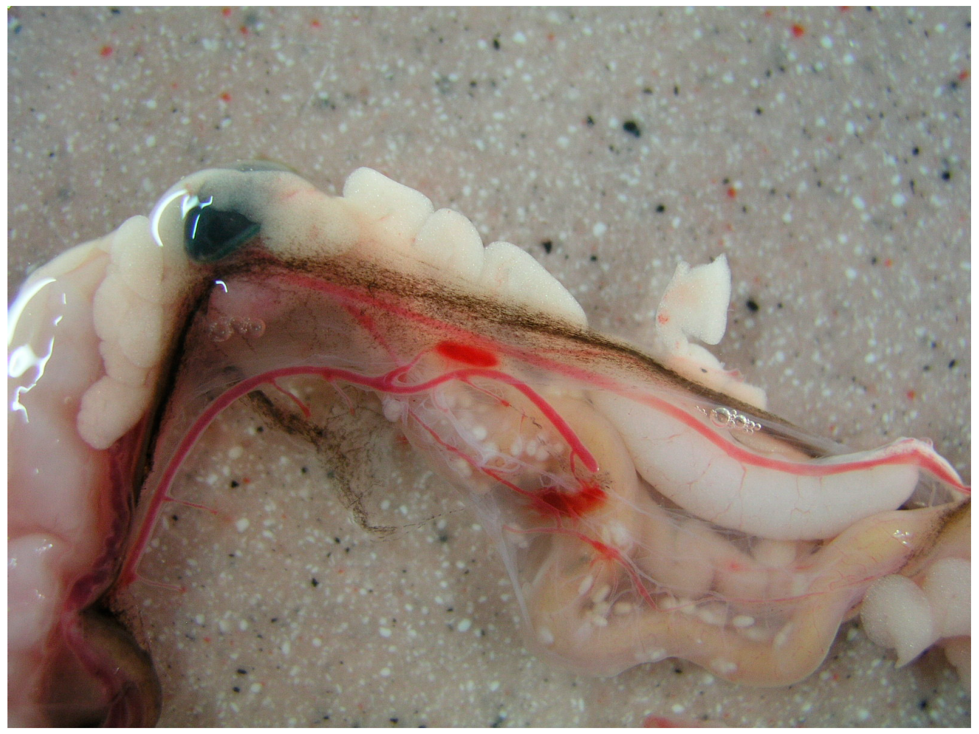
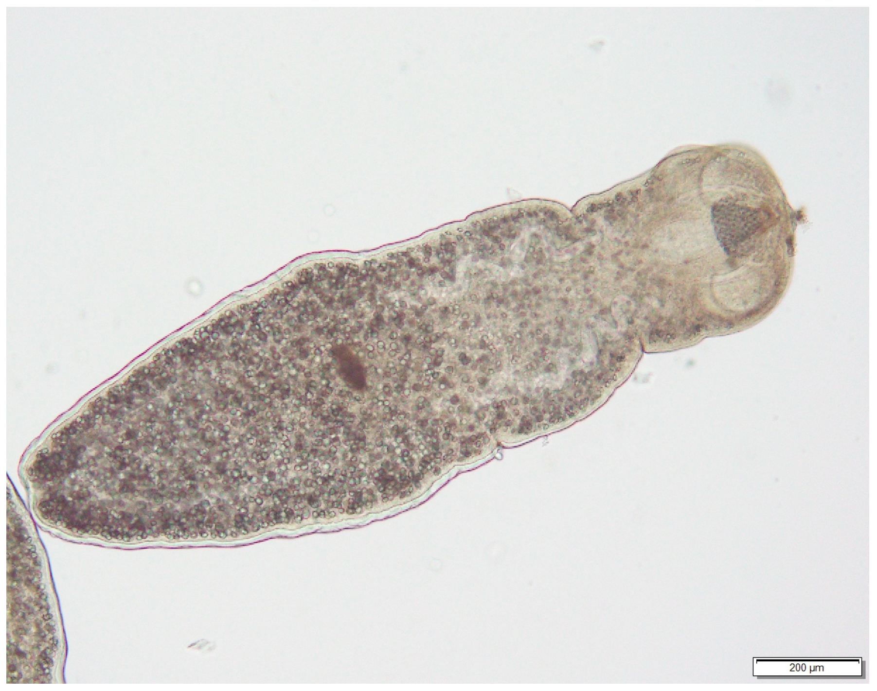
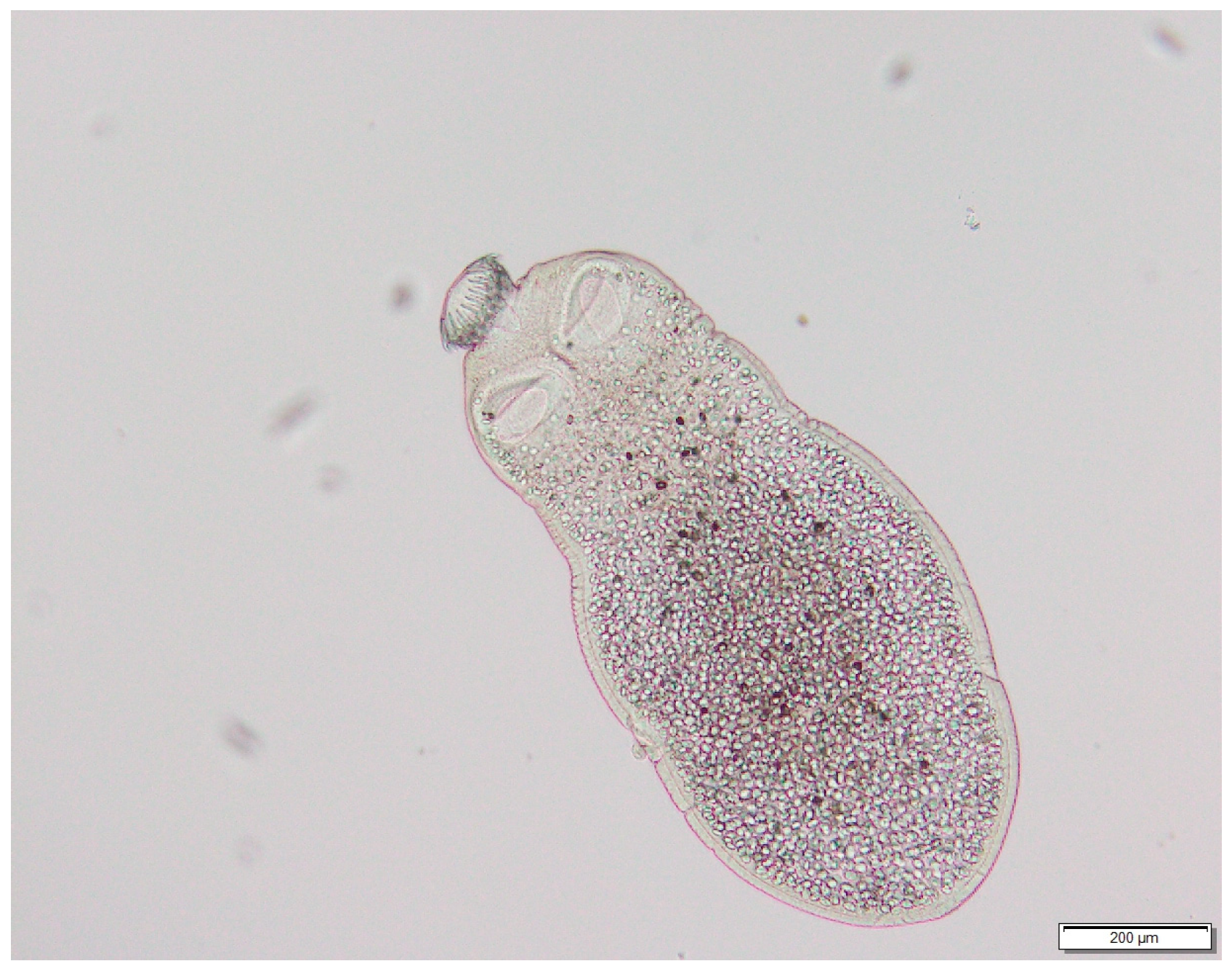
Most of these stages were located at the outer wall of the anterior small intestine or trapped by the mesentery (Figure 2). In the geckos, cysts were also seen under the capsule of the liver (Figure 3). The digestion in artificial gastric juice dissolved the cyst wall and scolices were seen after neutralization of the digestion fluid (Figure 4 and Figure 5).
Discrimination between the cysticercoids of J. pasqualei and J. gervaisi was not possible, since the scolex structures did not differ strikingly, and for this reason, these cysticercoids were reported as Joyeuxiella spp. Among indigenous snakes, the highest burdens of dipylidiid metacestodes were found in the saw-scaled vipers (5–210), Arabian horned vipers (3–75) and Schokari sand racers (2–63). The sand boas (E. jayakari) were negative for cestode larval stages.
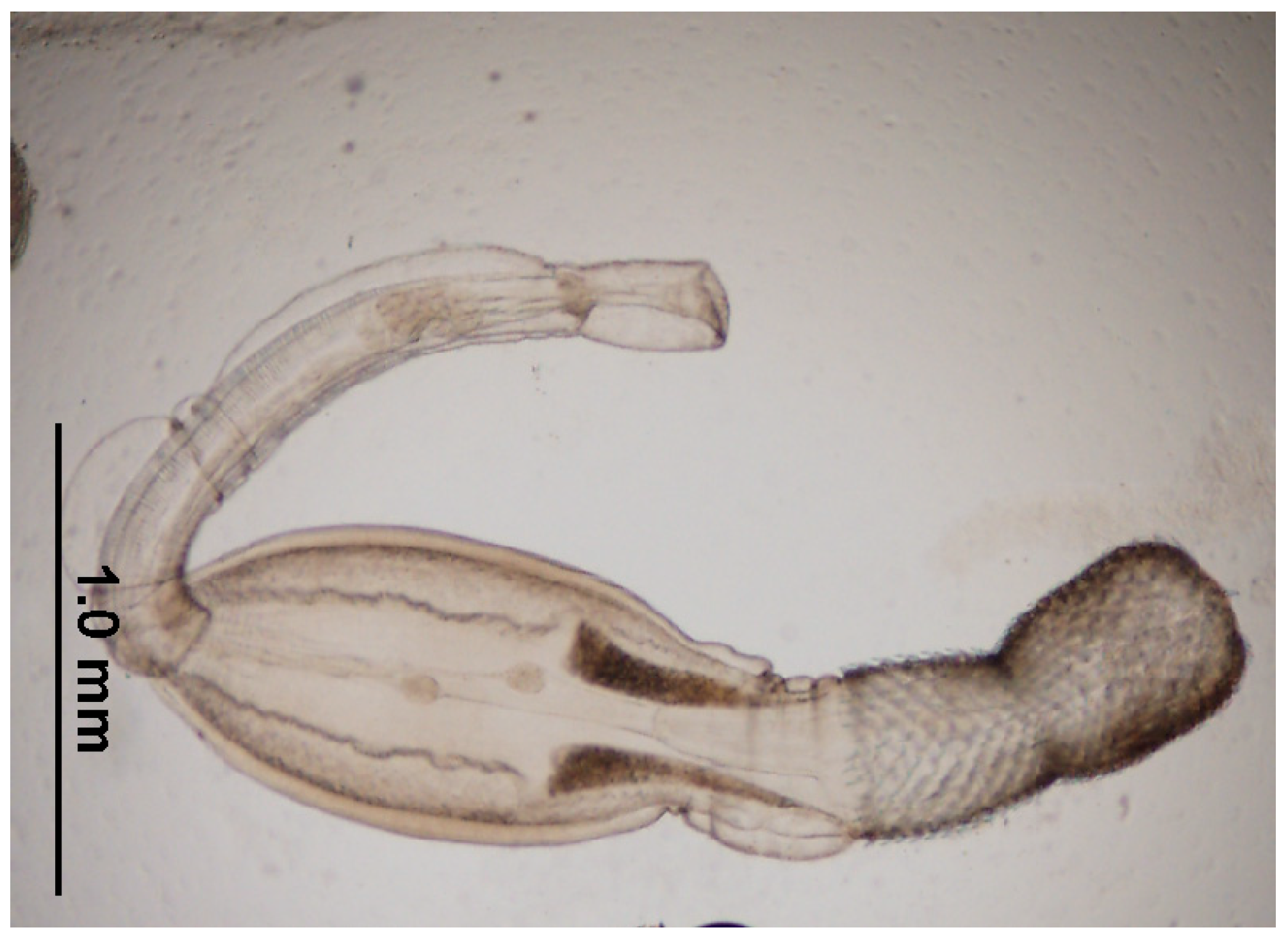
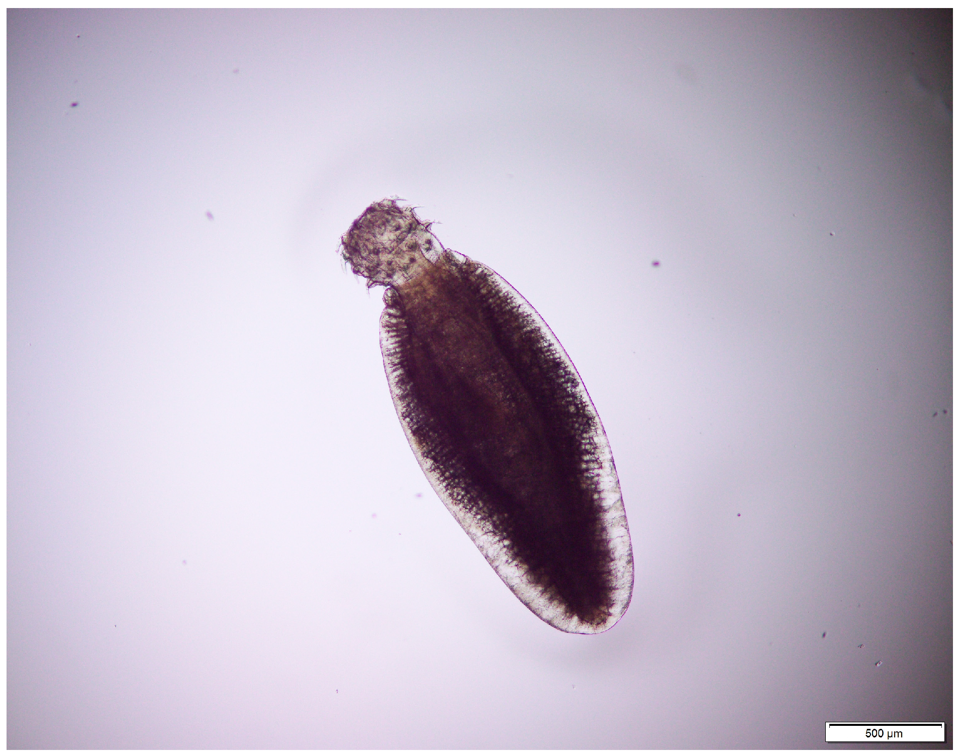
Based on the configuration and hook structures, two larval stages of acanthocephalans, Centrorhynchus sp. (Figure 6) and Macracanthorhynchus sp. (Figure 7), were detected in snakes only. These cystacanths most probably belonged to C. aluconis and M. catulinus, respectively.
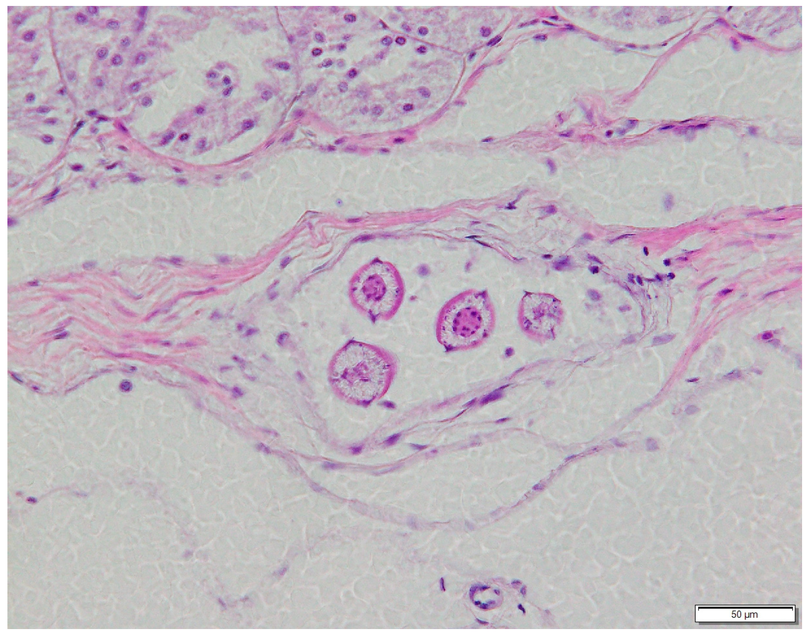
Two types of encapsulated nematode larvae that were found in the walls of the gastro-intestinal tracts of reptiles were spirurids but could not be determined to lower rank levels. Possibly, they belonged to Physocephalus dromedarii (Figure 8), a frequent gastric nematode of dromedaries in Dubai, and to a Rictulariidae species, perhaps to P. cahirensis (Figure 9).
Among the examined lizards, dipylidiid metacestodes were found only in both gecko species (H. flaviviridis and H. robustus), in numbers between one and three, while ocellated skinks and worm lizards were negative for cestode larvae.

4. Discussion

The results of this study showed that reptiles carry larval stages of (a) dipylidiid cestodes, (b) acanthocephalans and (c) nematodes. At least the genera Joyeuxiella and Diplopylidium, as well as the nematode P. cahirensis, were frequent intestinal helminths of feral cats in the Dubai Emirate. Single cats were also diagnosed with acanthocephalans, the cystacanths of which were seen in snakes.
The parasite fauna of stray domestic cats in the region has been investigated in Qatar [12,13,14], the UAE [8,9], Kuwait [15,16,17], Jordan [18], Egypt [19,20,21], Iraq [22,23,24,25,26] and Iran [27,28,29,30,31,32,33,34]. A literature review summarized 104 cat parasite studies for Turkey [35].
Due to the high prevalence of dipyliid cestodes with reptile involvement in their life cycles, the established parasite fauna of feral cats in Dubai differs strikingly from other reports on cat parasites in the region. Thus, in Qatar, a high H. taeniaeformis prevalence of 73.6% was recorded, and D. acanthotetra was the only dipylidiid species was found in 47.1% of the 658 dissected cats [14]. In the city of Mosul, in Iraq, J. pasqualei, D. nölleri and H. taeniaeformis were detected with a prevalence of 26%, 24% and 52%, respectively. In addition, Dipylidium caninum (64%), Spirometra mansonoides (24%), Diphyllobothrium sp. (12%) and Mesocestoides variabilis (16%) were found [23]. A more recent review of feline parasites in Iraq listed a total of 62 species of parasites, amongst them 17 cestode species [26]. The only cat survey carried out in Jordan revealed D. caninum in 12, H. taeniaeformes in three and Echinococcus granulosus in 10 out of 78 examined cats [18]. Examinations of cats in the northern Nile delta [20] and in the Cairo governorate of Egypt did not mention representatives of the genera Diplopylidium or Joyeuxiella [21]. This is surprising, since the strobilar stages of J. pasqualei and J. echinorhynchoides were described for the first time in cats [36] and foxes [37] in Egypt. Diplopylidium spp. and Joyeuxiella spp. were also not detected in cats examined in the Iranian cities of Tabriz [30], Azarshahr [31] and Ahar [32], while researchers in Bandar-e-Anzali [27], Kashan [29] and Zanjan [28] detected them with high prevalence and also mentioned the spirurid P. cahierensis. Research on cat parasites in Kuwait identified D. nölleri, D. acanthotetra, J. pasqualei and P. cahierensis in 54.6%, 45.4%, 38.3% and 34.6%, respectively [17]. In Turkey, J. pasqualei and D. nölleri were recorded mainly in the southern and eastern parts of the country [35]. All of the cited authors also mentioned H. taeniaeformis in different prevalences, but J. gervaisi (or under its junior synonym, J. fuhrmanni) and the two acanthocephalans have not been reported in cats in the region, except for Dubai. Nearly all of the cited researchers listed D. caninum, Mesocestoides spp. and Physaloptera praeputiale as frequent cat parasites. Neither these helminths nor J. echinorchynchoides we detected in cats from Dubai.
Contrary to M. catulinus, which is a parasite of canids and cats, C. aluconis is more often found in birds of prey and owls. For both acanthocephalan species, arthropods are intermediate hosts, and reptiles serve as paratenic hosts [38]. Apart from reptiles, the larval stage of C. aluconis was also detected in Macqueen’s bustards in Dubai [39].
Already in the first third of the 20th century, studies of the life cycle of Joyeuxiella spp. were carried out. By feeding larval stages from geckos to cats, the role of reptiles in the life cycle of these cestodes was experimentally proven [6,40]. But the way in which reptiles become infected remains unclear. It has already been assumed that insects are involved in the life cycle [41]. Recently, feeding experiments were carried out with mice and darkling beetles and their larvae, but all of the trials so far have given negative results [42].
With regards to reptiles, our own investigations showed that among the examined snakes, only sand boas did not harbor cestode larvae. The reason for this is the difference in diet. During the dissection of the stomachs of some of the vipers, remnants of locusts, beetles and solifuges were found. This was an indication that these snakes also feed on insects, while sand boas are not insectivorous.

5. Conclusions

Cestodes of the genera Joyeuxiella and Diplopylidium were present in cats in all three major habitats in Dubai with high prevalence and intensities. Snakes, except sand boas, were more frequently infected with larval stages of these tapeworms and showed higher larval burden compared to geckos, and all the examined skinks and worm lizards were negative for these parasites.
The absence of snakes in the city center of Dubai as a source of infection and a high prevalence of cestodes of the genera Joyeuxiella and Diplopylidium in feral cats allowed the conclusion that certain insects (cockroaches, crickets or others) that frequently can be seen in town and are eaten by reptiles, as well as by cats, are the real intermediate hosts, and that reptiles are paratenic hosts. Subsequent feeding experiments must be carried out to prove this hypothesis.

Author Contributions

Conceptualization, R.K.S.; methodology, R.K.S. and S.S.; formal analysis, R.K.S.; methodology, R.K.S. and S.S.; validation, R.K.S. and S.S.; resources, R.K.S.; data curation, S.S.; original draft preparation, R.K.S.; review and editing, R.K.S. and S.S. All authors have read and agreed to the published version of the manuscript.

Funding

The author declares that the research was conducted in the absence of any commercial and/or financial relationships that could be construed as potential conflicts of interest.

Institutional Review Board Statement

The Ethical Animal Welfare Committee (EAWC), comprising six veterinarians from the Central Veterinary Research Laboratory (CVRL), Wadi Al Safa and Dubai Falcon Hospital (DFH), follows the legislation that was laid down by the Ministry of Climate Change and Environment (MOCCAE) in Ministerial Decision No. (384) of the year 2008 on the executive by-law of the Federal Law No. (16) of the year 2007 concerning Animal Welfare. The welfare of all animals, their treatment and experimental use at the above facilities is reviewed and approved by the EAWC.

Data Availability Statement

The raw data for this article will be made available by the author, without undue reservation.

Acknowledgments

The author is grateful to the veterinary section of Dubai municipality and various pet clinics for submitting carcasses of feral cats. My thanks go to P. Nagy from Emirates Industry for Camel Milk Products, to D. O’Donovan from Wadi al Safa, to K. Hyland from Wildlife Protection Office, to J. Els from Breeding Center for Endangered Arabian Wildlife and to many other people who sent killed snakes and lizards for examination.

Conflicts of Interest

The author declares no conflicts of interest.

References

  1. Schuster, R.K. Cestodes of the genera Diplopylidium and Joyeuxiella (Eucestoda: Dipylidiidae—A review of historical data, species inventory and geographical distribution. Sci. Parasitol. 2020, 21, 1–17. [Google Scholar]
  2. Witenberg, G. On the cestodes subfamily Dipylidiinae Stiles. Z. Parasitenk. 1932, 4, 542–584. [Google Scholar] [CrossRef]
  3. Marchi, M. Sopra una nuovo cestode trovato nell’ Ascalabotes mauritanicus. Atti Soc. Ital. Sci. Nat. 1872, 15, 505–506. [Google Scholar]
  4. Parona, C. Elmintologia sarda. Contribuzione allo studio dei vermi parassiti in animali di Sardegna. Ann. Mus. Civ. Genova 1886, 24, 275–384. [Google Scholar]
  5. Diamare, V. Bemerkungen ueber Dipylidienlarven. Zentralbl. Bact. Parasitenk. 1894, 14, 565–566. [Google Scholar]
  6. Parrot, L.; Joyeux, C. Les cysticercoids de Tarentola mauritanica L. et les tenias du chat. Bull. Soc. Pathol. Exot. 1920, 13, 687–695. [Google Scholar]
  7. Gardner, A.S. The Amphibians and Reptiles of Oman and the UAE; Edition Chimaira: Frankfurt am Main, Germany, 2013; 480p. [Google Scholar]
  8. Schuster, R.K.; Thomas, K.; Sivakumar, S.; O’Donovan, D. The parasite fauna of stray domestic cats (Felis catus) in Dubai, United Arab Emirates. Parasitol. Res. 2009, 105, 125–134. [Google Scholar] [CrossRef] [PubMed]
  9. Schuster, R.K.; Mustafa, M.B.; Chester, S.T.; Knaus, M. Efficacy of Broadline® (Merial) against naturally acquired infections with cestodes of the genus Joyeuxiella. Parasitol. Res. 2016, 115, 2679–2684. [Google Scholar] [CrossRef]
  10. Reiczigel, J.; Marozzi, M.; Fabian, I.; Rozsa, L. Biostatistics for parasitologists—A primer to Quantitative Parasitology. Trends Parasitol. 2019, 35, 277–281. [Google Scholar] [CrossRef]
  11. Schuster, R.K.; Mehmood, N.; Varcasia, A.V.; Veneziano, V. Redescription and resurrection of the status of Joyeuxiella gervaisi (Setti, 1895) (Eucestoda, Dipylidiidae). Helminthologia 2023, 60, 166–174. [Google Scholar] [CrossRef]
  12. Abu-Madi, M.A.; Al-Ahbabi, D.A.; Al-Mashhadani, M.M.; Al-Ibrahim, R.; Pal, P.; Lewis, J.W. Patterns of parasitic infections in faecal samples from stray cat populations in Qatar. J. Helminthol. 2007, 81, 281–286. [Google Scholar] [CrossRef] [PubMed]
  13. Abu-Madi, M.A.; Behnke, J.M.; Prabhaker, K.S.; Al-Ibrahim, R.; Lewis, J. Intestinal helminths of feral cat populations from urban and suburban districts of Qatar. Vet. Parasitol. 2010, 168, 284–292. [Google Scholar] [CrossRef]
  14. Abu-Madi, M.A.; Pal, P.; Al-Thani, A.; Lewis, J.W. Descriptive epidemiology of intestinal helminth parasites from stray cat populations in Qatar. J. Helminthol. 2008, 82, 59–68. [Google Scholar] [CrossRef]
  15. Abdul-Salam, J.; Baker, K. Prevalence of intestinal helminths in stray cats in Kuwait. Pakistan Vet. J. 1990, 10, 17–21. [Google Scholar]
  16. El-Azazy, O.M.E.; Abdou, N.-E.; Khalil, A.I.; Al-Batel, M.K.; Majeed, Q.A.; Henedi, A.A.M.; Tahrani, L.M. Potential Zoonotic Trematodes Recovered in Stray Cats from Kuwait Municipality, Kuwait. Korean J. Parasitol. 2015, 53, 279–287. [Google Scholar] [CrossRef] [PubMed]
  17. El-Azazy, O.M.E.; Abdou, N.-E.; Khalil, A.I.; Al-Batel, M.K.; Majeed, Q.A.; Henedi, A.A.M.; Tahrani, L.M.A. Cestodes and nematodes recorded in stray cats in Kuwait. Glob. Vet. 2016, 16, 111–118. [Google Scholar]
  18. Morsy, T.A.; Michael, S.A.; El-Disi, A.M. Cats as reservoir hosts of human parasites in Amman, Jordan. J. Egypt. Soc. Parasitol. 1980, 10, 5–18. [Google Scholar]
  19. Morsy, T.A.; Sadek, M.S.M.; Al-Hamid, M.Y. Intestinal parasites in stray cats in Cairo, Egypt. J. Egypt. Soc. Parasitol. 1981, 11, 331–345. [Google Scholar]
  20. Khalafalla, R.E. A survey study on gastrointestinal parasites of stray cats in northern region of Nile Delta, Egypt. PLoS ONE 2011, 6, e20283. [Google Scholar] [CrossRef]
  21. Ayoub, M.B. Parasitic infection in stray cats and dogs with special reference to ultrastructure of the recovered worms. Animal Health Res. J. 2014, 2, 165–178. [Google Scholar]
  22. Nihad, W.; Al-Khalidi, M.; Tafiq, I.; Al-Alousi, M.; Subber, A.H. Internal and external parasites in cats in Mosul, Iraq. J. Vet. Parasitol. 1988, 2, 137–138. [Google Scholar]
  23. Al-Obaidi, Q.T. Prevalence of internal helminthes in stray cats (Felis catus) in Mosul city, Mosul-Iraq. J. Anim. Vet. Adv. 2023, 11, 2732–2736. [Google Scholar] [CrossRef]
  24. Hadi, A.M.; Faraj, A.A. Role of domestic cats Felis catus as reservoir hosts of internal parasites and protozoa in Baghdad. Bull. Iraq Nat. Hist. Mus. 2014, 13, 89–94. [Google Scholar]
  25. Al-Aredhi, H.S. Prevalence of gastrointestinal parasites in domestic cats (Felis catus) in Al-Diwaniya province/Iraq. Int. J. Curr. Microbiol. App. Sci. 2015, 4, 166–171. [Google Scholar]
  26. Al-Tae, A.-R.A.; Al Rubaie, A.-R.L. Checklist of parasites of stray cats Felis catus of Iraq. Ibn Al-Haitham J. Pure Appl. Sci. 2017, 1782, 143–152. [Google Scholar] [CrossRef]
  27. Changizi, E.; Mobedi, I.; Salimi-Bejestani, M.R.; Rezaei-Doust, A. Gastrointestinal helmintic parasites in stray cats (Felis catus) from north of Iran. Iran. J. Parasitol. 2007, 2, 25–29. [Google Scholar]
  28. Esmaeilzadeh, M.; Shamsfard, M.; Kazemi, A.; Khalafi, S.A.; Altome, S.A. Prevalence of protozoa and gastrointestinal helminthes in stray cats in Zanjan Province, north-west of Iran. Iran. J. Parasitol. 2009, 4, 71–75. [Google Scholar]
  29. Arbabi, M.; Hoshyar, H. Gastrointestinal parasites of stray cats in Kashan, Iran. Trop. Biomed. 2009, 26, 16–22. [Google Scholar]
  30. Garedaghi, Y.; Firouzivand, Y. Prevalence of gastrointestinal parasites of domestic cats and its zoonotic importance in Tabriz city, Iran. Cibtech J. Zool. 2014, 3, 87–92. [Google Scholar]
  31. Hajipour, N.; Baran, A.I.; Yakhchali, M.; Banan Khojasteh, S.M.; Hesari, F.S.; Esmaeinejad, B.; Arimand, J. A survey study on gastrointestinal parasites of stray cats in Azarshahr, (East Azerbaijan province, Iran). J. Parasit. Dis. 2016, 40, 1255–1260. [Google Scholar] [CrossRef] [PubMed]
  32. Yakhchali, M.; Hajipour, N.; Malekzadeh-Viayeh, R.; Esmaeilnejad, B.; Nemati-Haravani, T.; Fathollahzadeh, M.; Jafari, R. Gastrointestinal helminths and ectoparasites in the stray cats (Felidae: Felis catus) of Ahar Municipality, Northwestern Iran. Iran. J. Parasitol. 2017, 12, 298–304. [Google Scholar]
  33. Soroushianfar, M.; Sadr, A.; Sazmand, A.; Dianaty, S.; Khedi, J.; Schuster, R.K.; Borji, H. Gastrointestinal parasites of cats in the Middle East (2000–2023): A literature review. Parasitol. Int. 2024, 102, 102919. [Google Scholar] [CrossRef]
  34. Ahmadi, A.; Oryyan, A.; Alidadi, S. Parasites of stray cats in Iran: A parasitological and histopathological study. Acta Parasitol. 2024, 69, 664–674. [Google Scholar] [CrossRef]
  35. Barih, Ö; Tuygun, T.; Gençay Topçu, E.B.; Umur, Ş. The parasites of cats in Türkiye. Turkiye Parazitol. Derg. 2023, 47, 190–199. [Google Scholar] [CrossRef]
  36. Diamare, V. Note su cestodi. Boll. Soc. Natur. Napoli 1893, 7, 9–13. [Google Scholar]
  37. Sonsino, P. Noticie elmintologiche. Atti della Societa Toscana di Scienze Naturali Residente in Piesa. Process. Verbali 1889, 6, 191–194. [Google Scholar]
  38. Chochlova, I.G. Acanthocephala of Terrestrical Animals of the Fauna of the USSR [Akantocefaly Nazemnych Zivotnych Fauny SSSR]; Izdatel’stvo Nauka: Moscow, Russia, 1986; 275p. (In Russian) [Google Scholar]
  39. Schuster, R.K.; Sivakumar, S.; Kinne, J. Parasite findings in the MacQueen’s bustard, Chlamydotis macqueenii (Grey, 1832), and considerations on the parasite fauna of bustards and the systematic position of some of the parasites. Vet. Parasitol. Reg. Stud. Rep. 2025, 57, 101146. [Google Scholar] [CrossRef] [PubMed]
  40. Popov, P. Sur le development de Diplopylidium skrjabini n. sp. Ann. Parasitol. 1935, 13, 322–326. [Google Scholar] [CrossRef]
  41. Lopez-Neyra, C.R. Sobre la evolucion de la Joyeuxia chyceri v. Ratz. Infeccion experimental de la salamanquesas. Bol. Real. Soc. Espan. Hist. Nat. 1927, 27, 398–399. [Google Scholar]
  42. Bezerra-Santos, M.A.; Mendoza-Roldan, J.A.; Lia, R.P.; Annoscia, G.; Schuster, R.; Varcasia, A.; Sgroi, G.; Modry, D.; Otranto, D. Description of Joyeuxiella pasqualei (Cestoda: Dipylidiidae) from an Italian domestic dog, with a call for further research on its first intermediate host. Parasitology 2022, 149, 1769–1774. [Google Scholar] [CrossRef]
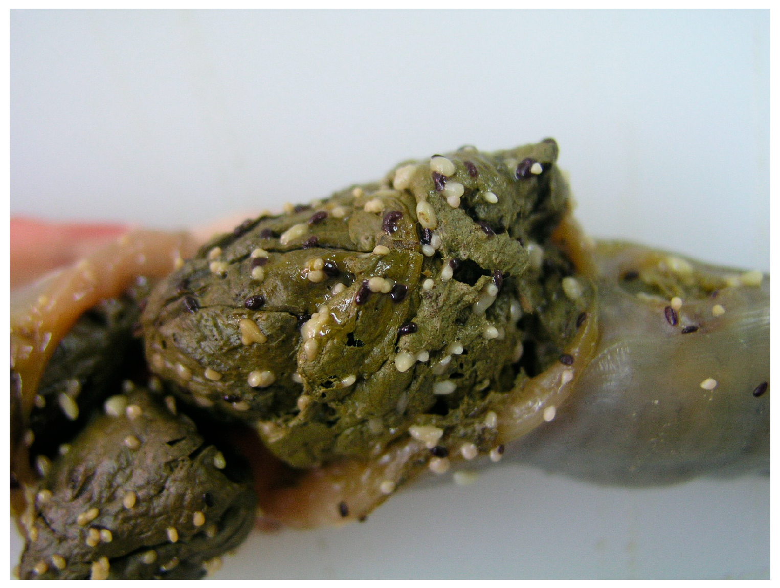
Figure 1. Rectal content of a heavily infected cat with dipylidiid cestodes. White proglottids belonged to Joyeuxiella spp., while dark-coloured structures are gravid segments of Diplopylidium nölleri.
Diversity 17 00578 g001
Figure 2. Dipylidiid cysts in the mesenterium and attached to the outer intestinal wall of a saw-scaled viper.
Diversity 17 00578 g002
Figure 3. Joyeuxiella cysts under the liver capsule of a gecko.
Diversity 17 00578 g003
Figure 4. Evaginated Joyeuxiella cysticercoid, rostellum retracted.
Diversity 17 00578 g004
Figure 5. Evaginated Diplopylidium nölleri cysticercoid.
Diversity 17 00578 g005
Figure 6. Centrorhynchus sp. cystacanthus with evaginated proboscis after artificial digestion of the intestine of a saw-scaled viper.
Diversity 17 00578 g006
Figure 7. Macracanthorhynchus sp. cystacanthus with evaginated proboscis after artificial digestion of the intestine of an Arabian horned viper.
Diversity 17 00578 g007
Figure 8. Third-stage larva of a spirurid (most probably Physocephalus dromedarii) after artificial digestion of the intestine of a saw-scaled viper.
Diversity 17 00578 g008
Figure 9. Rictulariidae larva (most probably Pterygodermatites cahirensis) in a histological section of the liver of a yellow-bellied house gecko.
Diversity 17 00578 g009
Table 1. Prevalence and burden of dipylidiid cestodes and the rictulariid nematode Pterygodermatites cahirensis in feral cats obtained from different habitats (city center n = 87, outskirts n = 141, desert n = 132) in Dubai. In the case of both Joyeuxiella species, the intensity was difficult to determine when juvenile specimens were present and for this reason, intensity numbers are given only for the genus Joyeyxiella. The table does not include Diplopylidium acanthotetra, Centrorhynchus aluconis or Macracanthorhynchus catulinus, since these helminths were detected in low numbers in cats mainly from desert habitats.
SpeciesHabitatNumber PositivesPrevalenceConfidence IntervalsAverage
Burden
Confidence Intervals
(%)UpperLowerUpperLower
Joyeuxiella
pasqualei
city center5563.252.273.3
outskirts7452.543.960.9
desert10277.369.284.1
all23164.259.069.1
Joyeuxiella
gervaisi
city center22.30.38.1
outskirts2316.310.323.5
desert5541.733.250.6
all8022.218.026.9
Joyeuxiella
spp.
city center5664.453.474.459.339.399
outskirts8157.448.865.758.342.585.7
desert10680.372.586.762.546.586.1
all24367.562.472.360.449.174.5
Diplopylidium
noelleri
city center2832.222.643.173.431.0148.0
outskirts3222.716.130.548.219.6151.0
desert7355.346.464.451.033.180.2
all13336.931.942.255.238.482.4
Pterygodermatites
cahirensis
city center4046.035.257.028.014.351.9
outskirts3625.518.633.613.28.522.1
desert4534.126.142.87.64.614.1
all12133.628.738.71611.125.1
Table 2. Prevalence and burden of dipylidiid cestodes and the rictulariid nematode Pterygodermatites cahierensis in adult (n = 279) and juvenile (n = 81) feral cats in Dubai. In the case of both Joyeuxiella species, the intensity was difficult to determine when juvenile specimens were present and for this reason, intensity numbers are given only for the genus Joyeyxiella.
Parasite SpeciesAge Group of CatsNumber PositivesPrevalenceConfidence IntervalsBurdenConfidence Intervals
(%)LowerUpperAverageLowerUpper
Joyeuxiella
pasqualei
adult20372.867.177.9
juvenile2834.624.346.0
Joyeuxiella
gervaisi
adult7828.022.833.6
juvenile22.50.38.6
Joyeuxiella
spp.
adult21476.771.381.560.348.677.8
juvenile2935.825.447.260.938.4103
Diplopylidium
noelleri
adult11842.336.448.355.937.783.5
juvenile1518.510.828.749.210.3192
Pterygodermatites
cahierensis
adult9734.829.240.712.38.719.5
juvenile2429.620.040.830.813.766.2
Table 3. Numbers of examined reptiles and their infection with larval helminths.
Reptile HostsNo of
Examined
Number of Hosts Infected with Larval Stages of
Joyeuxiella
spp.
Diplopylidium
noelleri
Spirurata
spp.
Centrorhynchus
sp.
Macracanthorhynchus
sp.
1.
snakes
Eryx jayakari1500400
Rhagerhis moilensis111010
Platyceps rhodorachis310121
Psammophis schokari1365360
Cerastes gasperetti512021
Echis carinatus262011381
Echis omanensis111101
Dolichophis jugularis222110
Total:66322213194
2.
Lizards
Hemidactylus flaviviridis32130300
Hemidactylus robustus930100
Chalcides ocellatus2200000
Diplometopon zarudnyi500200
Total:68160600
Disclaimer/Publisher’s Note: The statements, opinions and data contained in all publications are solely those of the individual author(s) and contributor(s) and not of MDPI and/or the editor(s). MDPI and/or the editor(s) disclaim responsibility for any injury to people or property resulting from any ideas, methods, instructions or products referred to in the content.