
Brain Disease-Modifying Effects of Radiofrequency as a Non-Contact Neuronal Stimulation Technology


Abstract
1. Introduction
2. The Effects of Non-Contact RF-EMF Stimulation on In Vitro Model Cells

| Non-Contact Stimulation Exposure | |||||
|---|---|---|---|---|---|
| Stimulation Type | Frequency and Intensity | Exposure Period | Cell Line | Effects | References |
| RF-EMF | 900 MHz EMF (AM 900 MHz, CM 900 MHz) | 10, 15 and 20 min | Primary cells (mouse olfactory bulbs, P2) | ↑ Cytoskeletal protein expression (GFAP, vimentin, nestin; CW 900 MHz for 15–20 min) ↓ Cytoskeletal protein expression (GFAP, vimentin, nestin; AM 900 MHz for 15–20 min) ↑ Caspase-3 expression (AM 900 MHz for 20 min) | [3] |
| 3.0 GHz 0.3/0.7 W/kg SAR | 60 min | Primary hippocampal neurons (rat embryonic hippocampi, E18) | ↓ Action potential ↑ Intracellular Ca2+ ↑ Synaptic activity (sEPSCs, sIPSCs) | [4] | |
| 918 Hz 0.2 W/kg SAR | 60 min | Primary astrocytes (rat neonatal brains and human fetal brains) | ↓ ROS (mitochondrial) ↓ NADPH oxidase activity | [5] | |
| 64/100 MHz, 0.4/0.6/0.9 W/kg SAR | 1–2 h/day for 4, 8 or 14 days | Primary human brain cells (human fetal brain tissue) | ↓ Aβ levels (Aβ40 and Aβ42, 64 MHz, 0.6 W/kg, 1 h/day for 14 days) | [6] | |
| 1800 MHz 1/2/4 W/kg SAR | 5 min on/10 min off for 1–3 days | Embryonic neural stem cells (mouse embryonic cortex, E13.5) | ↓ Neurite number, branching points, and total length ↓ Proneural genes (Ngn1, NeuroD expression) | [7] | |
| 50 Hz 1 MT | 24 h | SH-SY5Y cells (human neuroblastoma cells) | ↑ NOS activity ↑ O2− production ↑ TGF-β and IL-18BP expression | [8] | |
| 900 MHz 1 W/kg SAR | 24, 48, 72, 120 h | SN56 cells (mouse cholinergic neurons) Primary cortical neurons (rat) | ↑ β-thymosin mRNA ↓ Morphological maturation (neurites) | [9] | |
| 1800 MHz 0.23 W/kg SAR | 3 × 10 min/day for 2 days | SH-SY5Y cells (human neuroblastoma cells) | ↑ ROS levels ↑ Monomeric α-syn levels ↑ Cell death | [11] | |
| 1800 MHz 2 W/kg SAR | 5 min on/10 min off for 24 h | Primary cortical neurons (newborn SD rats) | ↑ ROS levels ↓ 8-OHdG levels ↓ Mitochondrial function ↓ mtDNA oxidative damage | [12] | |
| 1900 MHz | 2 h | Primary cortical neurons and astrocytes (ICR mouse embryonic, E15) | ↑ Apoptotic pathways (Caspase-2, Caspase-6, Asc) | [13] | |
| 2.45 GHz 6 W/kg SAR | 20 min | N9 cells (mouse microglial cells) | ↑ CD11b expression ↑ JAK2 and STAT3 phosphorylation ↑ Pro-inflammatory responses (TNF-α and iNOS) | [14] | |
| 1950 MHz 6 W/kg SAR | 2 h | HT22 cells (mouse hippocampal neuronal cells) | ↑ ROS levels ↑ Cell death ↑ Neurotoxicity | [15] | |
| 837 MHz (CDMA) 1950 MHz (W-CDMA) 2 W/kg SAR | 2 h | HT22 cells (mouse hippocampal neuronal cells) | No effect on Aβ-induced cytotoxicity, ROS production, or apoptosis | [16] | |
| 1950 MHz (W-CDMA) 6 W/kg SAR | 2 h/day over 3 days | HT22 cells (mouse hippocampal neuronal cells) SH-SY5Y cells (human neuroblastoma cells) | No effect on the expression levels of APP and BACE1 in either SH-SY5Y or HT22 cells. | [17] | |
| 935 MHz 4 W/kg SAR | On/off cycles of 120/120 s | SH-SY5Y cells (human neuroblastoma cells) N9 cells (mouse microglial cells) | No effect on the proportions of living, early apoptotic, or late apoptotic cells in either SH-SY5Y or N9 cells. | [18] | |
| 935 MHz 4 W/kg SAR | On/off cycles of 120/120 s for 24 h | SH-SY5Y cells (human neuroblastoma cells) | No effect on neuronal differentiation signaling pathways markers and mitochondrial fission and fusion markers | [19] | |
| 1800 MHz 4 W/kg SAR | 5 min on/10 min off for 1, 6, or 24 h | U251 and A172 cells (human glioblastoma) SH-SY5Y cells (human neuroblastoma cells) | No effect on cellular behavior. (cell cycle progression, cell proliferation, or cell viability) | [20] | |
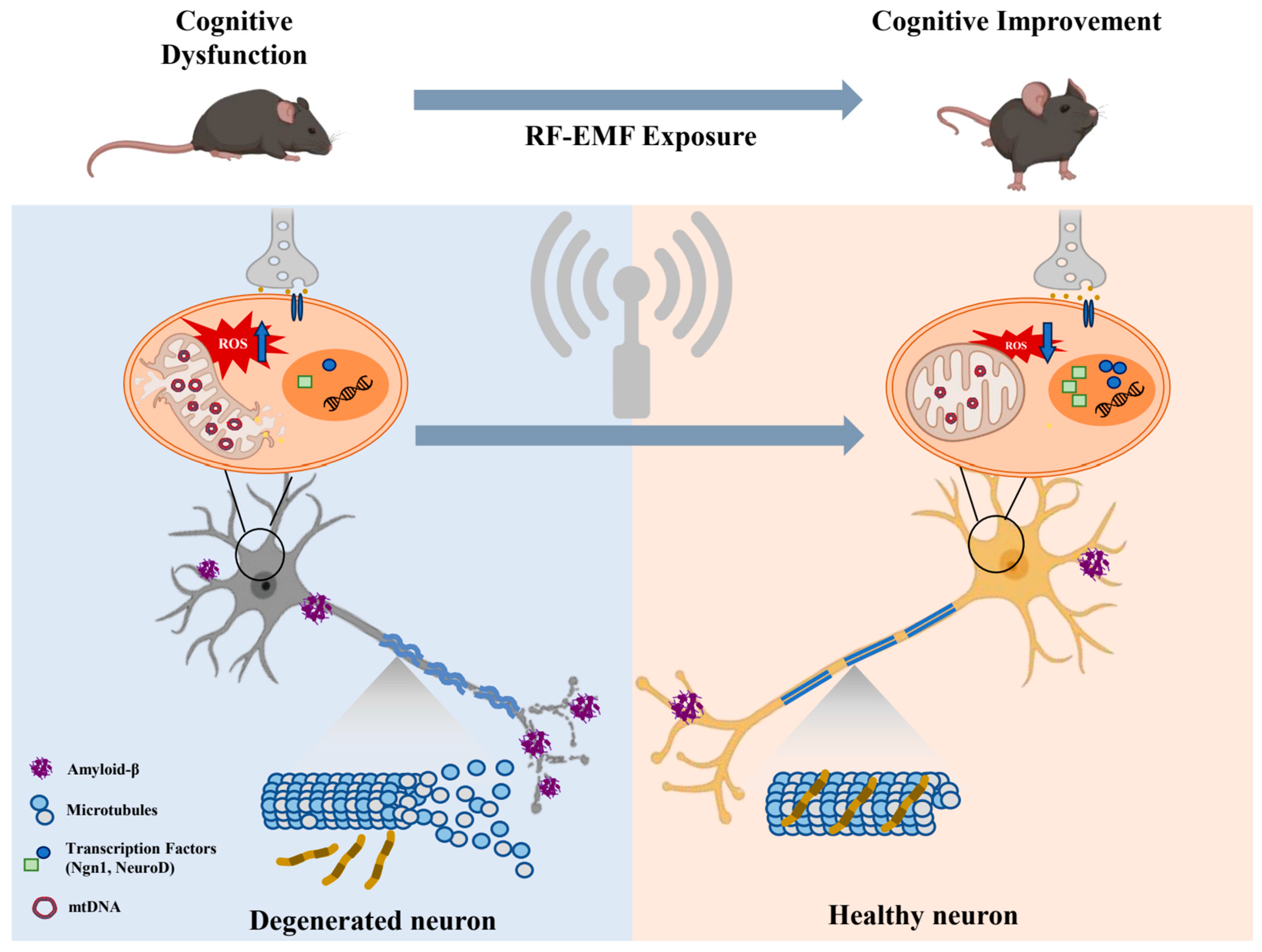
3. The Effects of Non-Contact RF-EMF Stimulation on Cognitive Behaviors and Molecular Mechanisms in In Vivo Animal Models
4. The Effects of Other Non-Contact Electrotherapeutic Stimulation Modalities in In Vitro and Model Systems
5. The Effects of Non-Contact RF-EMF Stimulation on the Physiological Process of Sleep in Human Subjects
6. The Effect of RF-EMF Stimulation on the Pathophysiology of the Human Brain
| Non-Contact Stimulation Exposure | |||||
|---|---|---|---|---|---|
| Stimulation Type | Frequency and Intensity | Exposure Periods | Participant | Effects | References |
| RF-EMF | 900 MHz 1 W/kg SAR | 15 min on/15 min off during 8 h sleep episode | Healthy young males (mean age: 22.6 years) | ↑ Non-REM sleep EEG power | [51] |
| 900 MHz 1 W/kg SAR | 30 min before a 3 h sleep episode | Healthy young males (mean age: 20–25 years) | Short exposure to EMF emitted by mobile phones affects brain physiology. | [52] | |
| 900 MHz 1 W/kg SAR | 30 min | Healthy young males (mean age: 20–25 years) | ↑ Regional cerebral blood flow in dorsolateral prefrontal cortex (rCBF) ↑ Alpha activity (EEG during wakefulness) | [53] | |
| 900 MHz 1 W/kg SAR | 15 min on/15 min off during 8 h sleep episode/ 30 min before a 3 h sleep episode | Healthy young males | ↑ Non-REM sleep EEG power | [54] | |
| 894.6 MHz 0.11 W/kg SAR | 30 min before sleep episode | Healthy individuals (males and females) | ↑ Rapid eye movement sleep | [55] | |
| 900 MHz | 15–20 min | Healthy young males and children | Induce short-term, reversible changes in human EEG | [56] | |
| 900 MHz 2 W/kg SAR | 30 min before sleep episode | Healthy young males (mean age: 23.2 years) | Affects non-rapid eye movement sleep and rapid eye movement sleep activity | [59] | |
| 900 MHz 0.2/5 W/kg SAR | 30 min before sleep episode | Healthy young males (mean age: 22.4 years) | Affects the non-REM sleep EEG and cognitive performance | [60] | |
| 900 MHz 1 W/kg SAR | 25 min | Healthy individuals (males and females; mean age: 18–30 years) | Exposure may affect human brain activity | [61] | |
| 894.6 MHz 0.11 W/kg SAR | 30 min before sleep episode | Healthy individuals (males and females; mean age: 27.9 years) | Affects the subsequent EEG spectral power during non-REM sleep | [62] | |
| 920 MHz 0/1/2 W/kg SAR | 30 min | Healthy individuals (males and females; mean age: 24.4 years) | The alpha power increases when the eyes open than eyes close EEG during RF-EMF exposure | [63] | |
| 900 MHz 2 W/kg SAR | 30 min before sleep episode | Healthy young males (mean age: 23.3 years) | No effect on sleep-related EEG activity | [64] | |
| 900 MHz 0.15 W/kg SAR | Over the night | Healthy young males (mean age: 19.9 years) | Affects brain activity during sleep and may interfering with cortical excitability renormalization and synaptic plasticity | [65] | |
| 2.45 GHz 6.4 mW/kg SAR | 8 h sleep episode | Healthy young males (mean age: 24.12 years) | No effect on EEG changes, but may improve declarative memory | [66] | |
| 3.5 GHz 0.037 ± 0.011 mW/kg (HASAR) 0.008 ± 0.019 mW/kg (BASAR) | 2 h | Healthy individuals (males and females; mean age: 26.6 years) | No effect on the EEG activity in healthy adults | [67] | |
| 1930–1990 MHz (3G UMTS) 1.6 W/kg SAR | 3 h/day) for 2 days | Healthy individuals (males and females; mean age: 18.6 years) | ↓ Sigma (11–12.75 Hz) power activity | [68] | |
| 900 MHz 1.4 W/kg SAR | - | Healthy individuals (males and females; mean age: 25 years) | Affects the correlation coefficients of the auditory evoked potentials (AEPs) | [76] | |
| 2140 MHz (UMTS) | 45 min | Healthy individuals (males and females; mean age: 37.7 years) | No effect on well-being or cognitive performance | [77] | |
| 900 MHz 1 W/kg SAR | 30/60 min | Healthy young males (mean age: 22.1 years) | Transiently affects cognitive performance and brain activity | [70] | |
| 2.14 GHz (CW, UMTS) | 45 min | Healthy individuals (adolescents and adults; mean age: 15–16 years; 25–40 years) | No effect on cognitive performance in healthy adolescents or adults | [71] | |
| 900 MHz 0.97/1.33 W/kg SAR | 25 min | Healthy young males (mean age: 23.47 years) | Enhances the ATP synthesis rates, thereby increasing carbohydrate intake. | [75] | |
7. Discussion
8. Conclusions
Author Contributions
Funding
Acknowledgments
Conflicts of Interest
References
- Bertagna, F.; Lewis, R.; Silva, S.R.P.; McFadden, J.; Jeevaratnam, K. Effects of electromagnetic fields on neuronal ion channels: A systematic review. Ann. N. Y. Acad. Sci. 2021, 1499, 82–103. [Google Scholar] [CrossRef] [PubMed]
- Zhu, R.; Sun, Z.; Li, C.; Ramakrishna, S.; Chiu, K.; He, L. Electrical stimulation affects neural stem cell fate and function in vitro. Exp. Neurol. 2019, 319, 112963. [Google Scholar] [CrossRef]
- Grasso, R.; Pellitteri, R.; Caravella, S.A.; Musumeci, F.; Raciti, G.; Scordino, A.; Sposito, G.; Triglia, A.; Campisi, A. Dynamic changes in cytoskeleton proteins of olfactory ensheathing cells induced by radiofrequency electromagnetic fields. J. Exp. Biol. 2020, 223, jeb217190. [Google Scholar] [CrossRef] [PubMed]
- Echchgadda, I.; Cantu, J.C.; Tolstykh, G.P.; Butterworth, J.W.; Payne, J.A.; Ibey, B.L. Changes in the excitability of primary hippocampal neurons following exposure to 3.0 GHz radiofrequency electromagnetic fields. Sci. Rep. 2022, 12, 3506. [Google Scholar] [CrossRef]
- Tsoy, A.; Saliev, T.; Abzhanova, E.; Turgambayeva, A.; Kaiyrlykyzy, A.; Akishev, M.; Saparbayev, S.; Umbayev, B.; Askarova, S. The effects of mobile phone radiofrequency electromagnetic fields on β-amyloid-induced oxidative stress in human and rat primary astrocytes. Neuroscience 2019, 408, 46–57. [Google Scholar] [CrossRef] [PubMed]
- Perez, F.P.; Maloney, B.; Chopra, N.; Morisaki, J.J.; Lahiri, D.K. Repeated electromagnetic field stimulation lowers amyloid-β peptide levels in primary human mixed brain tissue cultures. Sci. Rep. 2021, 11, 621. [Google Scholar] [CrossRef] [PubMed]
- Chen, C.; Ma, Q.; Liu, C.; Deng, P.; Zhu, G.; Zhang, L.; He, M.; Lu, Y.; Duan, W.; Pei, L. Exposure to 1800 MHz radiofrequency radiation impairs neurite outgrowth of embryonic neural stem cells. Sci. Rep. 2014, 4, 5103. [Google Scholar] [CrossRef]
- Reale, M.; Kamal, M.A.; Patruno, A.; Costantini, E.; D’Angelo, C.; Pesce, M.; Greig, N.H. Neuronal cellular responses to extremely low frequency electromagnetic field exposure: Implications regarding oxidative stress and neurodegeneration. PLoS ONE 2014, 9, e104973. [Google Scholar] [CrossRef] [PubMed]
- Del Vecchio, G.; Giuliani, A.; Fernandez, M.; Mesirca, P.; Bersani, F.; Pinto, R.; Ardoino, L.; Lovisolo, G.A.; Giardino, L.; Calzà, L. Continuous exposure to 900 MHz GSM-modulated EMF alters morphological maturation of neural cells. Neurosci. Lett. 2009, 455, 173–177. [Google Scholar] [CrossRef] [PubMed]
- Del Vecchio, G.; Giuliani, A.; Fernandez, M.; Mesirca, P.; Bersani, F.; Pinto, R.; Ardoino, L.; Lovisolo, G.A.; Giardino, L.; Calza, L. Effect of radiofrequency electromagnetic field exposure on in vitro models of neurodegenerative disease. Bioelectromagn. J. Bioelectromagn. Soc. Soc. Phys. Regul. Biol. Med. Eur. Bioelectromagn. Assoc. 2009, 30, 564–572. [Google Scholar] [CrossRef] [PubMed]
- Stefi, A.L.; Margaritis, L.H.; Skouroliakou, A.S.; Vassilacopoulou, D. Mobile phone electromagnetic radiation affects Amyloid Precursor Protein and α-synuclein metabolism in SH-SY5Y cells. Pathophysiology 2019, 26, 203–212. [Google Scholar] [CrossRef] [PubMed]
- Xu, S.; Zhou, Z.; Zhang, L.; Yu, Z.; Zhang, W.; Wang, Y.; Wang, X.; Li, M.; Chen, Y.; Chen, C. Exposure to 1800 MHz radiofrequency radiation induces oxidative damage to mitochondrial DNA in primary cultured neurons. Brain Res. 2010, 1311, 189–196. [Google Scholar] [CrossRef] [PubMed]
- Zhao, T.-Y.; Zou, S.-P.; Knapp, P.E. Exposure to cell phone radiation up-regulates apoptosis genes in primary cultures of neurons and astrocytes. Neurosci. Lett. 2007, 412, 34–38. [Google Scholar] [CrossRef] [PubMed]
- Yang, X.; He, G.; Hao, Y.; Chen, C.; Li, M.; Wang, Y.; Zhang, G.; Yu, Z. The role of the JAK2-STAT3 pathway in pro-inflammatory responses of EMF-stimulated N9 microglial cells. J. Neuroinflammation 2010, 7, 54. [Google Scholar] [CrossRef] [PubMed]
- Kim, J.-Y.; Kim, H.-J.; Kim, N.; Kwon, J.H.; Park, M.-J. Effects of radiofrequency field exposure on glutamate-induced oxidative stress in mouse hippocampal HT22 cells. Int. J. Radiat. Biol. 2017, 93, 249–256. [Google Scholar] [CrossRef] [PubMed]
- Lee, J.-S.; Kim, J.-Y.; Kim, H.-J.; Kim, J.C.; Lee, J.-S.; Kim, N.; Park, M.-J. Effects of combined radiofrequency field exposure on amyloid-beta–induced cytotoxicity in HT22 mouse hippocampal neurones. J. Radiat. Res. 2016, 57, 620–626. [Google Scholar] [CrossRef]
- Park, J.; Kwon, J.H.; Kim, N.; Song, K. Effects of 1950 MHz radiofrequency electromagnetic fields on Aβ processing in human neuroblastoma and mouse hippocampal neuronal cells. J. Radiat. Res. 2018, 59, 18–26. [Google Scholar] [CrossRef]
- Zielinski, J.; Ducray, A.D.; Moeller, A.M.; Murbach, M.; Kuster, N.; Mevissen, M. Effects of pulse-modulated radiofrequency magnetic field (RF-EMF) exposure on apoptosis, autophagy, oxidative stress and electron chain transport function in human neuroblastoma and murine microglial cells. Toxicol. Vitr. 2020, 68, 104963. [Google Scholar] [CrossRef]
- Von Niederhäusern, N.; Ducray, A.; Zielinski, J.; Murbach, M.; Mevissen, M. Effects of radiofrequency electromagnetic field exposure on neuronal differentiation and mitochondrial function in SH-SY5Y cells. Toxicol. Vitr. 2019, 61, 104609. [Google Scholar] [CrossRef]
- Su, L.; Wei, X.; Xu, Z.; Chen, G. RF-EMF exposure at 1800 MHz did not elicit DNA damage or abnormal cellular behaviors in different neurogenic cells. Bioelectromagnetics 2017, 38, 175–185. [Google Scholar] [CrossRef] [PubMed]
- Bok, J.; Ha, J.; Ahn, B.J.; Jang, Y. Disease-modifying effects of non-invasive electroceuticals on β-amyloid plaques and tau tangles for Alzheimer’s disease. Int. J. Mol. Sci. 2022, 24, 679. [Google Scholar] [CrossRef] [PubMed]
- Dragicevic, N.; Bradshaw, P.; Mamcarz, M.; Lin, X.; Wang, L.; Cao, C.; Arendash, G. Long-term electromagnetic field treatment enhances brain mitochondrial function of both Alzheimer’s transgenic mice and normal mice: A mechanism for electromagnetic field-induced cognitive benefit? Neuroscience 2011, 185, 135–149. [Google Scholar] [CrossRef]
- Arendash, G.W.; Sanchez-Ramos, J.; Mori, T.; Mamcarz, M.; Lin, X.; Runfeldt, M.; Wang, L.; Zhang, G.; Sava, V.; Tan, J. Electromagnetic field treatment protects against and reverses cognitive impairment in Alzheimer’s disease mice. J. Alzheimer’s Dis. 2010, 19, 191–210. [Google Scholar] [CrossRef]
- Son, Y.; Park, H.-J.; Jeong, Y.J.; Choi, H.-D.; Kim, N.; Lee, H.-J. Long-term radiofrequency electromagnetic fields exposure attenuates cognitive dysfunction in 5× FAD mice by regulating microglial function. Neural Regen. Res. 2023, 18, 2497–2503. [Google Scholar] [CrossRef] [PubMed]
- Son, Y.; Kim, J.S.; Jeong, Y.J.; Jeong, Y.K.; Kwon, J.H.; Choi, H.-D.; Pack, J.-K.; Kim, N.; Lee, Y.-S.; Lee, H.-J. Long-term RF exposure on behavior and cerebral glucose metabolism in 5xFAD mice. Neurosci. Lett. 2018, 666, 64–69. [Google Scholar] [CrossRef]
- Ji Jeong, Y.; Kang, G.-Y.; Hwa Kwon, J.; Choi, H.-D.; Pack, J.-K.; Kim, N.; Lee, Y.-S.; Lee, H.-J. 1950 MHz electromagnetic fields ameliorate Aβ pathology in Alzheimer’s disease mice. Curr. Alzheimer Res. 2015, 12, 481–492. [Google Scholar] [CrossRef]
- Arendash, G.W.; Mori, T.; Dorsey, M.; Gonzalez, R.; Tajiri, N.; Borlongan, C. Electromagnetic treatment to old Alzheimer’s mice reverses β-amyloid deposition, modifies cerebral blood flow, and provides selected cognitive benefit. PLoS ONE 2012, 7, e35751. [Google Scholar] [CrossRef]
- Banaceur, S.; Banasr, S.; Sakly, M.; Abdelmelek, H. Whole body exposure to 2.4 GHz WIFI signals: Effects on cognitive impairment in adult triple transgenic mouse models of Alzheimer’s disease (3xTg-AD). Behav. Brain Res. 2013, 240, 197–201. [Google Scholar] [CrossRef] [PubMed]
- Son, Y.; Jeong, Y.J.; Kwon, J.H.; Choi, H.D.; Pack, J.K.; Kim, N.; Lee, Y.S.; Lee, H.J. 1950 MHz radiofrequency electromagnetic fields do not aggravate memory deficits in 5xFAD mice. Bioelectromagnetics 2016, 37, 391–399. [Google Scholar] [CrossRef]
- Jiang, D.-P.; Li, J.; Zhang, J.; Xu, S.-L.; Kuang, F.; Lang, H.-Y.; Wang, Y.-F.; An, G.-Z.; Li, J.-H.; Guo, G.-Z. Electromagnetic pulse exposure induces overexpression of beta amyloid protein in rats. Arch. Med. Res. 2013, 44, 178–184. [Google Scholar] [CrossRef]
- Jiang, D.-P.; Li, J.-h.; Zhang, J.; Xu, S.-L.; Kuang, F.; Lang, H.-Y.; Wang, Y.-F.; An, G.-Z.; Li, J.; Guo, G.-Z. Long-term electromagnetic pulse exposure induces Abeta deposition and cognitive dysfunction through oxidative stress and overexpression of APP and BACE1. Brain Res. 2016, 1642, 10–19. [Google Scholar] [CrossRef] [PubMed]
- Barthélémy, A.; Mouchard, A.; Bouji, M.; Blazy, K.; Puigsegur, R.; Villégier, A.-S. Glial markers and emotional memory in rats following acute cerebral radiofrequency exposures. Environ. Sci. Pollut. Res. 2016, 23, 25343–25355. [Google Scholar] [CrossRef] [PubMed]
- Kim, J.H.; Yu, D.-H.; Huh, Y.H.; Lee, E.H.; Kim, H.-G.; Kim, H.R. Long-term exposure to 835 MHz RF-EMF induces hyperactivity, autophagy and demyelination in the cortical neurons of mice. Sci. Rep. 2017, 7, 41129. [Google Scholar] [CrossRef] [PubMed]
- Kim, J.H.; Sohn, U.D.; Kim, H.-G.; Kim, H.R. Exposure to 835 MHz RF-EMF decreases the expression of calcium channels, inhibits apoptosis, but induces autophagy in the mouse hippocampus. Korean J. Physiol. Pharmacol. Off. J. Korean Physiol. Soc. Korean Soc. Pharmacol. 2018, 22, 277. [Google Scholar] [CrossRef] [PubMed]
- Kim, J.H.; Lee, C.-H.; Kim, H.-G.; Kim, H.R. Decreased dopamine in striatum and difficult locomotor recovery from MPTP insult after exposure to radiofrequency electromagnetic fields. Sci. Rep. 2019, 9, 1201. [Google Scholar] [CrossRef]
- Obajuluwa, A.O.; Akinyemi, A.J.; Afolabi, O.B.; Adekoya, K.; Sanya, J.O.; Ishola, A.O. Exposure to radio-frequency electromagnetic waves alters acetylcholinesterase gene expression, exploratory and motor coordination-linked behaviour in male rats. Toxicol. Rep. 2017, 4, 530–534. [Google Scholar] [CrossRef] [PubMed]
- Jeong, Y.J.; Son, Y.; Han, N.-K.; Choi, H.-D.; Pack, J.-K.; Kim, N.; Lee, Y.-S.; Lee, H.-J. Impact of long-term RF-EMF on oxidative stress and neuroinflammation in aging brains of C57BL/6 mice. Int. J. Mol. Sci. 2018, 19, 2103. [Google Scholar] [CrossRef] [PubMed]
- Kesari, K.K.; Kumar, S.; Behari, J. 900-MHz microwave radiation promotes oxidation in rat brain. Electromagn. Biol. Med. 2011, 30, 219–234. [Google Scholar] [CrossRef]
- Dasdag, S.; Akdag, M.Z.; Erdal, M.E.; Erdal, N.; Ay, O.I.; Ay, M.E.; Yilmaz, S.G.; Tasdelen, B.; Yegin, K. Long term and excessive use of 900 MHz radiofrequency radiation alter microRNA expression in brain. Int. J. Radiat. Biol. 2015, 91, 306–311. [Google Scholar] [CrossRef]
- Sharma, A.; Shrivastava, S.; Shukla, S. Exposure of radiofrequency electromagnetic radiation on biochemical and pathological alterations. Neurol. India 2020, 68, 1092–1100. [Google Scholar] [CrossRef]
- Buss, S.S.; Fried, P.J.; Pascual-Leone, A. Therapeutic noninvasive brain stimulation in Alzheimer’s disease and related dementias. Curr. Opin. Neurol. 2019, 32, 292–304. [Google Scholar] [CrossRef]
- Wu, C.; Yang, L.; Feng, S.; Zhu, L.; Yang, L.; Liu, T.C.-Y.; Duan, R. Therapeutic non-invasive brain treatments in Alzheimer’s disease: Recent advances and challenges. Inflamm. Regen. 2022, 42, 31. [Google Scholar] [CrossRef]
- Ferreira, A.C.; Castellano, J.M. Leaving the lights on using gamma entrainment to protect against neurodegeneration. Neuron 2019, 102, 901–902. [Google Scholar] [CrossRef]
- Park, S.-S.; Park, H.-S.; Kim, C.-J.; Kang, H.-S.; Kim, D.-H.; Baek, S.-S.; Kim, T.-W. Physical exercise during exposure to 40-Hz light flicker improves cognitive functions in the 3xTg mouse model of Alzheimer’s disease. Alzheimer’s Res. Ther. 2020, 12, 62. [Google Scholar] [CrossRef] [PubMed]
- Martorell, A.J.; Paulson, A.L.; Suk, H.-J.; Abdurrob, F.; Drummond, G.T.; Guan, W.; Young, J.Z.; Kim, D.N.-W.; Kritskiy, O.; Barker, S.J. Multi-sensory gamma stimulation ameliorates Alzheimer’s-associated pathology and improves cognition. Cell 2019, 177, 256–271.e22. [Google Scholar] [CrossRef]
- Murdock, M.H.; Yang, C.-Y.; Sun, N.; Pao, P.-C.; Blanco-Duque, C.; Kahn, M.C.; Kim, T.; Lavoie, N.S.; Victor, M.B.; Islam, M.R. Multisensory gamma stimulation promotes glymphatic clearance of amyloid. Nature 2024, 627, 149–156. [Google Scholar] [CrossRef]
- Shen, Q.; Wu, X.; Zhang, Z.; Zhang, D.; Yang, S.; Xing, D. Gamma frequency light flicker regulates amyloid precursor protein trafficking for reducing β-amyloid load in Alzheimer’s disease model. Aging Cell 2022, 21, e13573. [Google Scholar] [CrossRef] [PubMed]
- Liu, Q.; Contreras, A.; Afaq, M.S.; Yang, W.; Hsu, D.K.; Russell, M.; Lyeth, B.; Zanto, T.P.; Zhao, M. Intensity-dependent gamma electrical stimulation regulates microglial activation, reduces beta-amyloid load, and facilitates memory in a mouse model of Alzheimer’s disease. Cell Biosci. 2023, 13, 138. [Google Scholar] [CrossRef]
- Soula, M.; Martín-Ávila, A.; Zhang, Y.; Dhingra, A.; Nitzan, N.; Sadowski, M.J.; Gan, W.-B.; Buzsáki, G. Forty-hertz light stimulation does not entrain native gamma oscillations in Alzheimer’s disease model mice. Nat. Neurosci. 2023, 26, 570–578. [Google Scholar] [CrossRef] [PubMed]
- Grosman-Dziewiszek, P.; Wiatrak, B.; Dziewiszek, W.; Jawień, P.; Mydlikowski, R.; Bolejko, R.; Szandruk-Bender, M.; Karuga-Kuźniewska, E.; Szeląg, A. Influence of 40 Hz and 100 Hz Vibration on SH-SY5Y Cells Growth and Differentiation—A Preliminary Study. Molecules 2022, 27, 3337. [Google Scholar] [CrossRef]
- Borbély, A.A.; Huber, R.; Graf, T.; Fuchs, B.; Gallmann, E.; Achermann, P. Pulsed high-frequency electromagnetic field affects human sleep and sleep electroencephalogram. Neurosci. Lett. 1999, 275, 207–210. [Google Scholar] [CrossRef]
- Huber, R.; Graf, T.; Cote, K.A.; Wittmann, L.; Gallmann, E.; Matter, D.; Schuderer, J.; Kuster, N.; Borbély, A.A.; Achermann, P. Exposure to pulsed high-frequency electromagnetic field during waking affects human sleep EEG. Neuroreport 2000, 11, 3321–3325. [Google Scholar] [CrossRef] [PubMed]
- Huber, R.; Treyer, V.; Borbely, A.; Schuderer, J.; Gottselig, J.; Landolt, H.P.; Werth, E.; Berthold, T.; Kuster, N.; Buck, A. Electromagnetic fields, such as those from mobile phones, alter regional cerebral blood flow and sleep and waking EEG. J. Sleep Res. 2002, 11, 289–295. [Google Scholar] [CrossRef]
- Huber, R.; Schuderer, J.; Graf, T.; Jütz, K.; Borbely, A.A.; Kuster, N.; Achermann, P. Radio frequency electromagnetic field exposure in humans: Estimation of SAR distribution in the brain, effects on sleep and heart rate. Bioelectromagnetics 2003, 24, 262–276. [Google Scholar] [CrossRef] [PubMed]
- Loughran, S.P.; Wood, A.W.; Barton, J.M.; Croft, R.J.; Thompson, B.; Stough, C. The effect of electromagnetic fields emitted by mobile phones on human sleep. Neuroreport 2005, 16, 1973–1976. [Google Scholar] [CrossRef] [PubMed]
- Kramarenko, A.V.; Tan, U. Effects of high-frequency electromagnetic fields on human EEG: A brain mapping study. Int. J. Neurosci. 2003, 113, 1007–1019. [Google Scholar] [CrossRef] [PubMed]
- Yanagi, S.; Sakamoto, M.; Nakano, T. Comparative study on pyruvate kinase activity-reducing action of various promoters of hepatocarcinogenesis. Int. J. Cancer 1986, 37, 459–464. [Google Scholar] [CrossRef] [PubMed]
- Kim, A.; Latchoumane, C.; Lee, S.; Kim, G.B.; Cheong, E.; Augustine, G.J.; Shin, H.-S. Optogenetically induced sleep spindle rhythms alter sleep architectures in mice. Proc. Natl. Acad. Sci. USA 2012, 109, 20673–20678. [Google Scholar] [CrossRef]
- Schmid, M.R.; Murbach, M.; Lustenberger, C.; Maire, M.; Kuster, N.; Achermann, P.; Loughran, S.P. Sleep EEG alterations: Effects of pulsed magnetic fields versus pulse-modulated radio frequency electromagnetic fields. J. Sleep Res. 2012, 21, 620–629. [Google Scholar] [CrossRef]
- Regel, S.J.; Tinguely, G.; Schuderer, J.; Adam, M.; Kuster, N.; Landolt, H.P.; Achermann, P. Pulsed radio-frequency electromagnetic fields: Dose-dependent effects on sleep, the sleep EEG and cognitive performance. J. Sleep Res. 2007, 16, 253–258. [Google Scholar] [CrossRef] [PubMed]
- D’Costa, H.; Trueman, G.; Tang, L.; Abdel-Rahman, U.; Abdel-Rahman, W.; Ong, K.; Cosic, I. Human brain wave activity during exposure to radiofrequency field emissions from mobile phones. Australas. Phys. Eng. Sci. Med. 2003, 26, 162–167. [Google Scholar] [CrossRef] [PubMed]
- Loughran, S.P.; McKenzie, R.J.; Jackson, M.L.; Howard, M.E.; Croft, R.J. Individual differences in the effects of mobile phone exposure on human sleep: Rethinking the problem. Bioelectromagnetics 2012, 33, 86–93. [Google Scholar] [CrossRef]
- Dalecki, A.; Verrender, A.; Loughran, S.P.; Croft, R.J. The effect of GSM electromagnetic field exposure on the waking electroencephalogram: Methodological influences. Bioelectromagnetics 2021, 42, 317–328. [Google Scholar] [CrossRef] [PubMed]
- Lustenberger, C.; Murbach, M.; Tüshaus, L.; Wehrle, F.; Kuster, N.; Achermann, P.; Huber, R. Inter-individual and intra-individual variation of the effects of pulsed RF EMF exposure on the human sleep EEG. Bioelectromagnetics 2015, 36, 169–177. [Google Scholar] [CrossRef] [PubMed]
- Lustenberger, C.; Murbach, M.; Dürr, R.; Schmid, M.R.; Kuster, N.; Achermann, P.; Huber, R. Stimulation of the brain with radiofrequency electromagnetic field pulses affects sleep-dependent performance improvement. Brain Stimul. 2013, 6, 805–811. [Google Scholar] [CrossRef]
- Bueno-Lopez, A.; Eggert, T.; Dorn, H.; Schmid, G.; Hirtl, R.; Danker-Hopfe, H. Effects of 2.45 GHz Wi-Fi exposure on sleep-dependent memory consolidation. J. Sleep Res. 2021, 30, e13224. [Google Scholar] [CrossRef] [PubMed]
- Jamal, L.; Yahia-Cherif, L.; Hugueville, L.; Mazet, P.; Lévêque, P.; Selmaoui, B. Assessment of electrical brain activity of healthy volunteers exposed to 3.5 GHz of 5G signals within environmental levels: A controlled–randomised study. Int. J. Environ. Res. Public Health 2023, 20, 6793. [Google Scholar] [CrossRef]
- Lowden, A.; Nagai, R.; Åkerstedt, T.; Hansson Mild, K.; Hillert, L. Effects of evening exposure to electromagnetic fields emitted by 3G mobile phones on health and night sleep EEG architecture. J. Sleep Res. 2019, 28, e12813. [Google Scholar] [CrossRef] [PubMed]
- Vecsei, Z.; Csathó, Á.; Thuróczy, G.; Hernádi, I. Effect of a single 30 min UMTS mobile phone-like exposure on the thermal pain threshold of young healthy volunteers. Bioelectromagnetics 2013, 34, 530–541. [Google Scholar] [CrossRef] [PubMed]
- Regel, S.J.; Gottselig, J.M.; Schuderer, J.; Tinguely, G.; Rétey, J.V.; Kuster, N.; Landolt, H.-P.; Achermann, P. Pulsed radio frequency radiation affects cognitive performance and the waking electroencephalogram. Neuroreport 2007, 18, 803–807. [Google Scholar] [CrossRef]
- Riddervold, I.S.; Pedersen, G.F.; Andersen, N.T.; Pedersen, A.D.; Andersen, J.B.; Zachariae, R.; Mølhave, L.; Sigsgaard, T.; Kjærgaard, S.K. Cognitive function and symptoms in adults and adolescents in relation to rf radiation from UMTS base stations. Bioelectromagn. J. Bioelectromagn. Soc. Soc. Phys. Regul. Biol. Med. Eur. Bioelectromagn. Assoc. 2008, 29, 257–267. [Google Scholar] [CrossRef]
- Fernández, C.; de Salles, A.; Sears, M.; Morris, R.; Davis, D. Absorption of wireless radiation in the child versus adult brain and eye from cell phone conversation or virtual reality. Environ. Res. 2018, 167, 694–699. [Google Scholar] [CrossRef] [PubMed]
- Volkow, N.D.; Tomasi, D.; Wang, G.-J.; Vaska, P.; Fowler, J.S.; Telang, F.; Alexoff, D.; Logan, J.; Wong, C. Effects of cell phone radiofrequency signal exposure on brain glucose metabolism. JAMA 2011, 305, 808–813. [Google Scholar] [CrossRef]
- Ferreri, F.; Curcio, G.; Pasqualetti, P.; De Gennaro, L.; Fini, R.; Rossini, P.M. Mobile phone emissions and human brain excitability. Ann. Neurol. Off. J. Am. Neurol. Assoc. Child Neurol. Soc. 2006, 60, 188–196. [Google Scholar] [CrossRef]
- Wardzinski, E.K.; Jauch-Chara, K.; Haars, S.; Melchert, U.H.; Scholand-Engler, H.G.; Oltmanns, K.M. Mobile phone radiation deflects brain energy homeostasis and prompts human food ingestion. Nutrients 2022, 14, 339. [Google Scholar] [CrossRef]
- Maby, E.; Jeannes, R.L.B.; Faucon, G.; Liegeois-Chauvel, C.; De Seze, R. Effects of GSM signals on auditory evoked responses. Bioelectromagn. J. Bioelectromagn. Soc. Soc. Phys. Regul. Biol. Med. Eur. Bioelectromagn. Assoc. 2005, 26, 341–350. [Google Scholar] [CrossRef]
- Regel, S.J.; Negovetic, S.; Röösli, M.; Berdiñas, V.; Schuderer, J.; Huss, A.; Lott, U.; Kuster, N.; Achermann, P. UMTS base station-like exposure, well-being, and cognitive performance. Environ. Health Perspect. 2006, 114, 1270–1275. [Google Scholar] [CrossRef]
| Non-Contact Stimulation Exposure | |||||
|---|---|---|---|---|---|
| Stimulation Type | Frequency and Intensity | Exposure Periods | Animal Models (Gender, Age) | Effects | References |
| RF-EMF | 918 MHz 0.25/1.05 W/kg SAR | 2 × 1 h/day for 1 month | APPsw/PS1 (Tg) (15–17 months) | ↑ Cognitive behavior (radial arm water maze) ↑ ATP production (147–159%) ↓ ROS level ↑ Complex IV activity (1133% in cortex, 1158% in hippocampus) | [22] |
| 918 MHz 0.25 W/kg SAR | 2 × 1 h/day for 2–8 months | AβPP/PS1 (Tg) (2–13 months) | ↑ Cognitive behavior (radial arm water maze and Y-maze) ↓ Aβ plaque burden (hippocampus and cortex) | [23] | |
| 1950 MHz 5 W/kg SAR | 2 h/day, 5 days/week for 6 months | 5xFAD (Tg) (female, 6–8 months) | ↑ Cognitive behavior (novel object recognition test and Y-maze) ↓ Aβ deposition No effect on the expression levels of genes associated with Aβ processing | [24] | |
| 1950 MHz 5 W/kg SAR | 2 h/day, 5 days/week for 8 months | 5xFAD (Tg) (female, 1.5 months) | ↑ Cognitive behavior (novel object recognition test) ↓ Anxiety-like behavior (open field test) ↑ Glucose metabolism (hippocampus and amygdala) | [25] | |
| 1950 MHz 5 W/kg SAR | 2 h/day, 5 days/week for 8 months | 5xFAD (Tg) (female, 1.5 months) | ↓ Aβ40 and Aβ42 levels (hippocampus and cortex) ↓ APP and BACE1 expression ↓ GFAP and Iba1 expression ↑ Memory performance (passive avoidance test and Y-maze) | [26]. | |
| 918 MHz 0.25/1.05 W/kg SAR | 2 h/day for 2 months | APPsw/PS1 (Tg) (21–27 months) | ↑ Cognitive behavior (Y-maze) ↓ Aβ plaque burden (hippocampus and cortex) ↑ Aβ disaggregation ↑ Energy metabolism ↓ Oxidative stress | [27] | |
| 2400 MHz 1.6 W/kg SAR | 2 h/day for 4 weeks | 3xTg-AD (male, 12 months) | ↑ Cognitive behavior (Barnes maze) ↓ Anxiety-like behavior (two-compartment box test) | [28] | |
| 1950 MHz 5 W/kg SAR | 2 h/day, 5 days/week for 3 months | 5xFAD (female, 1.5 months) | No effect on behavioral performance (Y-maze, Morris water maze, novel object recognition test, and open field test) | [29] | |
| 100/1000/10,000 pulses Do not show SAR value | Only one single exposure | Sprague Dawley rats (male, 2 months) | ↓ Cognitive behavior (Morris water maze) ↓ Aβ expression ↓ SOD activity and GSH content ↑ LC3-II expression | [30] | |
| 100/1000/10,000/100,000 pulses | 1–1000 s/day for 8 months | Sprague Dawley rats (male, 2 months) | ↓ Cognitive behavior (Morris water maze and Y-maze) ↑ Anxiety-like behavior (open field test and elevated plus maze) ↑ Aβ levels ↑ Oxidative stress (SOD, GSH, MDA) ↑ LC3-II expression | [31] | |
| 900 MHz 0/1.5/6 W/kg SAR | 0, 1.5, or 6.0 W/kg for 15 min or 6.0 W/kg for 45 min | Sprague Dawley rats (male, 6 weeks) | ↑ GFAP levels (striatum, 1.5 W/kg SAR) ↑ Cytosolic GFAP levels (hippocampus and olfactory bulb, 6 W/kg SAR) ↓ Cognitive behavior (fear conditioning test, 6 W/kg SAR) | [32] | |
| 835 MHz 4 W/kg SAR | 5 h/day, 5 days/week for 12 weeks | C57BL/6 mice (male, 6 weeks) | ↑ Locomotor activity (open field test) ↑ Beclin1, LC3B-II expression ↓ Bax and Bcl2 protein level | [33] | |
| 835 MHz 4 W/kg SAR | 5 h/day for 4 weeks | C57BL/6 mice (male, 6 weeks) | ↓ Voltage-gated calcium channel expression ↓ Bax ↑ Autophagy-related genes levels (Atg5, Atg9A, Beclin2, LC3B) | [34] | |
| 835 MHz 4 W/kg SAR | 5 h/day for 12 weeks | C57BL/6 mice (male, 6 weeks) | ↓ Synaptic Vesicle (SV) density ↓ Dopamine Levels ↓ TH Expression ↓ Locomotor activity (open field test and rotarod test) ↓ Synapsin I/II levels | [35] | |
| 2.5 GHz Do not show SAR value | 24 h/day for 4, 6 and 8 weeks | Albino rats (male, 4 weeks) | ↓ Exploratory behavior (open field test) ↓ Locomotor activity (rotarod test) ↓ AChE enzymatic activity ↑ AChE mRNA expression | [36] | |
| 1950 MHz 5 W/kg SAR | 2 h/day, 5 days/week for 8 months | C57BL/6J (female, 14 months) | No effect on oxidative stress, DNA damage, neuroinflammation, or apoptosis | [37] | |
| 900 MHz 0.9 W/kg SAR | 2 h/day for 45 days | Wistar rats (male, 35 days) | ↓ Antioxidant enzyme activity (GPx, SOD) ↑ Catalase activity ↑ ROS levels ↓ Protein kinase C (PKC) ↓ Melatonin Levels ↓ Apoptosis (caspase-3) ↑ Creatine kinase (CK) | [38] | |
| 900 MHz 0.036 W/kg SAR | 3 h/day, 7 days/week for 12 months | Wistar rats (male) | ↓ rno-miR-107 expression | [39] | |
| Microwave | 1800 MHz 0.433 W/kg SAR | 4 h/day, 5 days/week for 90 days | Wistar rats (male) | ↑ Oxidative stress (GSH) ↑ IL-6 and TNF-α expression ↑ DNA damage ↓ AChE activity | [40] |
| Non-Contact Stimulation Exposure | |||||
|---|---|---|---|---|---|
| Stimulation Type | Frequency and Intensity | Exposure Periods | Animal Models/ Cell Line | Effects | References |
| Light Flickering | 40 Hz | 6 days/week for 12 weeks | 3xTg-AD (male, 15 months) | ↓ Aβ and tau levels ↑ Cognitive behavior (Morris water maze, step through avoidance test) ↑ Bax and cleaved caspase 3 expression ↓ Bcl-2 expression | [44] |
| 40/80 Hz | 1 h/day for 7 days | 3xTg-AD (female, 6 months) | ↓ Aβ40 and Aβ42 levels ↑ Synaptophysin (PSD-95) | [47] | |
| 40 Hz | 1 h (Acute exposure) 1 h/day for 7 days (Chronic exposure) | APP/PS1 (male and female, 5–12 months) 5xFAD (male and female, 4–7 months) | No effect on AD pathology in APP/PS1 and 5xFAD mice. | [49] | |
| Multi-Sensory Gamma Stimulation | 40 Hz (Auditory and visual stimulation) | 1 h/day for 7 days (visual flicker stimulation) 20 min (Auditory tone train stimulation) | 5xFAD (male, 6 months) | ↓ Amyloid plaques (neocortex) ↑ Cognitive behavior (Morris water maze, novel object recognition) | [45] |
| 8/40/80 Hz (Auditory and visual stimulation) | 1 h | 5xFAD (male and female, 6 months) | ↓ Amyloid plaques (40 Hz) | [46] | |
| Gamma electrical stimulation | 40 Hz (25/50/100/200 µA) | 1 h/day for 4 weeks | 5xFAD (male, 3 months) | ↓ Aβ40 and Aβ42 levels ↑ Microglia cell counts ↑ Cognitive behavior (Morris water maze) | [48] |
| Low-magnitude low-frequency (LMLF) vibrations | 40/100 Hz | 8 h/day for 5 days | SH-SY5Y cells (human neuroblastoma cells) | ↑ Length of neurites ↑ Differentiation Levels | [50] |
Disclaimer/Publisher’s Note: The statements, opinions and data contained in all publications are solely those of the individual author(s) and contributor(s) and not of MDPI and/or the editor(s). MDPI and/or the editor(s) disclaim responsibility for any injury to people or property resulting from any ideas, methods, instructions or products referred to in the content. |
© 2025 by the authors. Licensee MDPI, Basel, Switzerland. This article is an open access article distributed under the terms and conditions of the Creative Commons Attribution (CC BY) license (https://creativecommons.org/licenses/by/4.0/).
Share and Cite
Sun, S.; Bok, J.; Jang, Y.; Seo, H. Brain Disease-Modifying Effects of Radiofrequency as a Non-Contact Neuronal Stimulation Technology. Int. J. Mol. Sci. 2025, 26, 2268. https://doi.org/10.3390/ijms26052268
Sun S, Bok J, Jang Y, Seo H. Brain Disease-Modifying Effects of Radiofrequency as a Non-Contact Neuronal Stimulation Technology. International Journal of Molecular Sciences. 2025; 26(5):2268. https://doi.org/10.3390/ijms26052268
Chicago/Turabian StyleSun, Shulei, Junsoo Bok, Yongwoo Jang, and Hyemyung Seo. 2025. "Brain Disease-Modifying Effects of Radiofrequency as a Non-Contact Neuronal Stimulation Technology" International Journal of Molecular Sciences 26, no. 5: 2268. https://doi.org/10.3390/ijms26052268
APA StyleSun, S., Bok, J., Jang, Y., & Seo, H. (2025). Brain Disease-Modifying Effects of Radiofrequency as a Non-Contact Neuronal Stimulation Technology. International Journal of Molecular Sciences, 26(5), 2268. https://doi.org/10.3390/ijms26052268