

Biological Activities and Phytochemical Screening of Thuja occidentalis Extracts with In Silico Approaches
 ,
,  , , ,
, , ,
Abstract

1. Introduction
2. Results
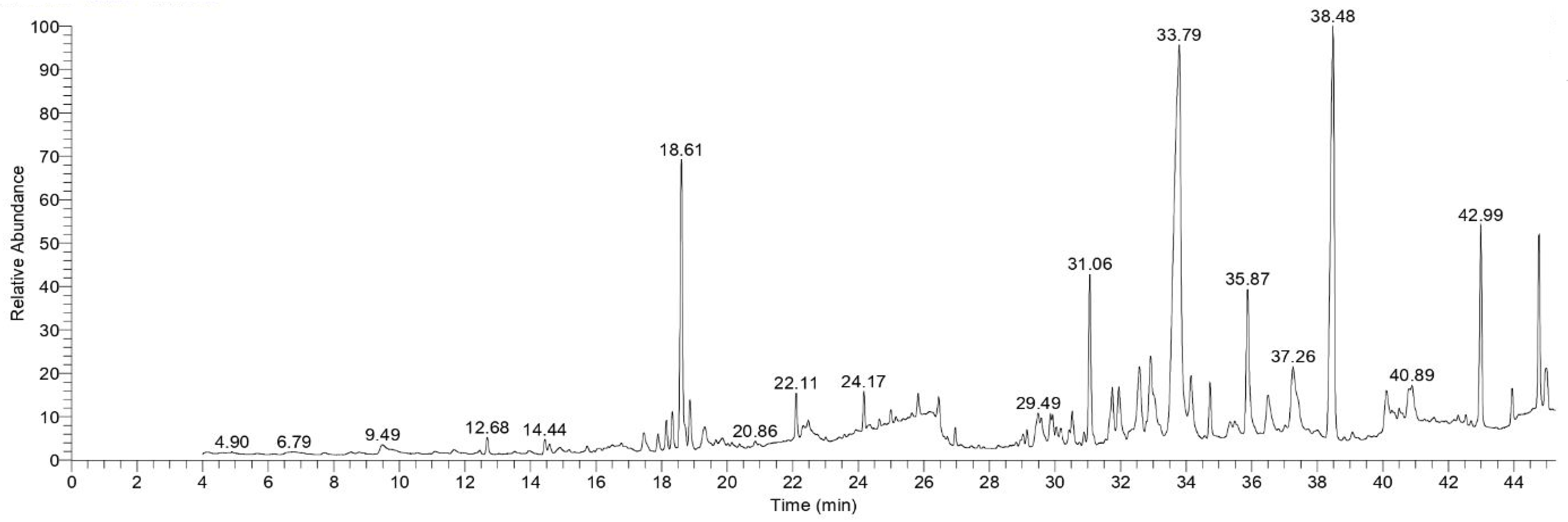
2.1. GC-MS Analysis
2.2. Biological Activities
2.2.1. Antiviral Assay
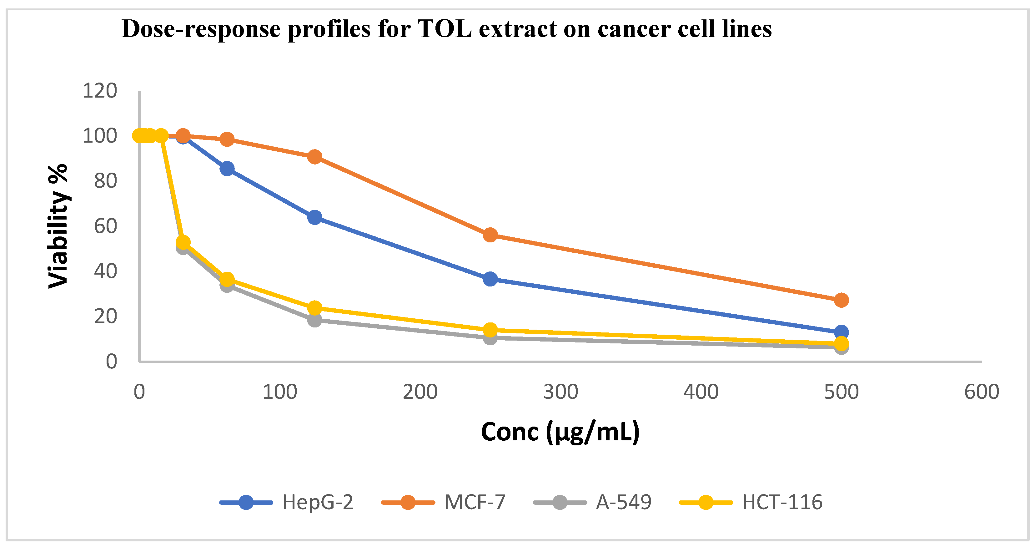
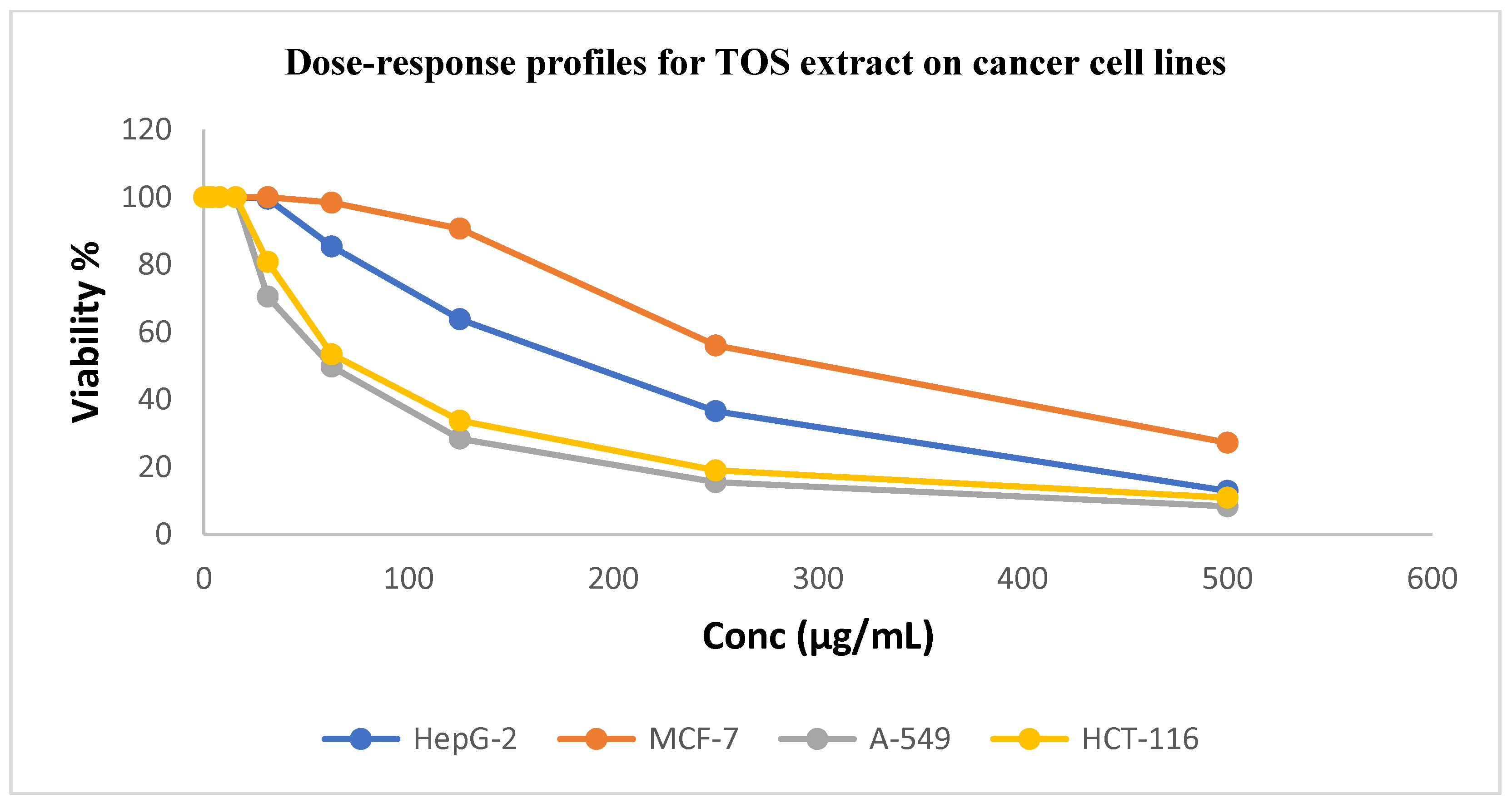
2.2.2. Cytotoxicity Studies
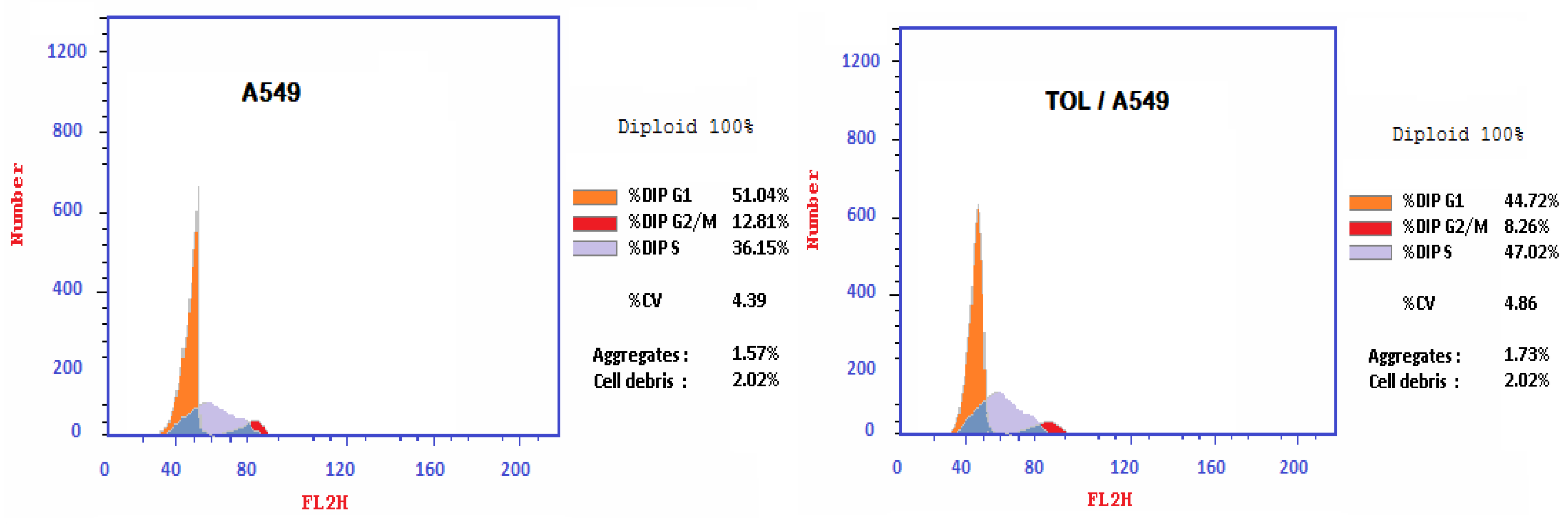
In A549 Cells, TOL Extract Produced S-Phase Cell Cycle Arrest
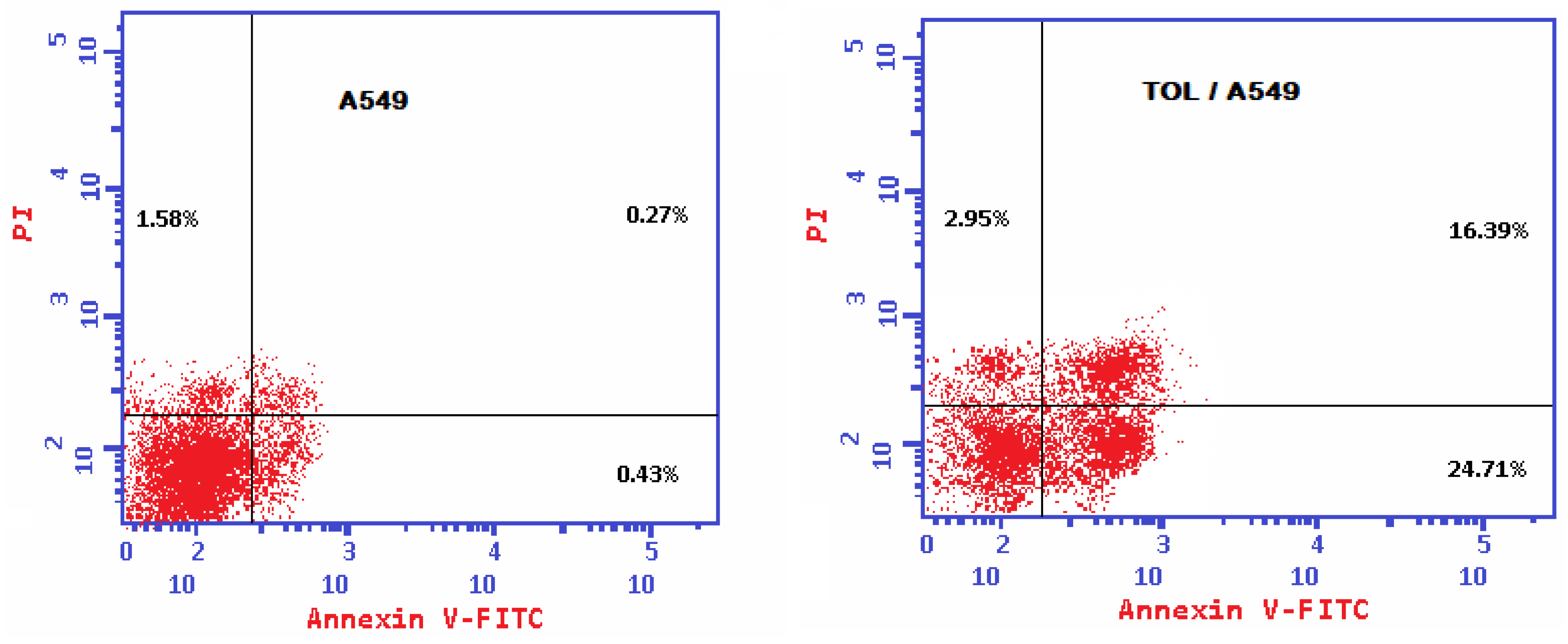
TOL Extract Induced Caspase-Dependent Apoptosis in A549 Cancer Cell Line, Which Was Mediated by p53
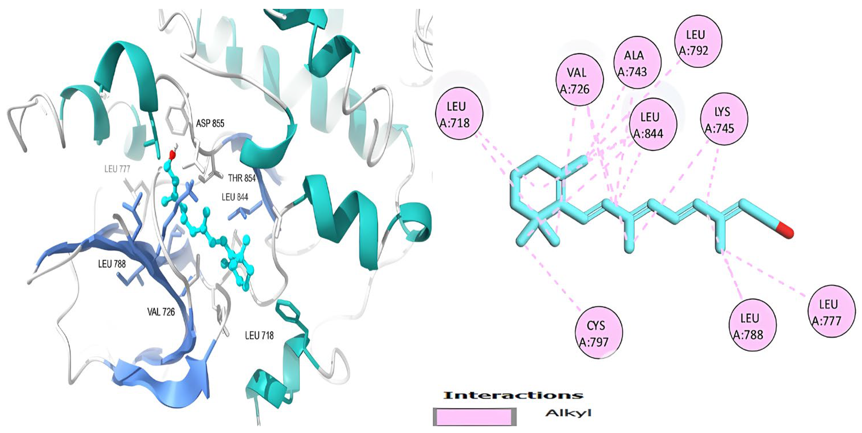
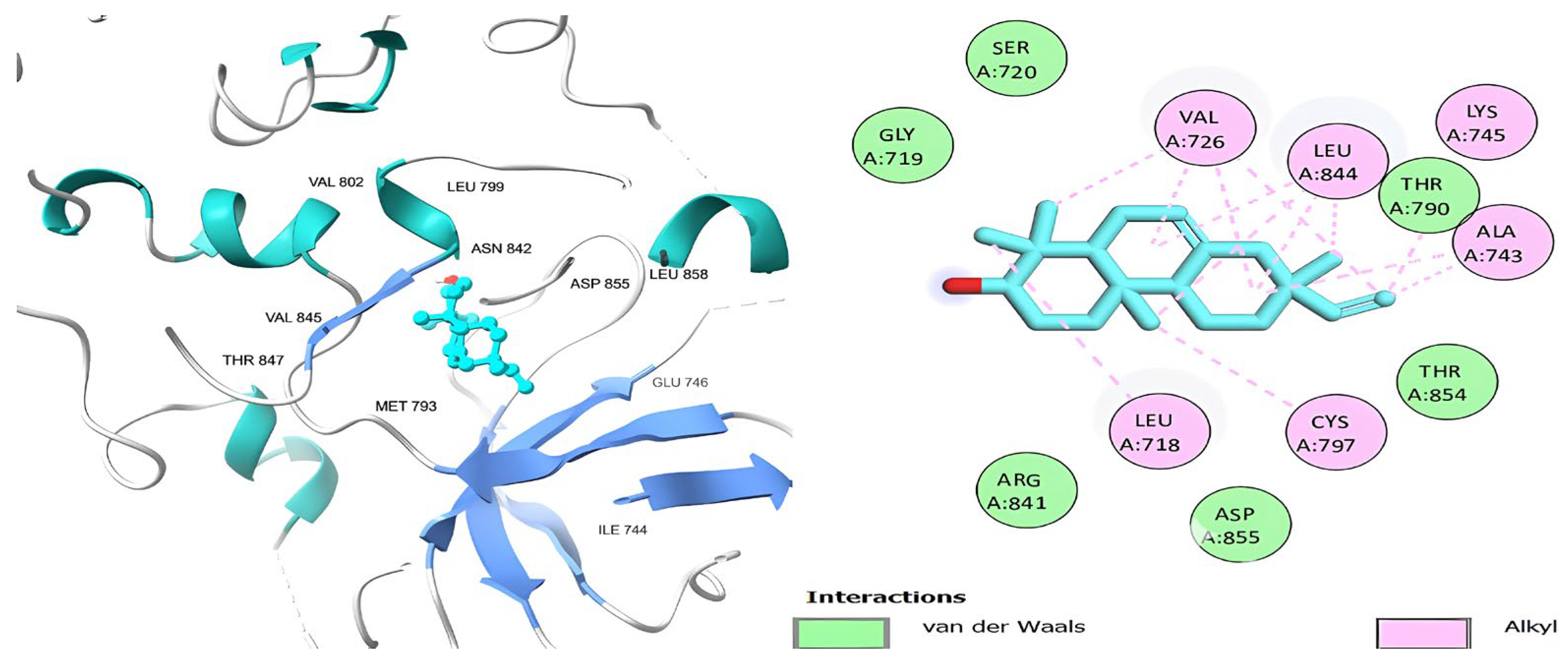
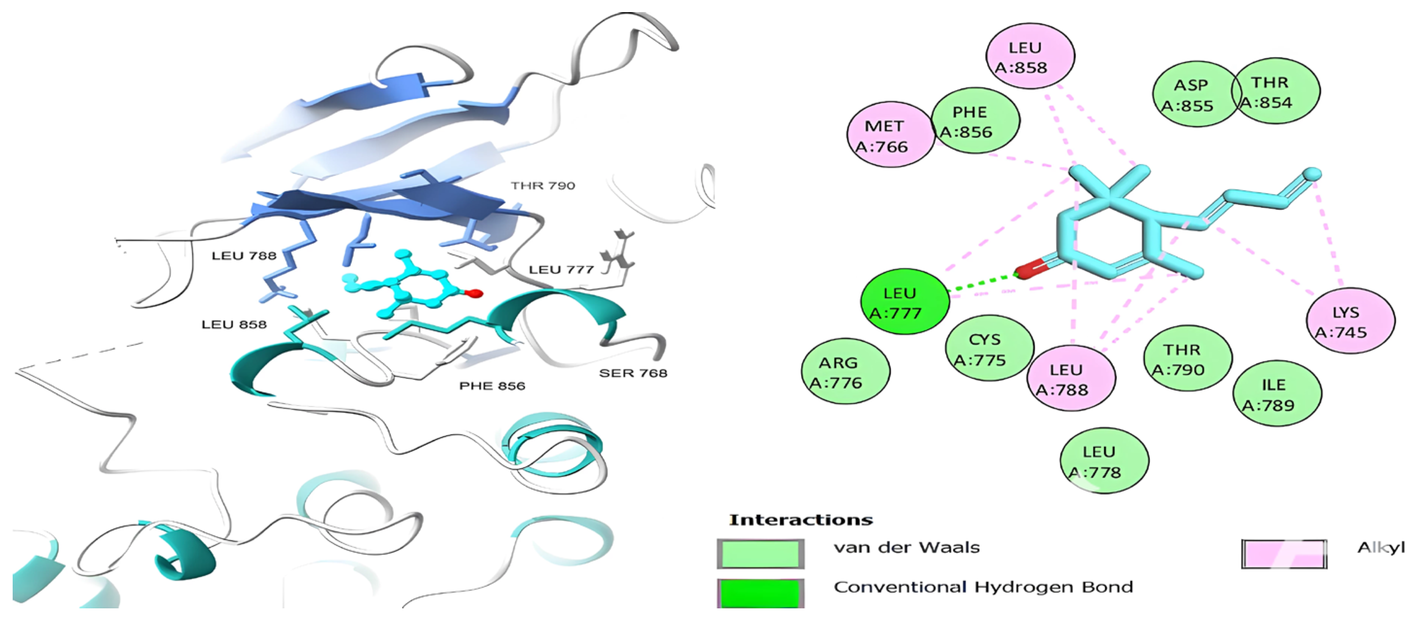
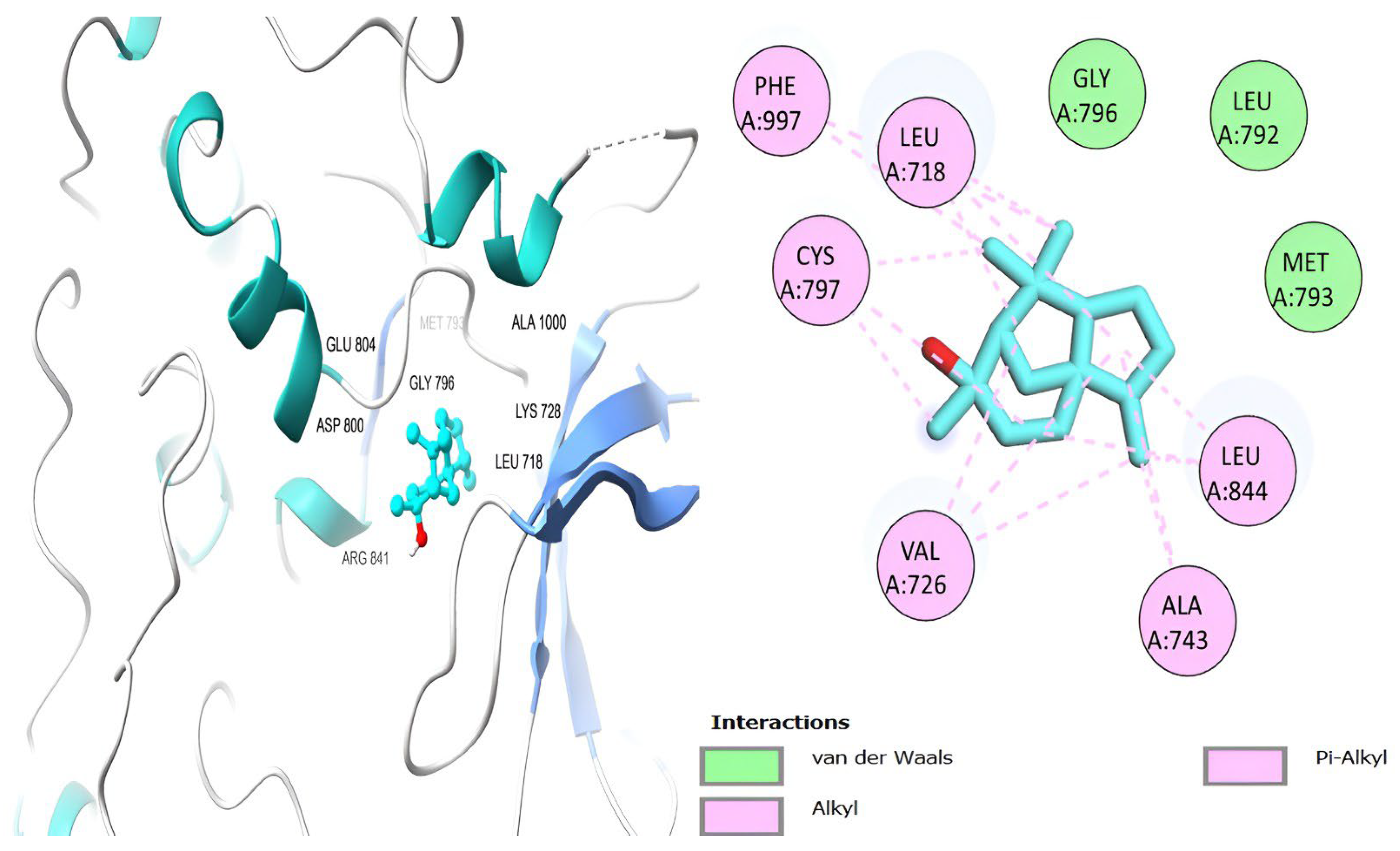
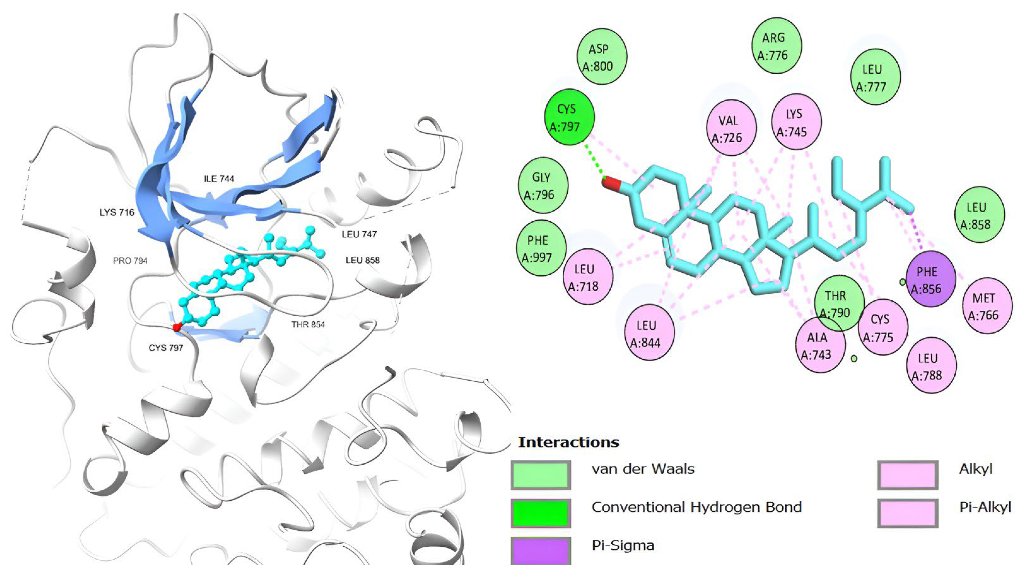
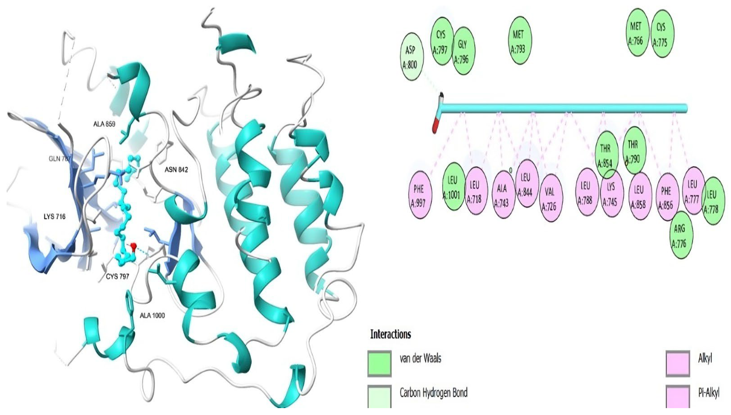
2.3. Molecular Docking
3. Discussion
4. Materials and Methods
4.1. Chemicals and Solvents
4.2. Collection and Extraction of Plant Material
4.3. GC-MS Analysis
4.4. Biological Assays
4.4.1. Antiviral Activity Assay
Viruses
Half-Maximal Cytotoxic Concentration (CC50) Determination
Half-Maximal Inhibitory Concentration (IC50) Determination
4.4.2. Cytotoxic Activity Assay
Cancer Cell Lines
Cell Viability Assay
Apoptosis Analysis (Annexin V-FITC Assay) of A549 Cells
Cell Cycle Analysis Using Flow Cytometry
Western Blot Analysis
4.5. Molecular Docking
4.6. Statistical Analysis
5. Conclusions
Author Contributions
Funding
Institutional Review Board Statement
Informed Consent Statement
Data Availability Statement
Conflicts of Interest
Abbreviations
| A549 | Human lung cancer cell line |
| ACC | Activity of complement-mediated cytotoxicity |
| ADCC | Cell-mediated antibody-dependent cytotoxicity |
| ATCC | American type culture collection |
| CC50 | Half-maximal cytotoxic concentration |
| CPE | Virus-induced cytopathic effect |
| DAPI | 4′,6-diamidino-2-phenylindole |
| DMEM | Dulbecco’s modified Eagle medium |
| DMSO | Dimethyl sulfoxide |
| EGFR-TK | Epidermal Growth Factor Receptor Tyrosine Kinase Domain |
| GC-MS | Gas chromatographic–mass spectrometry |
| H1N1 | Influenza A virus |
| HSV-2 | Herpes Simplex virus |
| HCT116 | Human colorectal cancer cell line |
| HepG2 | Human hepatocellular cancer cell line |
| IC50 | Half-maximal inhibitory concentration |
| MCF7 | Human breast cancer cell line |
| MDB | Microsoft Access Database |
| MDCK | Madin–Darby canine kidney cells |
| MOE | Molecular Operating Environment |
| MTT | 3-(4,5-dimethylthiazol-2-yl)-2,5-diphenyl-2H-tetrazolium bromide |
| M. Wt. | Molecular weight |
| NIST | National Institute of Standards and Technology |
| NK | Natural killer |
| PBS | Phosphate-buffered saline |
| RMSD | Root mean square deviation |
| RPMI | Roswell Park Memorial Institute Medium |
| RT | Room temperature |
| SD | Standard deviation |
| W19 | 4-{[4-(1-benzothiophen-4-yloxy)-3-chlorophenyl]amino}-N-(2-hydroxyethyl)-8,9-dihydro-7H-pyrimido[4,5-b]azepine-6-carboxamide |
References
- Caruntu, S.; Ciceu, A.; Olah, N.K.; Don, I.; Hermenean, A.; Cotoraci, C. Thuja occidentalis L. (Cupressaceae): Ethnobotany, phytochemistry and biological activity. Molecules 2020, 25, 5416. [Google Scholar] [CrossRef] [PubMed]
- Shen, B. A new golden age of natural products drug discovery. Cell 2015, 163, 1297–1300. [Google Scholar] [CrossRef] [PubMed]
- Harvey, A.L. Natural Products in Drug Discovery. Drug Discov. Today 2008, 13, 894–901. [Google Scholar] [CrossRef] [PubMed]
- Mahomoodally, M.F.; Atalay, A.; Picot, M.C.N.; Bender, O.; Celebi, E.; Mollica, A.; Zengin, G. Chemical, biological and molecular modelling analyses to probe into the pharmacological potential of Antidesma madagascariense Lam.: A multifunctional agent for developing novel therapeutic formulations. J. Pharm. Biomed. Anal 2018, 161, 425–435. [Google Scholar] [CrossRef] [PubMed]
- Bender, O.; Celik, I.; Dogain, R.; Atalay, A.; Shoman, M.E.; Ali, T.F.S.; Beshr, E.A.M.; Mohamed, M.; Alaaeldin, E.; Shawky, A.M.; et al. Vanillin-Based Indolin-2-one Derivative Bearing a Pyridyl Moiety as a Promising Anti-Breast Cancer Agent via Anti-Estrogenic Activity. ACS Omega 2003, 8, 6968–6981. [Google Scholar] [CrossRef]
- Chang, L.C.; Song, L.L.; Park, E.J.; Luyengi, L.; Lee, K.J.; Farnsworth, N.R.; Pezzuto, J.M.; Kinghorn, A.D. Bioactive Constituents of Thuja occidentalis. J. Nat. Prod. 2000, 63, 1235–1238. [Google Scholar] [CrossRef]
- Elsharkawy, E.R.; Ali, A.M. Effect of Drought Condition of North Region of Saudi Arabia on Accumulation of Chemical Compounds, Antimicrobial and Larvicidal Activity of Thuja orientalis. Orient. J. Chem. 2019, 35, 738–743. [Google Scholar] [CrossRef]
- Silva, I.S.; Nicolau, L.A.; Sousa, F.B.; de Araújo, S.; Oliveira, A.P.; Araújo, T.S.; Souza, L.K.; Martins, C.S.; Aquino, P.E.; Carvalho, L.L.; et al. Evaluation of anti-inflammatory potential of aqueous extract and polysaccharide fraction of Thuja occidentalis Linn. in mice. Int. J. Biol. Macromol. 2017, 105, 1105–1116. [Google Scholar] [CrossRef]
- Torres, A.; Vargas, Y.; Uribe, D.; Carrasco, C.; Torres, C.; Rocha, R.; Oyarzún, C.; San Martín, R.; Quezada, C. Pro-apoptotic and anti-angiogenic properties of the α/β-thujone fraction from Thuja occidentalis on glioblastoma cells. J. Neurooncol. 2016, 128, 9–19. [Google Scholar] [CrossRef]
- Sunila, E.S.; Hamsa, T.P.; Kuttan, G. Effect of Thuja occidentalis and its polysaccharide on cell-mediated immune responses and cytokine levels of metastatic tumor-bearing animals. Pharm. Biol. 2011, 49, 1065–1073. [Google Scholar] [CrossRef]
- Sunila, E.S.; Kuttan, G. A preliminary study on anti-metastatic activity of Thuja occidentalis L. in mice model. Immunopharmacol. Immunotoxicol. 2006, 28, 269–280. [Google Scholar] [CrossRef] [PubMed]
- Nazir, M.Z.; Chandel, S.; Sehgal, A. In vitro screening of antioxidant potential of Thuja occidentalis. J. Chem. Pharm. Res. 2016, 8, 283–286. [Google Scholar]
- Tsiri, D.; Graikou, K.; Pobłocka-Olech, I.; Krauze-Baranowska, M.; Spyropoulos, C.; Chinou, I. Chemosystematic value of the essential oil composition of Thuja species cultivated in Poland—Antimicrobial activity. Molecules 2009, 14, 4707–4715. [Google Scholar] [CrossRef] [PubMed]
- Bellili, S.; Aouadhi, C.; Dhifi, W.; Ghazghazi, H.; Jlassi, C.; Sadaka, C.; Beyrouthy, M.E.; Maaroufi, A.; Cherif, A.; Mnif, W. The influence of organs on biochemical properties of Tunisian Thuja occidentalis essential oils. Symmetry 2018, 10, 649. [Google Scholar] [CrossRef]
- Dubey, S.K.; Batra, A. Anti-diabetic activity of Thuja occidentalis Linn. Res. J. Pharm. Tech. 2008, 1, 362–365. [Google Scholar]
- Dubey, S.K.; Batra, A. Role of Phenolics in Anti-Atherosclerotic Property of Thuja occidentalis Linn. Ethnobot. Leafl. 2009, 2009, 12. [Google Scholar]
- Sanjita, D.; Ruchi, R. Antioxidant and gastro-protective properties of the fruits of Thuja occidentalis Linn. Asian J. Biochem. Pharm. Res. 2013, 3, 80–87. [Google Scholar]
- Beuscher, N.; Kopanski, L. Purification and biological characterization of antiviral substances from T. occidentalis. Planta Med. 1986, 6, 555–556. [Google Scholar] [CrossRef]
- Gohla, S.H.; Zeman, R.A.; Bögel, M.; Gohla, S.H.; Zeman, R.A.; Bögel, M.; Jurkiewicz, E.; Schrum, S.; Haubeck, H.D.; Schmitz, H.; et al. Modification of the in vitro replication of the human immunodeficiency virus HIV-1 by TPSg, a polysaccharide fraction isolated from the Cupressaceae Thuja occidentalis L. (Arborvitae). In Modern Trends in Human Leukemia IX, Haematology and Blood Transfusion; Neth, R., Frolova, E., Gallo, R.C., Greaves, M.F., Afanasiev, B.V., Elstner, E., Eds.; Springer: Berlin/Heidelberg, Germany, 1992; Volume 35. [Google Scholar]
- Bodinet, C.; Lindequist, U.; Teuscher, E.; Freudenstein, J. Effect of an orally applied herbal immune-modulator on cytokine induction and antibody response in normal and immunosuppressed mice. Phytomedicine 2002, 9, 606–613. [Google Scholar] [CrossRef]
- Siveen, K.S.; Kuttan, G. Thujone inhibits lung metastasis induced by B16F-10 melanoma cells in C57BL/6 mice. Can. J. Physiol. Pharmacol. 2011, 89, 691–703. [Google Scholar] [CrossRef]
- Stan, M.S.; Voicu, S.N.; Caruntu, S.; Nica, I.C.; Olah, N.K.; Burtescu, R.; Balta, C.; Rosu, M.; Herman, H.; Hermenean, A.; et al. Antioxidant and anti-inflammatory properties of a Thuja occidentalis mother tincture for the treatment of ulcerative colitis. Antioxidants 2019, 8, 416. [Google Scholar] [CrossRef] [PubMed]
- Asha, R.; Nisha, P.; Suneer, K.; Mythili, A.; Shafeeq, H.A.; Panneer, S.K.; Manikandan, P.; Shobana, C.S. In vitro activity of various potencies of homeopathic drug Thuja against molds involved in mycotic keratitis. Int. J. Pharm. Pharm. Sci. 2014, 6, 555–559. [Google Scholar]
- Alam, S.M.; Qureshi, M.A.; Jahan, N. Antimicrobial screening of some medicinal plants of Pakistan. Pak. J. Bot. 2010, 42, 4281–4284. [Google Scholar]
- Digrak, M.; Bagci, E.; Alma, M.H. Antibiotic action of seed lipids from five tree species grown in Turkey. Pharm. Biol. 2002, 40, 425–428. [Google Scholar] [CrossRef]
- Zasada, M.; Budzisz, E. Retinoids: Active molecules influencing skin structure formation in cosmetic and dermatological treatments. Postep. Dermatol. Alergol. 2019, 36, 392–397. [Google Scholar] [CrossRef]
- Guclu, G.; Tas, A.; Dincer, E.; Ucar, E.; Kaya, S.; Berisha, A.; Dural, E.; Silig, Y. Biological evaluation and in silico molecular docking studies of Abies cilicica (Antoine & Kotschy) Carrière) resin. J. Mol. Struct. 2023, 1288, 135740. [Google Scholar]
- Kyslychenko, V.; Karpiuk, U.; Diakonova, I.; Mohammad, S. Abu-Darwish. Phenolic Compounds and Terpenes in the Green Parts of Glycine Hispida. Adv. Environ. Biol. 2010, 4, 490–494. [Google Scholar]
- Ikhoon, O.H.; Woo, Y.; Yang, J.; Lee, P.S.; Mar, W.; Ki-Bong, O.; Shin, J. In vitro Na+/K+-ATPase inhibitory activity and antimicrobial activity of sesquiterpenes isolated from Thujopsis dolabrata. Arch. Pharm. Res. 2011, 34, 2141–2147. [Google Scholar]
- Chien, J.H.; Chang, K.F.; Lee, S.H.; Lee, C.J.; Chen, Y.T.; Lai, H.C.; Lu, Y.C.; Tsai, N.M. Cedrol restricts the growth of colorectal cancer in vitro and in vivo by inducing cell cycle arrest and caspase-dependent apoptotic cell death. Int. J. Med. Sci. 2022, 19, 1953–1964. [Google Scholar] [CrossRef]
- Zidan, K.; Nikhil, N.; Emran, T.B.; Mitra, S.; Islam, F.; Chandran, D.; Barua, J.; Khandaker, M.U.; Idris, A.M.; Rauf, A.; et al. Multifunctional roles and pharmacological potential of β-sitosterol: Emerging evidence toward clinical applications. Chem. Biol. Interact. 2022, 365, 110117. [Google Scholar]
- Katz, D.H.; Marcelletti, J.F.; Khalil, M.H.; Pope, L.E.; Katz, L.R. Antiviral activity of docosanol, an inhibitor of lipid enveloped viruses including Herpes Simplex. Proc. Nail. Acad. Sci. 1991, 88, 10825–10829. [Google Scholar] [CrossRef] [PubMed]
- Su, Y.C.; Hsu, K.P.; Wang, E.I.; Ho, C.L. Composition, anticancer, and antimicrobial activities in vitro of the heartwood essential oil of Cunninghamia lanceolata var. konishii from Taiwan. Nat. Prod. Commun. 2012, 7, 1245–1247. [Google Scholar] [CrossRef] [PubMed]
- Wang, X.; Sun, Z.; Gao, Y.; Liu, Q.S.; Yang, X.; Liang, J.; Ren, J.; Zhou, Q.; Jiang, G. 3-tert-Butyl-4-hydroxyanisole perturbs renal lipid metabolism in vitro by targeting androgen receptor-regulated de novo lipogenesis. Ecotoxicol. Environ. Saf. 2023, 258, 114979. [Google Scholar] [CrossRef] [PubMed]
- Fidyt, K.; Fiedorowicz, A.; Strządała, L.; Szumny, A. β-caryophyllene and β-caryophyllene oxide-natural compounds of anticancer and analgesic properties. Cancer Med. 2016, 5, 3007–3017. [Google Scholar] [CrossRef]
- Bozan, B.; Ozek, T.; Kurkcuoglu, M.; Kirimer, N.; Baser, K.; Husnu, C. Analysis of essential oil and headspace volatiles of the flowers of Pelargonium endlicherianum used as an anthelmintic in folk medicine. Planta Medica 1999, 65, 781–782. [Google Scholar] [CrossRef]
- Guo, J.; Li, M.; Zhao, Y.; Kang, S.-G.; Huang, K.; Tong, T. Dietary Supplementation of Cedryl Acetate Ameliorates Adiposity and Improves Glucose Homeostasis in High-Fat Diet-Fed Mice. Nutrients 2023, 15, 980. [Google Scholar] [CrossRef]
- Juárez-Rodríguez, M.M.; Cortes-López, H.; García-Contreras, R.; González-Pedrajo, B.; Díaz-Guerrero, M.; Martínez-Vázquez, M.; Rivera-Chávez, J.A.; Soto-Hernández, R.M.; Castillo-Juárez, I. Tetradecanoic Acids with Anti-Virulence Properties Increase the Pathogenicity of Pseudomonas aeruginosa in a Murine Cutaneous Infection Model. Front. Cell. Infect. Microbiol. 2021, 10, 597517. [Google Scholar] [CrossRef]
- Gonzalez-Rivera, M.L.; Barragan-Galvez, J.C.; Gasca-Martínez, D.; Hidalgo-Figueroa, S.; Isiordia-Espinoza, M.; Alonso-Castro, A.J. In Vivo Neuropharmacological Effects of Neophytadiene. Molecules 2023, 28, 3457. [Google Scholar] [CrossRef]
- Mazumder, K.; Nabila, A.; Aktar, A.; Farahnaky, A. Bioactive Variability and In Vitro and In Vivo Antioxidant Activity of Unprocessed and Processed Flour of Nine Cultivars of Australian lupin Species: A Comprehensive Substantiation. Antioxidants 2020, 9, 282. [Google Scholar] [CrossRef]
- Ibrahim, H.A.H.; El-Naggar, H.A.; El-Damhougy, K.A.; Bashar, N.A.E.; Abou Senna, F.M. Callyspongia crassa and C. siphonella (Porifera, Callyspongiidae) as a potential source for medical bioactive substances, Aqaba Gulf, Red Sea, Egypt. J. Bas. Appl. Zool. 2017, 78, 7. [Google Scholar] [CrossRef]
- Ganesh, M.; Mohankumar, M. Extraction and identification of bioactive components in Sida cordata (Burm. f.) using gas chromatography-mass spectrometry. J. Food Sci. Technol. 2017, 54, 3082–3091. [Google Scholar] [CrossRef] [PubMed]
- Amiranashvili, L.; Nadaraia, N.; Merlani, M.; Kamoutsis, C.; Petrou, A.; Geronikaki, A.; Pogodin, P.; Druzhilovskiy, D.; Poroikov, V.; Ciric, A.; et al. Antimicrobial Activity of Nitrogen-Containing 5-Alpha-androstane Derivatives: In Silico and Experimental Studies. Antibiotics 2020, 9, 224. [Google Scholar] [CrossRef] [PubMed]
- Gainche, M.; Ripoche, I.; Senejoux, F.; Cholet, J.; Ogeron, C.; Decombat, C.; Danton, O.; Delort, L.; Vareille-Delarbre, M.; Berry, A.; et al. Anti-Inflammatory and Cytotoxic Potential of New Phenanthrenoids from Luzula Sylvatica. Molecules 2020, 25, 2372. [Google Scholar] [CrossRef] [PubMed]
- Ain, Q.U.; Asas, S.; Jamal, A.; Arif, M.; Mahmood, H.M. Phytochemical analysis and Antifungal Activity of Gymnosperm against Fusarium Wilt of Banana. Appl. Ecol. Environ. Res. 2021, 19, 2477–2493. [Google Scholar] [CrossRef]
- Song, H.; Kim, J.; Shin, Y.K.; Kim, K.Y. Antibacterial activity of pimaric acid against the causative agent of American foulbrood, Paenibacillus larvae. J. Agric. Res. 2022, 61, 219–226. [Google Scholar] [CrossRef]
- Fuad, N.; Sekar, M.; Gan, S.; Lum, P.; Va, J.; Ravi, J.S. Lutein: A Comprehensive Review on its Chemical, Biological Activities and Therapeutic Potentials. Pharmacogn. J. 2020, 12, 1769–1778. [Google Scholar] [CrossRef]
- Choi, J.; Lee, E.; Lee, H.; Kim, K.; Ahn, K.; Shim, B.; Kim, N.; Song, M.; Baek, N.; Kim, S. Identification of campesterol from Chrysanthemum coronarium L. and its antiantiogenic activities. Phytother. Res. 2007, 21, 954–959. [Google Scholar] [CrossRef]
- RCSB PDB. Available online: https://www.rcsb.org/ligand/W19 (accessed on 16 January 2025).
- Alafnan, A.; Dogan, R.; Bender, O.; Celik, I.; Mollica, A.; Malik, J.; Rengasamy, K.; Break, M.; Khojali, W.; Alharby, T. Beta Elemene induces cytotoxic effects in FLT3 ITD-mutated acute myeloid leukemia by modulating apoptosis. Eur. Rev. Med. Pharmacol. Sci. 2023, 27, 3270–3287. [Google Scholar]
- Khazaei, S.; Abdul Hamid, R.; Ramachandran, V.; Mohd Esa, N.; Pandurangan, A.K.; Danazadeh, F.; Ismail, P. Cytotoxicity and Pro-apoptotic Effects of Allium atroviolaceum Flower Extract by Modulating Cell Cycle Arrest and Caspase Dependent and p53-Independent Pathway in Breast Cancer Cell Lines. Evid Based Complement Altern. Med. 2017, 2017, 1468957. [Google Scholar] [CrossRef]
- Chen, C.Y.; Liu, T.Z.; Tseng, W.C.; Lu, F.J.; Hung, R.P.; Chen, C.H. (−)-Anonaine induces apoptosis through Bax- and caspase-dependent pathways in human cervical cancer (HeLa) cells. Food Chem. Toxicol. 2008, 46, 2694–2702. [Google Scholar] [CrossRef]
- Al-Salman, H.N.K.; Ali, E.T.; Jabir, M.; Sulaiman, G.M.; Al-Jadaan, S.A.S. 2-Benzhydrylsulfinyl-N-hydroxyacetamide-Na extracted from fig as a novel cytotoxic and apoptosis inducer in SKOV-3 and AMJ-13 cell lines via P53 and caspase-8 pathway. Eur. Food Res. Technol. 2020, 246, 1591–1608. [Google Scholar] [CrossRef]
- Abouzied, A.S.; Abd-Rabo, M.M.; Huwaimel, B.; Almahmoud, S.A.; Almarshdi, A.A.; Alharbi, F.M.; Alenzi, S.S.; Albsher, B.N.; Alafnan, A. In Silico Pharmacokinetic Profiling of the Identified Bioactive Metabolites of Pergularia tomentosa L. Latex Extract and In Vitro Cytotoxic Activity via the Induction of Caspase-Dependent Apoptosis with S-Phase Arrest. Pharmaceuticals 2022, 15, 1132. [Google Scholar] [CrossRef] [PubMed]
- Mostafa, A.; Kandeil, A.; Elshaier, A.M.M.; Kutkat, O.; Moatasim, Y.; Rashad, A.A.; Shehata, M.; Gomaa, M.R.; Mahrous, N.; Mahmoud, S.H.; et al. FDA-Approved Drugs with Potent In Vitro Antiviral Activity against Severe Acute Respiratory Syndrome Coronavirus 2. Pharmaceuticals 2020, 13, 443. [Google Scholar] [CrossRef] [PubMed]
- Koutsoni, O.; Karampetsou, K.; Dotsika, E. In vitro screening of Antileishmanial Activity of natural product compounds: Determination of IC50, CC50 and SI values. Bio Protoc. 2019, 9, e3410. [Google Scholar] [CrossRef]
- AbouAitah, K.; Allayh, A.K.; Wojnarowicz, J.; Shaker, Y.M.; Swiderska-Sroda, A.; Lojkowski, W. Nano-formulation Composed of Ellagic Acid and Functionalized Zinc Oxide Nanoparticles Inactivates DNA and RNA Viruses. Pharmaceutics 2021, 13, 2174. [Google Scholar] [CrossRef]
- Mosmann, T. Rapid colorimetric assay for cellular growth and survival: Application to proliferation and cytotoxicity assays. J. Immunol. Methods 1983, 65, 55–63. [Google Scholar] [CrossRef]
- Darzynkiewicz, Z. Critical Aspects in Analysis of Cellular DNA Content. Curr. Protoc. Cytom. 2010, 52, 7.2.1–7.2.8. [Google Scholar] [CrossRef]
- Mahmood, T.; Yang, P.C. Western Blot: Theory and Trouble Shooting. North Am. J. Med. Sci. 2012, 4, 429–434. [Google Scholar]
- Chemical Computing Group Inc. Molecular Operating Environment (MOE); Chemical Computing Group Inc.: Montreal, QC, Canada, 2016. [Google Scholar]
- Alghamdi, A.; Abouzied, A.S.; Alamri, A.; Anwar, S.; Ansari, M.; Khadra, I.; Zaki, Y.H.; Gomha, S.M. Synthesis, Molecular Docking, and Dynamic Simulation Targeting Main Protease (Mpro) of New, Thiazole Clubbed Pyridine Scaffolds as Potential COVID-19 Inhibitors. Curr. Issues Mol. Biol. 2023, 45, 1422–1442. [Google Scholar] [CrossRef]
- Kawakita, Y.; Seto, M.; Ohashi, T.; Tamura, T.; Yusa, T.; Miki, H.; Iwata, H.; Kamiguchi, H.; Tanaka, T.; Sogabe, S.; et al. Design and synthesis of novel pyrimido [4,5-b]azepine derivatives as HER2/EGFR dual inhibitors. Bioorg. Med. Chem. 2013, 21, 2250–2261. [Google Scholar] [CrossRef]
- Abouzied, A.S.; Al-Humaidi, J.Y.; Bazaid, A.S.; Qanash, H.; Binsaleh, N.K.; Alamri, A.; Ibrahim, S.M.; Gomha, S.M. Synthesis, Molecular Docking Study, and Cytotoxicity Evaluation of Some Novel 1,3,4-Thiadiazole as Well as 1,3-Thiazole Derivatives Bearing a Pyridine Moiety. Molecules 2022, 27, 6368. [Google Scholar] [CrossRef] [PubMed]
- Mohammed, H.A.; Abouzied, A.S.; Mohammed, S.A.A.; Khan, R.A. In Vivo and In Silico Analgesic Activity of Ficus populifolia Extract Containing 2-O-β-D-(3′, 4′,6′-Tri-acetyl)-glucopyranosyl-3-methyl Pentanoic Acid. Int. J. Mol. Sci. 2023, 24, 2270. [Google Scholar] [CrossRef] [PubMed]
- Huwaimel, B.; Abouzied, A.S.; Anwar, S.; Elaasser, M.M.; Almahmoud, S.A.; Alshammari, B.; Alrdaian, D.; Alshammari, R.Q. Novel landmarks on the journey from natural products to pharmaceutical formulations: Phytochemical, biological, toxicological and computational activities of Satureja hortensis L. Food Chem. Toxicol. 2023, 179, 113969. [Google Scholar] [CrossRef] [PubMed]
- Aroua, L.M.; Alosaimi, A.H.; Alminderej, F.M.; Messaoudi, S.; Mohammed, H.A.; Almahmoud, S.A.; Chigurupati, S.; Albadri, A.E.A.E.; Mekni, N.H. Synthesis, Molecular Docking, and Bioactivity Study of Novel Hybrid Benzimidazole Urea Derivatives: A Promising α-Amylase and α-Glucosidase Inhibitor Candidate with Antioxidant Activity. Pharmaceutics 2023, 15, 457. [Google Scholar] [CrossRef] [PubMed]
- Al-Humaidi, J.Y.; Gomha, S.M.; Riyadh, S.M.; Ibrahim, M.S.; Zaki, E.A.; Abolibda, T.Z.; Jefri, O.A.; Abouzied, A.S. Synthesis, Biological Evaluation, and Molecular Docking of Novel Azolylhydrazonothiazoles as Potential Anticancer Agents. ACS Omega 2023, 8, 34044–34058. [Google Scholar] [CrossRef]
- Matos, B.; Howl, J.; Jerónimo, C.; Fardilha, M. The disruption of protein-protein interactions as a therapeutic strategy for prostate cancer. Pharmacol. Res. 2020, 161, 105145. [Google Scholar] [CrossRef]
- Joerger, A.C.; Fersht, A.R. The p53 Pathway: Origins, Inactivation in Cancer, and Emerging Therapeutic Approaches. Annu. Rev. Biochem. 2016, 85, 375–404. [Google Scholar] [CrossRef]
- McConkey, B.J.; Sobolev, V.; Edelman, M. The Performance of Current Methods in Ligand–Protein Docking. Curr. Sci. 2002, 83, 845–856. [Google Scholar]
- Wells, J.A.; McClendon, C.L. Reaching for high-hanging fruit in drug discovery at protein-protein interfaces. Nature 2007, 450, 1001–1009. [Google Scholar] [CrossRef]

| GC-MS Analysis of TOL | |||||
|---|---|---|---|---|---|
| Plant Extracts | Compound Name | M.Wt (amu) | RT (min) | Area % | Biological Activity |
| TOL | Retinol | 286 | 35.87 | 4.38 | Antioxidant effect [26] |
| Podocarp-7-en-3β-ol, 13β-methyl-13-vinyl | 288 | 31.06 | 4.65 | Anticancer effect [27] | |
| Megastigmatrienone | 190 | 18.86 | 1.27 | Antiproliferative effect [28] | |
| Cedrol | 222 | 18.33 | 1.01 | Antibacterial, antitumor effect [29,30] | |
| β-Sitosterol | 414 | 44.76 | 4.28 | Anticancer effect [31] | |
| Docosanol | 326 | 42.99 | 5.39 | Antiviral effect [32] | |
| Cedrene | 204 | 14.44 | 0.46 | Anticancer effect [33] | |
| 3-tert-Butyl-4-hydroxyanisole | 180 | 17.47 | 0.75 | Antihyperlipidemia [34] | |
| Caryophyllene oxide | 220 | 18.15 | 0.78 | Anticancer and analgesic [35] | |
| Gleenol | 222 | 19.32 | 0.70 | Termiticidal, antihelminitic [36] | |
| Cedryl acetate | 264 | 22.11 | 1.15 | Anti-obesity [37] | |
| Tetradecanoic acid | 228 | 22.48 | 0.64 | Antibacterial and antivirulence [38] | |
| Neophytadiene | 278 | 24.17 | 0.88 | Antidepressant and sedative [39] | |
| n-Hexadecanoic acid | 256 | 26.46 | 0.83 | Antioxidant [40] | |
| 5,8,11,14-Eicosatetraenoic acid, methyl ester | 318 | 29.48 | 1.08 | Antimicrobial and antiinflammatory [41] | |
| trans-13-Octadecenoic acid | 282 | 29.58 | 0.49 | Antioxidant [42] | |
| Androstan-3-one, 17-(Acetyloxy)-, (5à,17á) | 332 | 30.18 | 0.37 | Antimicrobial activity [43] | |
| 1-Phenanthrenemethanol | 286 | 31.75 | 1.73 | Cytotoxic and antiinflammatory [44] | |
| Podocarp-7-en-3-one | 286 | 31.95 | 1.46 | Antifungal [45] | |
| Pimaric acid | 302 | 32.92 | 2.48 | Antibacterial [46] | |
| Lutein | 568 | 40.8 | 0.95 | Antioxidant, antiinflammatory [47] | |
| Campesterol | 400 | 43.94 | 0.91 | Anticancer, cholesterol lowering [48] | |
| Plant Extract | % Inhibition of Virus | ||
|---|---|---|---|
| HSV-2 | H1N1 | Standard Ribavirin | |
| TOL | |||
| Concentration (100 μg/mL) | 17.70 | 16.20 | 24.50 (HSV-2) |
| 22.40 (H1N1) | |||
| Concentration (50 μg/mL) | 8.10 | 6.70 | 12.10 (HSV-2) |
| 10.70 (H1N1) | |||
| IC50 * (μg/mL) | 305 ± 3.6 | 912 ± 2.7 | 282 ± 1.9 (HSV-2) |
| 867 ± 1.7 (H1N1) | |||
| TOS | |||
| Concentration (100 μg/mL) | 3.8 | 2.1 | 24.50 (HSV-2) |
| 22.40 (H1N1) | |||
| Concentration (50 μg/mL) | 0 | 0 | 12.10 (HSV-2) |
| 10.70 (H1N1) | |||
| IC50 * (μg/mL) | 1530 ± 3.7 | 1790 ± 4.0 | 282 ± 1.9 (HSV-2) |
| 867 ± 1.7 (H1N1) | |||
| IC50 (μg/mL) a | |||||
|---|---|---|---|---|---|
| HepG2 | A549 | HCT116 | MCF7 | ||
| Extract | TOL | >100 | 18.45 ± 1.2 | 24.90 ± 1.7 | >100 |
| TOS | >100 | 33.61 ± 2.0 | 45.30 ± 2.4 | >100 | |
| Doxorubicin | 1.17 ± 0.1 | 1.44 ± 0.3 | 1.16 ± 0.2 | 2.39 ± 0.3 | |
| Selectivity Index (SI) a | |||||
|---|---|---|---|---|---|
| HepG2 | A549 | HCT116 | MCF7 | ||
| Extract | TOL | -------- | 3.70 | 2.74 | -------- |
| TOS | -------- | 3.92 | 2.90 | -------- | |
| MRC5 Leaves | 68.32 ± 2.1 | ||||
| IC50 b (μg/mL) Stem | 131.78 ± 3.0 | ||||
| Samples | Protein Expression * (μg/mL) | |||
|---|---|---|---|---|
| Bax | BCl2 | Caspase-3 | P-53 | |
| Control (A549 cancer cell line—untreated) | 3.20 ± 0.4 | 7.53 ± 0.4 | 69.31 ± 3.1 | 3.81 ± 0.2 |
| TOL | 14.20 ± 0.4 | 3.95 ± 0.3 | 141.85 ± 4.0 | 11.90 ± 1.7 |
| Compound | Binding Scores (kcal/mol) | Hydrogen Bond Interactions | Distance (Å) | Hydrophobic Interactions | Distance (Å) |
|---|---|---|---|---|---|
| Retinol | −9.30 | VAL 726 | 3.83, 3.84 | ||
| LYS 745 | 3.76 | ||||
| ALA 743 | 3.75, 3.94 | ||||
| LEU 718 | 3.58 | ||||
| LEU 844 | 3.87 | ||||
| Podocarp-7-en-3β-ol, 13β-methyl-13-vinyl | −8.20 | VAL 726 | 3.81 | ||
| ALA 743 | 3.88 | ||||
| LYS 745 | 3.78 | ||||
| THR 790 | 3.43 | ||||
| THR 854 | 3.81 | ||||
| Megastigmatrienone | −7.00 | LEU 777 | −2.54 | ALA 743 | 3.84 |
| LYS 745 | 3.77 | ||||
| LEU 858 | 3.91 | ||||
| Cedrol | −7.00 | LEU 718 | 3.78 | ||
| VAL 726 | 3.83 | ||||
| MET 793 | 3.82 | ||||
| CYS 797 | 3.91 | ||||
| LEU 844 | 3.12 | ||||
| β-Sitosterol | −10.60 | CYS 797 | 2.95 | LEU 718 | 3.21 |
| PHE 856 | 3.02 | ||||
| VAL 726 | 3.91 | ||||
| ALA 743 | 3.25 | ||||
| THR 790 | 3.12 | ||||
| LEU 844 | 3.78 | ||||
| THR 790 | 3.81 | ||||
| Docosanol | −6.20 | ASP 800 | 3.41 | THR 790 | 3.81 |
| LEU 718 | 3.43 | ||||
| ARG 841 | 3.65 | ||||
| LEU 844 | 3.82 | ||||
| LYS 745 | 3.48 | ||||
| ALA 743 | 3.18 | ||||
| W19 [49] | −10.50 | MET 793 SER 720 | 2.51 2.29 | PHE 856 | 3.88 |
| LEU 788 | 3.82 | ||||
| LYS 745 | 3.48 | ||||
| LEU 844 | 3.93 | ||||
| ALA 743 | 3.81 | ||||
| VAL 726 | 3.68 |
Disclaimer/Publisher’s Note: The statements, opinions and data contained in all publications are solely those of the individual author(s) and contributor(s) and not of MDPI and/or the editor(s). MDPI and/or the editor(s) disclaim responsibility for any injury to people or property resulting from any ideas, methods, instructions or products referred to in the content. |
© 2025 by the authors. Licensee MDPI, Basel, Switzerland. This article is an open access article distributed under the terms and conditions of the Creative Commons Attribution (CC BY) license (https://creativecommons.org/licenses/by/4.0/).
Share and Cite
Younes, K.; Abouzied, A.; Alqarni, S.; Elkashlan, A.; Hussein, W.; Alhathal, R.; Albsher, R.; Alshammari, S.; Huwaimel, B. Biological Activities and Phytochemical Screening of Thuja occidentalis Extracts with In Silico Approaches. Int. J. Mol. Sci. 2025, 26, 939. https://doi.org/10.3390/ijms26030939
Younes K, Abouzied A, Alqarni S, Elkashlan A, Hussein W, Alhathal R, Albsher R, Alshammari S, Huwaimel B. Biological Activities and Phytochemical Screening of Thuja occidentalis Extracts with In Silico Approaches. International Journal of Molecular Sciences. 2025; 26(3):939. https://doi.org/10.3390/ijms26030939
Chicago/Turabian StyleYounes, Kareem, Amr Abouzied, Saad Alqarni, Akram Elkashlan, Weiam Hussein, Rawabi Alhathal, Rahaf Albsher, Sarah Alshammari, and Bader Huwaimel. 2025. "Biological Activities and Phytochemical Screening of Thuja occidentalis Extracts with In Silico Approaches" International Journal of Molecular Sciences 26, no. 3: 939. https://doi.org/10.3390/ijms26030939
APA StyleYounes, K., Abouzied, A., Alqarni, S., Elkashlan, A., Hussein, W., Alhathal, R., Albsher, R., Alshammari, S., & Huwaimel, B. (2025). Biological Activities and Phytochemical Screening of Thuja occidentalis Extracts with In Silico Approaches. International Journal of Molecular Sciences, 26(3), 939. https://doi.org/10.3390/ijms26030939