Molecular Alterations and Pathways in Intrahepatic Cholangiocarcinoma: Available Evidence and New Perspectives
 , ,
, ,  , , , and
Abstract
1. Introduction
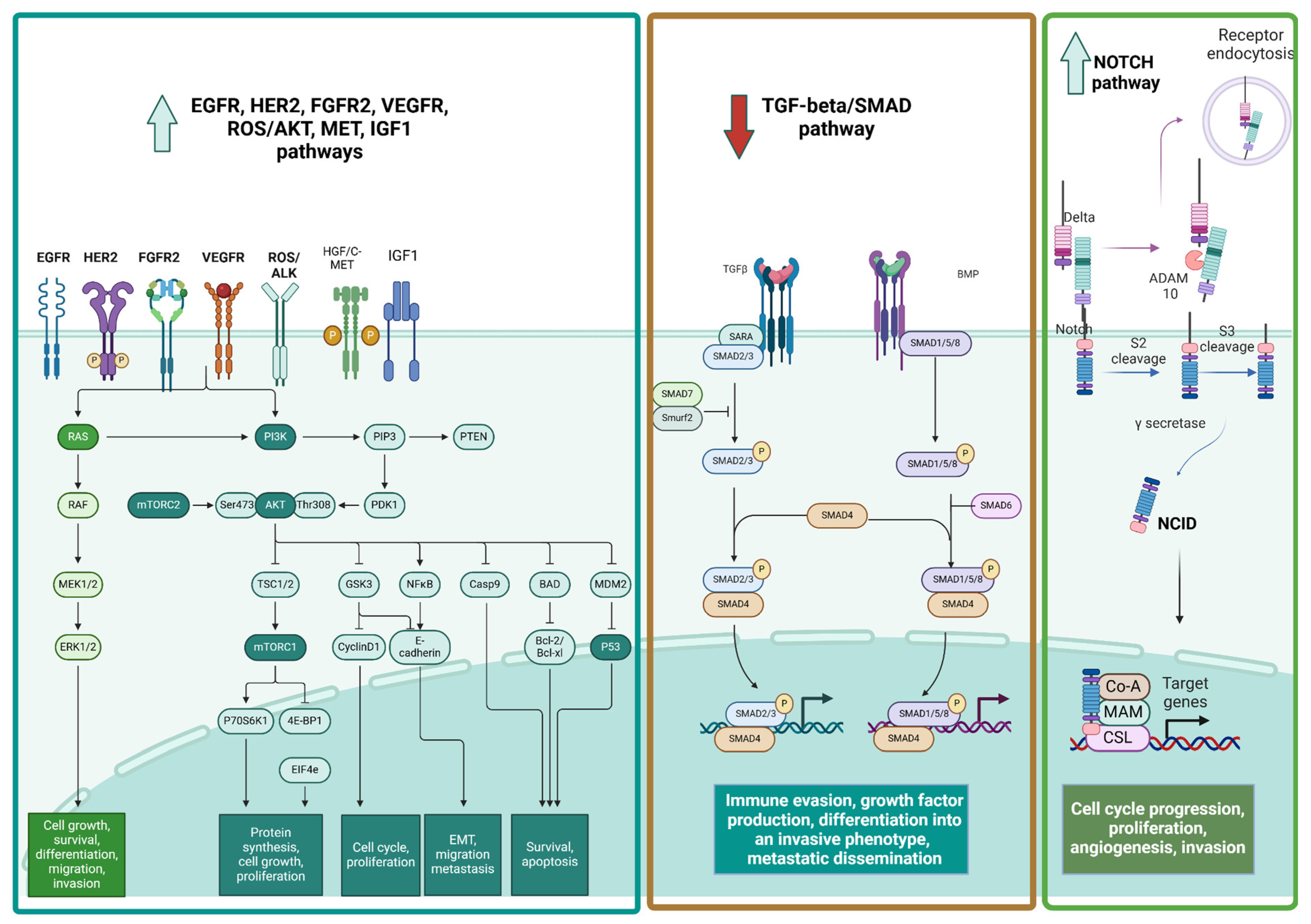
2. Cell Surface Receptors
2.1. HER Family
2.2. FGF Receptors
2.3. PDGF Receptors
2.4. VEGF Receptors
2.5. IGF Receptors
2.6. TGF Receptors
2.7. Notch Receptors
2.8. HGF/c-MET
2.9. ALK/ROS1 Receptors
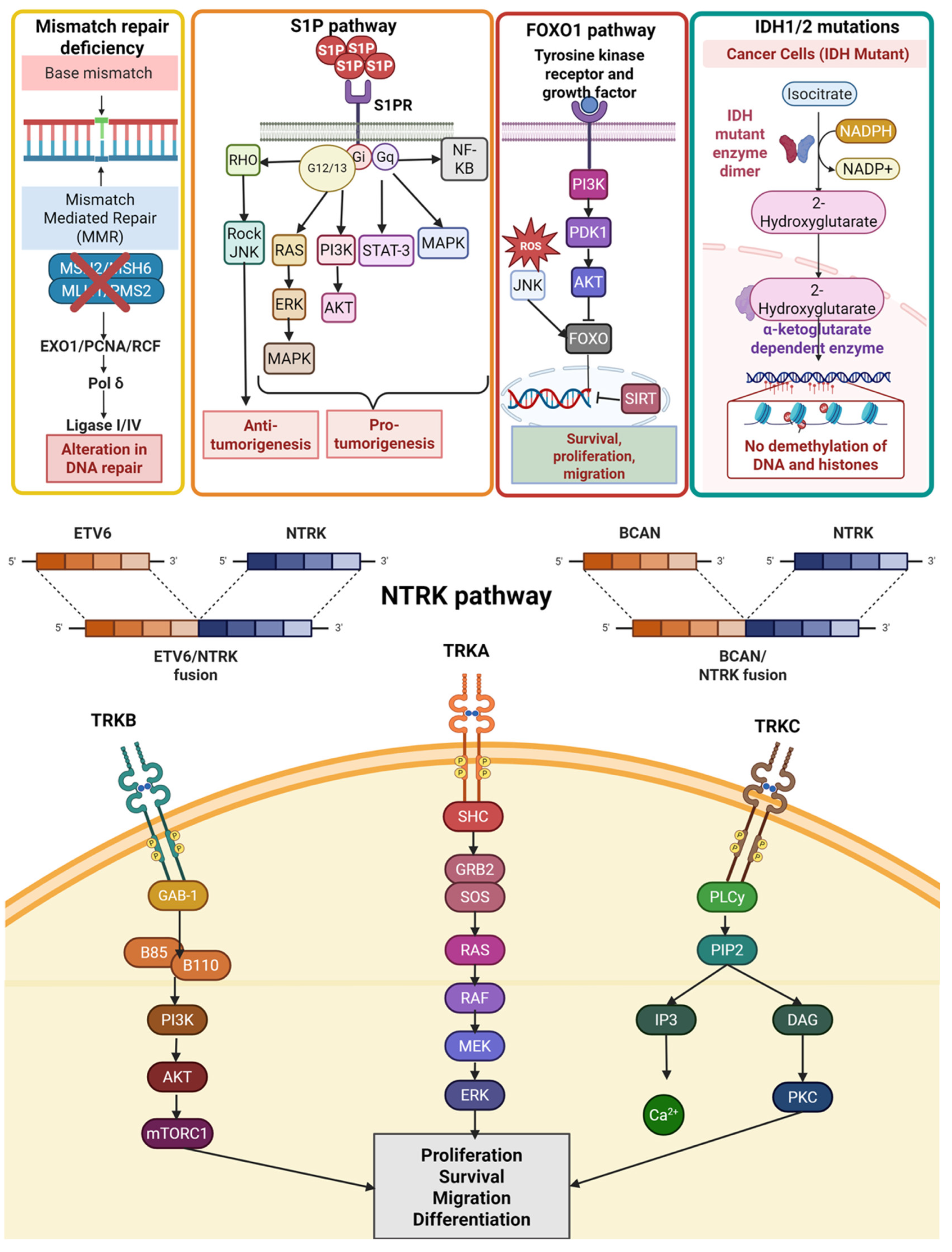
2.10. TRK Receptors
2.11. RET Receptor
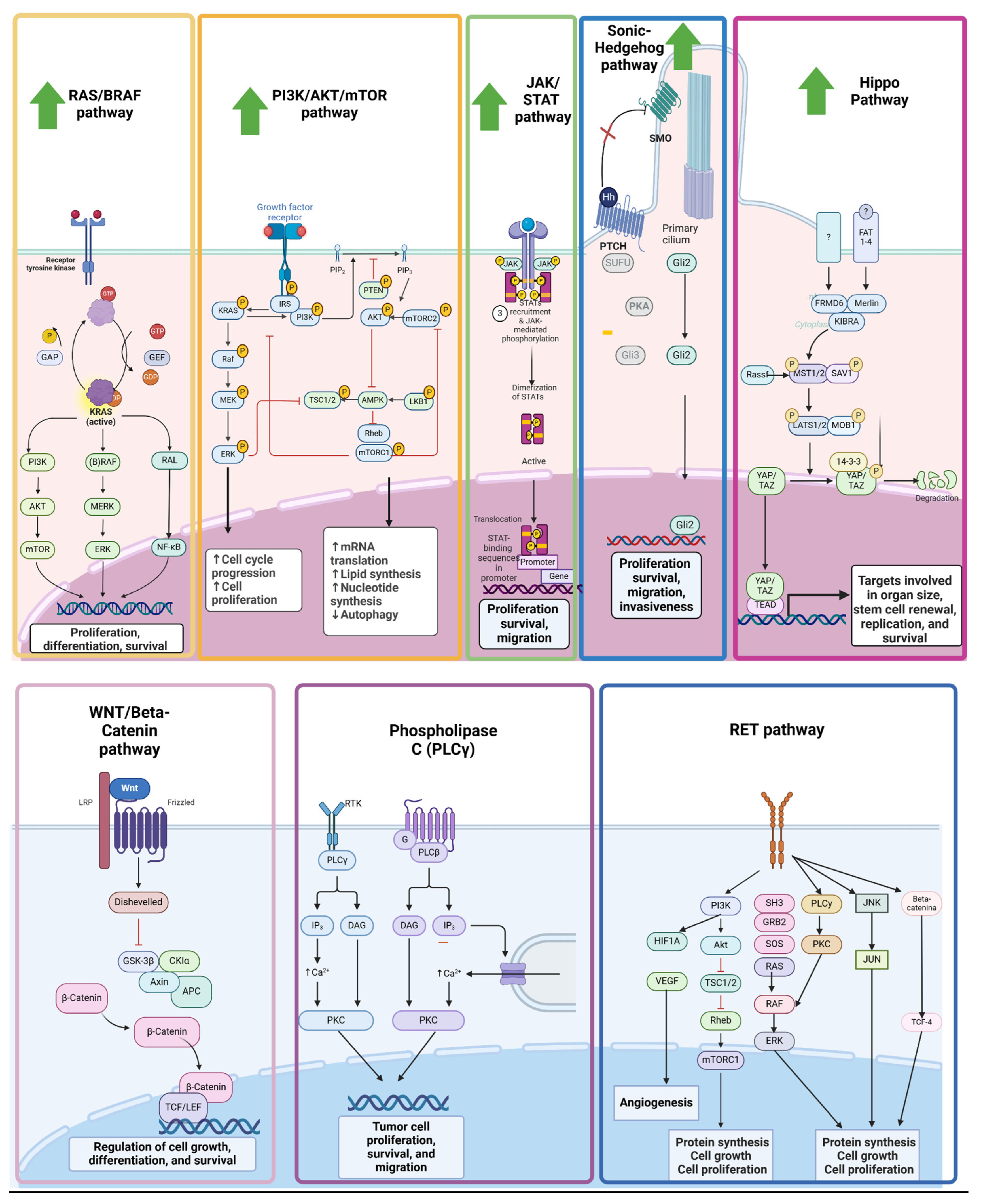
3. Cellular Signaling Pathways
3.1. KRAS/BRAF/MEK/ERK Pathway
3.2. PI3K/AKT/mTOR Pathway
3.3. JAK/STAT Pathway
3.4. Phospholipase C (PLCγ) Pathway
3.5. HIPPO/YAP1 Pathway
3.6. Sonic Hedgehog (SHH) Pathway
3.7. Wnt/B-Catenin Pathway
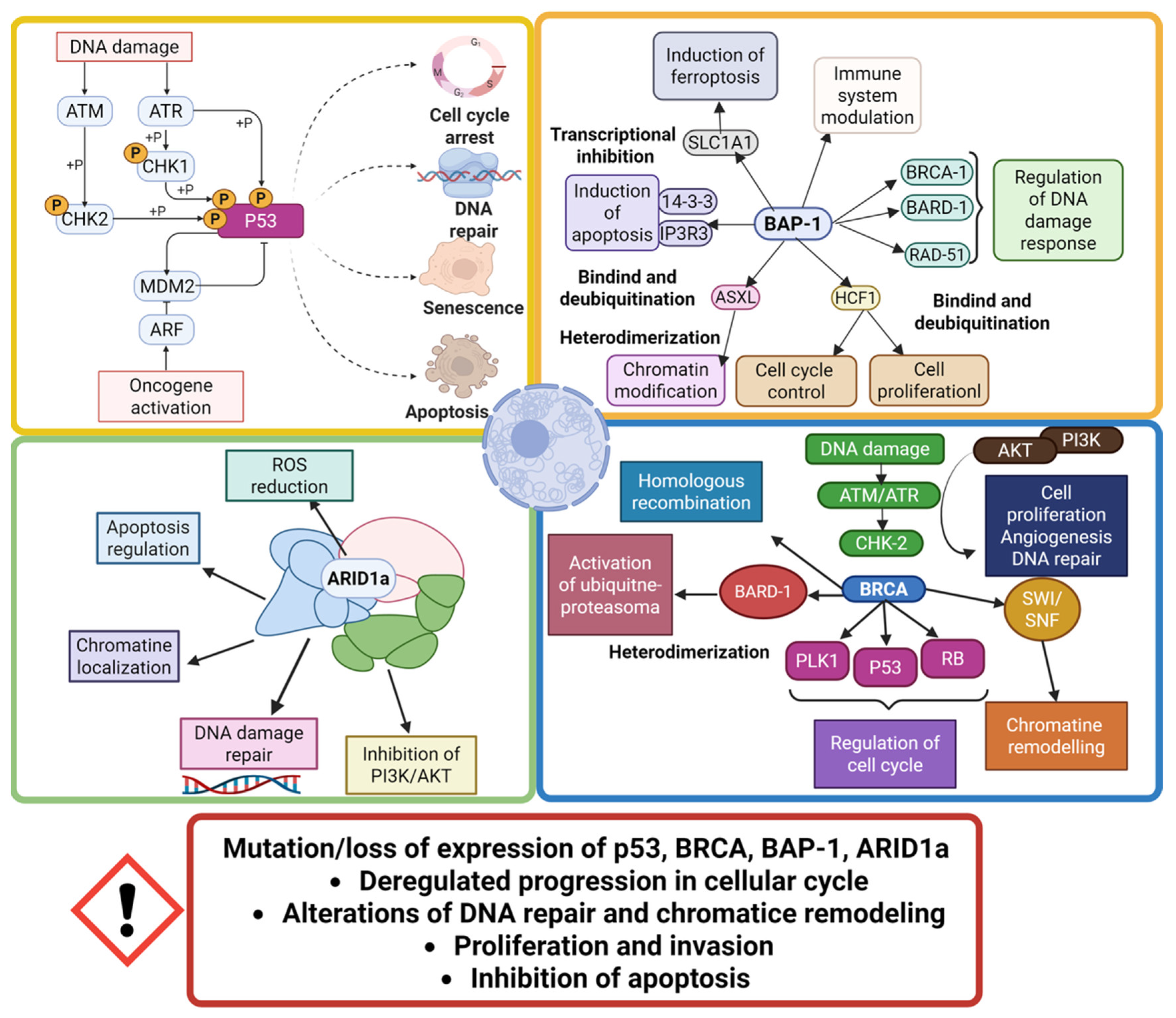
4. Tumor Suppressor Genes
4.1. TP53
4.2. PTEN—CDKN2A
4.3. BRCA
4.4. BAP-1
4.5. SWI/SNF Pathway
5. Metabolic and Other Pathways
5.1. S1P
5.2. FOXO1
5.3. IDH1/IDH2 Mutations
5.4. Mismatch Repair Deficiency Pathway
6. Conclusions
Supplementary Materials
Author Contributions
Funding
Institutional Review Board Statement
Informed Consent Statement
Data Availability Statement
Conflicts of Interest
Abbreviations
| ADAM | A disintegrin and metalloproteinase |
| ADP | Adenosine diphosphate |
| AKT | Protein kinase B |
| ALDH1A3 | Aldehyde Dehydrogenase 1 Family Member A3 |
| ALK | Anaplastic lymphoma kinase |
| AP-1 | Activator protein 1 |
| ARID1A | AT-Rich Interaction Domain 1A |
| AT | Ataxia Telangiectasia |
| ATR | Ataxia Telangiectasia Rad3-related protein |
| AXIN1 | Axis inhibition protein 1 |
| BAF | BRG1/BRM associated factor |
| BAFF | B cell activating factor |
| BAP1 | BRCA1 associated protein-1 |
| BP1 | Homebox protein 1 |
| BRCA | Breast cancer gene |
| BRG1 | Brahma-related gene 1 |
| BTC | Biliary tract cancer |
| C-MYC | Cellular MYC |
| CA19-9 | Carbohydrate antigen 19-9 |
| CCA | Cholangiocarcinoma |
| CCL5 | Chemokine (C-C motif) ligand 5 |
| CD24 | Cluster of differentiation 24 |
| CDKN2A | Cyclin-dependent kinase inhibitor 2A |
| CNS | Central nervous system |
| COX2 | Cyclooxygenase-2 |
| CTNBB1 | Catenin beta-1 |
| CXCL12 | Chemokine (C-X-C motif) ligand 12 |
| CXCR4 | C-X-C chemokine receptor type 4 |
| D2HG | D-2 hydroxyglutarate |
| DHH | Desert Hedgehog |
| DNA | Deoxyribonucleic acid |
| DUB | Deubiquitinating enzyme |
| eCCA | Extrahepatic cholangiocarcinoma |
| EGF | Epidermal growth factor |
| EGFR | Epidermal growth factor receptor |
| EMT | Epithelial-to-mesenchymal transition |
| ERBB2 | Human Epidermal growth factor 2 |
| ERK | Extracellular signal-regulated kinase |
| ESMO | European Society for Medical Oncology |
| FGF | Fibroblast Growth Factor |
| FGFR | Fibroblast Growth Factor Receptor |
| FOXO | Forkhead box transcription factors |
| GBC | Gallbladder cancer |
| GH | Growth hormone |
| GLI | GLI family zinc finger transcription factors |
| GOF | Gain-of-function |
| GTP | Guanosine triphosphate |
| HCC | Hepatocellular carcinoma |
| HES | Enhancer of split |
| HGF | Hepatocyte growth factor |
| HH | Hedgehog |
| HIF | Hypoxia-inducible factor |
| HR | Homologous recombination |
| iCCA | intrahepatic cholangiocarcinoma |
| IDH | Isocitrate Dehydrogenase |
| IGF | Insulin-like growth factor |
| IGFR | Insulin-like growth factor receptor |
| IHC | Immunohistochemistry |
| IL | Interleukin |
| IMR-1 | Inhibitor of Mastermind Recruitment-1 |
| JAG | Jagged |
| JAK | Janus Kinase |
| JNK | c-Jun N-terminal kinase |
| KCSG | Korean Cancer Study Group |
| KRAS | Kirsten Rat Sarcoma viral oncogene homolog |
| LATS | Large Tumor Suppressor |
| LIF | Leukemia inhibitory factor |
| LOF | Loss-of-function |
| MAML-1 | Mastermind-like proteins |
| MAP | Mitogen-activated protein |
| MAPK | Mitogen-activated protein kinase |
| MMR | Mismatch repair |
| MSI | Microsatellite instability |
| MTOR | Mammalian target of rapamycin |
| MUC4 | Mucin 4 |
| NRAS | Neuroblastoma RAS viral oncogene homolog |
| NADP | Nicotinamide adenine dinucleotide phosphate |
| NCCN | National Comprehensive Cancer Network |
| NEJM | New England Journal of Medicine |
| NF-kB | Nuclear factor kappa B |
| NGS | Next Generation Sequencing |
| NICD | Notch intracellular domain |
| NOTCH | Notch receptor |
| NTRK | Neurotrophic Tyrosine Receptor Kinase |
| ORR | Objective response rate |
| OS | Overall survival |
| PARP | Poly(ADP-ribose) polymerase |
| PBRM-1 | Polybromo 1 |
| PD-1 | Programmed cell death protein 1 |
| PD-L1 | Programmed cell death protein ligand 1 |
| PDGF | Platelet-derived growth factor |
| PDGFR | Platelet-derived growth factor receptor |
| PDK1 | Phosphoinositide-dependent kinases |
| PFS | Progression Free Survival |
| PGF | Placental growth factor |
| PI3K | Phosphatidylinositol 3-kinase |
| PIAS | Protein inhibitors of activated STAT proteins |
| PIP2 | Phosphatidylinositol-4,5-bisphosphate |
| PIP3 | Phosphatidylinositol-3,4,5-bisphosphate |
| PTCH1 | Protein patched homolog 1 |
| PTEN | Phosphatase and tensin homolog |
| RBP-J | Recombination Signal Binding Protein for Immunoglobulin Kappa J |
| RET | REarranged during Transfection |
| RNA | Ribonucleic acid |
| ROS1 | c-ros oncogene 1 |
| RTK | Receptor tyrosine kinase |
| S1P | Sphingosine-1-phosphate |
| SDF-1 | Stromal-Derived Factor-1 |
| SHH | Sonic Hedgehog |
| SOCS | Suppressors of cytokine signaling |
| SPHK1 | Sphingosine kinase 1 |
| SREBP1 | Sterol regulatory element-binding proteins |
| STAT | Signal Transducer and Activator of Transcription |
| SUFU | Suppressor of fused homolog |
| SWI/SNF | Switch/Sucrose non fermentable |
| TAZ | Transcriptional co-activator with PDZ-binding motif |
| TCA | Taurocholate |
| TGF | Transforming growth factor beta |
| TKI | Tyrosine kinase inhibitor |
| TNF | Tumor necrosis factor |
| TP53 | Tumor Protein p53 |
| TRAF6 | TNF receptor-associated factor 6 |
| TRAIL | Tumor necrosis factor-related apoptosis inducing ligand |
| TSC1 | Tuberous sclerosis complex |
| VEGF | Vascular endothelial-derived growth factor |
| VEGFR | Vascular endothelial growth factor receptor |
| YAP | Yes-associated protein |
| ZEB1 | Zinc finger E-box-binding homeobox 1 |
References
- Ghosn, M.; Kourie, H.R.; Tabchi, S. Gastrointestinal Cancers in the Era of Theranostics: Updates and Future Perspectives. World J. Gastroenterol. 2015, 21, 8473–8477. [Google Scholar] [CrossRef]
- Noel, M.S.; Hezel, A.F. New and Emerging Treatment Options for Biliary Tract Cancer. OncoTargets Ther. 2013, 6, 1545–1552. [Google Scholar] [CrossRef] [PubMed][Green Version]
- Sasaki, T.; Isayama, H.; Nakai, Y.; Koike, K. Current Status of Chemotherapy for the Treatment of Advanced Biliary Tract Cancer. Korean J. Intern. Med. 2013, 28, 515–524. [Google Scholar] [CrossRef]
- Ghosn, M.; Kourie, H.R.; El Rassy, E.; Chebib, R.; El Karak, F.; Hanna, C.; Nasr, D. Optimum Chemotherapy for the Management of Advanced Biliary Tract Cancer. World J. Gastroenterol. 2015, 21, 4121–4125. [Google Scholar] [CrossRef] [PubMed]
- Razumilava, N.; Gores, G.J. Cholangiocarcinoma. Lancet 2014, 383, 2168–2179. [Google Scholar] [CrossRef]
- Oh, D.-Y.; Ruth, H.A.; Qin, S.; Chen, L.-T.; Okusaka, T.; Vogel, A.; Kim, J.W.; Suksombooncharoen, T.; Ah, L.M.; Kitano, M.; et al. Durvalumab plus Gemcitabine and Cisplatin in Advanced Biliary Tract Cancer. NEJM Evid. 2022, 1, EVIDoa2200015. [Google Scholar] [CrossRef]
- Kelley, R.K.; Ueno, M.; Yoo, C.; Finn, R.S.; Furuse, J.; Ren, Z.; Yau, T.; Klümpen, H.-J.; Chan, S.L.; Ozaka, M.; et al. Pembrolizumab in Combination with Gemcitabine and Cisplatin Compared with Gemcitabine and Cisplatin Alone for Patients with Advanced Biliary Tract Cancer (KEYNOTE-966): A Randomised, Double-Blind, Placebo-Controlled, Phase 3 Trial. Lancet 2023, 401, 1853–1865, Erratum in Lancet 2024, 403, 1140. https://doi.org/10.1016/S0140-6736(24)00545-2. [Google Scholar] [CrossRef] [PubMed]
- Cardinale, V.; Carpino, G.; Reid, L.; Gaudio, E.; Alvaro, D. Multiple Cells of Origin in Cholangiocarcinoma Underlie Biological, Epidemiological and Clinical Heterogeneity. World J. Gastrointest. Oncol. 2012, 4, 94–102. [Google Scholar] [CrossRef]
- Moeini, A.; Haber, P.K.; Sia, D. Cell of Origin in Biliary Tract Cancers and Clinical Implications. JHEP Rep. 2021, 3, 100226. [Google Scholar] [CrossRef]
- Farshidfar, F.; Zheng, S.; Gingras, M.-C.; Newton, Y.; Shih, J.; Robertson, A.G.; Hinoue, T.; Hoadley, K.A.; Gibb, E.A.; Roszik, J.; et al. Integrative Genomic Analysis of Cholangiocarcinoma Identifies Distinct IDH-Mutant Molecular Profiles. Cell Rep. 2017, 18, 2780–2794, Erratum in Cell Rep. 2017, 19, 2878–2880. https://doi.org/10.1016/j.celrep.2017.06.008. [Google Scholar] [CrossRef]
- Ojesina, A.I.; Lichtenstein, L.; Freeman, S.S.; Pedamallu, C.S.; Imaz-Rosshandler, I.; Pugh, T.J.; Cherniack, A.D.; Ambrogio, L.; Cibulskis, K.; Bertelsen, B.; et al. Landscape of Genomic Alterations in Cervical Carcinomas. Nature 2014, 506, 371–375. [Google Scholar] [CrossRef]
- Vogel, A.; Bridgewater, J.; Edeline, J.; Kelley, R.K.; Klümpen, H.J.; Malka, D.; Primrose, J.N.; Rimassa, L.; Stenzinger, A.; Valle, J.W.; et al. Biliary Tract Cancer: ESMO Clinical Practice Guideline for Diagnosis, Treatment and Follow-Up. Ann. Oncol. 2023, 34, 127–140. [Google Scholar] [CrossRef] [PubMed]
- Benson, A.B.; D’Angelica, M.I.; Abrams, T.; Abbott, D.E.; Ahmed, A.; Anaya, D.A.; Anders, R.; Are, C.; Bachini, M.; Binder, D.; et al. NCCN Guidelines® Insights: Biliary Tract Cancers, Version 2.2023. J. Natl. Compr. Cancer Netw. 2023, 21, 694–704. [Google Scholar] [CrossRef] [PubMed]
- Roskoski, R. ErbB/HER Protein-Tyrosine Kinases: Structures and Small Molecule Inhibitors. Pharmacol. Res. 2014, 87, 42–59. [Google Scholar] [CrossRef]
- Zhang, H.; Berezov, A.; Wang, Q.; Zhang, G.; Drebin, J.; Murali, R.; Greene, M.I. ErbB Receptors: From Oncogenes to Targeted Cancer Therapies. J. Clin. Investig. 2007, 117, 2051–2058. [Google Scholar] [CrossRef]
- Nakazawa, K.; Dobashi, Y.; Suzuki, S.; Fujii, H.; Takeda, Y.; Ooi, A. Amplification and Overexpression of C-erbB-2, Epidermal Growth Factor Receptor, and c-Met in Biliary Tract Cancers. J. Pathol. 2005, 206, 356–365. [Google Scholar] [CrossRef] [PubMed]
- Zhang, C.; Xu, H.; Zhou, Z.; Tian, Y.; Cao, X.; Cheng, G.; Liu, Q. Blocking of the EGFR-STAT3 Signaling Pathway Through Afatinib Treatment Inhibited the Intrahepatic Cholangiocarcinoma. Exp. Ther. Med. 2018, 15, 4995–5000. [Google Scholar] [CrossRef]
- Banales, J.M.; Marin, J.J.G.; Lamarca, A.; Rodrigues, P.M.; Khan, S.A.; Roberts, L.R.; Cardinale, V.; Carpino, G.; Andersen, J.B.; Braconi, C.; et al. Cholangiocarcinoma 2020: The next Horizon in Mechanisms and Management. Nat. Rev. Gastroenterol. Hepatol. 2020, 17, 557–588. [Google Scholar] [CrossRef]
- Garrett, T.P.J.; McKern, N.M.; Lou, M.; Elleman, T.C.; Adams, T.E.; Lovrecz, G.O.; Kofler, M.; Jorissen, R.N.; Nice, E.C.; Burgess, A.W.; et al. The Crystal Structure of a Truncated ErbB2 Ectodomain Reveals an Active Conformation, Poised to Interact with Other ErbB Receptors. Mol. Cell 2003, 11, 495–505. [Google Scholar] [CrossRef]
- Hudis, C.A. Trastuzumab--Mechanism of Action and Use in Clinical Practice. N. Engl. J. Med. 2007, 357, 39–51. [Google Scholar] [CrossRef]
- Galdy, S.; Lamarca, A.; McNamara, M.G.; Hubner, R.A.; Cella, C.A.; Fazio, N.; Valle, J.W. HER2/HER3 Pathway in Biliary Tract Malignancies; Systematic Review and Meta-Analysis: A Potential Therapeutic Target? Cancer Metastasis Rev. 2017, 36, 141–157. [Google Scholar] [CrossRef]
- Swain, S.M.; Baselga, J.; Kim, S.-B.; Ro, J.; Semiglazov, V.; Campone, M.; Ciruelos, E.; Ferrero, J.-M.; Schneeweiss, A.; Heeson, S.; et al. Pertuzumab, Trastuzumab, and Docetaxel in HER2-Positive Metastatic Breast Cancer. N. Engl. J. Med. 2015, 372, 724–734. [Google Scholar] [CrossRef] [PubMed]
- Hurvitz, S.A.; Hegg, R.; Chung, W.-P.; Im, S.-A.; Jacot, W.; Ganju, V.; Chiu, J.W.Y.; Xu, B.; Hamilton, E.; Madhusudan, S.; et al. Trastuzumab Deruxtecan versus Trastuzumab Emtansine in Patients with HER2-Positive Metastatic Breast Cancer: Updated Results from DESTINY-Breast03, a Randomised, Open-Label, Phase 3 Trial. Lancet 2023, 401, 105–117, Erratum in Lancet 2023, 401, 556. https://doi.org/10.1016/S0140-6736(22)00045-9. [Google Scholar] [CrossRef] [PubMed]
- Bang, Y.-J.; Van Cutsem, E.; Feyereislova, A.; Chung, H.C.; Shen, L.; Sawaki, A.; Lordick, F.; Ohtsu, A.; Omuro, Y.; Satoh, T.; et al. Trastuzumab in Combination with Chemotherapy versus Chemotherapy Alone for Treatment of HER2-Positive Advanced Gastric or Gastro-Oesophageal Junction Cancer (ToGA): A Phase 3, Open-Label, Randomised Controlled Trial. Lancet 2010, 376, 687–697. [Google Scholar] [CrossRef] [PubMed]
- Shitara, K.; Bang, Y.-J.; Iwasa, S.; Sugimoto, N.; Ryu, M.-H.; Sakai, D.; Chung, H.-C.; Kawakami, H.; Yabusaki, H.; Lee, J.; et al. Trastuzumab Deruxtecan in Previously Treated HER2-Positive Gastric Cancer. N. Engl. J. Med. 2020, 382, 2419–2430. [Google Scholar] [CrossRef]
- Pahuja, K.B.; Nguyen, T.T.; Jaiswal, B.S.; Prabhash, K.; Thaker, T.M.; Senger, K.; Chaudhuri, S.; Kljavin, N.M.; Antony, A.; Phalke, S.; et al. Actionable Activating Oncogenic ERBB2/HER2 Transmembrane and Juxtamembrane Domain Mutations. Cancer Cell 2018, 34, 792–806.e5. [Google Scholar] [CrossRef]
- Jacobi, O.; Ross, J.S.; Goshen-Lago, T.; Haddad, R.; Moore, A.; Sulkes, A.; Brenner, B.; Ben-Aharon, I. ERBB2 Pathway in Biliary Tract Carcinoma: Clinical Implications of a Targetable Pathway. Oncol. Res. Treat. 2021, 44, 20–27. [Google Scholar] [CrossRef]
- Ahcene Djaballah, S.; Daniel, F.; Milani, A.; Ricagno, G.; Lonardi, S. HER2 in Colorectal Cancer: The Long and Winding Road from Negative Predictive Factor to Positive Actionable Target. Am. Soc. Clin. Oncol. Educ. Book 2022, 42, 219–232. [Google Scholar] [CrossRef]
- Yu, Y.; Yang, Y.; Li, H.; Fan, Y. Targeting HER2 Alterations in Non-Small Cell Lung Cancer: Therapeutic Breakthrough and Challenges. Cancer Treat. Rev. 2023, 114, 102520. [Google Scholar] [CrossRef]
- Nam, A.-R.; Kim, J.-W.; Cha, Y.; Ha, H.; Park, J.E.; Bang, J.-H.; Jin, M.H.; Lee, K.-H.; Kim, T.-Y.; Han, S.-W.; et al. Therapeutic Implication of HER2 in Advanced Biliary Tract Cancer. Oncotarget 2016, 7, 58007–58021. [Google Scholar] [CrossRef]
- Valle, J.W.; Lamarca, A.; Goyal, L.; Barriuso, J.; Zhu, A.X. New Horizons for Precision Medicine in Biliary Tract Cancers. Cancer Discov. 2017, 7, 943–962. [Google Scholar] [CrossRef]
- Kim, H.; Kim, R.; Kim, H.R.; Jo, H.; Kim, H.; Ha, S.Y.; Park, J.O.; Park, Y.S.; Kim, S.T. HER2 Aberrations as a Novel Marker in Advanced Biliary Tract Cancer. Front. Oncol. 2022, 12, 834104. [Google Scholar] [CrossRef] [PubMed]
- Lamarca, A.; Galdy, S.; Barriuso, J.; Moghadam, S.; Beckett, E.; Rogan, J.; Backen, A.; Billington, C.; McNamara, M.G.; Hubner, R.A.; et al. The HER3 Pathway as a Potential Target for Inhibition in Patients with Biliary Tract Cancers. PLoS ONE 2018, 13, e0206007, Erratum in PLoS ONE 2019, 14, e0212697. https://doi.org/10.1371/journal.pone.0212697. [Google Scholar] [CrossRef] [PubMed]
- Javle, M.; Borad, M.J.; Azad, N.S.; Kurzrock, R.; Abou-Alfa, G.K.; George, B.; Hainsworth, J.; Meric-Bernstam, F.; Swanton, C.; Sweeney, C.J.; et al. Pertuzumab and Trastuzumab for HER2-Positive, Metastatic Biliary Tract Cancer (MyPathway): A Multicentre, Open-Label, Phase 2a, Multiple Basket Study. Lancet Oncol. 2021, 22, 1290–1300. [Google Scholar] [CrossRef]
- Vivaldi, C.; Fornaro, L.; Ugolini, C.; Niccoli, C.; Musettini, G.; Pecora, I.; Cacciato Insilla, A.; Salani, F.; Pasquini, G.; Catanese, S.; et al. HER2 Overexpression as a Poor Prognostic Determinant in Resected Biliary Tract Cancer. Oncologist 2020, 25, 886–893. [Google Scholar] [CrossRef]
- Lowery, M.A.; Ptashkin, R.; Jordan, E.; Berger, M.F.; Zehir, A.; Capanu, M.; Kemeny, N.E.; O’Reilly, E.M.; El-Dika, I.; Jarnagin, W.R.; et al. Comprehensive Molecular Profiling of Intrahepatic and Extrahepatic Cholangiocarcinomas: Potential Targets for Intervention. Clin. Cancer Res. 2018, 24, 4154–4161. [Google Scholar] [CrossRef] [PubMed]
- Harding, J.J.; Fan, J.; Oh, D.-Y.; Choi, H.J.; Kim, J.W.; Chang, H.-M.; Bao, L.; Sun, H.-C.; Macarulla, T.; Xie, F.; et al. Zanidatamab for HER2-Amplified, Unresectable, Locally Advanced or Metastatic Biliary Tract Cancer (HERIZON-BTC-01): A Multicentre, Single-Arm, Phase 2b Study. Lancet Oncol. 2023, 24, 772–782. [Google Scholar] [CrossRef]
- Strickler, J.H.; Cercek, A.; Siena, S.; André, T.; Ng, K.; Van Cutsem, E.; Wu, C.; Paulson, A.S.; Hubbard, J.M.; Coveler, A.L.; et al. Tucatinib plus Trastuzumab for Chemotherapy-Refractory, HER2-Positive, RAS Wild-Type Unresectable or Metastatic Colorectal Cancer (MOUNTAINEER): A Multicentre, Open-Label, Phase 2 Study. Lancet Oncol. 2023, 24, 496–508. [Google Scholar] [CrossRef]
- Murthy, R.K.; Loi, S.; Okines, A.; Paplomata, E.; Hamilton, E.; Hurvitz, S.A.; Lin, N.U.; Borges, V.; Abramson, V.; Anders, C.; et al. Tucatinib, Trastuzumab, and Capecitabine for HER2-Positive Metastatic Breast Cancer. N. Engl. J. Med. 2020, 382, 597–609, Erratum in N. Engl. J. Med. 2020, 382, 586. https://doi.org/10.1056/NEJMx190039. [Google Scholar] [CrossRef]
- Lee, C.; Chon, H.J.; Cheon, J.; Lee, M.A.; Im, H.-S.; Jang, J.-S.; Kim, M.H.; Park, S.; Kang, B.; Hong, M.; et al. Trastuzumab plus FOLFOX for HER2-Positive Biliary Tract Cancer Refractory to Gemcitabine and Cisplatin: A Multi-Institutional Phase 2 Trial of the Korean Cancer Study Group (KCSG-HB19–14). Lancet Gastroenterol. Hepatol. 2023, 8, 56–65. [Google Scholar] [CrossRef]
- Ostwal, V.; Mandavkar, S.; Bhargava, P.; Srinivas, S.; Kapoor, A.; Shetty, O.; Kannan, S.; Chaugule, D.; Patil, R.; Parulekar, M.; et al. Trastuzumab Plus Gemcitabine-Cisplatin for Treatment-Naïve Human Epidermal Growth Factor Receptor 2-Positive Biliary Tract Adenocarcinoma: A Multicenter, Open-Label, Phase II Study (TAB). J. Clin. Oncol. 2024, 42, 800–807. [Google Scholar] [CrossRef] [PubMed]
- Meric-Bernstam, F.; Makker, V.; Oaknin, A.; Oh, D.-Y.; Banerjee, S.; González-Martín, A.; Jung, K.H.; Ługowska, I.; Manso, L.; Manzano, A.; et al. Efficacy and Safety of Trastuzumab Deruxtecan in Patients with HER2-Expressing Solid Tumors: Primary Results from the DESTINY-PanTumor02 Phase II Trial. J. Clin. Oncol. 2024, 42, 47–58. [Google Scholar] [CrossRef] [PubMed]
- Turner, N.; Grose, R. Fibroblast Growth Factor Signalling: From Development to Cancer. Nat. Rev. Cancer 2010, 10, 116–129. [Google Scholar] [CrossRef]
- Ornitz, D.M.; Itoh, N. The Fibroblast Growth Factor Signaling Pathway. Wiley Interdiscip. Rev. Dev. Biol. 2015, 4, 215–266. [Google Scholar] [CrossRef]
- Brooks, A.N.; Kilgour, E.; Smith, P.D. Molecular Pathways: Fibroblast Growth Factor Signaling: A New Therapeutic Opportunity in Cancer. Clin. Cancer Res. 2012, 18, 1855–1862. [Google Scholar] [CrossRef]
- Liu, S.; Weng, J.; Cao, M.; Zhou, Q.; Xu, M.; Xu, W.; Hu, Z.; Xu, M.; Dong, Q.; Sheng, X.; et al. FGFR2 Fusion/Rearrangement Is Associated with Favorable Prognosis and Immunoactivation in Patients with Intrahepatic Cholangiocarcinoma. Oncologist 2024, 29, e1734–e1747. [Google Scholar] [CrossRef] [PubMed]
- Buckarma, E.; De La Cruz, G.; Truty, M.; Nagorney, D.; Cleary, S.; Kendrick, M.; Borad, M.; Graham, R.P.; Gores, G.; Smoot, R. Impact of FGFR2 Gene Fusions on Survival of Patients with Intrahepatic Cholangiocarcinoma Following Curative Intent Resection. HPB 2022, 24, 1748–1756. [Google Scholar] [CrossRef]
- Vogel, A.; Sahai, V.; Hollebecque, A.; Vaccaro, G.; Melisi, D.; Al-Rajabi, R.; Paulson, S.; Borad, M.; Gallinson, D.; Murphy, A.; et al. O-2 Pemigatinib for Previously Treated Locally Advanced or Metastatic Cholangiocarcinoma: Final Results from FIGHT-202. Ann. Oncol. 2022, 33, S379. [Google Scholar] [CrossRef]
- Goyal, L.; Meric-Bernstam, F.; Hollebecque, A.; Valle, J.W.; Morizane, C.; Karasic, T.B.; Abrams, T.A.; Furuse, J.; Kelley, R.K.; Cassier, P.A.; et al. Futibatinib for FGFR2-Rearranged Intrahepatic Cholangiocarcinoma. N. Engl. J. Med. 2023, 388, 228–239. [Google Scholar] [CrossRef]
- Jung, S.-C.; Kang, D.; Ko, E.-A. Roles of PDGF/PDGFR Signaling in Various Organs. Korean J. Physiol. Pharmacol. 2025, 29, 139–155. [Google Scholar] [CrossRef] [PubMed]
- Andrae, J.; Gallini, R.; Betsholtz, C. Role of Platelet-Derived Growth Factors in Physiology and Medicine. Genes Dev. 2008, 22, 1276–1312. [Google Scholar] [CrossRef] [PubMed]
- Sia, D.; Hoshida, Y.; Villanueva, A.; Roayaie, S.; Ferrer, J.; Tabak, B.; Peix, J.; Sole, M.; Tovar, V.; Alsinet, C.; et al. Integrative Molecular Analysis of Intrahepatic Cholangiocarcinoma Reveals 2 Classes That Have Different Outcomes. Gastroenterology 2013, 144, 829–840. [Google Scholar] [CrossRef] [PubMed]
- Cadamuro, M.; Brivio, S.; Mertens, J.; Vismara, M.; Moncsek, A.; Milani, C.; Fingas, C.; Cristina Malerba, M.; Nardo, G.; Dall’Olmo, L.; et al. Platelet-Derived Growth Factor-D Enables Liver Myofibroblasts to Promote Tumor Lymphangiogenesis in Cholangiocarcinoma. J. Hepatol. 2019, 70, 700–709. [Google Scholar] [CrossRef]
- Fingas, C.D.; Mertens, J.C.; Razumilava, N.; Bronk, S.F.; Sirica, A.E.; Gores, G.J. Targeting PDGFR-β in Cholangiocarcinoma. Liver Int. 2012, 32, 400–409. [Google Scholar] [CrossRef]
- Ferrara, N.; Adamis, A.P. Ten Years of Anti-Vascular Endothelial Growth Factor Therapy. Nat. Rev. Drug Discov. 2016, 15, 385–403. [Google Scholar] [CrossRef]
- Apte, R.S.; Chen, D.S.; Ferrara, N. VEGF in Signaling and Disease: Beyond Discovery and Development. Cell 2019, 176, 1248–1264. [Google Scholar] [CrossRef]
- Cai, C.; Wang, X.; Fu, Q.; Chen, A. The VEGF Expression Associated with Prognosis in Patients with Intrahepatic Cholangiocarcinoma: A Systematic Review and Meta-Analysis. World J. Surg. Oncol. 2022, 20, 40. [Google Scholar] [CrossRef]
- Peng, M.; Li, H.; Cao, H.; Huang, Y.; Yu, W.; Shen, C.; Gu, J. Dual FGFR and VEGFR Inhibition Synergistically Restrain Hexokinase 2-Dependent Lymphangiogenesis and Immune Escape in Intrahepatic Cholangiocarcinoma. J. Gastroenterol. 2023, 58, 908–924. [Google Scholar] [CrossRef]
- Ghalehbandi, S.; Yuzugulen, J.; Pranjol, M.Z.I.; Pourgholami, M.H. The Role of VEGF in Cancer-Induced Angiogenesis and Research Progress of Drugs Targeting VEGF. Eur. J. Pharmacol. 2023, 949, 175586. [Google Scholar] [CrossRef]
- Calastri, M.C.J.; Ferreira, R.F.; Tenani, G.D.; Spinola, L.P.; Vieira, G.F.; Rabaça Roque Botelho, M.F.; Abrantes, A.M.C.; Tralhão, J.G.L.R.; De Brito, A.F.M.; Da Silva, R.F.; et al. Investigating VEGF. miR-145-3p, and miR-101-3p Expression in Patients with Cholangiocarcinoma. Asian Pac. J. Cancer Prev. 2022, 23, 2233–2241. [Google Scholar] [CrossRef] [PubMed]
- Yoshikawa, D.; Ojima, H.; Iwasaki, M.; Hiraoka, N.; Kosuge, T.; Kasai, S.; Hirohashi, S.; Shibata, T. Clinicopathological and Prognostic Significance of EGFR, VEGF, and HER2 Expression in Cholangiocarcinoma. Br. J. Cancer 2008, 98, 418–425. [Google Scholar] [CrossRef] [PubMed]
- Ntanasis-Stathopoulos, I.; Tsilimigras, D.I.; Gavriatopoulou, M.; Schizas, D.; Pawlik, T.M. Cholangiocarcinoma: Investigations into Pathway-Targeted Therapies. Expert Rev. Anticancer. Ther. 2020, 20, 765–773. [Google Scholar] [CrossRef]
- Kam, A.E.; Masood, A.; Shroff, R.T. Current and Emerging Therapies for Advanced Biliary Tract Cancers. Lancet Gastroenterol. Hepatol. 2021, 6, 956–969. [Google Scholar] [CrossRef]
- Kim, Y.; Song, J.; Kim, N.; Sim, T. Recent Progress in Emerging Molecular Targeted Therapies for Intrahepatic Cholangiocarcinoma. RSC Med. Chem. 2025, 16, 2314–2359. [Google Scholar] [CrossRef]
- Huang, Y.; Du, Z.; Kan, A.; He, M.; Li, H.; Lai, Z.; Wen, D.; Huang, L.; Li, Q.; Xu, L.; et al. Clinical and Biomarker Analyses of Hepatic Arterial Infusion Chemotherapy plus Lenvatinib and PD-1 Inhibitor for Patients with Advanced Intrahepatic Cholangiocarcinoma. Front. Immunol. 2024, 15, 1260191. [Google Scholar] [CrossRef]
- Merters, J.; Lamarca, A. Integrating Cytotoxic, Targeted and Immune Therapies for Cholangiocarcinoma. J. Hepatol. 2023, 78, 652–657. [Google Scholar] [CrossRef] [PubMed]
- Benmebarek, M.-R.; Oguz, C.; Seifert, M.; Ruf, B.; Myojin, Y.; Bauer, K.C.; Huang, P.; Ma, C.; Villamor-Payà, M.; Rodriguez-Matos, F.; et al. Anti-Vascular Endothelial Growth Factor Treatment Potentiates Immune Checkpoint Blockade Through a BAFF- and IL-12-Dependent Reprogramming of the TME. Immunity 2025, 58, 926–945.e10. [Google Scholar] [CrossRef] [PubMed]
- Jones, J.I.; Clemmons, D.R. Insulin-like Growth Factors and Their Binding Proteins: Biological Actions. Endocr. Rev. 1995, 16, 3–34. [Google Scholar] [CrossRef]
- Vaquero, J.; Lobe, C.; Tahraoui, S.; Clapéron, A.; Mergey, M.; Merabtene, F.; Wendum, D.; Coulouarn, C.; Housset, C.; Desbois-Mouthon, C.; et al. The IGF2/IR/IGF1R Pathway in Tumor Cells and Myofibroblasts Mediates Resistance to EGFR Inhibition in Cholangiocarcinoma. Clin. Cancer Res. 2018, 24, 4282–4296. [Google Scholar] [CrossRef]
- David, C.J.; Massagué, J. Contextual Determinants of TGFβ Action in Development, Immunity and Cancer. Nat. Rev. Mol. Cell Biol. 2018, 19, 419–435, Erratum in Nat. Rev. Mol. Cell Biol. 2018, 19, 479. https://doi.org/10.1038/s41580-018-0018-x. [Google Scholar] [CrossRef]
- Wang, J.; Shao, N.; Ding, X.; Tan, B.; Song, Q.; Wang, N.; Jia, Y.; Ling, H.; Cheng, Y. Crosstalk Between Transforming Growth Factor-β Signaling Pathway and Long Non-Coding RNAs in Cancer. Cancer Lett. 2016, 370, 296–301. [Google Scholar] [CrossRef]
- Heldin, C.-H.; Moustakas, A. Role of Smads in TGFβ Signaling. Cell Tissue Res. 2012, 347, 21–36. [Google Scholar] [CrossRef]
- Fouassier, L.; Marzioni, M.; Afonso, M.B.; Dooley, S.; Gaston, K.; Giannelli, G.; Rodrigues, C.M.P.; Lozano, E.; Mancarella, S.; Segatto, O.; et al. Signalling Networks in Cholangiocarcinoma: Molecular Pathogenesis, Targeted Therapies and Drug Resistance. Liver Int. 2019, 39 (Suppl. S1), 43–62. [Google Scholar] [CrossRef]
- Miyazono, K.; Katsuno, Y.; Koinuma, D.; Ehata, S.; Morikawa, M. Intracellular and Extracellular TGF-β Signaling in Cancer: Some Recent Topics. Front. Med. 2018, 12, 387–411. [Google Scholar] [CrossRef]
- Caja, L.; Dituri, F.; Mancarella, S.; Caballero-Diaz, D.; Moustakas, A.; Giannelli, G.; Fabregat, I. TGF-β and the Tissue Microenvironment: Relevance in Fibrosis and Cancer. Int. J. Mol. Sci. 2018, 19, 1294. [Google Scholar] [CrossRef]
- Thepmalee, C.; Panya, A.; Junking, M.; Chieochansin, T.; Yenchitsomanus, P.-T. Inhibition of IL-10 and TGF-β Receptors on Dendritic Cells Enhances Activation of Effector T-Cells to Kill Cholangiocarcinoma Cells. Hum. Vaccin. Immunother. 2018, 14, 1423–1431. [Google Scholar] [CrossRef]
- Shimizu, T.; Yokomuro, S.; Mizuguchi, Y.; Kawahigashi, Y.; Arima, Y.; Taniai, N.; Mamada, Y.; Yoshida, H.; Akimaru, K.; Tajiri, T. Effect of Transforming Growth Factor-Beta1 on Human Intrahepatic Cholangiocarcinoma Cell Growth. World J. Gastroenterol. 2006, 12, 6316–6324. [Google Scholar] [CrossRef] [PubMed]
- Zou, S.; Li, J.; Zhou, H.; Frech, C.; Jiang, X.; Chu, J.S.C.; Zhao, X.; Li, Y.; Li, Q.; Wang, H.; et al. Mutational Landscape of Intrahepatic Cholangiocarcinoma. Nat. Commun. 2014, 5, 5696. [Google Scholar] [CrossRef] [PubMed]
- Nakamura, H.; Arai, Y.; Totoki, Y.; Shirota, T.; Elzawahry, A.; Kato, M.; Hama, N.; Hosoda, F.; Urushidate, T.; Ohashi, S.; et al. Genomic Spectra of Biliary Tract Cancer. Nat. Genet. 2015, 47, 1003–1010. [Google Scholar] [CrossRef] [PubMed]
- Yan, X.; Zhang, W.; Zhang, B.; Liang, H.; Zhang, W.; Chen, X. Inactivation of Smad4 Is a Prognostic Factor in Intrahepatic Cholangiocarcinoma. Chin. Med. J. 2013, 126, 3039–3043. [Google Scholar] [CrossRef]
- Yoo, C.; Javle, M.M.; Verdaguer Mata, H.; de Braud, F.; Trojan, J.; Raoul, J.-L.; Kim, J.W.; Ueno, M.; Lee, C.-K.; Hijioka, S.; et al. Phase 2 Trial of Bintrafusp Alfa as Second-Line Therapy for Patients with Locally Advanced/Metastatic Biliary Tract Cancers. Hepatology 2023, 78, 758–770. [Google Scholar] [CrossRef]
- Che, L.; Fan, B.; Pilo, M.G.; Xu, Z.; Liu, Y.; Cigliano, A.; Cossu, A.; Palmieri, G.; Pascale, R.M.; Porcu, A.; et al. Jagged 1 Is a Major Notch Ligand Along Cholangiocarcinoma Development in Mice and Humans. Oncogenesis 2016, 5, e274. [Google Scholar] [CrossRef]
- Ranganathan, P.; Weaver, K.L.; Capobianco, A.J. Notch Signalling in Solid Tumours: A Little Bit of Everything but Not All the Time. Nat. Rev. Cancer 2011, 11, 338–351. [Google Scholar] [CrossRef]
- Ntziachristos, P.; Lim, J.S.; Sage, J.; Aifantis, I. From Fly Wings to Targeted Cancer Therapies: A Centennial for Notch Signaling. Cancer Cell 2014, 25, 318–334. [Google Scholar] [CrossRef]
- Yuan, X.; Wu, H.; Xu, H.; Xiong, H.; Chu, Q.; Yu, S.; Wu, G.S.; Wu, K. Notch Signaling: An Emerging Therapeutic Target for Cancer Treatment. Cancer Lett. 2015, 369, 20–27. [Google Scholar] [CrossRef] [PubMed]
- Zong, Y.; Panikkar, A.; Xu, J.; Antoniou, A.; Raynaud, P.; Lemaigre, F.; Stanger, B.Z. Notch Signaling Controls Liver Development by Regulating Biliary Differentiation. Development 2009, 136, 1727–1739. [Google Scholar] [CrossRef]
- Wu, W.-R.; Shi, X.-D.; Zhang, R.; Zhu, M.-S.; Xu, L.-B.; Yu, X.-H.; Zeng, H.; Wang, J.; Liu, C. Clinicopathological Significance of Aberrant Notch Receptors in Intrahepatic Cholangiocarcinoma. Int. J. Clin. Exp. Pathol. 2014, 7, 3272–3279. [Google Scholar] [PubMed]
- Aoki, S.; Mizuma, M.; Takahashi, Y.; Haji, Y.; Okada, R.; Abe, T.; Karasawa, H.; Tamai, K.; Okada, T.; Morikawa, T.; et al. Aberrant Activation of Notch Signaling in Extrahepatic Cholangiocarcinoma: Clinicopathological Features and Therapeutic Potential for Cancer Stem Cell-like Properties. BMC Cancer 2016, 16, 854. [Google Scholar] [CrossRef]
- Takebe, N.; Nguyen, D.; Yang, S.X. Targeting Notch Signaling Pathway in Cancer: Clinical Development Advances and Challenges. Pharmacol. Ther. 2014, 141, 140–149. [Google Scholar] [CrossRef] [PubMed]
- Massard, C.; Cassier, P.A.; Azaro, A.; Anderson, B.; Yuen, E.; Yu, D.; Oakley, G.; Benhadji, K.A.; Pant, S. A Phase 1b Study of Crenigacestat (LY3039478) in Combination with Gemcitabine and Cisplatin or Gemcitabine and Carboplatin in Patients with Advanced or Metastatic Solid Tumors. Cancer Chemother. Pharmacol. 2022, 90, 335–344. [Google Scholar] [CrossRef]
- Tchorz, J.S.; Kinter, J.; Müller, M.; Tornillo, L.; Heim, M.H.; Bettler, B. Notch2 Signaling Promotes Biliary Epithelial Cell Fate Specification and Tubulogenesis During Bile Duct Development in Mice. Hepatology 2009, 50, 871–879. [Google Scholar] [CrossRef] [PubMed]
- Zender, S.; Nickeleit, I.; Wuestefeld, T.; Sörensen, I.; Dauch, D.; Bozko, P.; El-Khatib, M.; Geffers, R.; Bektas, H.; Manns, M.P.; et al. A Critical Role for Notch Signaling in the Formation of Cholangiocellular Carcinomas. Cancer Cell 2013, 23, 784–795. [Google Scholar] [CrossRef] [PubMed]
- El Khatib, M.; Bozko, P.; Palagani, V.; Malek, N.P.; Wilkens, L.; Plentz, R.R. Activation of Notch Signaling Is Required for Cholangiocarcinoma Progression and Is Enhanced by Inactivation of P53 In Vivo. PLoS ONE 2013, 8, e77433, Erratum in PLoS ONE 2018, 13, e0206953. https://doi.org/10.1371/journal.pone.0206953. [Google Scholar] [CrossRef]
- Previs, R.A.; Coleman, R.L.; Harris, A.L.; Sood, A.K. Molecular Pathways: Translational and Therapeutic Implications of the Notch Signaling Pathway in Cancer. Clin. Cancer Res. 2015, 21, 955–961. [Google Scholar] [CrossRef]
- Wu, Y.; Cain-Hom, C.; Choy, L.; Hagenbeek, T.J.; de Leon, G.P.; Chen, Y.; Finkle, D.; Venook, R.; Wu, X.; Ridgway, J.; et al. Therapeutic Antibody Targeting of Individual Notch Receptors. Nature 2010, 464, 1052–1057. [Google Scholar] [CrossRef]
- Zhang, J.; Jiao, J.; Cermelli, S.; Muir, K.; Jung, K.H.; Zou, R.; Rashid, A.; Gagea, M.; Zabludoff, S.; Kalluri, R.; et al. miR-21 Inhibition Reduces Liver Fibrosis and Prevents Tumor Development by Inducing Apoptosis of CD24+ Progenitor Cells. Cancer Res. 2015, 75, 1859–1867. [Google Scholar] [CrossRef] [PubMed]
- Astudillo, L.; Da Silva, T.G.; Wang, Z.; Han, X.; Jin, K.; VanWye, J.; Zhu, X.; Weaver, K.; Oashi, T.; Lopes, P.E.M.; et al. The Small Molecule IMR-1 Inhibits the Notch Transcriptional Activation Complex to Suppress Tumorigenesis. Cancer Res. 2016, 76, 3593–3603. [Google Scholar] [CrossRef]
- Organ, S.L.; Tsao, M.-S. An Overview of the C-MET Signaling Pathway. Ther. Adv. Med. Oncol. 2011, 3, S7–S19. [Google Scholar] [CrossRef]
- Cecchi, F.; Rabe, D.C.; Bottaro, D.P. Targeting the HGF/Met Signalling Pathway in Cancer. Eur. J. Cancer 2010, 46, 1260–1270. [Google Scholar] [CrossRef]
- Terada, T.; Nakanuma, Y.; Sirica, A.E. Immunohistochemical Demonstration of MET Overexpression in Human Intrahepatic Cholangiocarcinoma and in Hepatolithiasis. Hum. Pathol. 1998, 29, 175–180. [Google Scholar] [CrossRef]
- Della Corte, C.M.; Viscardi, G.; Di Liello, R.; Fasano, M.; Martinelli, E.; Troiani, T.; Ciardiello, F.; Morgillo, F. Role and Targeting of Anaplastic Lymphoma Kinase in Cancer. Mol. Cancer 2018, 17, 30. [Google Scholar] [CrossRef]
- Lim, S.M.; Yoo, J.E.; Lim, K.H.; Meng Tai, D.W.; Cho, B.C.; Park, Y.N. Rare Incidence of ROS1 Rearrangement in Cholangiocarcinoma. Cancer Res. Treat. 2017, 49, 185–192. [Google Scholar] [CrossRef]
- Lee, K.-H.; Lee, K.-B.; Kim, T.-Y.; Han, S.-W.; Oh, D.-Y.; Im, S.-A.; Kim, T.-Y.; Yi, N.-J.; Lee, K.-W.; Suh, K.-S.; et al. Clinical and Pathological Significance of ROS1 Expression in Intrahepatic Cholangiocarcinoma. BMC Cancer 2015, 15, 721. [Google Scholar] [CrossRef] [PubMed]
- Mazzoni, F.; Petreni, P.; Vasile, E.; Panebianco, M.; Casadei-Gardini, A.; Negri, F.; Lunghi, A.; Pillozzi, S.; Vivaldi, C.; Gervasi, E.; et al. ROS1 Rearrangements Are Uncommon in Biliary Tract Cancers. Oncol. Lett. 2020, 20, 316. [Google Scholar] [CrossRef] [PubMed]
- Vaishnavi, A.; Le, A.T.; Doebele, R.C. TRKing down an Old Oncogene in a New Era of Targeted Therapy. Cancer Discov. 2015, 5, 25–34. [Google Scholar] [CrossRef]
- Marchiò, C.; Scaltriti, M.; Ladanyi, M.; Iafrate, A.J.; Bibeau, F.; Dietel, M.; Hechtman, J.F.; Troiani, T.; López-Rios, F.; Douillard, J.-Y.; et al. ESMO Recommendations on the Standard Methods to Detect NTRK Fusions in Daily Practice and Clinical Research. Ann. Oncol. 2019, 30, 1417–1427. [Google Scholar] [CrossRef] [PubMed]
- Gatalica, Z.; Xiu, J.; Swensen, J.; Vranic, S. Molecular Characterization of Cancers with NTRK Gene Fusions. Mod. Pathol. 2019, 32, 147–153. [Google Scholar] [CrossRef]
- Solomon, J.P.; Linkov, I.; Rosado, A.; Mullaney, K.; Rosen, E.Y.; Frosina, D.; Jungbluth, A.A.; Zehir, A.; Benayed, R.; Drilon, A.; et al. NTRK Fusion Detection Across Multiple Assays and 33,997 Cases: Diagnostic Implications and Pitfalls. Mod. Pathol. 2020, 33, 38–46. [Google Scholar] [CrossRef]
- Amatu, A.; Sartore-Bianchi, A.; Siena, S. NTRK Gene Fusions as Novel Targets of Cancer Therapy Across Multiple Tumour Types. ESMO Open 2016, 1, e000023. [Google Scholar] [CrossRef]
- Drilon, A.; Laetsch, T.W.; Kummar, S.; DuBois, S.G.; Lassen, U.N.; Demetri, G.D.; Nathenson, M.; Doebele, R.C.; Farago, A.F.; Pappo, A.S.; et al. Efficacy of Larotrectinib in TRK Fusion-Positive Cancers in Adults and Children. N. Engl. J. Med. 2018, 378, 731–739. [Google Scholar] [CrossRef]
- Doebele, R.C.; Drilon, A.; Paz-Ares, L.; Siena, S.; Shaw, A.T.; Farago, A.F.; Blakely, C.M.; Seto, T.; Cho, B.C.; Tosi, D.; et al. Entrectinib in Patients with Advanced or Metastatic NTRK Fusion-Positive Solid Tumours: Integrated Analysis of Three Phase 1-2 Trials. Lancet Oncol. 2020, 21, 271–282, Erratum in Lancet Oncol. 2021, 22, e428. https://doi.org/10.1016/S1470-2045(21)00538-6. [Google Scholar] [CrossRef] [PubMed]
- Subbiah, V.; Wolf, J.; Konda, B.; Kang, H.; Spira, A.; Weiss, J.; Takeda, M.; Ohe, Y.; Khan, S.; Ohashi, K.; et al. Tumour-Agnostic Efficacy and Safety of Selpercatinib in Patients with RET Fusion-Positive Solid Tumours Other than Lung or Thyroid Tumours (LIBRETTO-001): A Phase 1/2, Open-Label, Basket Trial. Lancet Oncol. 2022, 23, 1261–1273. [Google Scholar] [CrossRef]
- Rajalingam, K.; Schreck, R.; Rapp, U.R.; Albert, S. Ras Oncogenes and Their Downstream Targets. Biochim. Biophys. Acta 2007, 1773, 1177–1195. [Google Scholar] [CrossRef]
- McCubrey, J.A.; Steelman, L.S.; Chappell, W.H.; Abrams, S.L.; Wong, E.W.T.; Chang, F.; Lehmann, B.; Terrian, D.M.; Milella, M.; Tafuri, A.; et al. Roles of the Raf/MEK/ERK Pathway in Cell Growth, Malignant Transformation and Drug Resistance. Biochim. Biophys. Acta 2007, 1773, 1263–1284. [Google Scholar] [CrossRef]
- Ramos, J.W. The Regulation of Extracellular Signal-Regulated Kinase (ERK) in Mammalian Cells. Int. J. Biochem. Cell Biol. 2008, 40, 2707–2719. [Google Scholar] [CrossRef]
- Javle, M.; Bekaii-Saab, T.; Jain, A.; Wang, Y.; Kelley, R.K.; Wang, K.; Kang, H.C.; Catenacci, D.; Ali, S.; Krishnan, S.; et al. Biliary Cancer: Utility of next-Generation Sequencing for Clinical Management. Cancer 2016, 122, 3838–3847. [Google Scholar] [CrossRef] [PubMed]
- Robertson, S.; Hyder, O.; Dodson, R.; Nayar, S.K.; Poling, J.; Beierl, K.; Eshleman, J.R.; Lin, M.-T.; Pawlik, T.M.; Anders, R.A. The Frequency of KRAS and BRAF Mutations in Intrahepatic Cholangiocarcinomas and Their Correlation with Clinical Outcome. Hum. Pathol. 2013, 44, 2768–2773. [Google Scholar] [CrossRef] [PubMed]
- Xu, R.F.; Sun, J.P.; Zhang, S.R.; Zhu, G.S.; Li, L.B.; Liao, Y.L.; Xie, J.M.; Liao, W.J. KRAS and PIK3CA but Not BRAF Genes Are Frequently Mutated in Chinese Cholangiocarcinoma Patients. Biomed. Pharmacother. 2011, 65, 22–26. [Google Scholar] [CrossRef]
- Hyman, D.M.; Puzanov, I.; Subbiah, V.; Faris, J.E.; Chau, I.; Blay, J.-Y.; Wolf, J.; Raje, N.S.; Diamond, E.L.; Hollebecque, A.; et al. Vemurafenib in Multiple Nonmelanoma Cancers with BRAF V600 Mutations. N. Engl. J. Med. 2015, 373, 726–736, Erratum in N. Engl. J. Med. 2018, 379, 1585. [Google Scholar] [CrossRef]
- Subbiah, V.; Lassen, U.; Élez, E.; Italiano, A.; Curigliano, G.; Javle, M.; de Braud, F.; Prager, G.W.; Greil, R.; Stein, A.; et al. Dabrafenib plus Trametinib in Patients with BRAFV600E-Mutated Biliary Tract Cancer (ROAR): A Phase 2, Open-Label, Single-Arm, Multicentre Basket Trial. Lancet Oncol. 2020, 21, 1234–1243. [Google Scholar] [CrossRef]
- Deciphera Pharmaceuticals, LLC. A Phase 1/2, First-in-Human Study of DCC-3116 as Monotherapy and in Combination with RAS/MAPK Pathway Inhibitors in Patients with Advanced or Metastatic Solid Tumors with RAS/MAPK Pathway Mutations; clinicaltrials.gov; Deciphera Pharmaceuticals, LLC: Waltham, MA, USA, 2025. [Google Scholar]
- Ersahin, T.; Tuncbag, N.; Cetin-Atalay, R. The PI3K/AKT/mTOR Interactive Pathway. Mol. Biosyst. 2015, 11, 1946–1954. [Google Scholar] [CrossRef]
- Schmitz, K.J.; Lang, H.; Wohlschlaeger, J.; Sotiropoulos, G.C.; Reis, H.; Schmid, K.W.; Baba, H.A. AKT and ERK1/2 Signaling in Intrahepatic Cholangiocarcinoma. World J. Gastroenterol. 2007, 13, 6470–6477. [Google Scholar] [CrossRef]
- Porta, C.; Paglino, C.; Mosca, A. Targeting PI3K/Akt/mTOR Signaling in Cancer. Front. Oncol. 2014, 4, 64. [Google Scholar] [CrossRef]
- Laplante, M.; Sabatini, D.M. mTOR Signaling in Growth Control and Disease. Cell 2012, 149, 274–293. [Google Scholar] [CrossRef]
- Ocana, A.; Vera-Badillo, F.; Al-Mubarak, M.; Templeton, A.J.; Corrales-Sanchez, V.; Diez-Gonzalez, L.; Cuenca-Lopez, M.D.; Seruga, B.; Pandiella, A.; Amir, E. Activation of the PI3K/mTOR/AKT Pathway and Survival in Solid Tumors: Systematic Review and Meta-Analysis. PLoS ONE 2014, 9, e95219. [Google Scholar] [CrossRef]
- Roa, I.; Garcia, H.; Game, A.; de Toro, G.; de Aretxabala, X.; Javle, M. Somatic Mutations of PI3K in Early and Advanced Gallbladder Cancer: Additional Options for an Orphan Cancer. J. Mol. Diagn. 2016, 18, 388–394. [Google Scholar] [CrossRef] [PubMed]
- Yothaisong, S.; Dokduang, H.; Techasen, A.; Namwat, N.; Yongvanit, P.; Bhudhisawasdi, V.; Puapairoj, A.; Riggins, G.J.; Loilome, W. Increased Activation of PI3K/AKT Signaling Pathway Is Associated with Cholangiocarcinoma Metastasis and PI3K/mTOR Inhibition Presents a Possible Therapeutic Strategy. Tumour Biol. 2013, 34, 3637–3648. [Google Scholar] [CrossRef] [PubMed]
- Riener, M.-O.; Bawohl, M.; Clavien, P.-A.; Jochum, W. Rare PIK3CA Hotspot Mutations in Carcinomas of the Biliary Tract. Genes Chromosomes Cancer 2008, 47, 363–367. [Google Scholar] [CrossRef]
- Zhao, S.; Cao, Y.; Liu, S.-B.; Wang, X.-A.; Bao, R.-F.; Shu, Y.-J.; Hu, Y.-P.; Zhang, Y.-J.; Jiang, L.; Zhang, F.; et al. The E545K Mutation of PIK3CA Promotes Gallbladder Carcinoma Progression Through Enhanced Binding to EGFR. J. Exp. Clin. Cancer Res. 2016, 35, 97. [Google Scholar] [CrossRef]
- Deshpande, V.; Nduaguba, A.; Zimmerman, S.M.; Kehoe, S.M.; Macconaill, L.E.; Lauwers, G.Y.; Ferrone, C.; Bardeesy, N.; Zhu, A.X.; Hezel, A.F. Mutational Profiling Reveals PIK3CA Mutations in Gallbladder Carcinoma. BMC Cancer 2011, 11, 60. [Google Scholar] [CrossRef] [PubMed]
- Moon, A.; Chin, S.; Kim, H.K.; Kwak, J.J.; Koh, E.S.; Kim, Y.W.; Jang, K.-T. EGFR, COX2, p-AKT Expression and PIK3CA Mutation in Distal Extrahepatic Bile Duct Carcinoma. Pathology 2016, 48, 35–40. [Google Scholar] [CrossRef]
- Feng, F.; Cheng, Q.; Yang, L.; Zhang, D.; Ji, S.; Zhang, Q.; Lin, Y.; Li, F.; Xiong, L.; Liu, C.; et al. Guidance to Rational Use of Pharmaceuticals in Gallbladder Sarcomatoid Carcinoma Using Patient-Derived Cancer Cells and Whole Exome Sequencing. Oncotarget 2017, 8, 5349–5360. [Google Scholar] [CrossRef][Green Version]
- Lee, D.; Do, I.-G.; Choi, K.; Sung, C.O.; Jang, K.-T.; Choi, D.; Heo, J.S.; Choi, S.H.; Kim, J.; Park, J.Y.; et al. The Expression of Phospho-AKT1 and Phospho-MTOR Is Associated with a Favorable Prognosis Independent of PTEN Expression in Intrahepatic Cholangiocarcinomas. Mod. Pathol. 2012, 25, 131–139. [Google Scholar] [CrossRef]
- Leal, P.; García, P.; Sandoval, A.; Letelier, P.; Brebi, P.; Ili, C.; Álvarez, H.; Tapia, O.; Roa, J.C. Immunohistochemical Expression of Phospho-mTOR Is Associated with Poor Prognosis in Patients with Gallbladder Adenocarcinoma. Arch. Pathol. Lab. Med. 2013, 137, 552–557. [Google Scholar] [CrossRef] [PubMed]
- Roa, I.; de Toro, G.; Fernández, F.; Game, A.; Muñoz, S.; de Aretxabala, X.; Javle, M. Inactivation of Tumor Suppressor Gene Pten in Early and Advanced Gallbladder Cancer. Diagn. Pathol. 2015, 10, 148. [Google Scholar] [CrossRef] [PubMed]
- Liu, D.-C.; Yang, Z.-L. Overexpression of EZH2 and Loss of Expression of PTEN Is Associated with Invasion, Metastasis, and Poor Progression of Gallbladder Adenocarcinoma. Pathol. Res. Pract. 2011, 207, 472–478. [Google Scholar] [CrossRef] [PubMed]
- Tekcham, D.S.; Gupta, S.; Shrivastav, B.R.; Tiwari, P.K. Epigenetic Downregulation of PTEN in Gallbladder Cancer. J. Gastrointest. Cancer 2017, 48, 110–116. [Google Scholar] [CrossRef]
- Frampton, G.; Invernizzi, P.; Bernuzzi, F.; Pae, H.Y.; Quinn, M.; Horvat, D.; Galindo, C.; Huang, L.; McMillin, M.; Cooper, B.; et al. Interleukin-6-Driven Progranulin Expression Increases Cholangiocarcinoma Growth by an Akt-Dependent Mechanism. Gut 2012, 61, 268–277. [Google Scholar] [CrossRef]
- Zhong, W.; Tong, Y.; Li, Y.; Yuan, J.; Hu, S.; Hu, T.; Song, G. Mesenchymal Stem Cells in Inflammatory Microenvironment Potently Promote Metastatic Growth of Cholangiocarcinoma via Activating Akt/NF-κB Signaling by Paracrine CCL5. Oncotarget 2017, 8, 73693–73704. [Google Scholar] [CrossRef]
- Leelawat, K.; Leelawat, S.; Narong, S.; Hongeng, S. Roles of the MEK1/2 and AKT Pathways in CXCL12/CXCR4 Induced Cholangiocarcinoma Cell Invasion. World J. Gastroenterol. 2007, 13, 1561–1568. [Google Scholar] [CrossRef]
- Yamada, D.; Ilyas, S.I.; Razumilava, N.; Bronk, S.F.; Davila, J.I.; Champion, M.D.; Borad, M.J.; Bezerra, J.A.; Chen, X.; Gores, G.J. IL-33 Facilitates Oncogene-Induced Cholangiocarcinoma in Mice by an Interleukin-6-Sensitive Mechanism. Hepatology 2015, 61, 1627–1642. [Google Scholar] [CrossRef]
- Daorueang, D.; Thuwajit, P.; Roitrakul, S.; Laha, T.; Kaewkes, S.; Endo, Y.; Thuwajit, C. Secreted Opisthorchis viverrini Glutathione S-Transferase Regulates Cell Proliferation Through AKT and ERK Pathways in Cholangiocarcinoma. Parasitol. Int. 2012, 61, 155–161. [Google Scholar] [CrossRef]
- Wang, C.; Lei, H.; Tian, Y.; Shang, M.; Wu, Y.; Li, Y.; Zhao, L.; Shi, M.; Tang, X.; Chen, T.; et al. Clonorchis Sinensis Granulin: Identification, Immunolocalization, and Function in Promoting the Metastasis of Cholangiocarcinoma and Hepatocellular Carcinoma. Parasit. Vectors 2017, 10, 262. [Google Scholar] [CrossRef]
- Liu, R.; Zhao, R.; Zhou, X.; Liang, X.; Campbell, D.J.W.; Zhang, X.; Zhang, L.; Shi, R.; Wang, G.; Pandak, W.M.; et al. Conjugated Bile Acids Promote Cholangiocarcinoma Cell Invasive Growth Through Activation of Sphingosine 1-Phosphate Receptor 2. Hepatology 2014, 60, 908–918. [Google Scholar] [CrossRef]
- Hebenstreit, D.; Horejs-Hoeck, J.; Duschl, A. JAK/STAT-Dependent Gene Regulation by Cytokines. Drug News Perspect. 2005, 18, 243–249. [Google Scholar] [CrossRef]
- Heinrich, P.C.; Behrmann, I.; Haan, S.; Hermanns, H.M.; Müller-Newen, G.; Schaper, F. Principles of Interleukin (IL)-6-Type Cytokine Signalling and Its Regulation. Biochem. J. 2003, 374, 1–20. [Google Scholar] [CrossRef]
- Auernhammer, C.J.; Melmed, S. The Central Role of SOCS-3 in Integrating the Neuro-Immunoendocrine Interface. J. Clin. Investig. 2001, 108, 1735–1740. [Google Scholar] [CrossRef][Green Version]
- Akira, S. Roles of STAT3 Defined by Tissue-Specific Gene Targeting. Oncogene 2000, 19, 2607–2611. [Google Scholar] [CrossRef] [PubMed]
- Yu, H.; Jove, R. The STATs of Cancer--New Molecular Targets Come of Age. Nat. Rev. Cancer 2004, 4, 97–105. [Google Scholar] [CrossRef] [PubMed]
- Smirnova, O.V.; Ostroukhova, T.Y.; Bogorad, R.L. JAK-STAT Pathway in Carcinogenesis: Is It Relevant to Cholangiocarcinoma Progression? World J. Gastroenterol. 2007, 13, 6478–6491. [Google Scholar] [CrossRef] [PubMed]
- Fang, B. Genetic Interactions of STAT3 and Anticancer Drug Development. Cancers 2014, 6, 494–525. [Google Scholar] [CrossRef]
- Chen, M.-H.; Weng, J.-J.; Cheng, C.-T.; Wu, R.-C.; Huang, S.-C.; Wu, C.-E.; Chung, Y.-H.; Liu, C.-Y.; Chang, M.-H.; Chen, M.-H.; et al. ALDH1A3, the Major Aldehyde Dehydrogenase Isoform in Human Cholangiocarcinoma Cells, Affects Prognosis and Gemcitabine Resistance in Cholangiocarcinoma Patients. Clin. Cancer Res. 2016, 22, 4225–4235. [Google Scholar] [CrossRef]
- Chung, S.-Y.; Hung, Y.-P.; Pan, Y.-R.; Chang, Y.-C.; Wu, C.-E.; Hsu, D.S.-S.; Chang, P.M.-H.; Lu, M.-L.; Huang, C.-Y.F.; Su, Y.; et al. Ruxolitinib Combined with Gemcitabine Against Cholangiocarcinoma Growth via the JAK2/STAT1/3/ALDH1A3 Pathway. Biomedicines 2021, 9, 885. [Google Scholar] [CrossRef]
- Xu, A.; Suh, P.G.; Marmy-Conus, N.; Pearson, R.B.; Seok, O.Y.; Cocco, L.; Gilmour, R.S. Phosphorylation of Nuclear Phospholipase C Beta1 by Extracellular Signal-Regulated Kinase Mediates the Mitogenic Action of Insulin-like Growth Factor I. Mol. Cell Biol. 2001, 21, 2981–2990. [Google Scholar] [CrossRef] [PubMed][Green Version]
- Zhao, J.; Xu, G.; Qian, Y.W.D.; Li, Y.W.Y. Down-Regulation of miR-205 Promotes Stemness of Hepatocellular Carcinoma Cells by Targeting PLCβ1 and Increasing CD24 Expression. Neoplasma 2015, 62, 567–573. [Google Scholar] [CrossRef] [PubMed][Green Version]
- Wu, Y.-N.; Su, X.; Wang, X.-Q.; Liu, N.-N.; Xu, Z.-W. The Roles of Phospholipase C-β Related Signals in the Proliferation, Metastasis and Angiogenesis of Malignant Tumors, and the Corresponding Protective Measures. Front. Oncol. 2023, 13, 1231875. [Google Scholar] [CrossRef] [PubMed]
- Liang, S.; Guo, H.; Ma, K.; Li, X.; Wu, D.; Wang, Y.; Wang, W.; Zhang, S.; Cui, Y.; Liu, Y.; et al. A PLCB1-PI3K-AKT Signaling Axis Activates EMT to Promote Cholangiocarcinoma Progression. Cancer Res. 2021, 81, 5889–5903. [Google Scholar] [CrossRef]
- Sugimachi, K.; Nishio, M.; Aishima, S.; Kuroda, Y.; Iguchi, T.; Komatsu, H.; Hirata, H.; Sakimura, S.; Eguchi, H.; Bekki, Y.; et al. Altered Expression of Hippo Signaling Pathway Molecules in Intrahepatic Cholangiocarcinoma. Oncology 2017, 93, 67–74. [Google Scholar] [CrossRef]
- Sugihara, T.; Isomoto, H.; Gores, G.; Smoot, R. YAP and the Hippo Pathway in Cholangiocarcinoma. J. Gastroenterol. 2019, 54, 485–491. [Google Scholar] [CrossRef]
- Marti, P.; Stein, C.; Blumer, T.; Abraham, Y.; Dill, M.T.; Pikiolek, M.; Orsini, V.; Jurisic, G.; Megel, P.; Makowska, Z.; et al. YAP Promotes Proliferation, Chemoresistance, and Angiogenesis in Human Cholangiocarcinoma Through TEAD Transcription Factors. Hepatology 2015, 62, 1497–1510. [Google Scholar] [CrossRef] [PubMed]
- Pandolfi, S.; Stecca, B. Cooperative Integration Between HEDGEHOG-GLI Signalling and Other Oncogenic Pathways: Implications for Cancer Therapy. Expert Rev. Mol. Med. 2015, 17, e5. [Google Scholar] [CrossRef]
- Pietrobono, S.; Gagliardi, S.; Stecca, B. Non-Canonical Hedgehog Signaling Pathway in Cancer: Activation of GLI Transcription Factors Beyond Smoothened. Front. Genet. 2019, 10, 556. [Google Scholar] [CrossRef]
- Briscoe, J.; Thérond, P.P. The Mechanisms of Hedgehog Signalling and Its Roles in Development and Disease. Nat. Rev. Mol. Cell Biol. 2013, 14, 416–429. [Google Scholar] [CrossRef] [PubMed]
- Varjosalo, M.; Taipale, J. Hedgehog: Functions and Mechanisms. Genes Dev. 2008, 22, 2454–2472. [Google Scholar] [CrossRef] [PubMed]
- Gupta, S.; Takebe, N.; Lorusso, P. Targeting the Hedgehog Pathway in Cancer. Ther. Adv. Med. Oncol. 2010, 2, 237–250. [Google Scholar] [CrossRef]
- Ng, J.M.Y.; Curran, T. The Hedgehog’s Tale: Developing Strategies for Targeting Cancer. Nat. Rev. Cancer 2011, 11, 493–501. [Google Scholar] [CrossRef]
- Machado, M.V.; Diehl, A.M. Hedgehog Signalling in Liver Pathophysiology. J. Hepatol. 2018, 68, 550–562. [Google Scholar] [CrossRef]
- Jeng, K.-S.; Chang, C.-F.; Lin, S.-S. Sonic Hedgehog Signaling in Organogenesis, Tumors, and Tumor Microenvironments. Int. J. Mol. Sci. 2020, 21, 758. [Google Scholar] [CrossRef] [PubMed]
- Kim, Y.; Kim, M.-O.; Shin, J.S.; Park, S.H.; Kim, S.B.; Kim, J.; Park, S.C.; Han, C.J.; Ryu, J.K.; Yoon, Y.B.; et al. Hedgehog Signaling Between Cancer Cells and Hepatic Stellate Cells in Promoting Cholangiocarcinoma. Ann. Surg. Oncol. 2014, 21, 2684–2698. [Google Scholar] [CrossRef]
- Tang, L.; Tan, Y.-X.; Jiang, B.-G.; Pan, Y.-F.; Li, S.-X.; Yang, G.-Z.; Wang, M.; Wang, Q.; Zhang, J.; Zhou, W.-P.; et al. The Prognostic Significance and Therapeutic Potential of Hedgehog Signaling in Intrahepatic Cholangiocellular Carcinoma. Clin. Cancer Res. 2013, 19, 2014–2024. [Google Scholar] [CrossRef]
- Eichenmüller, M.; Gruner, I.; Hagl, B.; Häberle, B.; Müller-Höcker, J.; von Schweinitz, D.; Kappler, R. Blocking the Hedgehog Pathway Inhibits Hepatoblastoma Growth. Hepatology 2009, 49, 482–490. [Google Scholar] [CrossRef]
- Riedlinger, D.; Bahra, M.; Boas-Knoop, S.; Lippert, S.; Bradtmöller, M.; Guse, K.; Seehofer, D.; Bova, R.; Sauer, I.M.; Neuhaus, P.; et al. Hedgehog Pathway as a Potential Treatment Target in Human Cholangiocarcinoma. J. Hepatobiliary Pancreat. Sci. 2014, 21, 607–615. [Google Scholar] [CrossRef] [PubMed]
- El Khatib, M.; Kalnytska, A.; Palagani, V.; Kossatz, U.; Manns, M.P.; Malek, N.P.; Wilkens, L.; Plentz, R.R. Inhibition of Hedgehog Signaling Attenuates Carcinogenesis In Vitro and Increases Necrosis of Cholangiocellular Carcinoma. Hepatology 2013, 57, 1035–1045, Erratum in Hepatology 2014, 60, 771. [Google Scholar] [CrossRef]
- Kiesslich, T.; Mayr, C.; Wachter, J.; Bach, D.; Fuereder, J.; Wagner, A.; Alinger, B.; Pichler, M.; Di Fazio, P.; Ocker, M.; et al. Activated Hedgehog Pathway Is a Potential Target for Pharmacological Intervention in Biliary Tract Cancer. Mol. Cell. Biochem. 2014, 396, 257–268. [Google Scholar] [CrossRef]
- Al-Bahrani, R.; Nagamori, S.; Leng, R.; Petryk, A.; Sergi, C. Differential Expression of Sonic Hedgehog Protein in Human Hepatocellular Carcinoma and Intrahepatic Cholangiocarcinoma. Pathol. Oncol. Res. 2015, 21, 901–908. [Google Scholar] [CrossRef]
- Fingas, C.D.; Bronk, S.F.; Werneburg, N.W.; Mott, J.L.; Guicciardi, M.E.; Cazanave, S.C.; Mertens, J.C.; Sirica, A.E.; Gores, G.J. Myofibroblast-Derived PDGF-BB Promotes Hedgehog Survival Signaling in Cholangiocarcinoma Cells. Hepatology 2011, 54, 2076–2088. [Google Scholar] [CrossRef]
- Sekulic, A.; Migden, M.R.; Oro, A.E.; Dirix, L.; Lewis, K.D.; Hainsworth, J.D.; Solomon, J.A.; Yoo, S.; Arron, S.T.; Friedlander, P.A.; et al. Efficacy and Safety of Vismodegib in Advanced Basal-Cell Carcinoma. N. Engl. J. Med. 2012, 366, 2171–2179. [Google Scholar] [CrossRef] [PubMed]
- Dong, X.; Wang, C.; Chen, Z.; Zhao, W. Overcoming the Resistance Mechanisms of Smoothened Inhibitors. Drug Discov. Today 2018, 23, 704–710. [Google Scholar] [CrossRef] [PubMed]
- Zhang, Y.; Wang, X. Targeting the Wnt/β-Catenin Signaling Pathway in Cancer. J. Hematol. Oncol. 2020, 13, 165. [Google Scholar] [CrossRef]
- Rim, E.Y.; Clevers, H.; Nusse, R. The Wnt Pathway: From Signaling Mechanisms to Synthetic Modulators. Annu. Rev. Biochem. 2022, 91, 571–598. [Google Scholar] [CrossRef]
- Perugorria, M.J.; Olaizola, P.; Labiano, I.; Esparza-Baquer, A.; Marzioni, M.; Marin, J.J.G.; Bujanda, L.; Banales, J.M. Wnt-β-Catenin Signalling in Liver Development, Health and Disease. Nat. Rev. Gastroenterol. Hepatol. 2019, 16, 121–136. [Google Scholar] [CrossRef] [PubMed]
- Zhang, K.-S.; Zhou, Q.; Wang, Y.-F.; Liang, L.-J. Inhibition of Wnt Signaling Induces Cell Apoptosis and Suppresses Cell Proliferation in Cholangiocarcinoma Cells. Oncol. Rep. 2013, 30, 1430–1438. [Google Scholar] [CrossRef] [PubMed]
- Tokumoto, N.; Ikeda, S.; Ishizaki, Y.; Kurihara, T.; Ozaki, S.; Iseki, M.; Shimizu, Y.; Itamoto, T.; Arihiro, K.; Okajima, M.; et al. Immunohistochemical and Mutational Analyses of Wnt Signaling Components and Target Genes in Intrahepatic Cholangiocarcinomas. Int. J. Oncol. 2005, 27, 973–980. [Google Scholar] [CrossRef]
- Kabbara, K.W.; Cannon, T.; Winer, A.; Wadlow, R.C. Molecular Pathogenesis of Cholangiocarcinoma: Implications for Disease Classification and Therapy. Oncology 2022, 36, 492–498. [Google Scholar] [CrossRef]
- Ahn, K.S.; O’Brien, D.; Kang, Y.N.; Mounajjed, T.; Kim, Y.H.; Kim, T.-S.; Kocher, J.-P.A.; Allotey, L.K.; Borad, M.J.; Roberts, L.R.; et al. Prognostic Subclass of Intrahepatic Cholangiocarcinoma by Integrative Molecular-Clinical Analysis and Potential Targeted Approach. Hepatol. Int. 2019, 13, 490–500. [Google Scholar] [CrossRef]
- Marsh, V.; Davies, E.J.; Williams, G.T.; Clarke, A.R. PTEN Loss and KRAS Activation Cooperate in Murine Biliary Tract Malignancies. J. Pathol. 2013, 230, 165–173. [Google Scholar] [CrossRef]
- Lin, Y.-K.; Fang, Z.; Jiang, T.-Y.; Wan, Z.-H.; Pan, Y.-F.; Ma, Y.-H.; Shi, Y.-Y.; Tan, Y.-X.; Dong, L.-W.; Zhang, Y.-J.; et al. Combination of Kras Activation and PTEN Deletion Contributes to Murine Hepatopancreatic Ductal Malignancy. Cancer Lett. 2018, 421, 161–169. [Google Scholar] [CrossRef]
- Ross, J.S.; Wang, K.; Gay, L.; Al-Rohil, R.; Rand, J.V.; Jones, D.M.; Lee, H.J.; Sheehan, C.E.; Otto, G.A.; Palmer, G.; et al. New Routes to Targeted Therapy of Intrahepatic Cholangiocarcinomas Revealed by Next-Generation Sequencing. Oncologist 2014, 19, 235–242. [Google Scholar] [CrossRef]
- Nam, A.-R.; Yoon, J.; Jin, M.-H.; Bang, J.-H.; Oh, K.-S.; Seo, H.-R.; Kim, J.-M.; Kim, T.-Y.; Oh, D.-Y. ATR Inhibition Amplifies Antitumor Effects of Olaparib in Biliary Tract Cancer. Cancer Lett. 2021, 516, 38–47. [Google Scholar] [CrossRef]
- Johnson, D.E. The Ubiquitin-Proteasome System: Opportunities for Therapeutic Intervention in Solid Tumors. Endocr. Relat. Cancer 2015, 22, T1-17. [Google Scholar] [CrossRef] [PubMed]
- Murali, R.; Wiesner, T.; Scolyer, R.A. Tumours Associated with BAP1 Mutations. Pathology 2013, 45, 116–126. [Google Scholar] [CrossRef] [PubMed]
- Testa, J.R.; Cheung, M.; Pei, J.; Below, J.E.; Tan, Y.; Sementino, E.; Cox, N.J.; Dogan, A.U.; Pass, H.I.; Trusa, S.; et al. Germline BAP1 Mutations Predispose to Malignant Mesothelioma. Nat. Genet. 2011, 43, 1022–1025. [Google Scholar] [CrossRef] [PubMed]
- Battaglia, A. The Importance of Multidisciplinary Approach in Early Detection of BAP1 Tumor Predisposition Syndrome: Clinical Management and Risk Assessment. Clin. Med. Insights Oncol. 2014, 8, 37–47. [Google Scholar] [CrossRef]
- Wadt, K.a.W.; Aoude, L.G.; Johansson, P.; Solinas, A.; Pritchard, A.; Crainic, O.; Andersen, M.T.; Kiilgaard, J.F.; Heegaard, S.; Sunde, L.; et al. A Recurrent Germline BAP1 Mutation and Extension of the BAP1 Tumor Predisposition Spectrum to Include Basal Cell Carcinoma. Clin. Genet. 2015, 88, 267–272. [Google Scholar] [CrossRef] [PubMed]
- Betti, M.; Casalone, E.; Ferrante, D.; Romanelli, A.; Grosso, F.; Guarrera, S.; Righi, L.; Vatrano, S.; Pelosi, G.; Libener, R.; et al. Inference on Germline BAP1 Mutations and Asbestos Exposure from the Analysis of Familial and Sporadic Mesothelioma in a High-Risk Area. Genes Chromosomes Cancer 2015, 54, 51–62. [Google Scholar] [CrossRef]
- Yang, T.-J.; Li, T.-N.; Huang, R.-S.; Pan, M.Y.-C.; Lin, S.-Y.; Lin, S.; Wu, K.-P.; Wang, L.H.-C.; Hsu, S.-T.D. Tumor Suppressor BAP1 Nuclear Import Is Governed by Transportin-1. J. Cell Biol. 2022, 221, e202201094. [Google Scholar] [CrossRef]
- Jiao, Y.; Pawlik, T.M.; Anders, R.A.; Selaru, F.M.; Streppel, M.M.; Lucas, D.J.; Niknafs, N.; Guthrie, V.B.; Maitra, A.; Argani, P.; et al. Exome Sequencing Identifies Frequent Inactivating Mutations in BAP1, ARID1A and PBRM1 in Intrahepatic Cholangiocarcinomas. Nat. Genet. 2013, 45, 1470–1473. [Google Scholar] [CrossRef]
- Churi, C.R.; Shroff, R.; Wang, Y.; Rashid, A.; Kang, H.C.; Weatherly, J.; Zuo, M.; Zinner, R.; Hong, D.; Meric-Bernstam, F.; et al. Mutation Profiling in Cholangiocarcinoma: Prognostic and Therapeutic Implications. PLoS ONE 2014, 9, e115383. [Google Scholar] [CrossRef]
- Jusakul, A.; Cutcutache, I.; Yong, C.H.; Lim, J.Q.; Huang, M.N.; Padmanabhan, N.; Nellore, V.; Kongpetch, S.; Ng, A.W.T.; Ng, L.M.; et al. Whole-Genome and Epigenomic Landscapes of Etiologically Distinct Subtypes of Cholangiocarcinoma. Cancer Discov. 2017, 7, 1116–1135. [Google Scholar] [CrossRef]
- Simbolo, M.; Fassan, M.; Ruzzenente, A.; Mafficini, A.; Wood, L.D.; Corbo, V.; Melisi, D.; Malleo, G.; Vicentini, C.; Malpeli, G.; et al. Multigene Mutational Profiling of Cholangiocarcinomas Identifies Actionable Molecular Subgroups. Oncotarget 2014, 5, 2839–2852. [Google Scholar] [CrossRef]
- Shain, A.H.; Pollack, J.R. The Spectrum of SWI/SNF Mutations, Ubiquitous in Human Cancers. PLoS ONE 2013, 8, e55119. [Google Scholar] [CrossRef] [PubMed]
- Sarcognato, S.; Gringeri, E.; Fassan, M.; Di Giunta, M.; Maffeis, V.; Guzzardo, V.; Cillo, U.; Guido, M. Prognostic Role of BAP-1 and PBRM-1 Expression in Intrahepatic Cholangiocarcinoma. Virchows Arch. 2019, 474, 29–37. [Google Scholar] [CrossRef] [PubMed]
- Misumi, K.; Hayashi, A.; Shibahara, J.; Arita, J.; Sakamoto, Y.; Hasegawa, K.; Kokudo, N.; Fukayama, M. Intrahepatic Cholangiocarcinoma Frequently Shows Loss of BAP1 and PBRM1 Expression, and Demonstrates Specific Clinicopathological and Genetic Characteristics with BAP1 Loss. Histopathology 2017, 70, 766–774. [Google Scholar] [CrossRef] [PubMed]
- Fang, J.-Z.; Li, C.; Liu, X.-Y.; Hu, T.-T.; Fan, Z.-S.; Han, Z.-G. Hepatocyte-Specific Arid1a Deficiency Initiates Mouse Steatohepatitis and Hepatocellular Carcinoma. PLoS ONE 2015, 10, e0143042. [Google Scholar] [CrossRef]
- Shen, J.; Peng, Y.; Wei, L.; Zhang, W.; Yang, L.; Lan, L.; Kapoor, P.; Ju, Z.; Mo, Q.; Shih, I.-M.; et al. ARID1A Deficiency Impairs the DNA Damage Checkpoint and Sensitizes Cells to PARP Inhibitors. Cancer Discov. 2015, 5, 752–767. [Google Scholar] [CrossRef]
- Guo, B.; Friedland, S.C.; Alexander, W.; Myers, J.A.; Wang, W.; O’Dell, M.R.; Getman, M.; Whitney-Miller, C.L.; Agostini-Vulaj, D.; Huber, A.R.; et al. Arid1a Mutation Suppresses TGF-β Signaling and Induces Cholangiocarcinoma. Cell Rep. 2022, 40, 111253. [Google Scholar] [CrossRef]
- Lee, D.; Yu, E.J.; Ham, I.-H.; Hur, H.; Kim, Y.-S. AKT Inhibition Is an Effective Treatment Strategy in ARID1A-Deficient Gastric Cancer Cells. OncoTargets Ther. 2017, 10, 4153–4159. [Google Scholar] [CrossRef]
- Shen, J.; Ju, Z.; Zhao, W.; Wang, L.; Peng, Y.; Ge, Z.; Nagel, Z.D.; Zou, J.; Wang, C.; Kapoor, P.; et al. ARID1A Deficiency Promotes Mutability and Potentiates Therapeutic Antitumor Immunity Unleashed by Immune Checkpoint Blockade. Nat. Med. 2018, 24, 556–562. [Google Scholar] [CrossRef]
- Spiegel, S.; Milstien, S. The Outs and the Ins of Sphingosine-1-Phosphate in Immunity. Nat. Rev. Immunol. 2011, 11, 403–415. [Google Scholar] [CrossRef]
- Jin, L.; Liu, W.-R.; Tian, M.-X.; Fan, J.; Shi, Y.-H. The SphKs/S1P/S1PR1 Axis in Immunity and Cancer: More Ore to Be Mined. World J. Surg. Oncol. 2016, 14, 131. [Google Scholar] [CrossRef]
- Dumur, C.I.; Campbell, D.J.W.; DeWitt, J.L.; Oyesanya, R.A.; Sirica, A.E. Differential Gene Expression Profiling of Cultured Neu-Transformed versus Spontaneously-Transformed Rat Cholangiocytes and of Corresponding Cholangiocarcinomas. Exp. Mol. Pathol. 2010, 89, 227–235. [Google Scholar] [CrossRef]
- Greer, E.L.; Brunet, A. FOXO Transcription Factors at the Interface Between Longevity and Tumor Suppression. Oncogene 2005, 24, 7410–7425. [Google Scholar] [CrossRef]
- Guan, L.; Zhang, L.; Gong, Z.; Hou, X.; Xu, Y.; Feng, X.; Wang, H.; You, H. FoxO3 Inactivation Promotes Human Cholangiocarcinoma Tumorigenesis and Chemoresistance Through Keap1-Nrf2 Signaling. Hepatology 2016, 63, 1914–1927. [Google Scholar] [CrossRef]
- He, W.; Zhang, A.; Qi, L.; Na, C.; Jiang, R.; Fan, Z.; Chen, J. FOXO1, a Potential Therapeutic Target, Regulates Autophagic Flux, Oxidative Stress, Mitochondrial Dysfunction, and Apoptosis in Human Cholangiocarcinoma QBC939 Cells. Cell Physiol. Biochem. 2018, 45, 1506–1514. [Google Scholar] [CrossRef] [PubMed]
- Zhao, S.; Lin, Y.; Xu, W.; Jiang, W.; Zha, Z.; Wang, P.; Yu, W.; Li, Z.; Gong, L.; Peng, Y.; et al. Glioma-Derived Mutations in IDH1 Dominantly Inhibit IDH1 Catalytic Activity and Induce HIF-1alpha. Science 2009, 324, 261–265. [Google Scholar] [CrossRef]
- Dang, L.; White, D.W.; Gross, S.; Bennett, B.D.; Bittinger, M.A.; Driggers, E.M.; Fantin, V.R.; Jang, H.G.; Jin, S.; Keenan, M.C.; et al. Cancer-Associated IDH1 Mutations Produce 2-Hydroxyglutarate. Nature 2009, 462, 739–744. [Google Scholar] [CrossRef] [PubMed]
- Borger, D.R.; Tanabe, K.K.; Fan, K.C.; Lopez, H.U.; Fantin, V.R.; Straley, K.S.; Schenkein, D.P.; Hezel, A.F.; Ancukiewicz, M.; Liebman, H.M.; et al. Frequent Mutation of Isocitrate Dehydrogenase (IDH)1 and IDH2 in Cholangiocarcinoma Identified Through Broad-Based Tumor Genotyping. Oncologist 2012, 17, 72–79. [Google Scholar] [CrossRef]
- Jain, A.; Kwong, L.N.; Javle, M. Genomic Profiling of Biliary Tract Cancers and Implications for Clinical Practice. Curr. Treat. Options Oncol. 2016, 17, 58. [Google Scholar] [CrossRef]
- Grassian, A.R.; Pagliarini, R.; Chiang, D.Y. Mutations of Isocitrate Dehydrogenase 1 and 2 in Intrahepatic Cholangiocarcinoma. Curr. Opin. Gastroenterol. 2014, 30, 295–302. [Google Scholar] [CrossRef]
- Abou-Alfa, G.K.; Macarulla, T.; Javle, M.M.; Kelley, R.K.; Lubner, S.J.; Adeva, J.; Cleary, J.M.; Catenacci, D.V.; Borad, M.J.; Bridgewater, J.; et al. Ivosidenib in IDH1-Mutant, Chemotherapy-Refractory Cholangiocarcinoma (ClarIDHy): A Multicentre, Randomised, Double-Blind, Placebo-Controlled, Phase 3 Study. Lancet Oncol. 2020, 21, 796–807, Erratum in Lancet Oncol. 2024, 25, e61. https://doi.org/10.1016/S1470-2045(24)00013-5. [Google Scholar] [CrossRef] [PubMed]
- Tiwari, A.K.; Roy, H.K.; Lynch, H.T. Lynch Syndrome in the 21st Century: Clinical Perspectives. QJM 2016, 109, 151–158. [Google Scholar] [CrossRef] [PubMed]
- Le, D.T.; Uram, J.N.; Wang, H.; Bartlett, B.R.; Kemberling, H.; Eyring, A.D.; Skora, A.D.; Luber, B.S.; Azad, N.S.; Laheru, D.; et al. PD-1 Blockade in Tumors with Mismatch-Repair Deficiency. N. Engl. J. Med. 2015, 372, 2509–2520. [Google Scholar] [CrossRef]
- Ju, J.Y.; Dibbern, M.E.; Mahadevan, M.S.; Fan, J.; Kunk, P.R.; Stelow, E.B. Mismatch Repair Protein Deficiency/Microsatellite Instability Is Rare in Cholangiocarcinomas and Associated with Distinctive Morphologies. Am. J. Clin. Pathol. 2020, 153, 598–604. [Google Scholar] [CrossRef] [PubMed]
- Khuntikeo, N.; Padthaisong, S.; Loilome, W.; Klanrit, P.; Ratchatapusit, S.; Techasen, A.; Jareanrat, A.; Thanasukarn, V.; Srisuk, T.; Luvira, V.; et al. Mismatch Repair Deficiency Is a Prognostic Factor Predicting Good Survival of Opisthorchis viverrini-Associated Cholangiocarcinoma at Early Cancer Stage. Cancers 2023, 15, 4831. [Google Scholar] [CrossRef]
- Marabelle, A.; Le, D.T.; Ascierto, P.A.; Di Giacomo, A.M.; De Jesus-Acosta, A.; Delord, J.-P.; Geva, R.; Gottfried, M.; Penel, N.; Hansen, A.R.; et al. Efficacy of Pembrolizumab in Patients with Noncolorectal High Microsatellite Instability/Mismatch Repair-Deficient Cancer: Results from the Phase II KEYNOTE-158 Study. J. Clin. Oncol. 2020, 38, 1–10. [Google Scholar] [CrossRef]
- Mou, H.; Yu, L.; Liao, Q.; Hou, X.; Wu, Y.; Cui, Q.; Yan, N.; Ma, R.; Wang, L.; Yao, M.; et al. Successful Response to the Combination of Immunotherapy and Chemotherapy in Cholangiocarcinoma with High Tumour Mutational Burden and PD-L1 Expression: A Case Report. BMC Cancer 2018, 18, 1105. [Google Scholar] [CrossRef]
- Mohan, A.; Quingalahua, E.; Gunchick, V.; Paul, S.; Kumar-Sinha, C.; Crysler, O.; Zalupski, M.M.; Sahai, V. PARP Inhibitor Therapy in Patients with IDH1 Mutated Cholangiocarcinoma. Oncologist 2024, 29, 725–730. [Google Scholar] [CrossRef]
- Shah, M. NCT01713972—Dabrafenib and Pazopanib Hydrochloride in Treating Patients with Advanced Malignant Tumors. Available online: https://clin.larvol.com/trial-detail/NCT01713972 (accessed on 29 October 2025).
- Borbath, I.; Ceratti, A.; Verslype, C.; Demols, A.; Delaunoit, T.; Laurent, S.; Deleporte, A.; Vergauwe, P.; Van Maanen, A.; Sempoux, C.; et al. Combination of Gemcitabine and Cetuximab in Patients with Advanced Cholangiocarcinoma: A Phase II Study of the Belgian Group of Digestive Oncology. Ann. Oncol. 2013, 24, 2824–2829. [Google Scholar] [CrossRef]
- Malka, D.; Cervera, P.; Foulon, S. Gemcitabine and Oxaliplatin with or without Cetuximab in Advanced Biliary-Tract Cancer (BINGO): A Randomised, Open-Label, Non-Comparative Phase 2 Trial. Lancet Oncol. 2014, 15, 819–828. [Google Scholar] [CrossRef]
- Mazzaferro, V.; El-Rayes, B.F.; Droz Dit Busset, M. Derazantinib (ARQ 087) in Advanced or Inoperable FGFR2 Gene Fusion-Positive Intrahepatic Cholangiocarcinoma. Br. J. Cancer 2019, 120, 165–171. [Google Scholar] [CrossRef] [PubMed]
- Basilea Pharmaceutica. NCT03230318—Derazantinib in Subjects With FGFR2 Gene Fusion-, Mutation- or Amplification- Positive Inoperable or Advanced Intrahepatic Cholangiocarcinoma (FIDES-01). Available online: https://clinicaltrials.gov/study/NCT03230318 (accessed on 29 October 2025).
- Park, J.O.; Feng, Y.-H.; Su, W.-C. Erdafitinib in Asian Patients with Advanced Solid Tumors: An Open-Label, Single-Arm, Phase IIa Trial. BMC Cancer 2024, 24, 1006. [Google Scholar] [CrossRef]
- Bahleda, R.; Meric-Bernstam, F.; Goyal, L. Phase I, First-in-Human Study of Futibatinib, a Highly Selective, Irreversible FGFR1–4 Inhibitor in Patients with Advanced Solid Tumors. Ann. Oncol. 2020, 31, 1405–1412. [Google Scholar] [CrossRef]
- Javle, M.; Lowery, M.; Shroff, R.T. Phase II Study of BGJ398 in Patients with FGFR-Altered Advanced Cholangiocarcinoma. J. Clin. Oncol. 2018, 36, 276–282. [Google Scholar] [CrossRef]
- Makawita, S.; Abou-Alfa, G.K.; Roychowdhury, S. Infigratinib in Patients with Advanced Cholangiocarcinoma with FGFR2 Gene Fusions/Translocations: The PROOF 301 Trial. Future Oncol. 2020, 16, 2375–2384. [Google Scholar] [CrossRef]
- Bekaii-Saab, T.S.; Valle, J.W.; Van Cutsem, E. FIGHT-302: First-Line Pemigatinib vs Gemcitabine Plus Cisplatin for Advanced Cholangiocarcinoma with FGFR2 Rearrangements. Future Oncol. 2020, 16, 2385–2399. [Google Scholar] [CrossRef]
- Schuler, M.; Cho, B.C.; Sayehli, C.M. Rogaratinib in Patients with Advanced Cancers Selected by FGFR mRNA Expression: A Phase 1 Dose-Escalation and Dose-Expansion Study. Lancet Oncol. 2019, 20, 1454–1466. [Google Scholar] [CrossRef]
- Debiopharm International. NCT01948297—Debio 1347-101 Phase I Trial in Advanced Solid Tumours with Fibroblast Growth Factor Receptor (FGFR) Alterations. Available online: https://clinicaltrials.gov/study/NCT01948297 (accessed on 29 October 2025).
- Mayo Clinic. NCT02265341—Ponatinib Hydrochloride in Treating Patients with Advanced Biliary Cancer with FGFR2 Fusions. Available online: https://clinicaltrials.gov/study/NCT02265341 (accessed on 29 October 2025).
- Roychowdhury, S. NCT02272998—Ponatinib for Patients Whose Advanced Solid Tumor Cancer Has Activating Mutations Involving the Following Genes: FGFR1, FGFR2, FGFR3, FGFR4, RET, KIT. Available online: https://clinicaltrials.gov/study/NCT02272998 (accessed on 29 October 2025).
- Jazz Pharmaceuticals. NCT02892123—Trial of ZW25 (Zanidatamab) in Patients with Advanced HER2-Expressing Cancers. Available online: https://www.clinicaltrials.gov/study/NCT02892123 (accessed on 29 October 2025).
- Nakamura, Y.; Mizuno, N.; Sunakawa, Y. Tucatinib and Trastuzumab for Previously Treated Human Epidermal Growth Factor Receptor 2-Positive Metastatic Biliary Tract Cancer (SGNTUC-019): A Phase II Basket Study. J. Clin. Oncol. 2023, 41, 5569–5578. [Google Scholar] [CrossRef] [PubMed]
- Ohba, A.; Morizane, C.; Ueno, M. Multicenter Phase II Trial of Trastuzumab Deruxtecan for HER2-Positive Unresectable or Recurrent Biliary Tract Cancer: HERB Trial. Future Oncol. 2022, 18, 2351–2360. [Google Scholar] [CrossRef]
- Harding, J.J.; Piha-Paul, S.A.; Shah, R.H. Antitumour Activity of Neratinib in Patients with HER2-Mutant Advanced Biliary Tract Cancers. Nat. Commun. 2023, 14, 630. [Google Scholar] [CrossRef] [PubMed]
- Goyal, L.; Zheng, H.; Yurgelun, M.B. A Phase 2 and Biomarker Study of Cabozantinib in Patients with Advanced Cholangiocarcinoma. Cancer 2017, 123, 1979–1988. [Google Scholar] [CrossRef] [PubMed]
- Bridgewater, J.; Lopes, A.; Beare, S. A Phase 1b Study of Selumetinib in Combination with Cisplatin and Gemcitabine in Advanced or Metastatic Biliary Tract Cancer: The ABC-04 Study. BMC Cancer 2016, 16, 153, Erratum in BMC Cancer 2016, 16, 369. https://doi.org/10.1186/s12885-016-2403-1. [Google Scholar] [CrossRef]
- Kim, J.W.; Lee, K.-H.; Kim, J.-W. Enhanced Antitumor Effect of Binimetinib in Combination with Capecitabine for Biliary Tract Cancer Patients with Mutations in the RAS/RAF/MEK/ERK Pathway: Phase Ib Study. Br. J. Cancer 2019, 121, 332–339. [Google Scholar] [CrossRef]
- Kim, R.D.; McDonough, S.; El-Khoueiry, A.B. Randomised Phase II Trial (SWOG S1310) of Single Agent MEK Inhibitor Trametinib Versus 5-Fluorouracil or Capecitabine in Refractory Advanced Biliary Cancer. Eur. J. Cancer 2020, 130, 219–227. [Google Scholar] [CrossRef]
- Ikeda, M.; Ioka, T.; Fukutomi, A. Efficacy and Safety of Trametinib in Japanese Patients with Advanced Biliary Tract Cancers Refractory to Gemcitabine. Cancer Sci. 2018, 109, 215–224. [Google Scholar] [CrossRef]
- Lowery, M.A.; Bradley, M.; Chou, J.F. Binimetinib plus Gemcitabine and Cisplatin Phase I/II Trial in Patients with Advanced Biliary Cancers. Clin. Cancer Res. 2019, 25, 937–945. [Google Scholar] [CrossRef] [PubMed]
- Bahary, N. NCT02053376—A Phase 2 Trial of Regorafenib as A Single Agent in Advanced and Metastatic Biliary Tract Carcinoma/Cholangiocarcinoma Patients Who Have Failed First-line Chemotherapy. Available online: https://www.clinicaltrials.gov/study/NCT02053376 (accessed on 29 October 2025).
- Rizell, M.; Andersson, M.; Cahlin, C. Effects of the mTOR inhibitor sirolimus in patients with hepatocellular and cholangiocellular cancer. Int. J. Clin. Oncol. 2008, 13, 66–70. [Google Scholar] [CrossRef] [PubMed]
- Jung, K.S.; Lee, J.; Park, S.H. Pilot Study of Sirolimus in Patients with PIK3CA Mutant/Amplified Refractory Solid Cancer. Mol. Clin. Oncol. 2017, 7, 27–31. [Google Scholar] [CrossRef]
- Verzoni, E.; Pusceddu, S.; Buzzoni, R. Safety profile and treatment response of everolimus in different solid tumors: An observational study. Future Oncol. 2014, 10, 1611–1617, Erratum in Future Oncol. 2019, 15, 2435. https://doi.org/10.2217/fon.14.213. [Google Scholar] [CrossRef]
- Buzzoni, R.; Pusceddu, S.; Bajetta, E. Activity and Safety of RAD001 (Everolimus) in Patients Affected by Biliary Tract Cancer Progressing after Prior Chemotherapy: A Phase II ITMO Study. Ann. Oncol. 2014, 25, 1597–1603. [Google Scholar] [CrossRef]
- Lau, D.K.; Tay, R.Y.; Yeung, Y.H. Phase II Study of Everolimus (RAD001) Monotherapy as First-Line Treatment in Advanced Biliary Tract Cancer with Biomarker Exploration: The RADiChol Study. Br. J. Cancer 2018, 118, 966–971. [Google Scholar] [CrossRef] [PubMed]
- Kim, S.T.; Lee, J.; Park, S.H. Prospective Phase II Trial of Everolimus in PIK3CA Amplification/Mutation and/or PTEN Loss Patients with Advanced Solid Tumors Refractory to Standard Therapy. BMC Cancer 2017, 17, 211. [Google Scholar] [CrossRef] [PubMed]
- Costello, B.A.; Borad, M.J.; Qi, Y. Phase I Trial of Everolimus, Gemcitabine and Cisplatin in Patients with Solid Tumors. Invest. New Drugs 2014, 32, 710–716. [Google Scholar] [CrossRef] [PubMed]
- Bendell, J.C.; Rodon, J.; Burris, H.A. Phase I, Dose-Escalation Study of BKM120, an Oral Pan-Class I PI3K Inhibitor, in Patients with Advanced Solid Tumors. J. Clin. Oncol. 2012, 30, 282–290. [Google Scholar] [CrossRef]
- McRee, A.J.; Sanoff, H.K.; Carlson, C. A Phase I Trial of mFOLFOX6 Combined with the Oral PI3K Inhibitor BKM120 in Patients with Advanced Refractory Solid Tumors. Invest. New Drugs 2015, 33, 1225–1231. [Google Scholar] [CrossRef]
- Kim, R.D.; Alberts, S.R.; Peña, C. Phase I Dose-Escalation Study of Copanlisib in Combination with Gemcitabine or Cisplatin plus Gemcitabine in Patients with Advanced Cancer. Br. J. Cancer 2018, 118, 462–470. [Google Scholar] [CrossRef]
- Ahn, D.H.; Li, J.; Wei, L. Results of an Abbreviated Phase-II Study with the Akt Inhibitor MK-2206 in Patients with Advanced Biliary Cancer. Sci. Rep. 2015, 5, 12122. [Google Scholar] [CrossRef]
- Hollebecque, A.; Houédé, N.; Cohen, E.E.W. A Phase Ib Trial of LY2584702 Tosylate, a p70 S6 Inhibitor, in Combination with Erlotinib or Everolimus in Patients with Solid Tumours. Eur. J. Cancer 2014, 50, 876–884. [Google Scholar] [CrossRef] [PubMed]
- Azaro, A.; Rodon, J.; Calles, A. A First-in-Human Phase I Trial of LY2780301, a Dual p70 S6 Kinase and Akt Inhibitor, in Patients with Advanced or Metastatic Cancer. Invest. New Drugs 2015, 33, 710–719. [Google Scholar] [CrossRef]
- Angevin, E.; Cassier, P.A.; Italiano, A. Safety, Tolerability and Antitumour Activity of LY2780301 (p70S6K/AKT Inhibitor) in Combination with Gemcitabine in Molecularly Selected Patients with Advanced or Metastatic Cancer: A Phase IB Dose Escalation Study. Eur. J. Cancer 2017, 83, 194–202. [Google Scholar] [CrossRef]
- Tan, E.S.; Cao, B.; Kim, J. Phase 2 Study of Copanlisib in Combination with Gemcitabine and Cisplatin in Advanced Biliary Tract Cancers. Cancer 2021, 127, 1293–1300. [Google Scholar] [CrossRef]
- Javle, M.M.; Oh, D.-Y.; Ikeda, M. Varlitinib plus Capecitabine in Second-Line Advanced Biliary Tract Cancer: A Randomized, Phase II Study (TreeTopp). ESMO Open 2022, 7, 100314. [Google Scholar] [CrossRef] [PubMed]


Disclaimer/Publisher’s Note: The statements, opinions and data contained in all publications are solely those of the individual author(s) and contributor(s) and not of MDPI and/or the editor(s). MDPI and/or the editor(s) disclaim responsibility for any injury to people or property resulting from any ideas, methods, instructions or products referred to in the content. |
© 2025 by the authors. Licensee MDPI, Basel, Switzerland. This article is an open access article distributed under the terms and conditions of the Creative Commons Attribution (CC BY) license (https://creativecommons.org/licenses/by/4.0/).
Share and Cite
Astore, M.; Fabbri, L.; Monte, A.; Deiana, C.; Rizzo, A.; Tavolari, S.; Deserti, M.; Brandi, G.; Palloni, A.; Frega, G. Molecular Alterations and Pathways in Intrahepatic Cholangiocarcinoma: Available Evidence and New Perspectives. Int. J. Mol. Sci. 2025, 26, 11961. https://doi.org/10.3390/ijms262411961
Astore M, Fabbri L, Monte A, Deiana C, Rizzo A, Tavolari S, Deserti M, Brandi G, Palloni A, Frega G. Molecular Alterations and Pathways in Intrahepatic Cholangiocarcinoma: Available Evidence and New Perspectives. International Journal of Molecular Sciences. 2025; 26(24):11961. https://doi.org/10.3390/ijms262411961
Chicago/Turabian StyleAstore, Martina, Laura Fabbri, Andrea Monte, Chiara Deiana, Alessandro Rizzo, Simona Tavolari, Marzia Deserti, Giovanni Brandi, Andrea Palloni, and Giorgio Frega. 2025. "Molecular Alterations and Pathways in Intrahepatic Cholangiocarcinoma: Available Evidence and New Perspectives" International Journal of Molecular Sciences 26, no. 24: 11961. https://doi.org/10.3390/ijms262411961
APA StyleAstore, M., Fabbri, L., Monte, A., Deiana, C., Rizzo, A., Tavolari, S., Deserti, M., Brandi, G., Palloni, A., & Frega, G. (2025). Molecular Alterations and Pathways in Intrahepatic Cholangiocarcinoma: Available Evidence and New Perspectives. International Journal of Molecular Sciences, 26(24), 11961. https://doi.org/10.3390/ijms262411961