Abstract
High temperatures are one of the most important abiotic stressors affecting the survival and growth of American shad (Alosa sapidissima). Building on previous omics sequencing studies of A. sapidissima liver and gills under high temperature stress, this study focused on investigating the regulatory role of miR-1236-3p and its target gene hsp90b1. The results indicate that the full-length cDNA of the hsp90b1 gene is 2023 bp and comprises a 5’ end of 58 bp, a 3’ end of 84 bp, and a coding region of 1881 bp, encoding 626 amino acids. Sequence alignment and phylogenetic tree analysis reveal that the hsp90b1 sequence is highly conserved across species. In situ hybridization showed that hsp90b1 is mainly localized in the cytoplasm. Software prediction identified a potential binding site between miR-1236-3p and hsp90b1. Through the construction of wild-type and mutant 3’UTR hsp90b1 dual luciferase reporter plasmids, the targeted relationship between the two was confirmed. In addition, the spatiotemporal expression levels of the hsp90b1 was found to be highest in the multicellular stage and liver tissue at a cultivation temperature of 27 °C; miR-1236-3P was highly expressed in the hatching stage and heart tissue at 30 °C. These findings provide a theoretical foundation for further investigating the regulatory role of non-coding RNA in A. sapidissima heat stress and offer data for subsequent molecular breeding studies.
1. Introduction
Fish are ectothermic animals, and temperature fluctuations not only affect their physiological functions, but also influence the concentration of dissolved oxygen, resulting in accelerated metabolism, increased oxygen consumption and antioxidant enzyme activity, which ultimately leads to oxidative stress []. As fish face global warming and environmental degradation, heat tolerance has gradually evolved into a crucial physiological trait affecting their survival and distribution [,]. When the ambient temperatures exceed the optimal range for fish, the fish show a thermal stress reaction that affects their growth, metabolism, development, survival and reproduction [,]. In a high-temperature experiment with juvenile flounders (Paralichthys olivaceus), for example, heat shock treatment led to a significant increase in the expression of several immune-related genes such as interleukin-8 (IL-8), c-lysozyme (c-Lys) and immunoglobulin M (IgM)) in the liver and brain, which activated the immune system of the fish and increased their tolerance to high temperatures []. Low temperatures reduce the fluidity of tilapia cell membranes and enzyme activity, leading to slow growth, lethargy and even death. In response to cold stress, fish increase their unsaturated fatty acid content to improve membrane fluidity and alter enzyme activity in the body to maintain physiological functions []. Fang et al. [] found that the gut microbiota composition and metabolic profile of tsinling lenok trout (Brachymystax lenok tsinlingensis Li) changed significantly under heat stress conditions, with a marked decrease in the amount of glutathione, which is synthesized from glutamic acid and glycine, affecting growth and immune function. Heat shock proteins are protective proteins synthesized by cells in response to environmental factors such as temperature, salinity or dissolved oxygen content, and divided into six families based on their molecular weight: HSP40, HSP60, HSP70, HSP90, HSP110 and low molecular weight HSPs, and are often used as effective biomarkers of environmental stress and enable the assessment of the severity of damage in fish []. HSP70 is a highly efficient molecular chaperone that protects cells from damage caused by stress stimuli. Padmini et al. [] found, all markers for oxidative and nitrosative stress as well as the expression of HSP70 in Mugil cephalus showed significant seasonal variations, peaking in summer, and indicates that HSP70, which is overexpressed in fish hepatocytes under stress, can promote cell survival by counteracting the changes caused by oxidative and nitrosative stress. Studies on striped pufferfish (Takifugu fasciatus) revealed that HSPs exhibit different expression patterns under different stress conditions following exposure to low temperatures and biological stressors, and HSPs showed a sustained response to both cold stress and infection with Aeromonas hydrophila []. In our previous study on American shad (Alosa sapidissima) [], we also observed a significant increase in hsp90b1 expression with increasing temperature, suggesting its potential protective role in thermal stress responses of fish. However, the mechanisms underlying the involvement of hsp90b1 in the regulation of thermal stress and its interactions with other genes require further investigation.
A. sapidissima belongs to the order Clupeiformes, family Clupeidae, genus Alosa, and its morphology, body shape and nutritional value are highly similar to those of the Chinese shad (Tenualosa reevesii), which led to its introduction and gradual bred in China at the end of the 20th century [,]. A. sapidissima is a warm-water fish species with an optimal temperature range of 20~26 °C, and its growth and reproduction are significantly affected by fluctuations in water temperature []. Our previous studies found that the optimal incubation temperature for A. sapidissima is 18~20 °C. The blastula stage is a critical phase in the embryonic development of allis shad. From this stage onwards, gene expression related to cell formation and cell division is downregulated, while gene expression related to organogenesis, cell differentiation and other functional processes is significantly upregulated []. Furthermore, we performed multi-omics analysis on liver and gill tissue of A. sapidissima at different rearing temperatures and identified a large number of miRNAs (e.g., miR-125b, miR-92b), mRNAs (e.g., cat, alpl, etc.), proteins (e.g., HSP70, HSP90B1 and HSP5, etc.) and metabolites (e.g., L-serine, L-isoleucine, L-cystine, choline and betaine, etc.), suggesting that they can adapt to environmental changes under high temperature stress conditions by regulating gene expression and metabolic pathways [,,]. This also lays the foundation for further investigation into the molecular regulatory mechanisms of these miRNAs and their target genes.
MicroRNAs (miRNAs) are a class of important endogenous, non-coding small RNAs, typically 18 to 30 nucleotides in length, that are widely distributed in eukaryotic cells []. miRNAs regulate gene expression through complementary pairing with target mRNAs and are involved in diverse biological processes, including cell proliferation, differentiation, metabolism and stress responses [,]. During early development of zebrafish (Danio rerio), miR-181a-5p, which regulates mitochondrial biogenesis and respiration, was significantly upregulated in juvenile fish exposed to temperature fluctuations (28 ± 5 °C), suggesting that early-life exposure to diel thermal fluctuations induces lasting epigenetic changes []. In addition, Liu et al. [] found that the relative expression of miR-301a-3p in rainbow trout (Oncorhynchus mykiss) was significantly lower at 24 °C than at 18 °C, while the target gene hsp90b2 showed the opposite pattern. The aim of this study is to further validate the target genes of miR-1236-3p based on previous studies and to evaluate the potential role of miR-1236-3p in the liver of A. sapidissima by inhibiting hsp90b1 under heat stress conditions. The results of this study will provide useful information on the response of miRNAs in the liver of A. sapidissima under heat stress conditions and serve as a reference for future heat stress research in this species.
2. Results
2.1. Cloning and Sequence Characterization of the hsp90b1 Gene
Using RACE amplification, we successfully cloned the 2023 bp cDNA of the full-length hsp90b1 gene from the A. sapidissima, which comprises a 58 bp 5’ untranslated region (5’-UTR), an 84 bp 3’ untranslated region (3’-UTR), and a 1881 bp coding sequence (CDS). The CDS starts with the ATG codon and ends with the TGA codon and encodes a total of 626 amino acids (Figure S1). The prediction of the ProtParam software (https://web.expasy.org/protparam/) shows that the amino acid sequence of the HSP90B1 protein has a relative molecular mass of 71,975.34 Da, with a theoretical isoelectric point (pI) of 4.96. This sequence consists predominantly of hydrophilic amino acids containing 124 negatively charged residues (Asp + Glu) and 93 positively charged residues (Arg + Lys), which classifies it as an unstable hydrophilic protein (Figure S2a). The prediction of SignalP 4.0 software suggests that the first 21 amino acids of the protein sequence could form a signal peptide (Figure S2b); and no significant transmembrane structures are present (Figure S2c). Prediction of the secondary structure of the HSP90B1 protein revealed 50.80% (318) as α-helices, 14.86% (93) as extended strands, 3.99% (25) as β-turns, and 30.35% (190) by random coils (Figure S3).
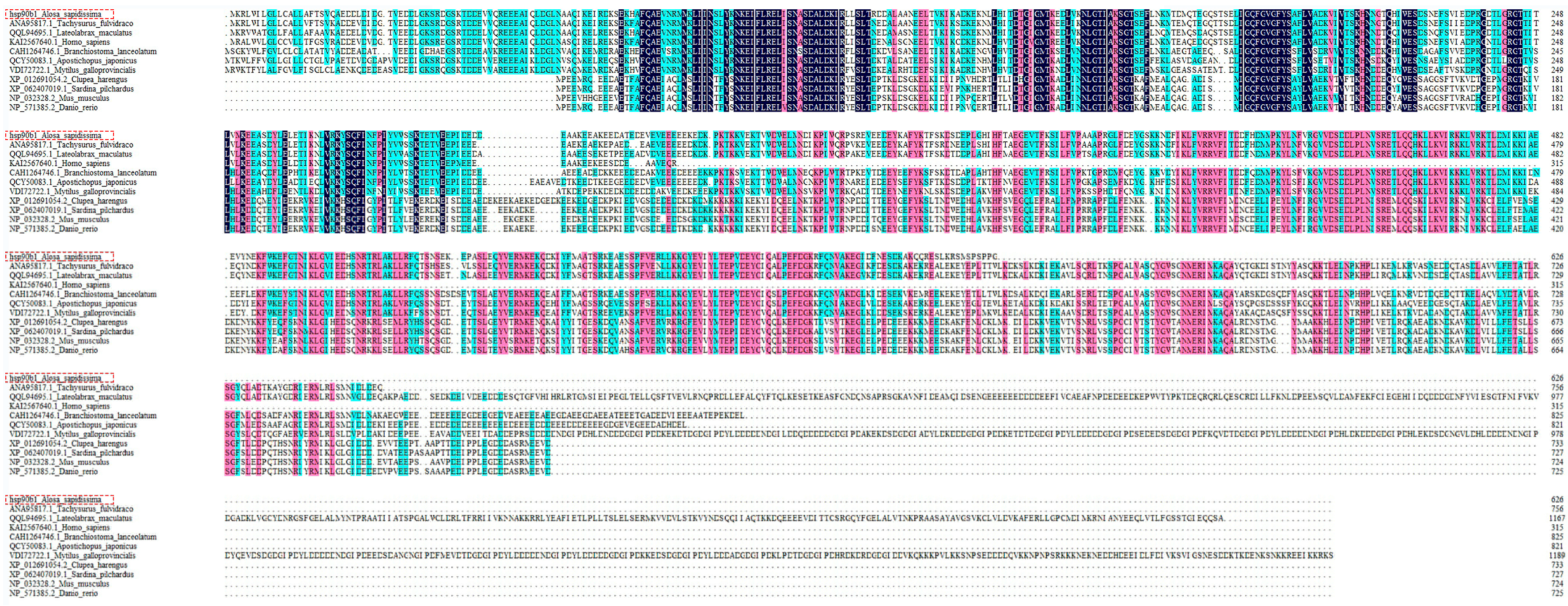
By comparing the amino acid sequence encoded by the hsp90b1 gene of the A. sapidissima with those of 10 other species, we found that the A. sapidissima has the highest homology with the Tachysurus fulvidraco at 89.71%, and a similarity of 88.10% and 85.61% with those of the Lateolabrax maculatus and Homo sapiens, respectively, and the similarity with those of the Clupea harengus, Sardina pilchardus, Mus musculus and Danio rerio was between 45% and 50% (Figure 1).

Figure 1.
Amino acid sequence alignment for A. sapidissima hsp90b1 and other species. Different colors indicate the similarity of the amino acid sequences: black: 100%; pink: 70%; cyan: 50% and more; white: below 50%. The red dashed box indicates the hsp90b1 gene information for the A. sapidissima.
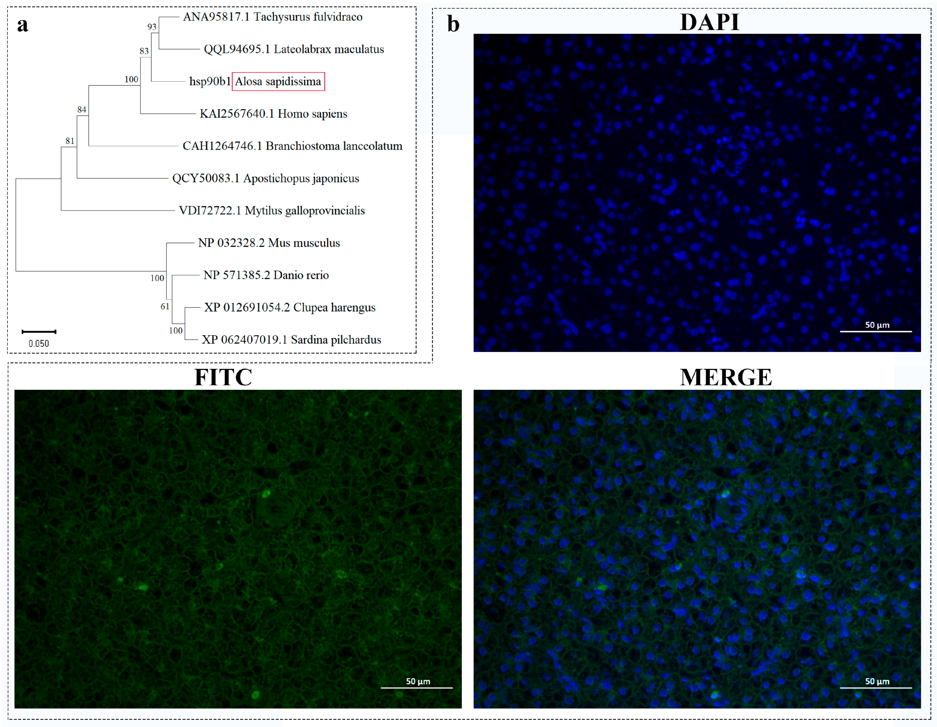
The results of the phylogenetic tree generated by the NJ method (Figure 2a) show that A. sapidissima forms a cluster with the Tachysurus fulvidraco and the Lateolabrax maculatus, indicating a close phylogenetic relationship. The function of the hsp90b1 gene is closely linked to its subcellular localization. To further clarify the role in the liver, we investigated its cellular distribution by fluorescence in situ hybridization, and the results showed that the hsp90b1 gene is mainly distributed in the cytoplasm of liver tissue cells (Figure 2b).

Figure 2.
Analysis of the sequence features of the hsp90b1 gene in the A. sapidissima. (a) Phylogenetic tree of HSP90B1 amino acid sequence of A. sapidissima. (b) Results of in situ hybridization analysis of the hsp90b1 gene in A. sapidissima (×400, 50 μm), with nuclei stained with DAPI (blue) and hsp90b1 signals stained with FITC (green). The red box represents the A. sapidissima.
2.2. Association Analysis Between miR-1236-3p and hsp90b1
Through target gene software prediction analysis, we identified two complementary sites in the 3’ UTR region of the hsp90b1 gene (...5’...GAG.GACAAGG...GGGAGAG...3’, ...5’...GGCAAG...GG.AGGAGG...3’...) that specifically bind miR-1236-3p (Figure 3a). We then performed a dual luciferase reporter assay to further test the relationship between them. By measuring the cell density of the group expressing the fluorescently labeled gene (PE-GFP), we indirectly evaluated the transfection efficiency. The results showed that the transfection efficiency was above 80% in all groups, indicating successful transfection of the plasmid (Figure S4). The results of luciferase activity detection are shown in Figure 3b. miR-146b decreased the fluorescence activity of the traf6 3’ UTR group by approximately 57% compared to miR-146b-NC, indicating that the experimental system was functioning normally. Recombinant plasmids hsp90b1-wt and hsp90b1-mut were also co-transfected into HEK 293T cells with miR-1236-3p mimic or mimic NC. Luciferase expression in the mimic NC + hsp90b1-wt and mimic NC + hsp90b1-mut groups showed no significant changes (p > 0.05). Co-transfection of miR-1236-3p-mimic with hsp90b1-wt resulted in inhibition of luciferase activity (p < 0.05), but co-transfection of miR-1236-3p mimic with hsp90b1-mut did not significantly affect luciferase activity (p > 0.05). These results suggest that miR-1236-3p is involved in the regulation of hsp90b1 expression by binding to the putative binding site in the 3’-UTR of the hsp90b1 gene.

Figure 3.
(a) Binding site analysis between hsp90b1 gene and miR-1236-3p. (b) Analysis of the target relationship of miR-1236-3p with hsp90b1 in HEK 293T cells using a dual luciferase reporter assay. Relative luciferase reporter expression was normalized to NC. Each treatment was repeated in triplicate. Data are presented as mean ± standard error. An asterisk (**) indicates p < 0.01 compared to the NC mimic, and “***” indicates p < 0.001.
2.3. Spatiotemporal Expression Properties of the hsp90b1 and miR-1236-3p
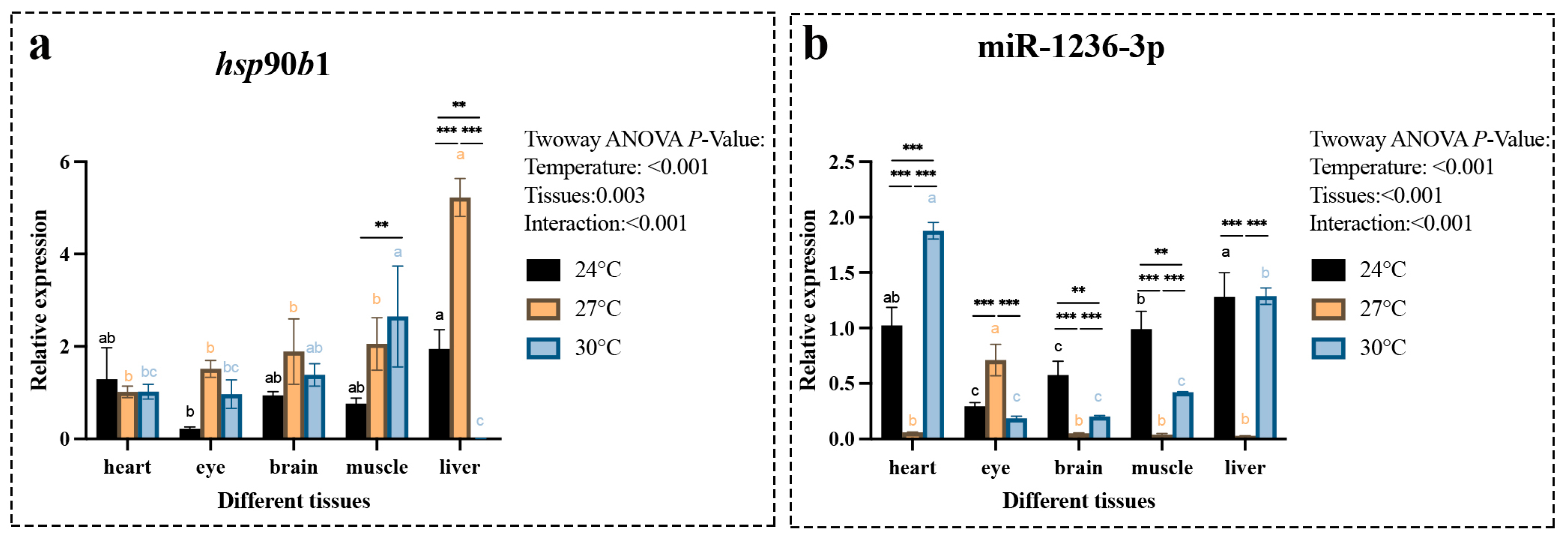
To further investigate the expression patterns of the hsp90b1 gene and miR-1236-3p, we systematically analyzed their expression levels in different tissues, temperature conditions and early developmental stages. The results showed significant differences in the overall expression trends of hsp90b1 gene and miR-1236-3p in various tissues under different temperature conditions. In particular, the expression of hsp90b1 gene was highest in liver tissue at 27 °C, followed by significantly reduced expression at 30 °C (Figure 4a). Comparison of hsp90b1 expression in different tissues at the same temperature revealed significantly higher levels in muscle tissue at 30°C compared to other tissues, while liver tissue had significantly higher expression than other tissues at 27 °C (Figure 4a). In addition, the expression of miR-1236-3p was significantly higher in heart tissue at 30 °C compared to other tissues and in liver tissue at 24 °C compared to other tissues (Figure 4b). At 27 °C, the expression of miR-1236-3p increased significantly in the eye, while it decreased extremely in the liver tissue (Figure 4b).

Figure 4.
Expression level of the hsp90b1 gene (a) and miR-1236-3p (b) in different tissues of the A. sapidissima under various temperature conditions. All parameter values represent the mean ± standard error of three replicate groups. Different letters within the same color column indicate significant differences between tissues (p < 0.05); “**” indicates significant differences (p < 0.01) within the same group across different temperature conditions, while “***” indicates p < 0.001.
We then examined the expression levels of hsp90b1 and miR-1236-3p during the major developmental stages of A. sapidissima at two different temperatures: 18 °C and 22 °C. The results showed that the gene expression of hsp90b1 peaked during the multicellular embryonic stage, followed by a significant decrease. It then increased significantly during the organogenesis stage before decreasing significantly again (Figure 5a). The expression level of miR-1236-3p remained low during the multicellular, blastula and gastrula stages. At 22 °C, it started to increase significantly during the neural crest stage. Subsequently, it gradually increased at both 18 °C and 22 °C during the organogenesis stage and reached its highest expression level during the hatching stage (Figure 5b).

Figure 5.
Expression level of the hsp90b1 gene (a) and miR-1236-3p (b) during different early developmental stages at 18 °C and 22 °C. All parameter values represent the mean ± standard error of three replicate groups. Different letters within the same color column indicate significant differences between developmental stages (p < 0.05); “*” denotes significant differences between temperature treatments at the same developmental stage (“*” means p < 0.05, “***” means p < 0.001). Multicellular stage (Mu), blastula stage (Bl), gastrula stage (Ga), neurula stage (Ne), organogenesis stage (Or) and hatching stage (Ha).
3. Discussion
Temperature is a crucial abiotic factor for aquatic organisms, affecting their embryonic development, physiological functions, survival and ability to maintain homeostasis in response to external stressors []. In view of the worsening effects of global climate change, the regulatory mechanisms of heat tolerance in aquatic organisms such as fish have become an important field of research []. Heat stress induces the production of heat shock proteins, which improve the ability of body cells to respond to external environmental stress, especially heat tolerance []. The hsp90b1 gene, a potential candidate and heat stress marker, maintains normal endoplasmic reticulum function by activating the response mechanism to unfolded proteins []. Thus, the expression of hsp90b1 was significantly increased under heat stress conditions in mice [], broiler chickens [] and rainbow trout []. Our previous studies [,] have also shown that the expression of hsp90b1 is closely correlated with temperature changes and increases significantly under high temperatures, suggesting that hsp90b1 may play an important protective role in the response of fish heat stress.
In this study, we cloned the complete cDNA sequence of the A. sapidissima hsp90b1 gene, which encodes 626 amino acids. We found that the protein encoded by this gene is a non-transmembrane protein that may be involved in biological processes such as transport and metabolism []. The secondary structure prediction suggests that the HSP90B1 protein consists of more than 50% α-helical domains. α-helices fulfill several functions in proteins: they maintain protein stability through intramolecular hydrogen bonding, ensure that proteins maintain their correct conformation under physiological conditions, and participate in protein-protein and protein-DNA interactions [,]. Furthermore, sequence alignments and phylogenetic tree between different species showed that the amino acid sequence of A. sapidissima hsp90b1 is highly conserved. Among them, we found that it is distantly related to zebrafish, which may be related to cumulative variations at key gene loci []. The results of in situ hybridization show that the hsp90b1 gene is mainly localized in the cytoplasm of liver tissue. This finding is consistent with the studies of Audouard et al. [], who reported high cytoplasmic expression of hsp90b1 in mouse cells. However, the specific molecular mechanism of its action requires further research.
Subsequently, we confirmed that miR-1236-3p can target and regulate hsp90b1 gene through bioinformatic software predictions and dual luciferase reporter assays, suggesting a potential interaction between the two genes that jointly influences the response of A. sapidissima to high-temperature stress. According to relevant studies, hsp90b1 is not only highly expressed in response to thermal stress, but is also closely associated with several intracellular signaling pathways that are critical for tumorigenesis and progression [,]. In nasopharyngeal carcinoma, for example, the expression of hsp90b1 is closely associated with tumor proliferation, glycolysis and angiogenesis. Inhibition of hsp90b1 leads to a significant reduction in tumor cell proliferation and the ability to metastasize []. Studies suggest that hsp90b1 is highly expressed in various cancers, with its overexpression often associated with poor prognosis []. Furthermore, hsp90b1 is involved as a molecular chaperone in processes such as protein refolding, environmental adaptation and immune responses and plays a fundamental role in the defense against various biotic and abiotic stress factors and in the maintenance of cellular homeostasis []. At the same time, hsp90b1 plays a crucial role in the cellular antioxidant stress response by helping cells resist oxidative damage and maintaining the stability of the intracellular environment by regulating the redox balance []. Saleh et al. [] demonstrated that silencing the hsp90 gene in Trichodina parasites significantly inhibits their growth and development. In experiments with common carp (Cyprinus carpio), spore-forming parasites treated with antisense oligonucleotides showed significantly reduced infectivity compared to the control group. This provides genetic evidence for the critical role of hsp90 in parasite growth and development and suggests that hsp90 could serve as a novel therapeutic target for successful disease control. In osteosarcoma research, qPCR analysis of miR-1236-3p expression revealed significant lower levels in osteosarcoma tissue compared to normal cartilage tissue, suggesting that miR-1236-3p may exert tumor suppressive effects []. These results indicate that hsp90b1 and miR-1236-3p play a critical role in cancer development and response to environmental stress. In addition, this study shows that both also exert key functions in heat stress responses; however, the specific molecular mechanisms and regulatory signaling pathways require further investigation.
In oviparous vertebrates, two critical events may occur between successful fertilization and hatching: sperm penetration of the egg, such as zona pellucida dissolution []; and the blastocyst stage during hatching, where embryo-derived cathepsins accelerate animal hatching []. In this study, we found that hsp90b1 exhibited the highest expression levels during the multicellular stage of embryonic development and in the liver at 27 °C. Since the multicellular stage is a critical period for neural crest differentiation, this suggests that the hsp90b1 gene may be involved in the process of neural crest differentiation []. In addition, hsp70 and hsp90 can regulate the expression of cellular heat shock proteins and are thought to contribute to abnormal embryonic development during incubation []. In this study, their expression levels were found to increase significantly when embryonic development progressed to the organogenesis stage, suggesting that HSP90B1 may be involved in the developmental processes of embryonic tissues and organs. miR-1236-3p has the highest expression levels during the hatching stage and in the heart at 30 °C. Its expression patterns during the developmental stages suggest that it plays a crucial role in the development and growth of A. sapidissima after hatching. Moreover, its expression level remains relatively high during the organ formation stage, suggesting that miR-1236-3p, like the hsp90b1 gene, may be involved in early embryonic development and play an important role in the formation of embryonic tissues and organs. These findings provide a theoretical basis for further research into the regulatory role of the hsp90b1 gene and miR-1236-3P during thermal stress in A. sapidissima.
4. Materials and Methods
4.1. Ethical Statement
This study was approved by the Bioethical Committee of the Freshwater Fisheries Research Center (FFRC) of the Chinese Academy of Fishery Sciences (CAFS) (BC2013863, 9/2013), and the fish were handled in accordance with the relevant guidelines of the Ministry of Science and Technology, Beijing, China (No. 398, 2006).
4.2. Experimental Fish and Sample Collection
The general experimental design of this study builds on our previous research on A. sapidissima []. The experimental fish were artificially bred second-year A. sapidissima obtained from the Yangzhong Base of Jiangsu Provincial Fisheries Technology Promotion Center. The water temperature was monitored throughout the experiment using a temperature control system. The different temperature gradients included a low temperature group (24.0 ± 0.5 °C), a medium temperature group (27.0 ± 0.5 °C) and a high temperature group (30.0 ± 0.5 °C). Shad of similar body size, without external injuries and with a robust body structure (average weight 354 ± 35 g) were selected. During sampling, three individuals were randomly selected from each temperature group. After anesthesia with phenoxyethanol (0.3 mL/L), liver, brain, heart, muscle and eye tissues were collected, washed with physiological saline, rapidly frozen in liquid nitrogen and brought to −80 °C for storage. In addition, two A. sapidissima were selected from the low-temperature group: one liver tissue sample was frozen in liquid nitrogen for gene cloning, and the other liver tissue sample was placed in 4% paraformaldehyde for in situ hybridization analysis. In addition, according to our previous embryonic stage sampling method [], samples were collected at the multicell (Mu), blastula (Bl), gastrula (Ga), neurula (Ne), organogenesis (Or) and hatching (Ha) stage under incubation conditions of 18 °C and 22 °C. These samples were quickly frozen in liquid nitrogen and brought to −80 °C for storage.
4.3. Cloning and In Situ Hybridization (ISH) Analysis
Based on the information about the hsp90b1 gene from previous transcriptomic data [], the open reading frame (ORF), 5′ and 3′ regions were amplified using the RACE method referring to []. Specific primers were designed using Primer Premier 5 (Table S1), and the A. sapidissima hsp90b1 gene sequence was amplified using a SMARTer™ RACE cDNA Amplification Kit (Clontech, Palo Alto, CA, USA). ProtScale (http://web.expasy.org/protscale/, 1 July 2024) was used to analyze the hydrophilicity/hydrophobicity of amino acid residues, ProtParam (https://web.expasy.org/protparam/, 10 July 2024) to predict the theoretical isoelectric point of protein molecules, DNAMAN 8.0 (Lynnon Co., Pointe-Claire, Canada) for alignment and multiple comparison of sequences and MEGA 7.0 [] to construct phylogenetic trees. The ISH probe was designed based on the cDNA sequences of the hsp90b1 gene with the sequences 5′-FAMCUUCUCCUUGUCAGCCUUAAUCUUCACGGUCAGU-3′. ISH analysis in different tissues was performed according to the previously method of Thisse et al. [].
4.4. Dual-Luciferase Reporter Assays
In our previous study [], we found by RNA-sequencing that the expression levels of miR-1236-3p and hsp90b1 in A. sapidissima liver differ at various temperatures. Here, we used TargetScan (http://www.targetscan.org, 5 July 2024) and RNAhybrid (https://omictools.com/rnahybrid-tool, 5 July 2024) to predict the binding site information. The 3’UTR region of the hsp90b1 gene was synthesized and cloned into the pmirGLO vector (Promega, Madison, WI, USA), downstream of the luciferase minigene. We then mutated the putative miRNA target sequence within the 3’UTR using the Site-Directed Mutagenesis Kit (Umibio, Shanghai, China).
Detailed information about the dual luciferase reporter assay can be found in our previous study []. More specifically, as follows: HEK 293T cells were seeded at a density of 1.0 × 10⁵ cells per well in a 48-well culture plate and cultured for 24 hours before transfection. The Lipofectamine 2000 reagent kit (Invitrogen, Carlsbad, CA, USA) was then used to co-transfect pmirGLO-hsp90b1-wt or pmirGLO-hsp90b1-mut plasmids with miR-1236-3p mimics or mimic negative controls (NC) into HEK293T cells according to the manufacturer’s instructions. In addition, miR-146b and traf6 genes were used as positive controls (PC). 48 hours after transfection, luciferase assays were performed using the Dual-Glo® Luciferase Assay System (Promega, WI, USA) according to the manufacturer’s instructions. Data collection methods were previously described in []. Renilla luciferase activity was normalized to firefly luciferase activity and expressed as a percentage of the control. Three biological replicates were used for each treatment.
4.5. RNA Isolation and Quantitative PCR
MiRNAs were extracted using a miRNeasy kit (Takara, Kyoto, Japan) and RNA was extracted using TRIzol® reagent (CWBio, Suzhou, China) according to the manufacturer’s protocol. The concentration was measured using a NanoDrop 2000UV-spectrophotometer (Thermo, Waltham, MA, USA), and the quality and integrity were checked by OD 260/280 and 1% agarose gel electrophoresis. Total RNA was reverse transcribed using Prime-Script RT Master Mix (Takara, Kyoto, Japan) and miRNAs were reverse transcribed using Mir-XTM miRNA First-strand Synthesis Kit (Takara, Kyoto, Japan). Primers were designed using Primer Premier 5 (Table S1). qRT-PCR was performed using SYBR Premix Ex TaqII (Takara, Kyoto, Japan) on a CFX-96 Real-time PCR System (Bio-Rad, Berkeley, CA, USA). All samples were run in triplicate, and the relative level of miRNAs and mRNAs was normalized to the amount of U6 and β-actin, respectively. Relative expression was calculated using the comparative threshold cycle (CT) method [], referred to as the 2−ΔΔCT algorithm.
4.6. Statistical Analysis
All results were expressed as means ± standard error of the mean. Statistical analysis was performed using SPSS 20.0 (SPSS Inc., Chicago, IL, USA). Prior to statistical analysis, all data were validated for normality (Shapiro–Wilk test) and homogeneity of variance (Levene test). The independent and interactive effects of dietary energy content and feeding regime were analyzed using a two-way ANOVA.
5. Conclusions
In this study, we successfully cloned the full-length hsp90b1 gene from A. sapidissima for the first time and found that it is mainly localized in the cytoplasm. Under heat stress conditions, the expression of hsp90b1 increases, while the expression of miR-1236-3p shows the opposite trend. Possibly, there is a negative feedback relationship between miR-1236-3p and hsp90b1, which is achieved by binding to the 3′-UTR regions of hsp90b1. Our results provide evidence for the role of miR-1236-3p in the liver and contribute to the understanding of miRNA-mediated gene regulatory mechanisms in American shad under heat stress.
Supplementary Materials
The following supporting information can be downloaded at https://www.mdpi.com/article/10.3390/ijms26209908/s1.
Author Contributions
Investigation, Formal analysis and Writing—original draft, M.L.; Investigation, Data curation, Y.L. and W.Z.; Resources, B.F. and W.X.; Investigation, Z.D. All authors have read and agreed to the published version of the manuscript.
Funding
This research was funded by the Seed Industry Revitalization Project of Jiangsu Province (JBGS [2021]131).
Institutional Review Board Statement
Not applicable.
Informed Consent Statement
Not applicable.
Data Availability Statement
The original contributions presented in this study are included in the article/Supplementary Material. Further inquiries can be directed to the corresponding author.
Acknowledgments
Our sincere thanks to Dai Fei of Jiangsu Jiuzhou Noah Agricultural Technology Co. (Changzhou City, Jiangsu Province) for providing the American shad experimental fry for her support.
Conflicts of Interest
The authors declare that they have no known competing financial interests or personal relationships that could have influenced the work in this paper.
Abbreviations
The following abbreviations are used in this manuscript:
HSP | Heat shock protein |
miRNA | MicroRNAs |
CDS | Coding sequence |
UTR | Untranslated region |
NJ | Neighbor joining |
qRT-PCR | Real-time quantitative reverse transcription polymerase chain reaction |
References
- Yadav, N.K.; Patel, A.B.; Singh, S.K.; Mehta, N.K.; Anand, V.; Lal, J.; Dekari, D.; Devi, N.C. Climate change effects on aquaculture production and its sustainable management through climate-resilient adaptation strategies: A review. Environ. Sci. Pollut. Res. Int. 2024, 31, 31731–31751. [Google Scholar] [CrossRef]
- Alfonso, S.; Gesto, M.; Sadoul, B. Temperature increase and its effects on fish stress physiology in the context of global warming. J. Fish. Biol. 2021, 98, 1496–1508. [Google Scholar] [CrossRef]
- Mugwanya, M.; Dawood, M.A.; Kimera, F.; Sewilam, H. Anthropogenic temperature fluctuations and their effect on aquaculture: A comprehensive review. Aquac. Fish. 2022, 7, 223–243. [Google Scholar] [CrossRef]
- Soyano, K.; Mushirobira, Y. The Mechanism of Low-Temperature Tolerance in Fish. Adv. Exp. Med. Biol. 2018, 1081, 149–164. [Google Scholar]
- Dahms, C.; Killen, S.S. Temperature change effects on marine fish range shifts: A meta-analysis of ecological and methodological predictors. Glob. Change Biol. 2023, 29, 4459–4479. [Google Scholar] [CrossRef]
- Lee, D.; Kim, K.H.; Park, J.W.; Lee, J.H.; Kim, J.H. High water temperature-mediated immune gene expression of olive flounder, Paralichthys olivaceus according to pre-stimulation at high temperatures. Environ. Toxicol. Pharmacol. 2023, 101, 104159. [Google Scholar] [CrossRef] [PubMed]
- Nobrega, R.O.; Batista, R.O.; Corrêa, C.F.; Mattioni, B.; Filer, K.; Pettigrew, J.E.; Fracalossi, D.M. Dietary supplementation of Aurantiochytrium sp. meal, a docosahexaenoic-acid source, promotes growth of Nile tilapia at a suboptimal low temperature. Aquaculture 2019, 507, 500–509. [Google Scholar] [CrossRef]
- Fang, M.; Lei, Z.; Ruilin, M.; Jing, W.; Leqiang, D. High temperature stress induced oxidative stress, gut inflammation and disordered metabolome and microbiome in tsinling lenok trout. Ecotoxicol. Environ. Saf. 2023, 266, 115607. [Google Scholar] [CrossRef] [PubMed]
- Jeyachandran, S.; Chellapandian, H.; Park, K.; Kwak, I.S. A Review on the Involvement of Heat Shock Proteins (Extrinsic Chaperones) in Response to Stress Conditions in Aquatic Organisms. Antioxidants 2023, 12, 1444. [Google Scholar] [CrossRef] [PubMed]
- Padmini, E.; Usha Rani, M. Impact of seasonal variation on HSP70 expression quantitated in stressed fish hepatocytes. Comp. Biochem. Physiol. B Biochem. Mol. Biol. 2008, 151, 278–285. [Google Scholar] [CrossRef]
- Zhang, W.; Qian, Z.; Ji, J.; Wang, T.; Yin, S.; Zhang, K. Characterization of HSP70 and HSP90 Gene Family in Takifugu fasciatus and Their Expression Profiles on Biotic and Abiotic Stresses Response. Genes 2024, 15, 1445. [Google Scholar] [CrossRef]
- Liu, Y.; Liang, Z.; Li, Y.; Zhu, W.; Feng, B.; Xu, W.; Fu, J.; Wei, P.; Luo, M.; Dong, Z. Integrated transcriptome and microRNA analysis reveals molecular responses to high-temperature stress in the liver of American shad (Alosa sapidissima). BMC Genom. 2024, 25, 656. [Google Scholar] [CrossRef] [PubMed]
- Higgs, D.M.; Plachta, D.T.; Rollo, A.K.; Singheiser, M.; Hasting, M.C.; Popper, A.N. Development of ultrasound detection in American shad (Alosa sapidissima). J. Exp. Biol. 2004, 207, 155–163. [Google Scholar] [CrossRef]
- Li, L.; Zhu, H.J.; Yi, X.Y.; Nie, Z.J.; Zheng, Y.; Yang, X.W.; Xu, P.; Yu, Y.; Xu, G. Study of the Quality and Nutritional Value of Alosa sapidissima in the Postmortem Process. Fishes 2022, 7, 302. [Google Scholar] [CrossRef]
- Gao, X.Q.; Liu, Z.F.; Guan, C.T.; Huang, B.; Lei, J.L.; Li, J.; Guo, Z.-L.; Wang, Y.-H.; Hong, L. Developmental changes in digestive enzyme activity in American shad, Alosa sapidissima, during early ontogeny. Fish Physiol. Biochem. 2017, 43, 397–409. [Google Scholar] [CrossRef]
- Liang, Z.; Miao, L.; Liu, Y.; Luo, M.; Feng, B.; Zhu, W.; Gong, Y.; Shi, X.; Li, Y.; Jiang, C.; et al. Effect of temperature on the embryonic development of American shad (Alosa sapidissima). J. Fish. China 2025, 49, 089104. (In Chinese) [Google Scholar]
- Luo, M.; Zhu, W.; Liang, Z.; Feng, B.; Xie, X.; Li, Y.; Liu, Y.; Shi, X.; Fu, J.; Miao, L.; et al. High-temperature stress response: Insights into the molecular regulation of American shad (Alosa sapidissima) using a multi-omics approach. Sci. Total Environ. 2024, 916, 170329. [Google Scholar] [CrossRef]
- Luo, M.; Feng, B.; Zhu, W.; Liang, Z.; Xu, W.; Fu, J.; Miao, L.; Dong, Z. Proteomics and metabolomics analysis of American shad (Alosa sapidissima) liver responses to heat stress. Comp. Biochem. Physiol. A Mol. Integr. Physiol. 2024, 296, 111686. [Google Scholar] [CrossRef] [PubMed]
- Tafrihi, M.; Hasheminasab, E. MiRNAs: Biology, Biogenesis, their Web-based Tools, and Databases. Microrna 2019, 8, 4–27. [Google Scholar] [CrossRef]
- Bizuayehu, T.T.; Babiak, I. MicroRNA in teleost fish. Genome Biol. Evol. 2014, 6, 1911–1937. [Google Scholar] [CrossRef]
- Dweep, H.; Showe, L.C.; Kossenkov, A.V. Functional Annotation of MicroRNAs Using Existing Resources. Methods Mol. Biol. 2022, 2257, 57–77. [Google Scholar]
- Gavarikar, S.; Craig, P.M. Developmental exposure to constant elevated temperatures and diel thermal variation alters microRNA expression and performance in zebrafish (Danio rerio). Comp. Biochem. Physiol. B Biochem. Mol. Biol. 2025, 279, 111122. [Google Scholar] [CrossRef]
- Liu, Z.; Ma, F.; Kang, Y.; Liu, X. Gene ssa-miR-301a-3p improves rainbow trout (Oncorhynchus mykiss) resistance to heat stress by targeting hsp90b2. PeerJ 2022, 10, e13476. [Google Scholar] [CrossRef] [PubMed]
- Zaman, T.; Fahad, T.M.; Rana, M.; Hossain, M.S.; Mamun, A.; Haque, M.A.; Sarker, A.; Islam, S.; Haque, M.-U.L.; Naz, T.; et al. Endosulfan affects embryonic development synergistically under elevated ambient temperature. Environ. Sci. Pollut. Res. Int. 2023, 30, 73393–73404. [Google Scholar] [CrossRef]
- Lim, M.Y.; Manzon, R.G.; Somers, C.M.; Boreham, D.R.; Wilson, J.Y. Impacts of temperature, morpholine, and chronic radiation on the embryonic development of round whitefish (Prosopium cylindraceum). Environ. Toxicol. Chem. 2018, 37, 2593–2608. [Google Scholar] [CrossRef] [PubMed]
- Zininga, T.; Ramatsui, L.; Shonhai, A. Heat Shock Proteins as Immunomodulants. Molecules 2018, 23, 2846. [Google Scholar] [CrossRef]
- Miao, Q.X.; Si, X.Y.; Xie, Y.J.; Chen, L.; Tang, X.F.; Zhang, H.F. Acute heat stress alters the expression of genes and proteins associated with the unfolded protein response pathway in the liver of broilers. Br. Poult. Sci. 2022, 63, 125–132. [Google Scholar] [CrossRef]
- Guo, H.; Yang, Y.; Qiao, Y.; He, J.; Yao, W.; Zheng, W. Heat stress affects fetal brain and intestinal function associated with the alterations of placental barrier in late pregnant mouse. Ecotoxicol. Environ. Saf. 2021, 227, 112916. [Google Scholar] [CrossRef]
- Barreto Sánchez, A.L.; Wang, Q.; Thiam, M.; Zhang, J.; Zhang, Q.; Zhang, N.; Li, Q.; Wen, J.; Zhao, G. Liver Transcriptome Response to Heat Stress in Beijing You Chickens and Guang Ming Broilers. Genes 2022, 13, 416. [Google Scholar] [CrossRef] [PubMed]
- Quan, J.; Kang, Y.; Li, L.; Zhao, G.; Sun, J.; Liu, Z. Proteome analysis of rainbow trout (Oncorhynchus mykiss) liver responses to chronic heat stress using DIA/SWATH. J. Proteom. 2021, 233, 104079. [Google Scholar] [CrossRef]
- Yu, H.; Yang, Z.; Sui, M.; Cui, C.; Hu, Y.; Hou, X.; Xing, Q.; Huang, X.; Bao, Z. Identification and Characterization of HSP90 Gene Family Reveals Involvement of HSP90, GRP94 and Not TRAP1 in Heat Stress Response in Chlamys farreri. Genes 2021, 12, 1592. [Google Scholar] [CrossRef]
- Giménez-Andrés, M.; Čopič, A.; Antonny, B. The Many Faces of Amphipathic Helices. Biomolecules 2018, 8, 45. [Google Scholar] [CrossRef]
- Zhang, Y.; Liu, Y.; Liu, Y.; Zuo, P.; Miao, S.; Hu, B.; Kang, Y.; Liu, W.; Yang, Q.; Ren, H.; et al. α-Helix-Mediated Protein Adhesion. J. Am. Chem. Soc. 2023, 145, 17125–17135. [Google Scholar] [CrossRef]
- Liu, J.; Wang, Z.; Hou, Y.P.; Liao, L.C. The Impact of STR Mutations on Kinship Identification. Fa Yi Xue Za Zhi 2024, 40, 484–491. [Google Scholar]
- Audouard, C.; Le Masson, F.; Charry, C.; Li, Z.; Christian, E.S. Oocyte-targeted deletion reveals that hsp90b1 is needed for the completion of first mitosis in mouse zygotes. PLoS ONE 2011, 6, e17109. [Google Scholar] [CrossRef]
- Hu, Y.; Yu, C.; Cheng, L.; Zhong, C.; An, J.; Zou, M.; Liu, B.; Gao, X. Flavokawain C inhibits glucose metabolism and tumor angiogenesis in nasopharyngeal carcinoma by targeting the HSP90B1/STAT3/HK2 signaling axis. Cancer Cell Int. 2024, 24, 158. [Google Scholar] [CrossRef]
- Wang, Y.; Wang, X. A Pan-Cancer Analysis of Heat-Shock Protein 90 Beta1 (HSP90B1) in Human Tumours. Biomolecules 2022, 12, 1377. [Google Scholar] [CrossRef] [PubMed]
- Chen, B.; Huang, Y.; He, S.; Yu, P.; Wu, L.; Peng., H. N6-methyladenosine modification in 18S rRNA promotes tumorigenesis and chemoresistance via HSF4b/HSP90B1/mutant p53 axis. Cell Chem. Biol. 2023, 30, 144–158. [Google Scholar] [CrossRef] [PubMed]
- Chen, M.; Chen, Y. Bioinformatics Analysis of Common Genetic and Molecular Traits and Association of Portal Hypertension with Pulmonary Hypertension. J. Healthc. Eng. 2022, 2022, 9237701. [Google Scholar] [CrossRef] [PubMed]
- Dong, W.H.; Liu, S.Y.; Ren, Y.P. Evolution and function of heat shock protein 90 in economic shellfish: A review. Dev. Comp. Immunol. 2025, 170, 105456. [Google Scholar] [CrossRef]
- Stanley, T.R.; Guisbert, K.S.K.; Perez, S.M.; Oneka, M.; Kernin, I.; Higgins, N.R.; Lobo, A.; Subasi, M.M.; Carroll, D.J.; Turingan, R.G.; et al. Stress response gene family expansions correlate with invasive potential in teleost fish. J. Exp. Biol. 2022, 225, jeb243263. [Google Scholar] [CrossRef] [PubMed]
- Saleh, M.; Abdel-Baki, A.S.; Dkhil, M.A.; El-Matbouli, M.; Al-Quraishy, S. Silencing of heat shock protein 90 (hsp90): Effect on development and infectivity of Ichthyophthirius multifiliis. BMC Vet. Res. 2023, 19, 62. [Google Scholar] [CrossRef]
- Li, J.; Chen, J.; Hu, Z.; Xu, W. MicroRNA-1236-3p inhibits human osteosarcoma growth. Oncol. Lett. 2020, 20, 367. [Google Scholar] [CrossRef] [PubMed]
- Trikić, M.Z.; Monk, P.; Roehl, H.; Partridge, L.J. Regulation of zebrafish hatching by tetraspanin cd63. PLoS ONE 2011, 6, e19683. [Google Scholar] [CrossRef]
- Tokuhiro, K.; Dean, J. Glycan-Independent Gamete Recognition Triggers Egg Zinc Sparks and ZP2 Cleavage to Prevent Polyspermy. Dev. Cell 2018, 46, 627–640. [Google Scholar] [CrossRef]
- Myers, S.M.; Mulligan, L.M. The RET receptor is linked to stress response pathways. Cancer Res. 2004, 64, 4453–4463. [Google Scholar] [CrossRef]
- Xiong, X.; Luo, S.; Wu, B.; Wang, J. Comparative Developmental Toxicity and Stress Protein Responses of Dimethyl Sulfoxide to Rare Minnow and Zebrafish Embryos/Larvae. Zebrafish 2017, 14, 60–68. [Google Scholar] [CrossRef]
- Liang, Z.; Feng, B.; Miao, L.; Zhu, W.; Lin, Y.; Luo, M.; Yang, S.; Wang, L.; Fu, J.; Chen, H.; et al. Combined Illumina and Pacbio sequencing technology on transcriptome analysis reveals several key regulations during the early development of American shad (Alosa sapidissima). Aquacult. Rep. 2022, 25, 101264. [Google Scholar] [CrossRef]
- Wang, L.M.; Bu, H.Y.; Song, F.B.; Zhu, W.B.; Fu, J.J.; Dong, Z.J. Characterization and functional analysis of slc7a11 gene, involved in skin color differentiation in the red tilapia. Comp. Biochem. Physiol. A Mol. Integr. Physiol. 2019, 236, 110529. [Google Scholar] [CrossRef]
- Kumar, S.; Stecher, G.; Tamura, K. MEGA7: Molecular Evolutionary Genetics Analysis Version 7.0 for Bigger Datasets. Mol. Biol. Evol. 2016, 33, 1870–1874. [Google Scholar] [CrossRef]
- Thisse, C.; Thisse, B. High-resolution in situ hybridization to whole-mount zebrafish embryos. Nat. Protoc. 2008, 3, 59–69. [Google Scholar] [CrossRef] [PubMed]
- Dong, Z.; Luo, M.; Wang, L.; Yin, H.; Zhu, W.; Fu, J. MicroRNA-206 Regulation of Skin Pigmentation in Koi Carp (Cyprinus carpio L.). Front. Genet. 2020, 11, 47. [Google Scholar] [CrossRef] [PubMed]
- Liu, X.; Wang, Y.; Liu, Z.; Kang, Y.; Ma, F.; Luo, Z.; Wang, J.; Huang, J. miR-434 and miR-242 have a potential role in heat stress response in rainbow trout (Oncorhynchus mykiss). J. Fish. Biol. 2021, 99, 1798–1803. [Google Scholar] [CrossRef] [PubMed]
- Livak, K.J.; Schmittgen, T.D. Analysis of relative gene expression data using real-time quantitative PCR and the 2(-Delta Delta C(T)) Method. Methods 2001, 25, 402–408. [Google Scholar] [CrossRef]
Disclaimer/Publisher’s Note: The statements, opinions and data contained in all publications are solely those of the individual author(s) and contributor(s) and not of MDPI and/or the editor(s). MDPI and/or the editor(s) disclaim responsibility for any injury to people or property resulting from any ideas, methods, instructions or products referred to in the content. |
© 2025 by the authors. Licensee MDPI, Basel, Switzerland. This article is an open access article distributed under the terms and conditions of the Creative Commons Attribution (CC BY) license (https://creativecommons.org/licenses/by/4.0/).