Article

Unscheduled m6A Deposition in RNA via m6ATP Incorporation by DNA Polymerases

1
Biochemistry Ph.D. Program, Florida International University, Miami, FL 33199, USA
2
Department of Physics, Florida International University, Miami, FL 33199, USA
3
Biomolecular Sciences Institute, Florida International University, Miami, FL 33199, USA
4
Department of Chemistry and Biochemistry, Florida International University, Miami, FL 33199, USA
*
Author to whom correspondence should be addressed.
Int. J. Mol. Sci. 2025, 26(19), 9263; https://doi.org/10.3390/ijms26199263
Submission received: 5 August 2025 / Revised: 18 September 2025 / Accepted: 21 September 2025 / Published: 23 September 2025

Abstract

N6-methyladenosine (m6A) is the most abundant modification of mRNA and plays a crucial role in mediating cellular functions, and it is associated with cancer and neurodegenerative diseases. Studies have shown that m6A is predominantly deposited on its consensus motif by the m6A writer proteins RNA methyltransferase METLL3/METLL14. However, it was found that nonconventional m6A deposition by other alternative pathways may also exist and can modulate epitranscriptomic regulation in cells. Thus, understanding the molecular mechanisms underlying nonconventional m6A deposition outside the canonical motifs will provide novel insights into the full scope of the functional impact of m6A. In this study, we discovered that m6ATP was efficiently incorporated by the repairing DNA polymerases pol β and pol η through RNA gap-filling synthesis on an RNA-DNA hybrid. Steady-state kinetics results showed that m6ATP was incorporated into RNA by the DNA polymerases with a comparable efficiency to ATP. AlphaFold3-assisted molecular dynamics simulations further elucidated the structural basis for the DNA polymerases to incorporate m6ATP into the RNA substrates by showing that the enzymes employed the unique base-stacking mechanism to govern the distance between the 3′-OH group of the 3′-terminus nucleotide of the primer and the 5′-α-phosphate of m6ATP to perform their catalysis. Furthermore, we detected a significant amount of m6ATP in human cells. We showed that the m6ATP level was associated with that of the oxidative stress biomarker 8-oxoGTP in cells, suggesting that unscheduled m6A deposition on RNA can be mediated by m6ATP incorporation that is associated with cellular oxidative stress. Our study sheds light on the unscheduled m6A deposition as a potential alternative mechanism for altering epitranscriptomic modifications.

1. Introduction

m6A is the most abundant internal modification identified in the eukaryotic mRNA [1]. m6A plays a central role in regulating gene expression by modulating RNA alternative splicing [2], stability [3,4], nuclear export [5], and protein translation [6]. Thus, as a critical epitranscriptional marker, m6A is involved in the regulation of cellular stress responses [7,8], disease status [9,10,11], and cancer development [12,13]. It has been shown that m6A exists in all the regions of mRNA but preferentially accumulates around the translation start site and the stop codons [14]. m6A-seq has revealed that most m6A is found within the consensus m6A motif, R/DRACH (1), in which R/D represents G or A, and H represents U or C or A. However, studies have shown that the m6A motifs are degenerate, with multiple variations occurring at the +/− 1, 2, and 3 positions [1,14,15,16]. Moreover, it was shown that m6ATP can also be deposited by another m6A writer, METTL16, at a different motif of UACAGAGAA [17]. All these findings suggest that there are diversified alternative pathways for noncanonical m6A deposition on RNA through the pathways mediated by known or unknown m6A methyltransferases, as well as non-methyltransferase-dependent mechanisms. It was shown that the locations of m6A play an essential role in regulating cellular functions. For example, m6A in the 5′-UTR can facilitate cap-independent translation by recruiting a specific m6A reader, eukaryotic initiation factor 3 (eIF3) [18], whereas m6A located around the stop codons and in the 3′-UTR is recognized by the m6A reader, YTHDF2, mediating RNA decay [3]. While m6A deposition by the RNA methyltransferases and m6A writer METTL3/METTL14 exhibits a preference for the R/DRACH motifs [19], the presence of m6A in noncanonical sites in RNA raises intriguing questions about whether there are alternative mechanisms for m6A RNA deposition. Understanding its underlying mechanisms is crucial for elucidating the entire regulatory role of m6A RNA modifications in cellular processes.
Previous studies have revealed that wild-type and mutant DNA polymerases can incorporate ribonucleoside triphosphates (rNTPs) into DNA through their compromised ability to exclude rNTPs [20]. Gao et al. have further demonstrated that the mutant MMLV reverse transcriptase can acquire RNA polymerase activity to incorporate rNTPs into RNA [21]. Recent studies have shown that pol η, a translesion synthesis DNA polymerase [22], which is essential in assisting replication DNA polymerases to bypass bulky DNA lesions during DNA replication [23], also exhibits RNA synthesis activity [24]. This unexpected capability of DNA polymerases to incorporate ribonucleotides suggests a broader spectrum of substrate recognition of repairing DNA polymerases and their functional flexibility and plasticity in processing both deoxyribonucleotides and ribonucleotides, mediating DNA repair, and potentially processing RNA damage intermediates, especially on RNA-DNA hybrids formed during gene transcription and DNA lagging-strand replication. Furthermore, it was found that RNA synthesis activity by pol η can be significantly increased by manganese (Mn2+) compared with its RNA synthesis activity in the presence of Mg2+ [25,26]. This further raises the question of whether repairing DNA polymerases can also incorporate m6ATP, the methylated ATP, into RNA through their RNA synthesis activity on DNA-RNA hybrids.
Although both the divalent metals Mg2+ and Mn2+ are essential cofactors for the catalysis of nucleotide incorporation of DNA polymerases [27], Mg2+ is the physiologically relevant metal cofactor for DNA polymerases [28,29]. This is because Mg2+ is abundant in cells and can provide the optimal conditions for efficient enzymatic activity of DNA polymerases and the high fidelity of DNA synthesis [30]. On the other hand, although Mn2+ is an essential trace metal in cells [28,29], at the level where it can significantly increase DNA polymerase activities, Mn2+ can also substantially reduce the stringency of nucleotide selection and fidelity of DNA polymerases, thereby promoting the misincorporation of nucleotides and mismatches [31]. This further suggests that Mn2+ can modulate the structures of DNA polymerases, making the enzymes more adaptable to various types of modifications of nucleotides, such as different configurations of ribose and base modifications. However, it is unknown if Mn2+ can cause pol η to incorporate the modified ribonucleotide m6ATP and if the metal can also induce the structural changes of the high-fidelity DNA repair polymerase, DNA polymerase β (pol β), the central component of the DNA base excision repair (BER) pathway [32,33], which also possesses deoxyribose-5-phosphate (5′-dRP) lyase activity [33], conferring pol β RNA synthesis activity and its incorporation of modified ribonucleotides such as m6ATP. Understanding the incorporation of m6ATP by the repairing DNA polymerases and the underlying mechanism can open a new avenue to reveal an alternative mechanism for m6A RNA deposition, in addition to the canonical m6A deposition at the R/DRACH motif by RNA methyltransferases. Therefore, it is crucial for deciphering the potential mechanisms of m6A deposition beyond its canonical methylation by the writer enzymes. We further hypothesized that repairing DNA polymerases such as pol β and pol η can incorporate m6ATP in the presence of Mg2+ and Mn2+. To test this hypothesis, we initially determined the activity of pol β and pol η in incorporating ATP and m6ATP into RNA using Mg2+ or Mn2+ as a cofactor. We then compared the efficiency of m6ATP incorporation with that of ATP by pol β and pol η using steady-state enzyme kinetics. We further revealed the structural basis of the incorporation of m6ATP by pol β and pol η with Mg2+ and Mn2+ using molecular modeling and molecular dynamics (MD) simulations. Finally, we determined the level of m6ATP in human cells. Our results showed that pol β only incorporated m6ATP in the presence of Mn2+ with high efficiency, whereas pol η efficiently incorporated m6ATP in the presence of both Mg2+ and Mn2+.

2. Results

2.1. Pol β Can Incorporate m6ATP into RNA in the Presence of Mn2+

Since previous studies have shown that pol η can synthesize RNA [24,25,26], we further hypothesized that RNA gaps resulting from oxidative RNA damage on RNA-DNA hybrids can allow the incorporation of m6ATP into RNA by the repairing DNA polymerases. To test this possibility, we initially determined pol β m6ATP and ATP incorporation into the 1 nt RNA gap and open-template substrates in the presence of 5 mM Mg2+. We found that pol β failed to incorporate either ATP or m6ATP on the gapped or open-template RNA in the presence of Mg2+ s. Since the previously reported structures of pol β with Mg2+ or Mn2+ have revealed that pol β adopts a more open structure in the presence of Mn2+ [34], we then tested whether pol β can incorporate m6ATP into RNA in the presence of Mn2+. The results showed that pol β incorporated m6ATP in both gapped and open-template RNA in the presence of 1 mM Mn2+ (Figure 1). Pol β at 50–200 nM inserted only one ATP or m6ATP into the 1 nt RNA gap substrate, with a much higher amount of ATP incorporation product (Figure 1A, lanes 2–4) than that of m6ATP (Figure 1A, lanes 6–8). On the open RNA template substrate, the same concentrations of pol β inserted two ATP or m6ATP (Figure 1B), with much more ATP incorporation products generated (Figure 1B lanes 2–4) than m6ATP incorporation products (Figure 1B, lanes 6–8). This indicated that m6A was extended by additional m6A incorporation in the presence of Mn2+. The results showed that m6ATP could be incorporated by pol β only in the presence of Mn2+, with a lower efficiency compared with its ATP incorporation. Moreover, we found that the extension of m6A by pol β resulted in m6A:G mismatch.

2.2. Incorporation of m6ATP into RNA by Pol η

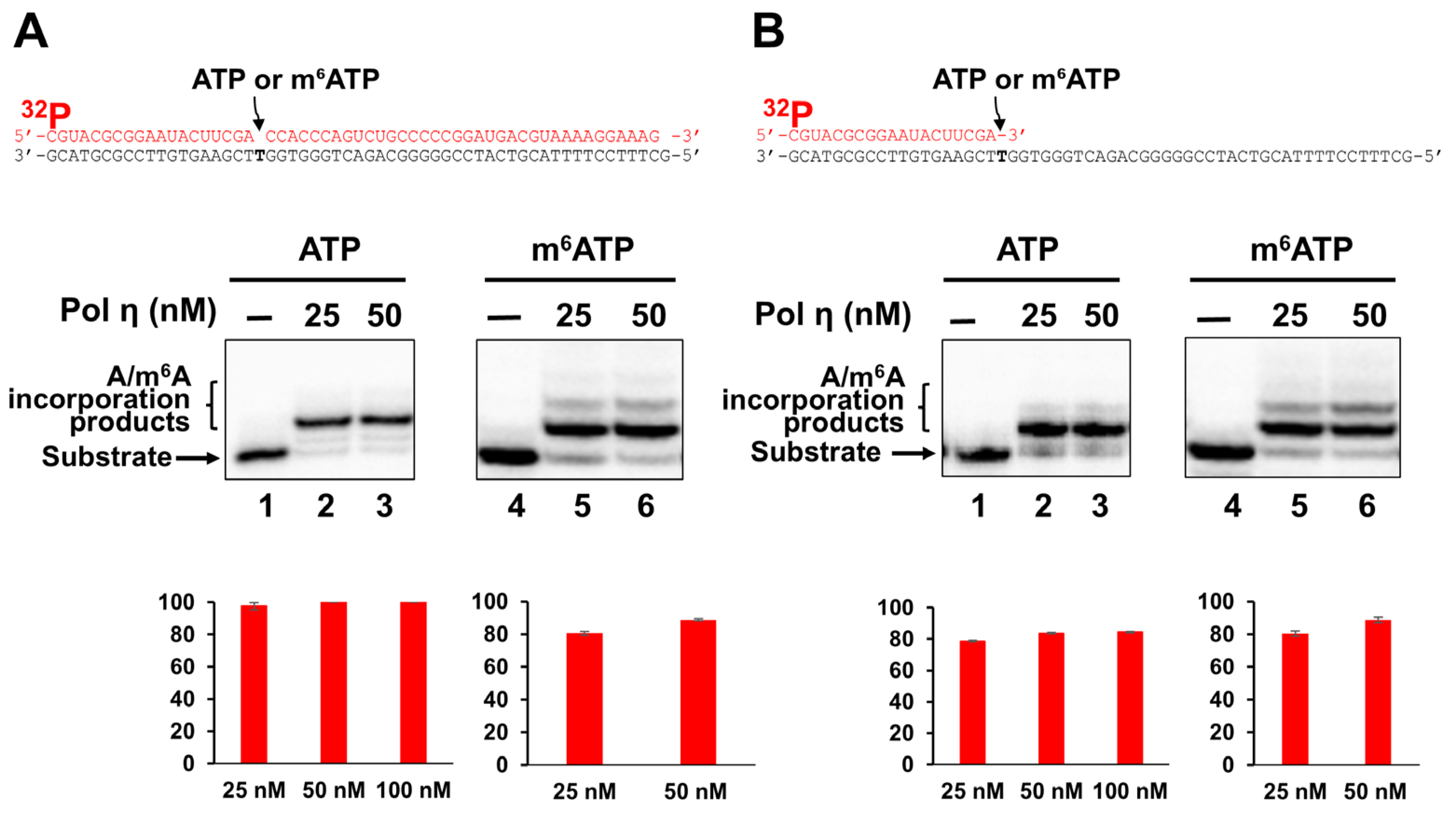
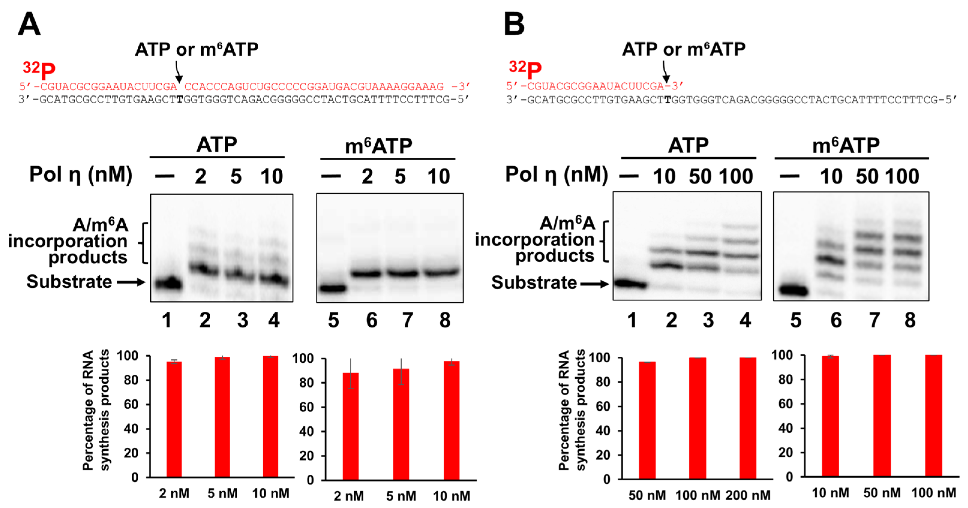
We next asked if pol η can also incorporate m6ATP into RNA. As a translesion DNA synthesis polymerase, pol η is known for its structural flexibility and tolerance of diverse nucleotides and DNA damage. Thus, we reasoned that pol η exhibits the ability to incorporate m6ATP in the presence of Mg2+. To test this, we examined the pol η incorporation of m6ATP on the 1 nt RNA gap and open-template RNA substrates in the presence of Mg2+ and Mn2+ and compared the incorporation activity with that of ATP incorporation (Figure 2). The results indicated that pol η at 25–50 nM efficiently incorporated ATP and m6ATP into both the 1 nt gap (Figure 2A, lanes 2–3 and 5–6) and open-template RNA substrates (Figure 2B, lanes 2–3 and 5–6) in the presence of Mg2+. Moreover, with both the RNA substrates, pol η extended m6A by incorporating additional m6A, creating a small amount of m6A:dG base mispairing product (Figure 2A, 2B, lanes 5–6). In the presence of Mn2+, pol η incorporated ATP and m6ATP in both gapped and open-template RNA efficiently (Figure 3). Surprisingly, on the RNA gapped substrate, pol η incorporation of ATP but not m6ATP also led to a small amount of A:dG mismatch (Figure 3A, compare lanes 2–4 with lanes 6–8). On the RNA open-template substrate, pol η generated multiple nucleotide misincorporation products with both ATP and m6ATP (Figure 3B, lanes 2–4 and 6–8), indicating a significantly reduced fidelity of ATP and m6ATP incorporation.

2.3. The Catalytic Efficiency of m6ATP Incorporation by Pol β and Pol η

Using steady-state kinetics, we further compared the efficiency of pol β and pol η incorporation of m6ATP with that of ATP in the presence of Mg2+ and Mn2+ (Figure 4). Given the fact that pol β and pol η preferential substrates are 1 nt gapped and open-template substrates, respectively, we performed the steady-state kinetics studies on the RNA synthesis by DNA polymerases. The results showed that pol β exhibited similar catalytic efficiency (kcat/Km) in inserting ATP and m6ATP, shown as 6.89 × 10⁻⁴ μM−1 min−1 for ATP and 4.49 × 10⁻⁴ μM−1 min−1 for m6ATP in the presence of Mn2+ (Figure 4A). In contrast, in the presence of 5 mM Mg2+, the efficiency of pol η incorporation of ATP on the RNA open template exhibited more than four-fold higher catalytic efficiency than that of m6ATP (Figure 4B). However, in the presence of 1 mM Mn2+, the catalytic efficiency of ATP and m6ATP incorporation by pol η was similar, with 6333.3 × 10−4 μM−1 min−1 for ATP incorporation and 5727.3 × 10−4 μM−1 min−1 for m6ATP incorporation. The results indicated that the presence of Mn2+ significantly stimulated the catalysis of m6ATP incorporation by pol η. The catalytic efficiency of pol η m6ATP incorporation was increased by nearly 200-fold by Mn2+ (compare the kcat/Km for ATP and m6ATP in panel B with kcat/Km of the nucleotides in panel C). The steady-state kinetics results revealed that pol η inserted m6ATP into the RNA open-template substrate with high efficiency, indicating that m6ATP was efficiently incorporated by pol η in the presence of Mg2+ and Mn2+.

2.4. The Structural Basis of m6ATP RNA Incorporation by Pol β and Pol η in the Presence of Mg2+ and Mn2+

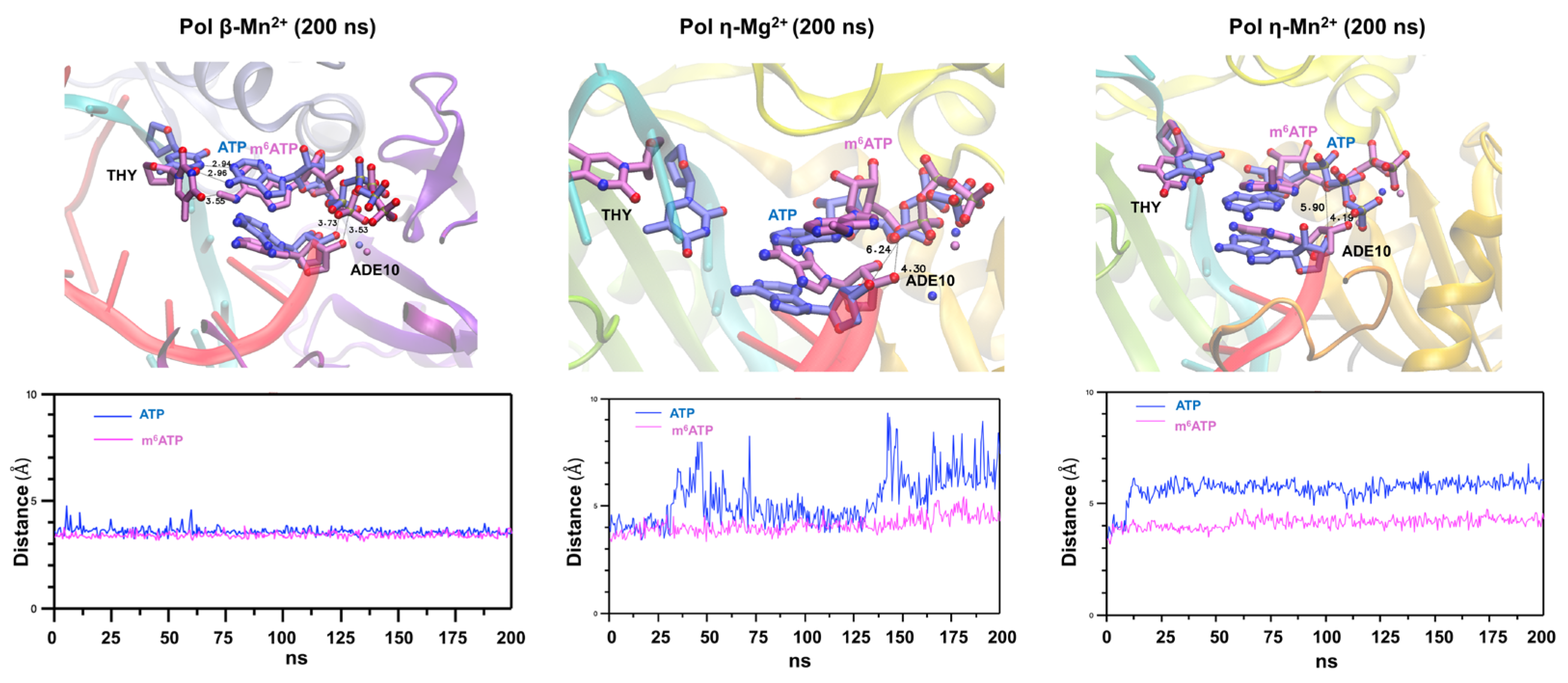
To further reveal the structural basis of the incorporation of m6ATP into RNA by pol β and pol η in the presence of Mn2+ or Mg2+, we conducted an AlphaFold3-assisted molecular dynamics (MD) simulation of m6ATP and ATP incorporation by the DNA polymerases using pol β (PDB ID 5TBB) [35] and pol η crystal structures (PDB ID 4J9N) [36], with the DNA sequences of the substrates replaced with the DNA and RNA sequences used in this study. We superimposed the structures of enzyme–substrate ternary complexes with m6ATP and ATP for both pol β and pol η to elucidate the molecular mechanisms underlying the different nucleotide incorporation efficiency by pol β and pol η in the presence of Mn2+ or Mg2+ (Figure 5). The results showed that pol β adopted a different conformation with m6ATP from that with ATP in the DNA-RNA-pol β-m6A/ATP ternary complex (Figure 5, the left panel). In the pol β-ATP-RNA substrate-Mn2+ ternary complex, ATP exhibited its base pair with the template T (Figure 5, the left panel). Surprisingly, m6ATP failed to base pair with the template T. Instead, it employed the purine ring to interact with the part of the purine ring of the adenosine at the 3′-terminus of the upstream primer, suggesting a base-stacking-like interaction (Figure 5, the left panel). This led to the almost identical distance between the 3′-OH group and α phosphate of 3.53 Å (ATP) and 3.75 Å (m6ATP). The RMSD analysis showed that the distance was stabilized between 3.5 Å and 4.5 Å (Figure 5, the left panel, the RMSD graph below the structure). On the other hand, in the pol η-substrate-ATP complex in the presence of Mg2+, ATP formed a hydrogen bond with the template T, resulting in 6.24 Å between the 3′-OH group of the RNA primer and the α phosphate of ATP (Figure 5, the panel in the middle). However, similar to the pol β-m6ATP ternary complex, m6ATP also failed to base pair with the template T in the pol η-m6ATP ternary complex. Instead, it exhibited a partial base-stacking interaction with the purine ring of the 3′-terminus A of the upstream primer. Consequently, this altered the distance between the 3′-OH group and the α-phosphate of m6ATP to 4.30 Å (Figure 5, the panel in the middle). The RMSD analysis showed that the distance between the 3′-OH group and the α-phosphate of ATP dynamically changed between 3.5 Å and 9 Å, suggesting an active molecular collision. However, the distance for m6ATP was stabilized within the range of 3.5 Å–5.5 Å (Figure 5, the graph below the panel in the middle). Interestingly, in the pol η-substrate-Mn2+ ternary complex, ATP failed to base pair with the template T, thereby increasing the distance to 5.90Å for the 3′-OH group and the α-phosphate of ATP (Figure 5, the panel on the right). However, the polymerase employed the base-stacking interaction of m6ATP with the purine ring of the 3′-terminus A, thereby shortening the distance of the 3′-OH group and the α-phosphate of m6ATP to 4.19Å (Figure 5, the panel on the right). The results were consistent with the RMSD results, showing that the distance for the 3′-OH group of the 3′-terminus nucleotide and the α-phosphate of ATP and m6ATP was stabilized at 6Å and 4Å, respectively (Figure 5, the right panel and the graph below the structure). The results further indicated that both pol β and pol η employed the same base-stacking-like strategy to sustain the distance between the 3′-OH and α-phosphate of m6ATP for the catalysis of m6ATP incorporation in the presence of Mn2+ and Mg2+.

2.5. A Significant Amount of m6ATP Is Detected in Human Normal and Cancer Cells and Is Associated with Cellular Oxidative Stress

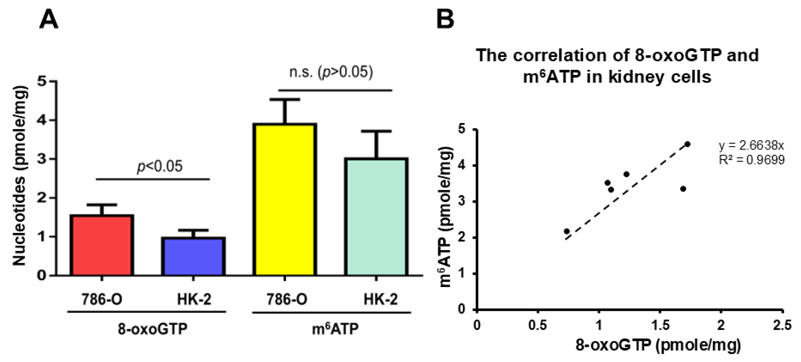
To determine if there exists m6ATP in the cellular nucleotide pool and if there is an association between the cellular m6ATP level and oxidative stress and its-induced RNA gaps, we detected the levels of m6ATP and 8-oxoGTP, the most frequently oxidized nucleotides in the nucleotide pool in human cells, which are often used as biomarkers of cellular oxidative stress. We used LC-MRM/MS to measure the cellular m6ATP and 8-oxoGTP levels in normal human kidney cells (HK-2) and kidney cancer cells (786-O). We found that m6ATP and 8-oxoGTP existed in the cells with 3 pmole/mg protein of m6ATP and 0.9 pmole/mg protein of 8-oxoGTP in normal kidney cells, HK-2, along with 3.9 pmole/mg protein of m6ATP and 1.5 pmole/mg protein of 8-oxoGTP in kidney cancer cells, 786-O (Figure 6A). Moreover, the results showed that m6ATP is 2.6–3-fold higher than 8-oxoGTP in the kidney cancer cells and normal kidney cells, indicating the existence of m6ATP in the nucleotide pool. Surprisingly, the results further revealed that the m6ATP and 8-oxoGTP levels in the kidney cancer 786-O cells were 1.3-fold (p < 0.05) and 1.7-fold (n.s., p > 0.05) higher than those in normal kidney cells, respectively, suggesting that there was a correlation between the cellular levels of m6ATP and 8-oxoGTP. Indeed, we found that a linear correlation between the levels of m6ATP and 8-oxoGTP in kidney cells was established, with the cellular m6ATP increasing with increased level of 8-oxoGTP (Figure 6B). Collectively, these results indicate that m6ATP can exist in the cellular nucleotide pool at a significant level, suggesting that it is elevated along with cellular oxidative stress and its resulting DNA and RNA damage.

3. Discussion

In this study, we provided the first evidence that the repair and translesion DNA polymerases pol β and pol η can incorporate m6ATP into RNA on the 1 nt RNA gap and RNA open-template substrates with comparable efficiency for their ATP incorporation (Figure 1, Figure 2, Figure 3 and Figure 4). Using MD, we further demonstrated that pol β and pol η incorporated m6ATP by employing a unique base-stacking-like mechanism with a part of the purine ring of the 3′-terminus adenosine of the upstream RNA primer in the presence of Mn2+ and Mg2+. The unique mechanism governed the distance between the 3′-OH group and the α-phosphate of m6ATP and its dynamic change, mediating the efficiency of m6ATP incorporation (Figure 5). Furthermore, we detected a significant amount of m6ATP in normal and cancer kidney cells. We showed that the cellular level of m6ATP increased with the increase in the amount of the oxidative stress biomarker 8-oxoGTP (Figure 6A,B). Our results supported the hypothetical model in which cellular oxidative stress from ROS can simultaneously stimulate the production of m6ATP in the cellular nucleotide pool and induce RNA damage, such as RNA gaps. Subsequently, RNA gaps are recognized by repairing DNA polymerases, such as pol η and pol β. The DNA polymerases then incorporate m6ATP into RNA through their RNA gap-filling synthesis activity on an RNA-DNA hybrid, leading to the unscheduled m6A deposition on RNA (Figure 7). There may be a scenario during which, after m6A is incorporated, the RNA strand is displaced by the reannealing of the non-template DNA to the template DNA strand, forcing the RNA strand to dissociate from the template. In this case, the RNA strand may be subject to RNA degradation, generating m6AMP that can be converted back into m6ATP in the nucleotide pool and reused for m6ATP RNA incorporation.
Here, we revealed a potential alternative mechanism for m6A RNA deposition that is independent of the RNA methyltransferase, METLL3/METTL14, but through m6ATP incorporation via oxidative RNA damage-induced RNA gaps by repairing DNA polymerases. We named the alternate pathway as an unscheduled m6A RNA deposition. Our study suggests that the unscheduled m6A deposition through RNA damage can potentially lead to the disruption of m6A RNA depositions that are usually regulated by m6A writers such as METTL3/METTL14, thereby altering cellular functions. Moreover, we found that incorporation of m6A by pol β and pol η also resulted in nucleotide misincorporation through its extension, especially on the RNA open-template substrate, which represents a large RNA gap on RNA-DNA hybrids (Figure 1B, lanes 6–8, Figure 2A, 2B, lanes 5–6, and Figure 3B, lanes 6–8). This can potentially reduce RNA integrity, protein–RNA interactions, and m6A-regulated cellular functions. Our study further suggests crosstalk between RNA modifications and DNA repair polymerase, implicating the interplay of RNA integrity, cellular oxidative stress, and epitranscriptional regulation on R-loops during gene transcription and DNA repair. Our findings will also provide new insights into an alternate mechanism of m6A RNA deposition, contributing to a broader understanding of the diversified functions of repairing DNA polymerases that bridge the crosstalk between the genome and epitranscriptome.
Our results suggest that repairing DNA polymerases can make use of RNA base damage, such as AP-site-induced RNA gaps, to incorporate m6ATP into RNA on RNA-DNA hybrids. It is possible that the incorporated m6A in RNA can be sustained in RNA via its extension by DNA polymerases (Figure 1B, Figure 2, and Figure 3), leading to an RNA nick that is subsequently sealed by an RNA ligase, as recently reported in [37]. Since AP sites are the most abundant form of RNA base damage that can arise from cellular spontaneous depurination/depyrimidination or exposure to endogenous and exogenous genotoxicants [38], this may increase the probability of m6ATP being incorporated into RNA via RNA damage. Recent studies have implicated the effects of RNA damage and repair on genome stability via RNA-DNA hybrids on R-loops, and several critical reviews have highlighted the importance of RNA damage and repair in modulating cellular gene expression and function [39,40,41]. Our findings further suggest that repairing DNA polymerases can mediate the crosstalk between the epitranscriptome and genome by playing a dual role in incorporating m6ATP and performing RNA-guided DNA gap-filling synthesis to repair DNA base damage [42] on RNA-DNA hybrids. Consequently, this may further allow DNA repair polymerases to coordinate their activities for DNA repair and m6A incorporation, thereby modulating epitranscriptomic modifications and maintaining genome integrity.
Structurally, the presence of the methyl group at the N6 position of m6ATP may cause steric hindrance to disrupt the base-pairing of the nucleotide with the template T. This may further alter the interaction of pol β and pol η with the DNA-RNA hybrid substrates, reducing the fidelity and efficiency of the DNA polymerases. Using AlphaFold3-assisted MD, we found that to adapt to the steric hindrance from the methyl group of m6ATP, pol β and pol η evolved a unique base-stacking or base-stacking-like mechanism by employing the purine ring of m6ATP to base stack with the part of the purine ring of the 3′-terminus adenosine of the upstream RNA primer (Figure 5). Consequently, this resulted in the stabilized distance between the 3′-OH group and the α-phosphate of m6ATP ranging between 3.5 and 4Å (Figure 5), thereby ensuring the success of the nucleophilic attack initiated by the 3′-OH group of the upstream primer and the catalysis of m6ATP incorporation. Furthermore, we found that in the pol β-RNA-DNA hybrid-ATP ternary complex in the presence of Mn2+, the distance only exhibited a small dynamic change ranging from 3.5 to 4.5Å during 6.25–62.5 ns as compared to the stabilized distance in the ternary complex with m6ATP (Figure 5, the panel on the left), leading to the similar catalytic efficiency of ATP and m6ATP incorporation by pol β (Figure 4A). Interestingly, for pol η, in the presence of Mg2+, although the distance between the 3′-OH group and α-phosphate of ATP underwent a large range of dynamic changes, ranging from 3.5Å to 9Å as compared with the change from 4 to 5 Å for m6ATP (Figure 5, the panel in the middle), the DNA polymerase still achieved more than four-fold higher efficiency in incorporating ATP than m6ATP in the presence of Mg2+ (Figure 4B). In the presence of Mn2+, pol η maintained the distance change for ATP from 4.5Å to 6Å and 4Å to 4.5Å for m6ATP during 200 ns, leading to a higher efficiency of ATP incorporation than m6ATP. These results suggest that the large dynamic change in the distance between the 3′-OH group and α-phosphate of ATP and m6ATP, rather than the distance per se, plays a crucial role in mediating the catalysis of the nucleotide incorporation into RNA. The results further demonstrate that the differences in the dynamic distance change between the 3′-OH group and the α-phosphate of the incoming nucleotides in pol β and pol η determined the efficiency of the molecular collision of the atoms, thereby governing the difference in the catalytic efficiency of ATP and m6ATP incorporation.
In this study, we detected a significant amount of m6ATP in normal kidney and kidney cancer cells (Figure 6A). We further identified a positive correlation between the levels of m6A and the oxidative stress biomarker 8-oxoGTP (Figure 6B), suggesting that the cellular m6ATP level can also serve as an effective response to oxidative stress. Furthermore, since oxidative stress that is signified by the massive production of cellular ROS can simultaneously damage DNA, RNA, and nucleotides triphosphate in the nucleotide pool, we suggest that an unscheduled m6A deposition can be mediated via m6ATP incorporation into RNA gaps by DNA polymerases upon RNA damage on RNA-DNA hybrids on R-loops induced by oxidative stress (Figure 7). Moreover, our results show that the level of m6ATP is 3- and 2.6-fold of 8-oxoGTP in normal kidney cells and kidney cancer cells (Figure 6A), suggesting that the level of m6ATP in the cellular nucleotide pool is significant and plays a critical role in modulating RNA modifications and m6A deposition in an RNA methyltransferase-independent mechanism. Since it was reported that m6A in DNA can be generated by incorporating m6dATP [43], our results suggest that m6ATP in the nucleotide pool can also be used as the precursor to be reduced into m6dATP for generating 6mA in DNA, leading to the maintenance of genome stability, as reported recently [44]. Since Musheev et al. have shown that m6AMP can be converted into m6ADP, which is then reduced to m6dADP by ribonucleotide diphosphate reductase, which is subsequently, phosphorylated into m6dATP, leading to the incorporation of 6mATP into DNA by DNA polymerases [43], it is possible that cellular m6ATP in the ribonucleotide pool can also be directly regenerated from m6AMP that results from the degradation of m6A-modified RNA. m6AMP can then be phosphorylated into m6ATP by a nucleotide kinase and recycled back into the nucleotide pool. Thus, our study opens a new avenue for revealing the novel role of m6A in mediating the interplay between epitranscriptomic and genomic stability. It should be noted that our results show that kidney cancer cells exhibited higher m6ATP and 8-oxoGTP levels than normal kidney cells, without a statistically significant difference (p > 0.05) for m6ATP but with a statistically significant difference for 8-oxoGTP (p < 0.05) (Figure 6A). This further indicates that more biological replicates will be needed to determine the statistical significance of the difference in m6ATP between normal and cancer kidney cells.
It has been reported that in mammalian cells, the intracellular concentrations are approximately ATP 3152 ± 1698 µM, GTP 468 ± 224 µM, UTP 567 ± 460 µM, and CTP, 278 ± 242 µM [45]. In contrast, the concentrations of dNTPs exhibit different ranges: dATP 24 ± 22 µM, dGTP 5.2 ± 4.5 µM, dCTP 29 ± 19 µM, and dTTP 37 ± 30 µM [45]. Based on the fact that a single mammalian kidney cell has a volume of 96 μm3 per cell (use rat kidney duct cells as an example), as reported in [46], according to our results, we estimated that the concentrations of m6ATP in 786-O cells and normal kidney HK-2 cells were 33.8 ± 4.9 nM and 26.1 ± 5.5 nM, respectively. Thus, the m6ATP concentration in the human kidney cells was about 1000-fold lower than the level of dATP in cells. Since Musheev et al. have found that m6dATP at a lower concentration than m6ATP can be incorporated into the genomic DNA by DNA polymerases in cells [43], our results suggest that it is possible that m6ATP at a relatively higher concentration than m6dATP in cells can also be incorporated into RNA on RNA-DNA hybrids of R-loops.
In cells, RNA gaps with a DNA template can be induced on R-loops. This can result from RNA base damage such as RNA abasic sites that are generated by cellular oxidative stress and RNA base alkylation [38]. Subsequently, RNA abasic sites on RNA-DNA hybrids can be cleaved by human AP endonuclease 1 (APE1) at the 5′-end of the abasic sites, leading to the formation of RNA gaps on RNA-DNA hybrids [47]. It is possible that RNA gaps also serve as the substrates for RNase H1 on R-loops, leading to coordination between RNase H1 and DNA repair polymerase-mediated m6ATP incorporation. However, it is unlikely that RNA gaps are used as the substrate of RNA polymerases. This is because RNA polymerases recognize the template DNA as the substrate and move along with the template DNA to perform RNA synthesis. Thus, it is unlikely that RNA polymerases can readily move back to bind RNA gaps on RNA-DNA hybrids. Here, we discovered an alternative role of repairing DNA polymerases with their RNA m6ATP incorporation activity due to their substrate plasticity. Studies on understanding the coordination among DNA repair polymerases and RNA processing enzymes in modulating m6ATP incorporation shall be conducted in the future.
It was reported that the concentration of Mn2+ in human cells is typically low and that the physiological concentration of Mn2+ in human HeLa cells is 1.14 ± 0.15 μM [48]. This concentration is significantly lower than the concentration used in our experiments (1 mM), which can represent an Mn2+ genotoxic level. Although we did not detect any m6ATP incorporation by the DNA polymerases at the physiological concentration of Mn2+, it remains possible that the incorporation of m6ATP by pol β and pol η in cells can be induced at the physiological concentration of Mn2+ of 1.14 μM by cooperating with their cofactors. The importance of m6ATP incorporation by DNA repair polymerases in the presence of their cofactors under physiological and pathological concentrations of Mn2+ needs to be elucidated in the future.
Our MD results showed that the methyl group on m6ATP disrupted the standard Watson–Crick base pairing between m6ATP and the template T in the catalytic center of the DNA polymerases by forcing the purine ring m6ATP to turn away from the template T, leading to reduced efficiency of m6ATP incorporation. Simultaneously, the methyl group promoted the base-stacking interaction of m6ATP with the upstream primer 3′-terminus A through hydrophobic and van der Waals effects, thereby sustaining the distance between the 3′-OH of the upstream primer and the α-phosphate of m6ATP needed for catalysis of the nucleotide incorporation. Our results further indicate that the methyl group at the 6-position of m6ATP forced the purine ring of the nucleotide to form base-stacking interactions with the part of the aromatic ring of 3-terminus A of the upstream primer (Figure 5) via π–π interactions, which stabilizes the orientation of the 3′-OH group of the upstream RNA primer. This further suggests that the upstream primer 3′-terminus nucleotide does not strictly need to be adenine to form its base-stacking interaction with the purine ring of m6ATP. The effects of various types of ribonucleotides at the 3′-terminus on the base-stacking interaction with m6ATP warrant a study in the future.
However, it should be noted that our study has several limitations. First, while our results provide new insights into the potential mechanism of unscheduled m6A deposition mediated by repairing DNA polymerases, which was associated with oxidative stress, its physiological functions on epitranscriptomic regulation and genome stability remain to be elucidated in vivo using animal models or advanced cellular models in the future. In addition, we could not exclude the possibility that the association between m6ATP and 8-oxoGTP may be affected by some potential cofounding factors in cells. The causal correlation between oxidative-stress-induced 8-oxoGTP and m6ATP and their effects on cellular functions and disease progression needs to be further determined. Second, the conditions employed in our in vitro RNA-DNA hybrid-based approaches may not fully reflect those in complex biological systems. However, our results suggest the possibility of m6ATP RNA incorporation by DNA repair polymerases in cells. This is supported by the fact that pol β and pol η are well known to adapt to the noncanonical substrates to tolerate and incorporate oxidized, damaged, and other types of modified nucleotides [49,50]. Moreover, our results show that pol η exhibited efficient incorporation of m6ATP in RNA at a physiological concentration of Mg2+, suggesting the potential role of the DNA polymerase in incorporating modified ribonucleotide in RNA in cells. Also, it is possible that in cells, the repair DNA polymerases may enrich m6ATP and Mg2+ or Mn2+ at the enzyme catalytic center to a level comparable to that used in our experiments, thereby fulfilling their efficient catalysis for m6ATP incorporation in RNA. The mechanisms of m6ATP RNA incorporation by repairing DNA polymerases via RNA damage need to be further elucidated in vivo. Third, in our study, the levels of m6ATP and 8-oxoGTP were detected only in limited types of cells, the normal and cancer kidney cells. This may limit the application of our conclusions to a broader range of cell types, including healthy and diseased cells. In addition, our results have a limitation in revealing the relative abundance of m6ATP and 8-oxoGTP in the cellular nucleotide pool due to the lack of information on the total amount of ATP and GTP in the kidney cell lines. Future studies should also determine the percentage of chemically modified ribonucleotides among the total amount of all four types of ribonucleotides in the cellular nucleotide pool to elucidate the roles of the modified nucleotides in modulating cellular functions. Furthermore, future studies on addressing all these limitations will reveal the biological significance and impact of the unscheduled m6A deposition, opening a new avenue for understanding the crosstalk between epitranscriptome and genome via m6A incorporation and DNA repair mediated by DNA polymerases on R-loops.

4. Materials and Methods

4.1. Materials

ATP (100 mM) and m6ATP (100 mM) were purchased from Fisher Scientific (Waltham, PA, USA) and TriLink BioTechnologies (San Diego, CA, USA), respectively. The radionucleotide, 32P-ATP (6000 µCi/mmol), was purchased from Revvity Inc. (Waltham, MA, USA). DNA and RNA oligonucleotides were synthesized by Eurofins Genomics (Louisville, KY, USA). Micro Bio-Spin 6 chromatography columns were from Bio-Rad Laboratories (Hercules, CA, USA). Human pol β protein was expressed and purified as described previously [51]. Purified human pol η protein was a generous gift from Dr. Wei Yang from the National Institute of Diabetes and Digestive and Kidney Diseases (NIDDK)/National Institutes of Health (NIH) [36]. BER reaction buffer was made with 50 mM Tris-HCl, pH 7.5, 50 mM KCl, 0.1 mM EDTA, 0.1 mg/mL bovine serum albumin (BSA), and 0.01% Nonidet P-40 (NP-40). Moreover, 2× stopping buffer was made of 95% deionized formamide and 10 mM EDTA, 0.25% bromophenol blue (Sigma-Aldrich, St. Louis, MO, USA), and 0.25% (w/v) xylene cyanol FF (Sigma-Aldrich, St. Louis, MO, USA).

4.2. RNA-DNA Hybrid Oligonucleotide Substrates

An RNA-DNA hybrid substrate containing a 1 nt RNA gap was designed to mimic an RNA damage intermediate to test the 1 nt RNA gap-filling synthesis. An open-template RNA substrate was designed to mimic a large RNA gap generated from RNA damage. The 1 nt gap substrate was constructed by annealing the 5’-end 32P-ATP radiolabeled 19 nt upstream RNA primer (5′-CGUACGCGGAAUACUUCGA-3′) and 36 nt downstream RNA primer (5′-CCACCCAGUCUGCCCCCGGAUGACGUAAAAGGAAAG -3′) with the 57 nt DNA template containing a dT base positioned opposite to the 1 nt gap (5′-GCTTTCCTTTTACGTCATCCGGGGGCAGACTGGGTGGTTCGAAGTGTTCCGCGTACG-3′. The 1 nt gap substrate was assembled by annealing the upstream and downstream RNA primers with the template strand at a molar ratio of 1:3:3. An open-template RNA-DNA hybrid substrate was constructed by annealing the 5’-end 32P-ATP-radiolabeled 19 nt upstream RNA primer with the 56 nt DNA template at a molar ratio at 1:3. The RNA and DNA oligonucleotides were annealed in 1× annealing buffer containing 10 mM Tris pH 7.5, 100 mM NaCl, and 1 mM EDTA. The substrate annealing mixture was then subjected to 95 °C for 5 min and slowly cooled down to room temperature at 25 °C.

4.3. Determination of RNA Synthesis Activity by DNA Polymerases

The DNA-templated RNA synthesis activities of pol β and pol η were measured by incubating 50 nM RNA-DNA hybrid substrates with increasing concentrations of the DNA polymerases in the presence of 500 μM NTPs at 37 °C for 60 min in the reaction mixture (20 µL containing 5 mM of Mg2+ or 1 mM of Mn2+ in the BER reaction buffer with 50 mM Tris-HCl, pH 7.5, 0.1 mM EDTA, 50 mM KCl, 0.01% NP-40 and 0.1 mg/mL BSA). Under these conditions, DNA polymerases were able to achieve high ribonucleotide incorporation efficiency, as reported in previous studies [52,53]. The reactions were terminated by adding a 2× stop buffer (10 mM EDTA, 95% deionized formamide, and 0.25% (w/v) each of bromophenol blue and xylene cyanol FF). Samples were then denatured at 95 °C for 5 min and subjected to 15% urea-denaturing polyacrylamide gel electrophoresis. The substrates and products were detected using a Pharos FX Plus PhosphorImager (Bio-Rad Laboratories, Hercules, CA, USA). All the experiments were repeated independently three times.

4.4. Determination of the Efficiency of ATP and m6ATP Incorporation in RNA by Pol β and Pol η Using Steady-State Kinetics

The efficiency of ATP and m6ATP incorporation in RNA by pol β and pol η was determined using the steady-state enzyme kinetics of RNA synthesis at a fixed concentration of the DNA polymerases, RNA-DNA hybrid substrate, and metal cofactor (Mg2+ or Mn2+) with increasing concentrations of ATP or m6ATP substrates. The steady-state kinetics of ATP and m6ATP incorporation by pol β was determined using 50 nM of 1 nt gap substrate, 200 nM of pol β, and 1 mM of Mn2+ with increasing concentrations of ATP and m6ATP (10–100 μM). The steady-state kinetics of RNA synthesis by pol η was determined using 50 nM of open-template RNA substrate, and either 25 nM of pol η, 5 mM of Mg2+ with increasing concentrations of ATP and m6ATP (10–1000 μM) or 10 nM of pol η, 1 mM of Mn2+ with increasing concentrations of ATP and m6ATP (0.5–5 μM). The enzymes and substrates were incubated at 37 °C for different time intervals (0 to 15 min for pol β and pol η-Mg2+, 0–10 min for pol η-Mn2+) in the reaction mixture (20 μL). The reactions were terminated using a 2× stop buffer, and the reaction mixture was heated for 5 min at 95 °C. Substrates and products were separated using a 15% urea-denaturing polyacrylamide gel and detected by the Pharos FX Plus PhosphorImager. The Vmax, Km, and kcat of the DNA polymerases were calculated using the Enzyme Kinetics Module of the Prism-GraphPad software version 9.0.2. All the experiments were repeated at least three times.

4.5. Molecular Dynamics Simulation of the Ternary Complexes of Pol β or Pol η-RNA-DNA Hybrid-NTPs

Protein–RNA-DNA hybrid-NTP ternary complexes were modeled using AlphaFold3 [54] by incorporating the amino acid sequences for human pol β (residues 1–335) or pol η (residues 1–432), along with the 1 nt RNA gap substrate for pol β and RNA open-template substrate for pol η containing the DNA template and up- and downstream primers with the sequences in this study. An ATP molecule and two divalent metal ions, Mg2+ or Mn2+ were also included in the input structures. The resulting models were structurally aligned to the crystal structures of the pol β ternary complex (PDB: 5TBB) [35] and pol η (PDB: 4J9N) [36,55] for validation and refinement. To generate complexes containing m6ATP, the ATP coordinates from the AlphaFold3-generated protein–RNA-DNA ternary complexes were modified using Avogadro2 [56]. A methyl group was added at the N6 position of the adenine to yield m6ATP, which substituted the ATP molecule in the original models. The pol β and pol η with the substrates were then set up for MD simulations using CHARMM-GUI [57,58,59], with the structures solvated in the TIP3P water model in a cubic box. Ions were added to achieve concentrations of 0.15 M KCl and 50 mM MgCl2 for the complex containing Mg2+ and 10 mM MnCl2 for the complex containing Mn2+. The GPU version of NAMD 3.0b2 [60] was used to simulate the systems using Charmm36m force field [61,62]. Simulations began with 10,000 steps of minimization and 250 ps of equilibration at 303 K and 1 atm pressure. The temperature was maintained at 37 °C using Langevin temperature coupling with a damping coefficient of 1/ps. The pressure was kept constant using a Nose–Hoover Langevin piston [63] with a 50-femtoseconds (fs) period and 25 fs decay. The Particle Mesh Ewald method (PME) [64] was used for long-range electrostatic interactions with periodic boundary conditions. All the covalently bonded hydrogen atoms were restrained with the ShakeH algorithm [65]. A 200 ns unconstrained production run was performed for each system using a 2 fs/step. Visual molecular dynamics (VMD) was used to analyze the trajectories.

4.6. Cellular m6ATP Level Measurement

The levels of m6ATP and 8-oxoGTP in human kidney cells (1.2 × 10⁶ cells) (HK-2, ATCC, Manassas, VA, USA) and human kidney cancer cells (1.2 × 10⁶ cells) (786-O, ATCC, Manassas, VA, USA) were determined by liquid chromatography–multiple reaction monitoring mass spectrometry analysis (LC-MRM/MS by Creative Proteomics (Shirley, NY, USA). The LC-MRM/MS platform used by Creative Proteomics (Shirley, NY, USA) employed UPLC–QTRAP 6500+, which was operated in ESI-negative mode with an ammonium acetate/acetonitrile binary gradient. The technique ionized the samples and collected the sample ions in a negative-ion mode. This allowed us to use the retention time to distinguish the methylation group located at different atoms of RNA bases. This approach is commonly used for nucleotide isomer separation and detection, as previously reported [66]. Thus, the method used by Creative Proteomics in our study allowed the differentiation of m6ATP from other forms of methylated ATP in cells with an identical m/z value. In our study, cell pellets from the biological triplicates of two types of cells were used for the experiments. A mixed standard solution containing the synthesized m6ATP and 8-oxGTP was used as the internal standard (IS). The standard curves were created via linear regression using the data obtained from these standard solutions. Cell pellets were thawed on ice, resuspended in 15 μL of liquid, and diluted to a final volume of 100 μL by adding 80% acetonitrile. Cells were lysed using an MM 400 mixer mill at 30 Hz for 3 min. Cell lysates were then centrifuged at 21,000× g for 10 min to obtain the supernatant. The residual protein pellets were subjected to protein quantification using the standard BCA (bicinchoninic acid) assay. Aliquots (10 μL) of both the cell samples and standard solution were injected into a HILIC column for LC-MRM/MS analysis, which was performed on a Waters UPLC system coupled with a Sciex QTRAP 6500+ mass spectrometer operated in negative-ion detection mode. Chromatographic separation was achieved using binary solvent gradient elution with ammonium acetate buffer and acetonitrile as the mobile phases. The levels of m6ATP and 8-oxoGTP in the cell samples were determined by interpolating the standard curve at pmole/mg protein, where “mg protein” refers to the amount of protein per mg protein among the total amount of protein from the total 1.2 × 10⁶ cells of each type of cell. The protein concentrations were measured from the same sample after extraction using a standardized BCA assay.

5. Conclusions

In this study, for the first time, we identified a novel mechanism by which m6A was deposited into RNA through the incorporation of m6ATP by DNA repair and translesion polymerases pol β and pol η through their RNA gap-filling synthesis. We further demonstrated that m6ATP was efficiently incorporated into RNA gap and open-template RNA substrates via a unique base-stacking-like mechanism. Further analysis of the cellular m6ATP levels showed their positive correlation with the oxidative stress biomarker 8-oxoGTP, suggesting an association of m6ATP and its RNA incorporation with cellular oxidative stress. Our findings suggest an interplay among DNA repair, RNA integrity, and genome stability.

Author Contributions

Conceptualization, F.Q. and Y.L.; methodology, F.Q., Y.L., J.F. and P.C.; software, J.F. and P.C.; validation, F.Q., Y.L., J.F. and P.C.; formal analysis, F.Q., Y.L., J.F. and P.C.; investigation, F.Q., J.F., Y.L. and P.C.; resources, Y.L. and P.C.; data curation, F.Q. and J.F.; writing—original draft preparation, F.Q., J.F., Y.L. and P.C.; writing—review and editing, F.Q., Y.L., J.F. and P.C.; visualization, F.Q., J.F., P.C. and Y.L.; supervision, Y.L. and P.C.; project administration, Y.L.; funding acquisition, Y.L. All authors have read and agreed to the published version of the manuscript.

Funding

This research was funded by the National Institutes of Health under grants R03ES035200 and R03ES035200–01S1 to Y.L.

Data Availability Statement

All the data will be deposited into a publicly accessible repository, zenodo: 10.5281/zenodo.16741489.

Acknowledgments

We thank the members of the Liu Laboratory at Florida International University for their insightful comments on the work.

Conflicts of Interest

The authors declare no conflicts of interest.

References

  1. Dominissini, D.; Moshitch-Moshkovitz, S.; Schwartz, S.; Salmon-Divon, M.; Ungar, L.; Osenberg, S.; Cesarkas, K.; Jacob-Hirsch, J.; Amariglio, N.; Kupiec, M.; et al. Topology of the human and mouse m6A RNA methylomes revealed by m6A-seq. Nature 2012, 485, 201–206. [Google Scholar] [CrossRef] [PubMed]
  2. Roundtree, I.A.; He, C. Nuclear m(6)A Reader YTHDC1 Regulates mRNA Splicing. Trends Genet. 2016, 32, 320–321. [Google Scholar] [CrossRef] [PubMed]
  3. Du, H.; Zhao, Y.; He, J.; Zhang, Y.; Xi, H.; Liu, M.; Ma, J.; Wu, L. YTHDF2 destabilizes m(6)A-containing RNA through direct recruitment of the CCR4-NOT deadenylase complex. Nat. Commun. 2016, 7, 12626. [Google Scholar] [CrossRef]
  4. Huang, H.; Weng, H.; Sun, W.; Qin, X.; Shi, H.; Wu, H.; Zhao, B.S.; Mesquita, A.; Liu, C.; Yuan, C.L.; et al. Recognition of RNA N(6)-methyladenosine by IGF2BP proteins enhances mRNA stability and translation. Nat. Cell Biol. 2018, 20, 285–295. [Google Scholar] [CrossRef] [PubMed]
  5. Qiao, Y.; Sun, Q.; Chen, X.; He, L.; Wang, D.; Su, R.; Xue, Y.; Sun, H.; Wang, H. Nuclear m6A reader YTHDC1 promotes muscle stem cell activation/proliferation by regulating mRNA splicing and nuclear export. eLife 2023, 12, e82703. [Google Scholar] [CrossRef]
  6. Du, H.; Zou, N.Y.; Zuo, H.L.; Zhang, X.Y.; Zhu, S.C. YTHDF3 mediates HNF1alpha regulation of cervical cancer radio-resistance by promoting RAD51D translation in an m6A-dependent manner. FEBS J. 2023, 290, 1920–1935. [Google Scholar] [CrossRef]
  7. Lee, K.P.; Liu, K.; Kim, E.Y.; Medina-Puche, L.; Dong, H.; Di, M.; Singh, R.M.; Li, M.; Qi, S.; Meng, Z.; et al. The m6A reader ECT1 drives mRNA sequestration to dampen salicylic acid-dependent stress responses in Arabidopsis. Plant Cell 2024, 36, 746–763. [Google Scholar] [CrossRef]
  8. Cai, Z.; Tang, Q.; Song, P.; Tian, E.; Yang, J.; Jia, G. The m6A reader ECT8 is an abiotic stress sensor that accelerates mRNA decay in Arabidopsis. Plant Cell 2024, 36, 2908–2926. [Google Scholar] [CrossRef]
  9. Yin, H.; Ju, Z.; Zheng, M.; Zhang, X.; Zuo, W.; Wang, Y.; Ding, X.; Zhang, X.; Peng, Y.; Li, J.; et al. Loss of the m6A methyltransferase METTL3 in monocyte-derived macrophages ameliorates Alzheimer’s disease pathology in mice. PLoS Biol. 2023, 21, e3002017. [Google Scholar] [CrossRef]
  10. Wang, J.N.; Wang, F.; Ke, J.; Li, Z.; Xu, C.H.; Yang, Q.; Chen, X.; He, X.Y.; He, Y.; Suo, X.G.; et al. Inhibition of METTL3 attenuates renal injury and inflammation by alleviating TAB3 m6A modifications via IGF2BP2-dependent mechanisms. Sci. Transl. Med. 2022, 14, eabk2709. [Google Scholar] [CrossRef]
  11. Zhou, C.; She, X.; Gu, C.; Hu, Y.; Ma, M.; Qiu, Q.; Sun, T.; Xu, X.; Chen, H.; Zheng, Z. FTO fuels diabetes-induced vascular endothelial dysfunction associated with inflammation by erasing m6A methylation of TNIP1. J. Clin. Investig. 2023, 133, e160517. [Google Scholar] [CrossRef]
  12. Wang, S.; Nie, J.; Xu, K.; Liu, Y.; Tong, W.; Li, A.; Zuo, W.; Liu, Z.; Yang, F. YY1 is regulated by ALKBH5-mediated m6A modification and promotes autophagy and cancer progression through targeting ATG4B. Aging 2023, 15, 9590–9613. [Google Scholar] [CrossRef] [PubMed]
  13. Huang, C.; Zhou, S.; Zhang, C.; Jin, Y.; Xu, G.; Zhou, L.; Ding, G.; Pang, T.; Jia, S.; Cao, L. ZC3H13-mediated N6-methyladenosine modification of PHF10 is impaired by fisetin which inhibits the DNA damage response in pancreatic cancer. Cancer Lett. 2022, 530, 16–28. [Google Scholar] [CrossRef] [PubMed]
  14. Meyer, K.D.; Saletore, Y.; Zumbo, P.; Elemento, O.; Mason, C.E.; Jaffrey, S.R. Comprehensive analysis of mRNA methylation reveals enrichment in 3′ UTRs and near stop codons. Cell 2012, 149, 1635–1646. [Google Scholar] [CrossRef] [PubMed]
  15. Linder, B.; Grozhik, A.V.; Olarerin-George, A.O.; Meydan, C.; Mason, C.E.; Jaffrey, S.R. Single-nucleotide-resolution mapping of m6A and m6Am throughout the transcriptome. Nat. Methods 2015, 12, 767–772. [Google Scholar] [CrossRef]
  16. Fu, Y.; Dominissini, D.; Rechavi, G.; He, C. Gene expression regulation mediated through reversible m(6)A RNA methylation. Nat. Rev. Genet. 2014, 15, 293–306. [Google Scholar] [CrossRef]
  17. Flamand, M.N.; Tegowski, M.; Meyer, K.D. The Proteins of mRNA Modification: Writers, Readers, and Erasers. Annu. Rev. Biochem. 2023, 92, 145–173. [Google Scholar] [CrossRef]
  18. Meyer, K.D.; Patil, D.P.; Zhou, J.; Zinoviev, A.; Skabkin, M.A.; Elemento, O.; Pestova, T.V.; Qian, S.B.; Jaffrey, S.R. 5′ UTR m(6)A Promotes Cap-Independent Translation. Cell 2015, 163, 999–1010. [Google Scholar] [CrossRef]
  19. Liu, J.; Yue, Y.; Han, D.; Wang, X.; Fu, Y.; Zhang, L.; Jia, G.; Yu, M.; Lu, Z.; Deng, X.; et al. A METTL3-METTL14 complex mediates mammalian nuclear RNA N6-adenosine methylation. Nat. Chem. Biol. 2014, 10, 93–95. [Google Scholar] [CrossRef]
  20. Brown, J.A.; Suo, Z. Unlocking the sugar “steric gate” of DNA polymerases. Biochemistry 2011, 50, 1135–1142. [Google Scholar] [CrossRef]
  21. Gao, G.; Orlova, M.; Georgiadis, M.M.; Hendrickson, W.A.; Goff, S.P. Conferring RNA polymerase activity to a DNA polymerase: A single residue in reverse transcriptase controls substrate selection. Proc. Natl. Acad. Sci. USA 1997, 94, 407–411. [Google Scholar] [CrossRef] [PubMed]
  22. Trincao, J.; Johnson, R.E.; Escalante, C.R.; Prakash, S.; Prakash, L.; Aggarwal, A.K. Structure of the catalytic core of S. cerevisiae DNA polymerase eta: Implications for translesion DNA synthesis. Mol. Cell 2001, 8, 417–426. [Google Scholar] [CrossRef] [PubMed]
  23. Johnson, R.E.; Prakash, S.; Prakash, L. Efficient bypass of a thymine-thymine dimer by yeast DNA polymerase, Poleta. Science 1999, 283, 1001–1004. [Google Scholar] [CrossRef]
  24. Gali, V.K.; Balint, E.; Serbyn, N.; Frittmann, O.; Stutz, F.; Unk, I. Translesion synthesis DNA polymerase eta exhibits a specific RNA extension activity and a transcription-associated function. Sci. Rep. 2017, 7, 13055. [Google Scholar] [CrossRef]
  25. Balint, E.; Unk, I. Selective Metal Ion Utilization Contributes to the Transformation of the Activity of Yeast Polymerase eta from DNA Polymerization toward RNA Polymerization. Int. J. Mol. Sci. 2020, 21, 8248. [Google Scholar] [CrossRef]
  26. Balint, E.; Unk, I. Manganese Is a Strong Specific Activator of the RNA Synthetic Activity of Human Poleta. Int. J. Mol. Sci. 2021, 23, 230. [Google Scholar] [CrossRef] [PubMed]
  27. Vashishtha, A.K.; Wang, J.; Konigsberg, W.H. Different Divalent Cations Alter the Kinetics and Fidelity of DNA Polymerases. J. Biol. Chem. 2016, 291, 20869–20875. [Google Scholar] [CrossRef]
  28. Tholey, G.; Ledig, M.; Mandel, P.; Sargentini, L.; Frivold, A.H.; Leroy, M.; Grippo, A.A.; Wedler, F.C. Concentrations of physiologically important metal ions in glial cells cultured from chick cerebral cortex. Neurochem. Res. 1988, 13, 45–50. [Google Scholar] [CrossRef]
  29. Tholey, G.; Ledig, M.; Kopp, P.; Sargentini-Maier, L.; Leroy, M.; Grippo, A.A.; Wedler, F.C. Levels and sub-cellular distribution of physiologically important metal ions in neuronal cells cultured from chick embryo cerebral cortex. Neurochem. Res. 1988, 13, 1163–1167. [Google Scholar] [CrossRef]
  30. Balint, E.; Unk, I. For the Better or for the Worse? The Effect of Manganese on the Activity of Eukaryotic DNA Polymerases. Int. J. Mol. Sci. 2023, 25, 363. [Google Scholar] [CrossRef]
  31. Vashishtha, A.K.; Konigsberg, W.H. The effect of different divalent cations on the kinetics and fidelity of Bacillus stearothermophilus DNA polymerase. AIMS Biophys. 2018, 5, 125–143. [Google Scholar] [CrossRef]
  32. Sawyer, D.L.; Sweasy, J.B. DNA Polymerase beta in the Context of Cancer. Crit. Rev. Oncog. 2022, 27, 17–33. [Google Scholar] [CrossRef]
  33. Beard, W.A.; Wilson, S.H. Structure and mechanism of DNA polymerase Beta. Chem. Rev. 2006, 106, 361–382. [Google Scholar]
  34. Pelletier, H.; Sawaya, M.R.; Wolfle, W.; Wilson, S.H.; Kraut, J. A structural basis for metal ion mutagenicity and nucleotide selectivity in human DNA polymerase beta. Biochemistry 1996, 35, 12762–12777. [Google Scholar] [CrossRef]
  35. Reed, A.J.; Vyas, R.; Raper, A.T.; Suo, Z. Structural Insights into the Post-Chemistry Steps of Nucleotide Incorporation Catalyzed by a DNA Polymerase. J. Am. Chem. Soc. 2017, 139, 465–471. [Google Scholar] [CrossRef]
  36. Weng, P.J.; Gao, Y.; Gregory, M.T.; Wang, P.; Wang, Y.; Yang, W. Bypassing a 8,5′-cyclo-2′-deoxyadenosine lesion by human DNA polymerase eta at atomic resolution. Proc. Natl. Acad. Sci. USA 2018, 115, 10660–10665. [Google Scholar] [CrossRef]
  37. Yuan, Y.; Stumpf, F.M.; Schlor, L.A.; Schmidt, O.P.; Saumer, P.; Huber, L.B.; Frese, M.; Hollmuller, E.; Scheffner, M.; Stengel, F.; et al. Chemoproteomic discovery of a human RNA ligase. Nat. Commun. 2023, 14, 842. [Google Scholar] [CrossRef] [PubMed]
  38. Prashar, T.; De La Selle, F.; Hudak, K.A. Abasic RNA: Its formation and potential role in cellular stress response. RNA Biol. 2023, 20, 348–358. [Google Scholar] [CrossRef] [PubMed]
  39. Yan, L.L.; Zaher, H.S. How do cells cope with RNA damage and its consequences? J. Biol. Chem. 2019, 294, 15158–15171. [Google Scholar] [CrossRef]
  40. Cordes, J.; Zhao, S.; Engel, C.M.; Stingele, J. Cellular responses to RNA damage. Cell 2025, 188, 885–900. [Google Scholar] [CrossRef] [PubMed]
  41. Olatunji, M.; Liu, Y. RNA damage and its implications in genome stability. DNA Repair 2025, 147, 103821. [Google Scholar] [CrossRef] [PubMed]
  42. Tsegay, P.S.; Hernandez, D.; Qu, F.; Olatunji, M.; Mamun, Y.; Chapagain, P.; Liu, Y. RNA-guided DNA base damage repair via DNA polymerase-mediated nick translation. Nucleic Acids Res. 2023, 51, 166–181. [Google Scholar] [CrossRef] [PubMed]
  43. Musheev, M.U.; Baumgartner, A.; Krebs, L.; Niehrs, C. The origin of genomic N(6)-methyl-deoxyadenosine in mammalian cells. Nat. Chem. Biol. 2020, 16, 630–634. [Google Scholar] [CrossRef]
  44. Conti, B.A.; Novikov, L.; Tong, D.; Xiang, Q.; Vigil, S.; McLellan, T.J.; Nguyen, C.; De La Cruz, N.; Veettil, R.T.; Pradhan, P.; et al. N6-methyladenosine in DNA promotes genome stability. eLife 2025, 13, RP101626. [Google Scholar] [CrossRef] [PubMed]
  45. Traut, T.W. Physiological concentrations of purines and pyrimidines. Mol. Cell Biochem. 1994, 140, 1–22. [Google Scholar] [CrossRef]
  46. Solenov, E.I. Cell volume and sodium content in rat kidney collecting duct principal cells during hypotonic shock. J. Biophys. 2008, 2008, 420963. [Google Scholar] [CrossRef]
  47. Liu, Y.; Rodriguez, Y.; Ross, R.L.; Zhao, R.; Watts, J.A.; Grunseich, C.; Bruzel, A.; Li, D.; Burdick, J.T.; Prasad, R.; et al. RNA abasic sites in yeast and human cells. Proc. Natl. Acad. Sci. USA 2020, 117, 20689–20695. [Google Scholar] [CrossRef]
  48. Kahali, S.; Das, S.K.; Kumar, R.; Gupta, K.; Kundu, R.; Bhattacharya, B.; Nath, A.; Venkatramani, R.; Datta, A. A water-soluble, cell-permeable Mn(ii) sensor enables visualization of manganese dynamics in live mammalian cells. Chem. Sci. 2024, 15, 10753–10769. [Google Scholar] [CrossRef]
  49. Tsegay, P.S.; Hernandez, D.; Brache, C.; Chatgilialoglu, C.; Krokidis, M.G.; Chapagain, P.; Liu, Y. Incorporation of 5′,8-cyclo-2′deoxyadenosines by DNA repair polymerases via base excision repair. DNA Repair 2022, 109, 103258. [Google Scholar] [CrossRef]
  50. Shimizu, M.; Gruz, P.; Kamiya, H.; Masutani, C.; Xu, Y.; Usui, Y.; Sugiyama, H.; Harashima, H.; Hanaoka, F.; Nohmi, T. Efficient and erroneous incorporation of oxidized DNA precursors by human DNA polymerase eta. Biochemistry 2007, 46, 5515–5522. [Google Scholar] [CrossRef]
  51. Beaver, J.M.; Lai, Y.; Xu, M.; Casin, A.H.; Laverde, E.E.; Liu, Y. AP endonuclease 1 prevents trinucleotide repeat expansion via a novel mechanism during base excision repair. Nucleic Acids Res. 2015, 43, 5948–5960. [Google Scholar] [CrossRef]
  52. Cavanaugh, N.A.; Beard, W.A.; Wilson, S.H. DNA polymerase beta ribonucleotide discrimination: Insertion, misinsertion, extension, and coding. J. Biol. Chem. 2010, 285, 24457–24465. [Google Scholar] [CrossRef]
  53. Su, Y.; Egli, M.; Guengerich, F.P. Mechanism of Ribonucleotide Incorporation by Human DNA Polymerase eta. J. Biol. Chem. 2016, 291, 3747–3756. [Google Scholar] [CrossRef]
  54. Abramson, J.; Adler, J.; Dunger, J.; Evans, R.; Green, T.; Pritzel, A.; Ronneberger, O.; Willmore, L.; Ballard, A.J.; Bambrick, J.; et al. Accurate structure prediction of biomolecular interactions with AlphaFold 3. Nature 2024, 630, 493–500. [Google Scholar] [CrossRef]
  55. Zhao, Y.; Gregory, M.T.; Biertumpfel, C.; Hua, Y.J.; Hanaoka, F.; Yang, W. Mechanism of somatic hypermutation at the WA motif by human DNA polymerase eta. Proc. Natl. Acad. Sci. USA 2013, 110, 8146–8151. [Google Scholar] [CrossRef] [PubMed]
  56. Hanwell, M.D.; Curtis, D.E.; Lonie, D.C.; Vandermeersch, T.; Zurek, E.; Hutchison, G.R. Avogadro: An advanced semantic chemical editor, visualization, and analysis platform. J. Cheminform. 2012, 4, 17. [Google Scholar] [CrossRef] [PubMed]
  57. Lee, J.; Cheng, X.; Swails, J.M.; Yeom, M.S.; Eastman, P.K.; Lemkul, J.A.; Wei, S.; Buckner, J.; Jeong, J.C.; Qi, Y.; et al. CHARMM-GUI Input Generator for NAMD, GROMACS, AMBER, OpenMM, and CHARMM/OpenMM Simulations Using the CHARMM36 Additive Force Field. J. Chem. Theory Comput. 2016, 12, 405–413. [Google Scholar] [CrossRef]
  58. Jo, S.; Kim, T.; Iyer, V.G.; Im, W. CHARMM-GUI: A web-based graphical user interface for CHARMM. J. Comput. Chem. 2008, 29, 1859–1865. [Google Scholar] [CrossRef] [PubMed]
  59. Brooks, B.R.; Brooks, C.L., 3rd; Mackerell, A.D., Jr.; Nilsson, L.; Petrella, R.J.; Roux, B.; Won, Y.; Archontis, G.; Bartels, C.; Boresch, S.; et al. CHARMM: The biomolecular simulation program. J. Comput. Chem. 2009, 30, 1545–1614. [Google Scholar] [CrossRef]
  60. Phillips, J.C.; Braun, R.; Wang, W.; Gumbart, J.; Tajkhorshid, E.; Villa, E.; Chipot, C.; Skeel, R.D.; Kale, L.; Schulten, K. Scalable molecular dynamics with NAMD. J. Comput. Chem. 2005, 26, 1781–1802. [Google Scholar] [CrossRef]
  61. Huang, J.; MacKerell, A.D., Jr. CHARMM36 all-atom additive protein force field: Validation based on comparison to NMR data. J. Comput. Chem. 2013, 34, 2135–2145. [Google Scholar] [CrossRef] [PubMed]
  62. Huang, J.; MacKerell, A.D., Jr. Force field development and simulations of intrinsically disordered proteins. Curr. Opin. Struct. Biol. 2018, 48, 40–48. [Google Scholar] [CrossRef]
  63. Nose, S.; Klein, M.L. Constant-temperature-constant-pressure molecular-dynamics calculations for molecular solids: Application to solid nitrogen at high pressure. Phys. Rev. B Condens. Matter 1986, 33, 339–342. [Google Scholar] [CrossRef] [PubMed]
  64. Essmann, U.; Perera, L.; Berkowitz, M.L.; Darden, T.; Lee, H.; Pedersen, L.G. A smooth particle mesh Ewald method. J. Chem. Phys. 1995, 103, 8577–8593. [Google Scholar] [CrossRef]
  65. Ryckaert, J.-P.; Ciccotti, G.; Berendsen, H.J.C. Numerical integration of the cartesian equations of motion of a system with constraints: Molecular dynamics of n-alkanes. J. Comput. Phys. 1977, 23, 327–341. [Google Scholar] [CrossRef]
  66. Jiang, H.P.; Xiong, J.; Liu, F.L.; Ma, C.J.; Tang, X.L.; Yuan, B.F.; Feng, Y.Q. Modified nucleoside triphosphates exist in mammals. Chem. Sci. 2018, 9, 4160–4167. [Google Scholar] [CrossRef]
Figure 1. Incorporation of ATP and m6ATP into RNA by pol β in the presence of Mn2+. DNA-templated 1 nt RNA gap and open-template RNA substrates (50 nM) were incubated with increasing concentrations of pol β (50, 100, and 200 nM) and 500 μM ATP or m6ATP at 37 °C for 60 min in the presence of 1 mM Mn2+. Panel (A) represents the pol β RNA synthesis resulting from a 1 nt RNA gap substrate and the quantification of the RNA synthesis products. Panel (B) represents the pol β RNA synthesis products on the open RNA template substrate. Lanes 1 and 5 represent substrate only. Lanes 2–4 represent pol β ATP incorporation on the gap or open-templated RNA substrate. Lanes 6–8 represent pol β m6ATP incorporation on the gap or open-templated RNA substrate. The synthesized RNA products are indicated by arrows on the left side of the gel images. The percentage of the pol β RNA synthesis products was calculated and illustrated in the bar charts below the gel images. All experiments were performed at least in triplicate.
Ijms 26 09263 g001
Figure 2. The incorporation of m6ATP into RNA by pol η in the presence of Mg2+. DNA-templated 1 nt RNA gap substrate and RNA open-template substrate (50 nM) were incubated with increasing concentrations of pol η (25–50 nM) at 37C for 60 min in the presence of 500 μM ATP or m6ATP and 5 mM Mg2+. Panel (A) represents the pol η RNA synthesis on the 1 nt RNA gap substrate and the quantification of the RNA synthesis products. Panel (B) represents the pol η RNA synthesis products on the open RNA template substrate. Lanes 1 and 4 represent substrate only. Lanes 2–3 represent pol η ATP incorporation on the gap (panel A) or open-templated RNA substrate. Lanes 5–6 represent pol η m6ATP incorporation on gap or open-templated RNA substrate (panel B). The synthesized RNA products are indicated by arrows on the left side of the gel images. The percentage of the pol η RNA synthesis products was calculated and illustrated in the bar charts below the gel images. All experiments were performed at least in triplicate.
Ijms 26 09263 g002
Figure 3. The incorporation of ATP and m6ATP into RNA by pol η in the presence of Mn2+. DNA-templated 1 nt RNA gapped and open-template substrates (50 nM) were incubated with increasing concentrations of pol η (2–10 nM for the RNA gap substrate and 10–100 nM for the RNA open-template substrate) at 37 °C for 60 min in the presence of 500 μM ATP or m6ATP and 1 mM Mn2+. Panel (A) represents the results of pol η RNA incorporation of ATP and m6ATP on the 1 nt RNA-gap substrate. Panel (B) illustrates the results of pol η incorporation of ATP and m6ATP on the RNA open-template substrate. The percentage of RNA synthesis products is illustrated in the bar charts below the gel images. Lanes 1 and 5 represent substrate only. Lanes 2–4 represent pol η ATP incorporation on gap or open-templated RNA substrate. Lanes 6–8 represent m6ATP incorporation by pol η on gap or open-templated RNA substrate. The synthesized RNA products are indicated by arrows on the left side of the gel images. All experiments were performed at least in triplicate.
Ijms 26 09263 g003
Figure 4. Steady-state kinetics of ATP and m6ATP incorporation by pol β and pol η in the presence of Mn2+ and Mg2+. The steady-state kinetics of pol β incorporation of ATP and m6ATP in the presence of 1 mM Mn2+ (A) and pol η incorporation of ATP and m6ATP in the presence of 5 mM Mg2+ (B) and 1 mM Mn2+ (C) were measured with various concentrations of ATP or m6ATP using the 1 nt RNA gap substrate (pol β) or RNA open-template substrate (pol η). The experiments were performed at least in triplicate.
Ijms 26 09263 g004
Figure 5. Structural basis of the incorporation of m6ATP by pol β and pol η. MD experiments were performed with the pol β-1 nt RNA gap-ATP or m6ATP ternary complexes in the presence of 10 mM Mn2+ and pol η-RNA open-template-ATP or m6ATP ternary complexes in the presence of 50 mM Mg2+ (the panel in the middle) or in the presence of 10 mM Mn2+ (the panel on the right). The MD experiments were performed for 200 ns. The dynamic changes in the distance between the 3′-hydroxyl group of the 3′-terminus primer and α-phosphate of ATP or m6ATP were determined and illustrated as RMSD (Å) in the graphs shown below the enzyme–substrate ternary complex structures.
Ijms 26 09263 g005
Figure 6. The level of m6ATP and its association with the oxidative stress biomarker 8-oxoGTP in kidney cells. The m6ATP and 8-oxoGTP levels in kidney cancer cells (786-O) and normal kidney cells (HK-2) were measured using liquid chromatography–multiple reaction monitoring mass spectrometry analysis, as described in the Materials and Methods. (A) The levels of 8-oxoGTP and m6ATP (pmole/mg protein) in 786-O and HK-2 cells were illustrated using a bar chart. (B) The correlation between the levels of m6ATP and 8-oxoGTP in 786-O and HK2 cells was created by plotting the level of m6ATP (Y-axis) and 8-oxoGTP (X-axis) using a linear regression. The results were obtained from three biological replicates of 786-O and HK-2 cells.
Ijms 26 09263 g006
Figure 7. Unscheduled m6A deposition through m6ATP incorporation by DNA polymerases induced by oxidative stress. Cellular oxidative stress stimulates the production of m6ATP in the nucleotide pool and induces RNA base damage, such as RNA gaps on an RNA-DNA hybrid during gene transcription. DNA polymerases such as pol β and pol η can then make use of the RNA gaps and take m6ATP from the ribonucleotide pool to incorporate m6ATP into the RNA strand through their RNA gap-filling synthesis. Subsequently, this results in the unscheduled m6A deposition into RNA through the maintenance of RNA integrity by DNA repair polymerases.
Ijms 26 09263 g007
Disclaimer/Publisher’s Note: The statements, opinions and data contained in all publications are solely those of the individual author(s) and contributor(s) and not of MDPI and/or the editor(s). MDPI and/or the editor(s) disclaim responsibility for any injury to people or property resulting from any ideas, methods, instructions or products referred to in the content.