Article

Soybean Calmodulin-Binding Transcription Activators, GmCAMTA2 and GmCAMTA8, Coordinate the Circadian Regulation of Developmental Processes and Drought Stress Responses

1
Plant Molecular Biology and Biotechnology Research Center, Gyeongsang National University, Jinju 52828, Republic of Korea
2
Division of Applied Life Science (BK21 Four), Gyeongsang National University, Jinju 52828, Republic of Korea
3
Institute of Agriculture and Life Science, Gyeongsang National University, Jinju 52828, Republic of Korea
*
Authors to whom correspondence should be addressed.
These authors contributed equally to this work.
Int. J. Mol. Sci. 2023, 24(14), 11477; https://doi.org/10.3390/ijms241411477
Submission received: 24 May 2023 / Revised: 3 July 2023 / Accepted: 12 July 2023 / Published: 14 July 2023
(This article belongs to the Special Issue Molecular Insight of Plants Response to Drought Stress)

Abstract

:
The calmodulin-binding transcription activators (CAMTAs) mediate transcriptional regulation of development, growth, and responses to various environmental stresses in plants. To understand the biological roles of soybean CAMTA (GmCAMTA) family members in response to abiotic stresses, we characterized expression patterns of 15 GmCAMTA genes in response to various abiotic stresses. The GmCAMTA genes exhibited distinct circadian regulation expression patterns and were differently expressed in response to salt, drought, and cold stresses. Interestingly, the expression levels of GmCAMTA2, GmCAMTA8, and GmCAMTA12 were higher in stem tissue than in other soybean tissues. To determine the roles of GmCAMTAs in the regulation of developmental processes and stress responses, we isolated GmCAMTA2 and GmCAMTA8 cDNAs from soybean and generated Arabidopsis overexpressing transgenic plants. The GmCAMTA2-OX and GmCAMTA8-OX plants showed hypersensitivity to drought stress. The water in the leaves of GmCAMTA2-OX and GmCAMTA8-OX plants was lost faster than that in wild-type (WT) plants under drought-stress conditions. In addition, stress-responsive genes were down-regulated in the GmCAMTA2-OX and GmCAMTA8-OX plants under drought stress conditions compared to WT plants. Our results suggest that GmCAMTA2 and GmCAMTA8 genes are regulated by circadian rhythms and function as negative regulators in development and drought stress responses.

1. Introduction

The Ca2+-dependent signal cascades in plants physiologically regulate the division and elongation of cells, stomatal movement, and developmental processes in plant tolerance or adaptation responses to abiotic stresses [1,2]. These signal cascades included calmodulin (CaM) and calmodulin-like proteins (CMLs), calcineurin B-like proteins (CBLs), and calcium-dependent protein kinases (CDPKs), playing a role as Ca2+ sensors [1]. The CaMs with one or more EF-hand Ca2+-binding motifs are associated with a multitude of biochemical reactions, such as the control of transcriptional expression and enzyme activity [2,3]. Although the Ca2+ ion lacks cellular levels to activate CaM under natural conditions, when exposed to abiotic stresses, the Ca2+ levels are rapidly increased, generating the Ca2+ signals, and there is a boost in the activity of CaM [4]. CaM is the best characterized of the Ca2+ receptors, and the Ca2+–CaM complex regulates the ion channels, transporters, transcription factors (TFs), and various enzymes, including protein kinase, phosphatases, and metabolic enzymes [5,6]. The CaM-driven TFs, such as calmodulin-binding transcription activators (CAMTAs), DREB, MYB, WRKY, NAC, bZIP, bHLH, and MADS-box play a pivotal role in multiple cellular processes, including plant stress responses and developmental processes [1,6].
The expression patterns of CAMTAs show significant differences in the extensive developmental stages and plant stress responses [1,6]. In Arabidopsis, the expression of AtCAMTA6 (AT3G16940) shows a high level in mature leaves and siliques compared with other tissues [6]. In addition, the expression levels of AtCAMTA2 (AT5G64220) in seeds and young leaves are significantly lower than those in other tissues [6]. In tomato (Solanum lycopersicum), PtCAMTA4 (JN566050) shows high levels of transcripts in turning and orange fruit stages, while PtCAMTA2 (JN566047) is highly expressed in all fruit developmental stages except the mature green and breaker stages [7]. In the tea (Camellia sinensis) plant, the expression of CsCAMTA1 (TEA025813.1) is at higher levels in old leaves than those in other tissues [8]. In black cottonwood (Populus trichocarpa), PtCAMTA1 (POPTR_0001s13700) is highly expressed in the mature leaves, while PtCAMTA2 (POPTR_0005s07660) and PtCAMTA3 (POPTR_0007s05410) are weakly expressed in the roots compared to other tissues [9]. The expression levels of all seven rice OsCAMTA genes are displayed more in leaves after flowering compared with before flowering [6]. The MuCAMTA1 (Musa acuminate; LOC1039776) and phavuCAMTA1 (Phaseolus vulgaris; PHAVU_006G206400g) genes are highly expressed under drought stress conditions [10,11]. In the root of maize (Zea mays), the expression of ZmCAMTA1 (GRMZM2G171600) is significantly decreased to cold stress, but increased in response to salt stress and jasmonic acid (JA) treatment [12]. In Medicago truncatula, the expression of MtCAMTA7 (Medtr8g090205) is induced by ABA treatment [13]. Therefore, the expression of CAMTA genes presents substantial specificity in the developmental stages and tissues, as well as in stress responses.
CAMTAs in plants are known to be positive or negative regulators in plant development processes, as well as in biotic and abiotic stress responses [14,15,16]. AtCAMTA1 (AT5G09410) positively regulates the ABA-dependent response for enhancing plant tolerance to drought stress [14]. The atcamta1 mutants show hypersensitivity to drought stress by regulating stress-responsive genes, such as RD26, ERD7, RAB18, LTPs, and COR78 [14]. AtCAMTA3 (AT2G22300) functions as a negative regulator in plant defense responses against bacteria [17] and fungi [18]. The atcamta3 mutants show enhanced resistance to Pseudomonas syringae pv. tomato DC3000 (PstDC3000) and Botrytis cinerea by the high expression of pathogenesis-related (PR) genes [17,18]. In contrast, AtCAMTA3 acts as a positive regulator in plant tolerance to freezing stress [15]. A mutation of atcamta3 weakens the freezing tolerance caused by reducing the cold-induced accumulation of CBF gene transcript levels during exposure to low temperatures [15]. AtCAMTA1 and AtCAMTA5 (AT4G16150) positively regulate the pollen developmental process by increasing the expression of Arabidopsis V-PPase 1 (AVP1) [19]. Transcriptome profiling using a camta6 mutant under salt stress shows that 638 upregulated and 1242 downregulated genes were involved in AtCAMTA6-dependent manners [20]. In rice, OsCBT/OsCAMTA5 (LOC_Os07g30774) plays a negative role in the plant defense response against pathogens with the high expression of PR genes, such as PR1, PR4, PR10, and PBZ [21]. Rice oscbt-1 mutants show enhanced resistance against both rice blast fungus (Magnaporthe grisea) and bacteria (Xanthomonas oryzae) [21]. Soybean GmCAMTA12 (Glyma.17G031900) regulates the expression of its regulatory network-related genes with the CGCG/CGTTG motif under drought stress in Arabidopsis and soybean hairy roots [22]. Ectopic expression of peach (Prunus persica) CAMTA gene, PpCAMTA1 (Prupe.1G108700) in Arabidopsis atcamta2/3 double mutants suppresses salicylic acid (SA) biosynthesis and the expression of SA-related genes in the plant defense response against PstDC3000 [23]. Thus, CAMTAs in plants play crucial roles as transcription factors in both biotic and abiotic stress responses.
Drought stress leads to ABA accumulation in ABA-dependent drought stress response, such as the closing of stomata [24]. The ABA levels in plants oscillate energetically during the daytime, reaching maximum levels of ABA towards noon, then decreasing to the lowest level during the night [25]. Rhythmic expression of ABA-responsive genes, such as β-glucosidase (BG1) [25] and 9-cis-epoxycarotenoid dioxygenase 3 (NCED3) [26], control the stomata opening for minimizing water loss [27]. In addition, plant drought tolerance was regulated by circadian core oscillators [27]. These core oscillators included Lov kelch protein 2 (LKP2) [28], Pseudo-response regulator proteins (PRR5, 7, and 9) [29], Gigantea (GI) [30], and Timing of CAB 1 (TOC1) [31]. Mutation or overexpression of these genes shows sensitive or tolerant phenotypes when exposed to drought stress [28,29,30,31]. These studies suggest that rhythmic accumulation of ABA levels and rhythmic expression of drought-responsive genes have an effect on determining the stress-sensitive or tolerant phenotype to drought stress.
The 15 GmCAMTAs have been identified in soybeans by the SoyBase database (http://soybase.org/, accessed on 8 November 2022) (Supplementary Table S1), and the analysis of their gene expression shows that the expression patterns of GmCAMTAs were altered during abiotic stress (salt, drought, cold, and oxidative) and hormone (ABA, SA, and JA) responses [32]. Recently, it was reported that GmCAMTA12 plays an important role in the tolerance responses of Arabidopsis and soybeans to drought stress [22]. However, the functional characterization of other GmCAMTA members is still needed to cover their roles in soybean development processes and stress tolerance responses. In this study, we showed that soybean GmCAMTA2 and GmCAMTA8 function in the developmental processes and drought stress response.

2. Results

2.1. Circadian Rhythms Affected the Expression Patterns of GmCAMTAs under Long-Day Conditions

The stress-responsive genes in plants are rhythmically expressed in stress signaling, thereby producing daily cellular, metabolic, and physiological rhythms [24]. To investigate the influence on the transcripts of GmCAMTAs by daily circadian rhythms, the expression patterns of GmCAMTAs under long-day conditions (16 h light/8 h dark) were measured. Quantitative RT-PCR analyses showed that the expression of 15 GmCAMTAs accumulated with the onset of light and reached maximum access levels of their expression at ZT9 to ZT15 (Figure 1). After the light was turned off (ZT18 to ZT20), the expression levels of most GmCAMTAs significantly decreased more than approximately two-fold under light conditions (Figure 1). However, the expression levels of GmCAMTA3, GmCAMTA5, GmCAMTA6, GmCAMTA7, and GmCAMTA13 at ZT18 and ZT20 were subtly reduced less than two-fold than between ZT9 and ZT15 (Figure 1). The second peak of GmCAMTAs was detected at the end of the night, between ZT21 and ZT24, even though it was weaker than the first peak between ZT9 to ZT15. In addition, in silico analysis using the Diurnal database tool showed the diurnal expression of GmCAMTA2, GmCAMTA5, GmCAMTA8, GmCAMTA12, and GmCAMTA13 (Supplementary Figure S1). Unfortunately, the diurnal expression changes of other GmCAMTAs were not available in the database. Therefore, it is suggested that the transcripts of GmCAMTAs are regulated according to circadian rhythms in the daily clock.

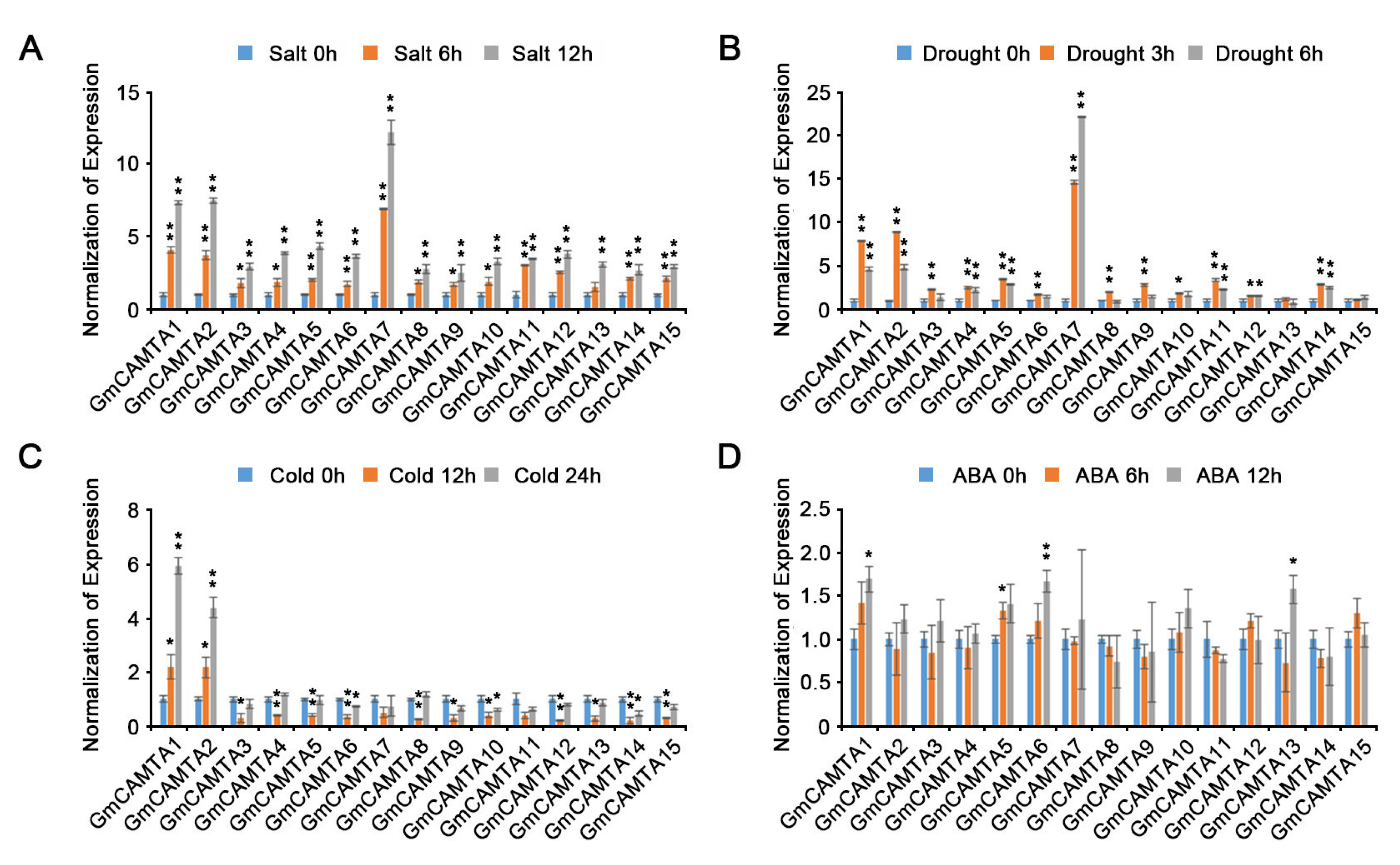
2.2. Various Abiotic Stresses Affected the Expression of GmCAMTAs

Previously, a few studies reported that the expression of GmCAMTAs is regulated by abiotic stresses and hormone treatment in soybean [22,32]. However, previous results had not considered the rhythmical changes of GmCAMTAs expression in abiotic stress responses. To accurately investigate the transcript changes of GmCAMTAs in response to abiotic stress treatment in view of the rhythmical changes, we measured the relative expression values of GmCAMTA genes under both non-stress and abiotic stress conditions; then, we carried out the normalization of the relative expression values measured under abiotic stress conditions, with the relative expression values at each time point measured under non-stress conditions. When soybean seedlings were exposed to salt stress, all of the GmCAMTAs were highly expressed at early times, and their expression levels increased gradually as time passed (Figure 2A). The expression levels of most GmCAMTAs in drought stress responses were significantly increased compared to those under non-stress conditions, except GmCAMTA13 and GmCAMTA15 (Figure 2B). Interestingly, GmCAMTA1 and GmCAMTA2 were highly expressed to cold stress; however, the expression levels of other GmCAMTAs were reduced during cold stress treatment (Figure 2C). ABA did not influence the expression of most GmCAMTAs in soybean, although GmCAMTA1, GmCAMTA5, GmCAMTA6, and GmCAMTA13 were weakly expressed under exogenous ABA conditions (Figure 2D). Thus, the results indicate that the gene expressions of GmCAMTAs were differently influenced by various abiotic stresses.

2.3. The Transcripts of GmCAMTAs had Substantial Specificity in Developmental Processes

Plant developmental processes are associated with abiotic stress responses through impressive biochemical, physiological, and morphological changes [33]. We tested the changes in GmCAMTA expression levels in different tissues using qRT-PCR analysis. Many GmCAMTAs were expressed in the roots; however, GmCAMTA2, GmCAMTA7, GmCAMTA9, GmCAMTA14, and GmCAMTA15 were weakly expressed (Figure 3, Supplementary Figure S2A). In the leaves, the expression of most GmCAMTAs was highly induced, except for those of GmCAMTA2, GmCAMTA8, and GmCAMTA12 (Figure 3, Supplementary Figure S2B). Interestingly, GmCAMTA2, GmCAMTA8, and GmCAMTA12 were relatively highly expressed in the stem tissues as compared to other GmCAMTAs (Figure 3, Supplementary Figure S2C). Unlike the expression patterns of GmCAMTAs in roots, leaves, and stem tissues, the expression levels of GmCAMTAs were similar in the flower (Figure 3, Supplementary Figure S2D). Our results indicated that GmCAMTAs were selectively expressed in each of the plant tissues, thereby showing substantial specificity in the developmental stages.

2.4. Overexpression of GmCAMTA2 and GmCAMTA8 Reduced Drought Tolerance in Arabidopsis

GmCAMTA2, GmCAMTA8, and GmCAMTA12 were highly expressed in the stem (Figure 3). The overexpression of GmCAMTA12 in Arabidopsis and soybean enhance plant tolerance to drought stress [22]. To investigate the functions of GmCAMTA2 and GmCAMTA8 in drought stress responses, we generated Arabidopsis transgenic plants with overexpressed GmCAMTA2 and GmCAMTA8 under the CAMV 35S promoter. To establish the expression levels of GmCAMTA2 and GmCAMTA8 in Arabidopsis transgenic plants, we performed an RT-PCR analysis. According to the results of RT-PCR, we selected three individual lines, each with different expression levels of GmCAMTA2 (GmCAMTA2-OX #1, #4, and #8 lines; Supplementary Figure S3A) and GmCAMTA8 (GmCAMTA8-OX #5, #8, and #9 lines; Supplementary Figure S3B). We examined the tolerance of Arabidopsis transgenic plants overexpressing GmCAMTA2 and GmCAMTA8 during exposure to drought stress (Figure 4). In natural conditions, GmCAMTA2-OX and GmCAMTA8-OX plants were similar in growth and development compared with WT and empty vector-overexpressing (Vector-OX) Arabidopsis plants (Figure 4A). GmCAMTA2-OX and GmCAMTA8-OX plants showed hypersensitivity compared with WT and Vector-OX plants under drought stress and re-watering conditions (Figure 4A). The survival rates after re-watering under drought stress conditions were lower in the GmCAMTA2-OX (approximately 2.78 to 13.89%) and GmCAMTA8-OX (approximately 2.78 to 11.11%) than the WT (approximately 72.22 to 79.17%) and Vector-OX (approximately 70.83 to 80.56%) (Figure 4B). To confirm the drought-sensitive phenotypes in the GmCAMTA2-OX and GmCAMTA8-OX plants, we measured the water loss from detached leaves during exposure to drought stress. The water loss of the GmCAMTA2-OX and GmCAMTA8-OX plants was significantly higher than those of the WT and Vector-OX plants in response to drought stress (Figure 4C). These results suggested that the overexpression of GmCAMTA2 and GmCAMTA8 reduced plant tolerance to drought stress, and GmCAMTA2 and GmCAMTA8 function as negative regulators in drought stress response.

2.5. Overexpression of GmCAMTA2 and GmCAMTA8 Regulated the Transcription of Stress-Responsive Genes in Drought Stress Responses

To investigate whether overexpression of GmCAMTA2 and GmCAMTA8 affected the expression patterns of stress-responsive genes during drought stress responses, we performed qRT-PCR analysis of various stress-responsive genes (Figure 5). The expression levels of ABA-induced genes, AtRD29A and AtRD29B, were significantly decreased in GmCAMTA2-OX (AtRD29A; approximately 0.97- to 1.67-fold, AtRD29B; approximately 1.18- to 1.69-fold) and GmCAMTA8-OX (AtRD29A; approximately 1.18- to 1.61-fold, AtRD29B; approximately 0.94- to 1.53-fold) compared with those in WT plants (AtRD29A; approximately 2.61-fold, AtRD29B; approximately 3.60-fold) at the 60 min time point during drought stress. The Pyrroline-5-Carboxylate Synthase 2 (P5CS2) gene was regulated in the biosynthesis of proline, which is an antioxidant in water stress response [34]. The expression of AtP5CS2 was weakly decreased in GmCAMTA2-OX (approximately 0.75- to 0.82-fold) and GmCAMTA8-OX (approximately 0.62- to 0.88-fold) compared with those in WT plants (approximately 1.23-fold) at the 60 min time point during drought stress. In addition, the expression of the stress-related marker gene AtKIN1 was decreased in GmCAMTA2-OX (approximately 1.60- to 1.92-fold) and GmCAMTA8-OX (approximately 1.37- to 1.50-fold) compared with those in WT plants (approximately 2.76-fold) at the 60 min time point during drought stress. These results suggested that the overexpression of GmCAMTA2 and GmCAMTA8 down-regulated the expression of stress-responsive genes during drought stress in Arabidopsis. Thus, GmCAMTA2 and GmCAMTA8 act as negative regulators in drought stress responses.

3. Discussion

The characterization of various CAMTA genes as core transcription factors with CaM binding sites has been reported in recent years by genome-wide analysis and expression profiles [6,7,8,9,10,11,12,13,16,32]. Although 15 GmCAMTA genes in soybeans have been identified, the functions of GmCAMTAs remained almost unknown, except for GmCAMTA12 [22]. In this study, we showed that the expression of GmCAMTA genes was controlled by circadian rhythms (Figure 1). Previous studies of CAMTA genes have not indicated the circadian regulation of CAMTA gene expression. Plant cellular responses, such as gene expression, development, and metabolic processes, were circadian regulated by circadian oscillators [35,36]. Circadian responses in plants were attributable to cellular oscillations in the intracellular Ca2+ concentration ([Ca2+]) in the surrounding environment, including photoperiod and light intensity [37,38]. The calmodulin-like 24 (CML24), one of the Ca2+-binding proteins, regulated the circadian period in Ca2+-dependent signaling [36]. The most important properties of CAMTAs in plants have a high correlation with CaM proteins, as a Ca2+ sensor, to regulate the gene expression in Ca2+-dependent plant responses [1]. These facts were important to explain that the circadian regulation of GmCAMTAs transcripts was mediated by the cellular oscillation of [Ca2+].
Many CAMTAs in crops have been directly or indirectly associated with various developmental and cellular processes, whether through autonomously regulating a major transcription factor or targeting other transcription factors [1]. The fifteen GmCAMTAs in soybean [32], seven PtCAMTAs in Populus trichocarpa [9], nine ZmCAMTAs in maize [12], seven MtCAMTAs in Medicago truncatula [13], and fifteen VvCAMTAs in Vitis vinifera [39] were endowed with substantial specificity in the developmental stages by highly tissue-specific expression. Root-specific-induced PtCAMTA2 and PtCAMTA3 genes were negatively regulated under cold stress in a short time [9]. In this study, we showed that GmCAMTA2, GmCAMTA8, and GmCAMTA12 were especially highly expressed in stem tissues compared to other GmCAMTAs (Figure 3). The growth and development of plant tissues, such as flowers, stems, and leaves, were greatly affected by abiotic stress [40]. The tissue-specific expression pattern of GmCAMTAs is an important characteristic for investigating the functions of GmCAMTAs in diverse molecular processes against environmental stresses. Since plants are constantly exposed to environmental stimuli during their lifetime, they have developed various adaptive mechanisms between the developmental process and stress responses [41,42].
Drought stress is an important restricting factor for plant growth and developmental processes, holding both expansion and elongation down [41]. Since drought stress triggers low turgor pressure in the cell, the osmotic maintenance of cell turgor in plants is of great importance for plant growth and survival [41]. In many crops, soybeans show a highly sensitive response to drought stress through various morphological changes [42]. When exposed to drought stress, soybean shows momentous changes in most of the tissue morphology, such as a reduction of newly developed branches and trifoliate leaves, growth inhibition of newly emerging leaves, and reduction in leaf area via repression of lamina expansion [42]. Drought stress has a negative effect on the developmental processes of stems and leaves by decreasing their elongation and expansion [41]. In particular, drought stress often leads to the expression of common morphogenesis- and stress-responsive genes through molecular mechanisms, including reactive oxygen species (ROS)- and phytohormone-dependent signaling [40]. ATP-dependent metalloprotease 4 (AtFtsH4) played a tissue-specific role in maintaining the stem cell activity of shoot apical meristem (SAM) stages by ROS accumulation under high-temperature conditions [43]. MscS-like 2 (MSL2) and MSL3 were mediated to cell proliferation by regulation of WUSCHEL (WUS) gene expression in osmotic stress responses [44]. The mutation of NADPH genes showed unstable shoot development by decreasing the stem cell population [45]. In the case of CAMTA genes, the relationships between development and stress responses have been studied more in fruit species than in crops. The tomato SISR2 and SISR3L transcriptions were affected by ethylene signaling during fruit development and ripening [7]. The expression of peach PpCAMTA1 was induced in cold stress, but PpCAMTA3 was repressed with UV-B irradiation during fruit development [23]. Our results indicated that GmCAMTA2, GmCAMTA8, and GmCAMTA12 were significantly expressed in both stem tissues and drought stress responses (Figure 2 and Figure 3). When GmCAMTA2 and GmCAMTA8 were overexpressed in Arabidopsis, GmCAMTA2-OX, and GmCAMTA8-OX displayed the sensitive phenotype under drought stress conditions by losing water faster than WT plant (Figure 4). These results suggest that GmCAMTA2 and GmCAMTA8 act as negative regulators in drought stress responses. Considering the tolerant phenotype of Arabidopsis transgenic plants produced by GmCAMTA12 overexpression [22] and sensitive phenotypes by GmCAMTA2 and GmCAMTA8 overexpression (Figure 4) under drought stress conditions, GmCAMTA2, GmCAMTA8, and GmCAMTA12 may be associated with drought tolerance response by different regulatory mechanisms. Our results suggest that GmCAMTA2 and GmCAMTA8 play an important role in both the development process and drought stress responses by circadian regulation.

4. Materials and Methods

4.1. Plant Materials and Growth Conditions

We used the soybean (Glycine max cv. Williams 82) and Arabidopsis (Arabidopsis thaliana Col-0 ecotype) plants in this study. The soybean plants were cultured in a growth chamber at 28 °C under long-day conditions (16 h light/8 h dark), and Arabidopsis plants in a growth chamber at 22 °C under long-day conditions (16 h light/8 h dark). To test the expression levels of GmCAMTAs, the two-week-old soybean plants were hydroponically cultured in the 1/2 MS medium with circadian rhythms. We used the two-week-old soybean plants in the 1/2 MS medium for various abiotic stress treatments. For salt stress treatment, the two-week-old soybean plants were transferred uniformly in the 1/2 MS liquid medium containing 150 mM NaCl (salt stress). For cold stress treatment, the two-week-old soybean plants in the 1/2 MS medium were placed on ice (approximately 2 °C). For ABA hormonal treatment, the two-week-old soybean plants were transferred uniformly in the 1/2 MS liquid medium. And then, 100 µM ABA was sprayed on the leaves of soybean plants. To treat soybean or Arabidopsis plants to drought stress, the two-week-old soybean or 10-day-old Arabidopsis plants, after removing the medium, were transferred to the Petri dish. And then, plants were placed in a growth chamber under long-day conditions (16 h light/8 h dark). Stress treatments were applied at ZT 3. After stress treatments, we performed the plant sampling at indicated different times. For the expression analysis of GmCAMTAs in different tissues, the soybean plants, after sowing seeds, were grown in the greenhouse under the seasonal cultivation period.

4.2. Identification of GmCAMTA Genes in Soybean

The GmCAMTA genes were identified from the soybean genome database at Phytozome version 13 (https://phytozome-next.jgi.doe.gov/, accessed on 1 July 2019). The specific primers for the qRT-PCR and RT-PCR analyses were designed with the IDT (Integrated DNA Technologies) service website (https://sg.idtdna.com/pages, accessed on 10 August 2020) based on the sequence information of GmCAMTAs (Supplementary Table S2). The full-length coding sequences of GmCAMTA2 (3309 bp) and GmCAMTA8 (2736 bp) were amplified by PCR using cDNA from soybean plants, and then cloned into the pMLBart binary vector under the control of the CaMV 35S promoter, which contained a BASTA resistance gene (Supplementary Figure S3). We confirmed the full-length coding sequences through DNA sequencing analysis.

4.3. Generation of GmCMATA-Overexpressing Arabidopsis Transgenic Plants

The pMLBart-GmCAMTA2 and pMLBart-GmCAMTA8 plasmids were transformed into Arabidopsis plants using the Agrobacterium-mediated floral-dipping methods. The GmCAMTA2- and GmCAMTA8-overexpressing transgenic plants were selected on the soil by spraying with 0.2% BASTA herbicide. The expression levels of GmCAMTA2 and GmCAMTA8 in Arabidopsis transgenic plants were analyzed using reverse transcription PCR (RT-PCR) with gene-specific primers (Supplementary Table S2).

4.4. Physiological Assay of Drought Stress

To test the physiological phenotypes under drought stress conditions, two-week-old plants grown in soil with sufficient water were not watered for 13 days. After re-watering, the recovery of the drought-treated plants was monitored. The assays for drought sensitivity were thrice-replicated experiments using at least 12 plants for each line in each experiment. To measure the transpirational water loss, the shoots of 4-week-old plants in the soil were detached from the root. After being placed on Petri dishes, it was weighed immediately. The fresh weights of detached leaves were measured periodically at the indicated times, and the percentages of water loss were calculated. The water loss assays were replicated three times, using at least four plants for each line in each experiment.

4.5. Analysis of Gene Expression

The total RNA was isolated from each plant sample using the RNeasy Plant Mini Kit (Qiagen, Hilden, Germany), according to the manufacturer’s instructions. To remove any genomic DNA contaminants, purified total RNA was treated with DNaseI (Sigma-Aldrich, St. Louis, MO, USA). For the RT-PCR and quantitative RT-PCR (qRT-PCR) analyses, 1 µg of total RNA was used for cDNA synthesis using the RevertAid First Strand cDNA Synthesis Kit (Thermo Fisher Scientific, Waltham, MA, USA), in accordance with the manufacturer’s protocol. The qRT-PCR analysis was performed using the QuantiSpeed SYBR No-ROX Kit (PhileKorea, Seoul, Republic of Korea), and the relative gene expression levels were automatically calculated using the CFX384 real-time PCR detection system (Bio-Rad Laboratories, Hercules, CA, USA). The expression levels of soybean GmTUBULIN and Arabidopsis AtTUBULIN2 were used as the internal control. The qRT-PCR experiments were performed in three independent replicates. The gene-specific primers used are listed in Supplementary Table S2. To represent the heatmap, qRT-PCR data for each GmCAMTA gene were normalized on the GmTUBULIN, and the expression heatmap was performed with the Microsoft Excel program.

5. Conclusions

In summary, we characterized the biological roles of 15 GmCAMTA genes in soybean (Glycine max cv. Williams 82). A total of 15 GmCAMTAs were significantly expressed in accordance with circadian rhythms in the daily clock. The expression levels of most GmCAMTAs were induced during various abiotic stresses, including salt, drought, and cold stress, except ABA. Interestingly, GmCAMTA2, GmCAMTA8, and GmCAMTA12 were more highly expressed in stem tissues. The overexpression of GmCAMTA2 and GmCAMTA8 in Arabidopsis suppressed plant tolerance under drought-stress conditions. The expression levels of stress-responsive genes were dramatically reduced by overexpressing GmCAMTA2 and GmCAMTA8 in drought stress responses. These results suggest that GmCAMTA2 and GmCAMTA8 function in tissue-specific and plant stress responses through rhythmic regulation under drought stress conditions.

Supplementary Materials

The supporting information can be downloaded at: https://www.mdpi.com/article/10.3390/ijms241411477/s1.

Author Contributions

D.B., H.M.C., H.J.C. and M.C.K. designed and performed the experiments, analyzed data, and wrote the manuscript. Y.J.C., B.J.J., S.H.L. and M.S.P. performed some experiments. M.C.K. discussed and commented on the results and revised the manuscript. All authors have read and agreed to the published version of the manuscript.

Funding

This research was supported by the Basic Science Research Program through the National Research Foundation of Korea (NRF), funded by the Ministry of Education (2015R1A6A1A03031413 (M.C.K.), 2020R1I1A1A01067256 (H.J.C.), 2020R1A6A1A03044344 (D.B.) and 2022R1I1A1A01072247 (D.B.)).

Institutional Review Board Statement

Not applicable.

Informed Consent Statement

Not applicable.

Data Availability Statement

Data supporting the reported results can be made available upon request to the corresponding author.

Conflicts of Interest

The authors declare no conflict of interest.

References

  1. Iqbal, Z.; Shariq Iqbal, M.; Singh, S.P.; Buaboocha, T. Ca2+/Calmodulin complex triggers CAMTA transcriptional machinery under stress in plants: Signaling cascade and molecular regulation. Front. Plant Sci. 2020, 11, 598327. [Google Scholar] [CrossRef] [PubMed]
  2. Kudla, J.; Becker, D.; Grill, E.; Hedrich, R.; Hippler, M.; Kummer, U.; Parniske, M.; Romeis, T.; Schumacher, K. Advances and current challenges in calcium signaling. New Phytol. 2018, 218, 414–431. [Google Scholar] [CrossRef] [PubMed]
  3. Day, I.S.; Reddy, V.S.; Shad Ali, G.; Reddy, A.S. Analysis of EF-hand-containing proteins in Arabidopsis. Genome Biol. 2002, 3, RESEARCH0056. [Google Scholar] [CrossRef] [PubMed]
  4. Sun, Q.P.; Guo, Y.; Sun, Y.; Sun, D.Y.; Wang, X.J. Influx of extracellular Ca2+ involved in jasmonic-acid-induced elevation of [Ca2+]cyt and JR1 expression in Arabidopsis thaliana. J. Plant Res. 2006, 119, 343–350. [Google Scholar] [CrossRef] [PubMed]
  5. Poovaiah, B.W.; Du, L. Calcium signaling: Decoding mechanism of calcium signatures. New Phytol. 2018, 217, 1394–1396. [Google Scholar] [CrossRef] [PubMed]
  6. Xiao, P.; Feng, J.W.; Zhu, X.T.; Gao, J. Evolution analyses of CAMTA transcription factor in plants and its enhancing effect on cold-tolerance. Front. Plant Sci. 2021, 12, 758187. [Google Scholar] [CrossRef]
  7. Yang, T.; Peng, H.; Whitaker, B.D.; Conway, W.S. Characterization of a calcium/calmodulin-regulated SR/CAMTA gene family during tomato fruit development and ripening. BMC Plant Biol. 2012, 12, 19. [Google Scholar] [CrossRef]
  8. Zhou, Q.; Zhao, M.; Xing, F.; Mao, G.; Wang, Y.; Dai, Y.; Niu, M.; Yuan, H. Identification and expression analysis of CAMTA genes in tea plant reveal their complex regulatory role in stress responses. Front. Plant Sci. 2022, 13, 910768. [Google Scholar] [CrossRef]
  9. Wei, M.; Xu, X.; Li, C. Identification and expression of CAMTA genes in Populus trichocarpa under biotic and abiotic stress. Sci. Rep. 2017, 7, 17910. [Google Scholar] [CrossRef]
  10. Meer, L.; Mumtaz, S.; Labbo, A.M.; Khan, M.J.; Sadiq, I. Genome-wide identification and expression analysis of calmodulin-binding transcription activator genes in banana under drought stress. Sci. Hortic. 2019, 244, 10–14. [Google Scholar] [CrossRef]
  11. Saeidi, K.; Zare, N.; Baghizadeh, A.; Asghari-Zakaria, R. Phaseolus vulgaris genome possesses CAMTA genes, and phavuCAMTA1 contributes to the drought tolerance. J. Genet. 2019, 98, 31. [Google Scholar] [CrossRef]
  12. Yue, R.; Lu, C.; Sun, T.; Peng, T.; Han, X.; Qi, J.; Yan, S.; Tie, S. Identification and expression profiling analysis of calmodulin-binding transcription activator genes in maize (Zea mays L.) under abiotic and biotic stresses. Front. Plant Sci. 2015, 6, 576. [Google Scholar] [CrossRef] [PubMed]
  13. Yang, Y.; Sun, T.; Xu, L.; Pi, E.; Wang, S.; Wang, H.; Shen, C. Genome-wide identification of CAMTA gene family members in Medicago truncatula and their expression during root nodule symbiosis and hormone treatments. Front. Plant Sci. 2015, 6, 459. [Google Scholar] [CrossRef]
  14. Pandey, N.; Ranjan, A.; Pant, P.; Tripathi, R.K.; Ateek, F.; Pandey, H.P.; Patre, U.V.; Sawant, S.V. CAMTA 1 regulates drought responses in Arabidopsis thaliana. BMC Genom. 2013, 14, 216. [Google Scholar] [CrossRef]
  15. Doherty, C.J.; Van Buskirk, H.A.; Myers, S.J.; Thomashow, M.F. Roles for Arabidopsis CAMTA transcription factors in cold-regulated gene expression and freezing tolerance. Plant Cell 2009, 21, 972–984. [Google Scholar] [CrossRef] [PubMed]
  16. Noman, M.; Aysha, J.; Ketehouli, T.; Yang, J.; Du, L.; Wang, F.; Li, H. Calmodulin binding transcription activators: An interplay between calcium signalling and plant stress tolerance. J. Plant Physiol. 2021, 256, 153327. [Google Scholar] [CrossRef]
  17. Du, L.; Ali, G.S.; Simons, K.A.; Hou, J.; Yang, T.; Reddy, A.S.; Poovaiah, B.W. Ca2+/calmodulin regulates salicylic-acid-mediated plant immunity. Nature 2009, 457, 1154–1158. [Google Scholar] [CrossRef] [PubMed]
  18. Galon, Y.; Nave, R.; Boyce, J.M.; Nachmias, D.; Knight, M.R.; Fromm, H. Calmodulin-binding transcription activator (CAMTA) 3 mediates biotic defense responses in Arabidopsis. FEBS Lett. 2008, 582, 943–948. [Google Scholar] [CrossRef] [PubMed]
  19. Mitsuda, N.; Isono, T.; Sato, M.H. Arabidopsis CAMTA family proteins enhance V-PPase expression in pollen. Plant Cell Physiol. 2003, 44, 975–981. [Google Scholar] [CrossRef]
  20. Shkolnik, D.; Finkler, A.; Pasmanik-Chor, M.; Fromm, H. Calmodulin-binding transcription activator 6: A key regulator of Na+ homeostasis during germination. Plant Physiol. 2019, 180, 1101–1118. [Google Scholar] [CrossRef]
  21. Koo, S.C.; Choi, M.S.; Chun, H.J.; Shin, D.B.; Park, B.S.; Kim, Y.H.; Park, H.M.; Seo, H.S.; Song, J.T.; Kang, K.Y.; et al. The calmodulin-binding transcription factor OsCBT suppresses defense responses to pathogens in rice. Mol. Cells 2009, 27, 563–570. [Google Scholar] [CrossRef]
  22. Noman, M.; Jameel, A.; Qiang, W.D.; Ahmad, N.; Liu, W.C.; Wang, F.W.; Li, H.Y. Overexpression of GmCAMTA12 enhanced drought tolerance in Arabidopsis and soybean. Int. J. Mol. Sci. 2019, 20, 4849. [Google Scholar] [CrossRef]
  23. Yang, C.; Li, Z.; Cao, X.; Duan, W.; Wei, C.; Zhang, C.; Jiang, D.; Li, M.; Chen, K.; Qiao, Y.; et al. Genome-wide analysis of calmodulin binding transcription activator (CAMTA) gene family in peach (Prunus persica L. Batsch) and ectopic expression of PpCAMTA1 in Arabidopsis camta2,3 mutant restore plant development. Int. J. Mol. Sci. 2022, 23, 10500. [Google Scholar] [CrossRef] [PubMed]
  24. Xu, X.; Yuan, L.; Xie, Q. The circadian clock ticks in plant stress responses. Stress Biol. 2022, 2, 15. [Google Scholar] [CrossRef]
  25. Lee, K.H.; Piao, H.L.; Kim, H.Y.; Choi, S.M.; Jiang, F.; Hartung, W.; Hwang, I.; Kwak, J.M.; Lee, I.J.; Hwang, I. Activation of glucosidase via stress-induced polymerization rapidly increases active pools of abscisic acid. Cell 2006, 126, 1109–1120. [Google Scholar] [CrossRef]
  26. Adams, S.; Grundy, J.; Veflingstad, S.R.; Dyer, N.P.; Hannah, M.A.; Ott, S.; Carré, I.A. Circadian control of abscisic acid biosynthesis and signalling pathways revealed by genome-wide analysis of LHY binding targets. New Phytol. 2018, 220, 893–907. [Google Scholar] [CrossRef] [PubMed]
  27. Grundy, J.; Stoker, C.; Carré, I.A. Circadian regulation of abiotic stress tolerance in plants. Front. Plant Sci. 2015, 6, 648. [Google Scholar] [CrossRef] [PubMed]
  28. Miyazaki, Y.; Abe, H.; Takase, T.; Kobayashi, M.; Kiyosue, T. Overexpression of LOV KELCH protein 2 confers dehydration tolerance and is associated with enhanced expression of dehydration-inducible genes in Arabidopsis thaliana. Plant Cell Rep. 2015, 34, 843–852. [Google Scholar] [CrossRef]
  29. Nakamichi, N.; Kita, M.; Ito, S.; Yamashino, T.; Mizuno, T. Pseudoresponse regulators, PRR9, PRR7 and PRR5, together play essential roles close to the circadian clock of Arabidopsis thaliana. Plant Cell Physiol. 2005, 46, 686–698. [Google Scholar] [CrossRef]
  30. Baek, D.; Kim, W.Y.; Cha, J.Y.; Park, H.J.; Shin, G.; Park, J.; Lim, C.J.; Chun, H.J.; Li, N.; Kim, D.H.; et al. The Gigantea-Enhanced EM level complex enhances drought tolerance via regulation of abscisic acid synthesis. Plant Physiol. 2020, 184, 443–458. [Google Scholar] [CrossRef]
  31. Legnaioli, T.; Cuevas, J.; Mas, P. TOC1 functions as a molecular switch connecting the circadian clock with plant responses to drought. EMBO J. 2009, 28, 3745–3757. [Google Scholar] [CrossRef]
  32. Wang, G.; Zeng, H.; Hu, X.; Zhu, Y.; Chen, Y.; Shen, C.; Wang, H.; Poovaiah, B.W.; Du, L. Identification and expression analyses of calmodulin-binding transcription activator genes in soybean. Plant Soil. 2015, 386, 205–221. [Google Scholar] [CrossRef]
  33. Ma, X.; Su, Z.; Ma, H. Molecular genetic analyses of abiotic stress responses during plant reproductive development. J. Exp. Bot. 2020, 71, 2870–2885. [Google Scholar] [CrossRef] [PubMed]
  34. Sabbioni, G.; Funck, D.; Forlani, G. Enzymology and regulation of δ1-pyrroline-5-carboxylate synthetase 2 from rice. Front. Plant Sci. 2021, 12, 672702. [Google Scholar] [CrossRef]
  35. Más, P. Circadian clock function in Arabidopsis thaliana: Time beyond transcription. Trends Cell Biol. 2008, 18, 273–281. [Google Scholar] [CrossRef] [PubMed]
  36. Martí Ruiz, M.C.; Hubbard, K.E.; Gardner, M.J.; Jung, H.J.; Aubry, S.; Hotta, C.T.; Mohd-Noh, N.I.; Robertson, F.C.; Hearn, T.J.; Tsai, Y.C.; et al. Circadian oscillations of cytosolic free calcium regulate the Arabidopsis circadian clock. Nat. Plants 2018, 4, 690–698. [Google Scholar] [CrossRef]
  37. Love, J.; Dodd, A.N.; Webb, A.A. Circadian and diurnal calcium oscillations encode photoperiodic information in Arabidopsis. Plant Cell 2004, 16, 956–966. [Google Scholar] [CrossRef]
  38. Tataroglu, O.; Zhao, X.; Busza, A.; Ling, J.; O’Neill, J.S.; Emery, P. Calcium and SOL protease mediate temperature resetting of circadian clocks. Cell. 2015, 163, 1214–1224. [Google Scholar] [CrossRef]
  39. Shangguan, L.; Wang, X.; Leng, X.; Liu, D.; Ren, G.; Tao, R.; Zhang, C.; Fang, J. Identification and bioinformatic analysis of signal responsive/calmodulin-binding transcription activators gene models in Vitis vinifera. Mol. Biol. Rep. 2014, 41, 2937–2949. [Google Scholar] [CrossRef]
  40. Zhu, J.K. Abiotic Stress Signaling and Responses in Plants. Cell 2016, 167, 313–324. [Google Scholar] [CrossRef]
  41. Jaleel, C.A.; Manivannan, P.; Wahid, A.; Farooq, M.; Al-Juburi, H.J.; Somasundararam, R.; Panneerselvam, R. Drought stress in plants: A review on morphological characteristics and pigments composition. Int. J. Agric. Biol. 2009, 11, 100–105. Available online: https://www.researchgate.net/publication/253008137 (accessed on 10 July 2023).
  42. Mangena, P. Water stress: Morphological and anatomic changes in soybean (Glycine max L.) plants. In Plant, Abiotic Stress and Responses to Climate Change; Andjelkovic, V., Ed.; Intech Open: London, UK, 2018; pp. 9–31. [Google Scholar] [CrossRef]
  43. Lee, H. Stem cell maintenance and abiotic stress response in shoot apical meristem for developmental plasticity. J. Plant Biol. 2018, 61, 358–365. [Google Scholar] [CrossRef]
  44. Dolzblasz, A.; Smakowska, E.; Gola, E.M.; Sokołowska, K.; Kicia, M.; Janska, H. The mitochondrial protease AtFTSH4 safeguards Arabidopsis shoot apical meristem function. Sci. Rep. 2016, 6, 28315. [Google Scholar] [CrossRef] [PubMed]
  45. Wilson, M.E.; Mixdorf, M.; Berg, R.H.; Haswell, E.S. Plastid osmotic stress influences cell differentiation at the plant shoot apex. Development 2016, 143, 3382–3393. [Google Scholar] [CrossRef] [PubMed]
Figure 1. Transcriptional influence of GmCAMTAs in circadian rhythms. The expression levels of GmCAMTAs were analyzed for circadian regulation using qRT-PCR. The two-week-old seedlings of wild-type (WT) soybeans (Glycine max cv. Williams 82) were grown under long-day conditions (16 h light/8 h dark). Zeitgeber time (ZT) means a time of turning on a light. The white and black areas mark the approximate division of light and dark. The qRT-PCR analysis was performed with each GmCAMTA-specific primer (Supplementary Table S2). GmTUBULIN was used as an internal control for normalization. Error bars represent the SD of three biological replicates, each with three technical replicates.
Ijms 24 11477 g001
Figure 2. Effects of various abiotic stresses on the expression of GmCAMTAs. The expression levels of GmCAMTAs were analyzed in soybean seedlings during exposure to various abiotic stresses using qRT-PCR. The total RNA was extracted from two-week-old seedlings of WT soybeans under different stresses: (A) salt (150 mM NaCl), (B) drought, (C) cold (approximately 2 °C), and (D) 100 µM ABA. The qRT-PCR analysis was performed with each GmCAMTA-specific primer (Supplementary Table S2). GmTUBULIN was used as an internal control, and then the values of the expression levels were calculated by expression values of GmCAMTAs under non-stress conditions for normalization. Error bars represent the SD of three biological replicates, each with three technical replicates. Asterisks indicate significant differences compared with the expression of the zero indicated time point by Student’s t-test (* p-value  <  0.05, ** p-value  <  0.01).
Ijms 24 11477 g002
Figure 3. Heatmap analysis of GmCAMTAs expression in soybean tissues. The heatmap analysis based on qRT-PCR results showed the relative expression levels of GmCAMTAs in different tissues of soybean plants. The total RNA was extracted from various tissues, including roots, leaves, stem, and flower, at the V4 stages of WT soybeans grown under long-day conditions (16 h light/8 h dark). The qRT-PCR analysis was performed with each GmCAMTA-specific primer (Supplementary Table S2). GmTUBULIN was used as an internal control for normalization.
Ijms 24 11477 g003
Figure 4. Effect of drought stress on GmCAMTA2 and GmCAMTA8 overexpressing transgenic Arabidopsis. (A) Phenotypes of GmCAMTA2-OX and GmCAMTA8-OX compared with WT and Vector-OX plants in response to drought stress and re-watering. The plants were grown in soil with sufficient water for two weeks (upper panels), then water was withheld for thirteen days (middle panels). The drought-stressed plants were then re-watered for one day (bottom panels). Each experiment comprised at least 12 plants, and three replicates were performed. (B) Comparison of survival rates (%) among GmCAMTA2-OX, GmCAMTA8-OX, Vector-OX, and WT plants against drought stress. Each experiment comprised at least 12 plants, and error bars represent the SD of three biological replicates. Asterisks indicate significant differences compared with WT plants by Student’s t-test (** p-value  <  0.01). (C) Analysis of water loss from detached leaves of three-week-old GmCAMTA2-OX, GmCAMTA8-OX, Vector-OX, and WT plants under drought stress conditions. Average values of water loss were measured at the indicated time points. Each experiment comprised at least four plants, and error bars represent the SD of three biological replicates. Asterisks indicate significant differences compared with WT plants by Student’s t-test (* p-value  <  0.05, ** p-value  <  0.01).
Ijms 24 11477 g004
Figure 5. Effect on the transcriptions of Arabidopsis stress-responsive genes by the overexpression of GmCAMTA2 and GmCAMTA8 in drought stress responses. The expression levels of stress-responsive genes were analyzed using qRT-PCR in GmCAMTA2-OX and GmCAMTA8-OX plants under drought-stress conditions. Total RNA was extracted from 10-day-old GmCAMTA2-OX and GmCAMTA8-OX Arabidopsis at indicated time points to drought stress. The qRT-PCR analysis was performed with each gene-specific primer of the Arabidopsis stress-responsive genes, AtRD29A, AtRD29B, AtP5CS2, and AtKIN1 (Supplementary Table S2). The expression of Arabidopsis AtTUBULIN2 was used as an internal control for normalization. Error bars represent the SD of two biological replicates, each with three technical replicates. Asterisks indicate significant differences compared with expression levels in WT plants by Student’s t-test (* p-value  <  0.05, ** p-value  <  0.01).
Ijms 24 11477 g005
Disclaimer/Publisher’s Note: The statements, opinions and data contained in all publications are solely those of the individual author(s) and contributor(s) and not of MDPI and/or the editor(s). MDPI and/or the editor(s) disclaim responsibility for any injury to people or property resulting from any ideas, methods, instructions or products referred to in the content.

