
The Potential of Polyelectrolyte Multilayer Films as Drug Delivery Materials


Abstract
1. Introduction
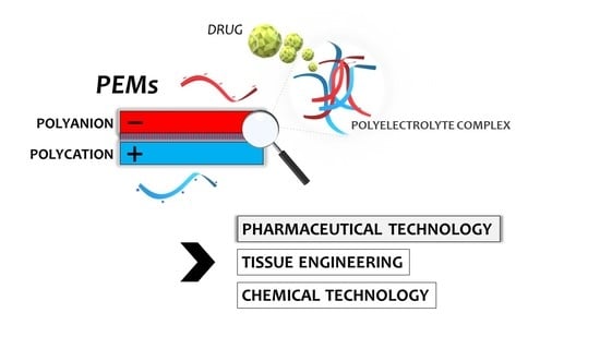
1.1. PECs
1.2. Potential of PEMs
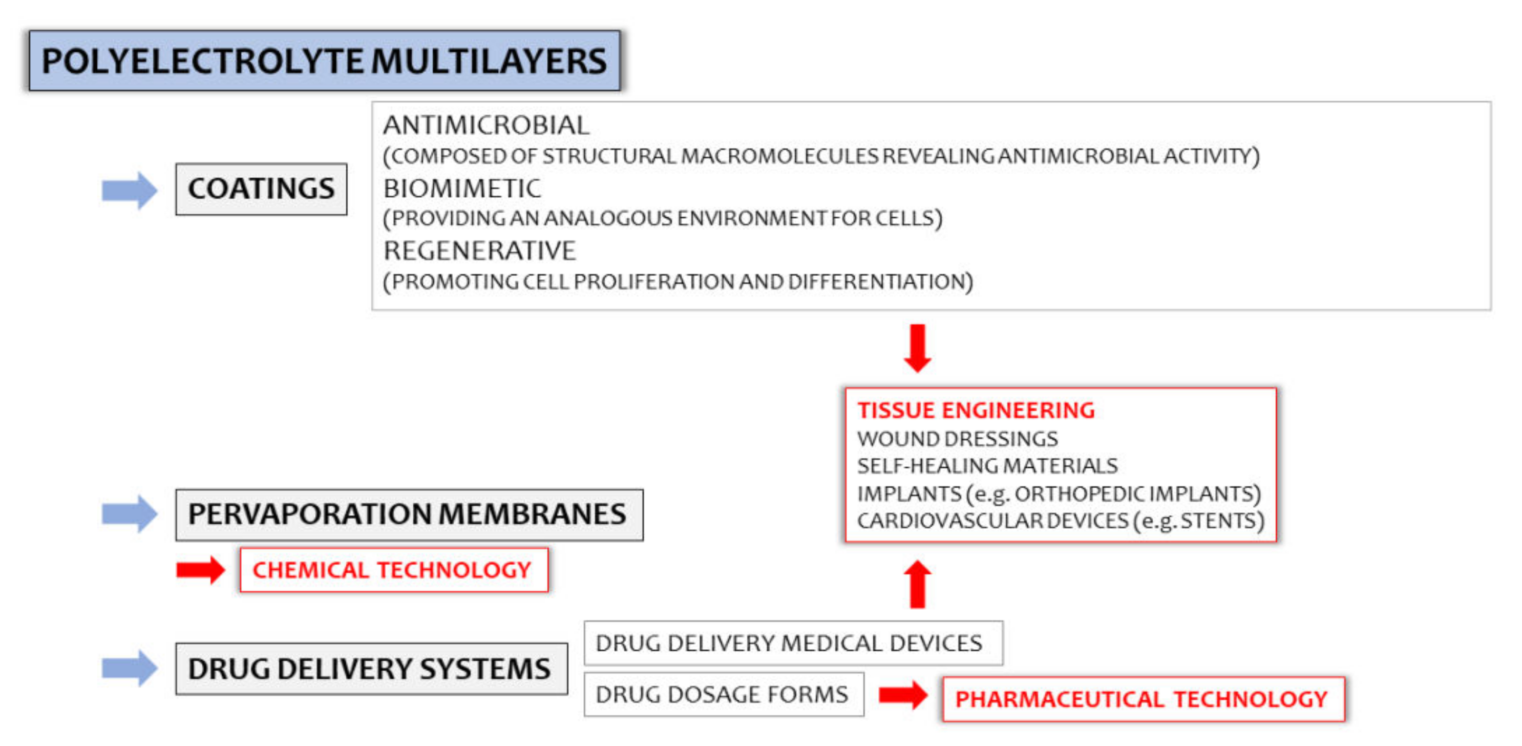
1.3. Physicochemical Background of PEM Formation
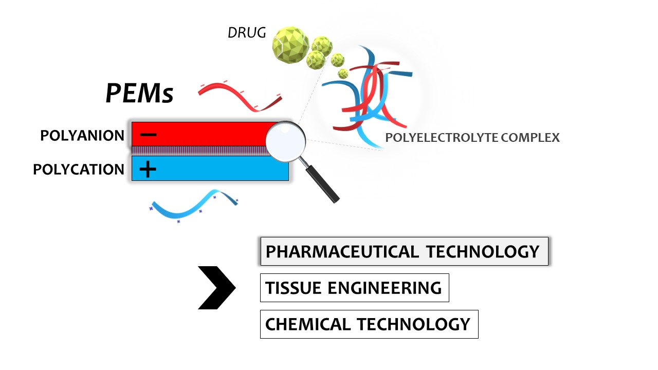
2. Applicability of PEMs
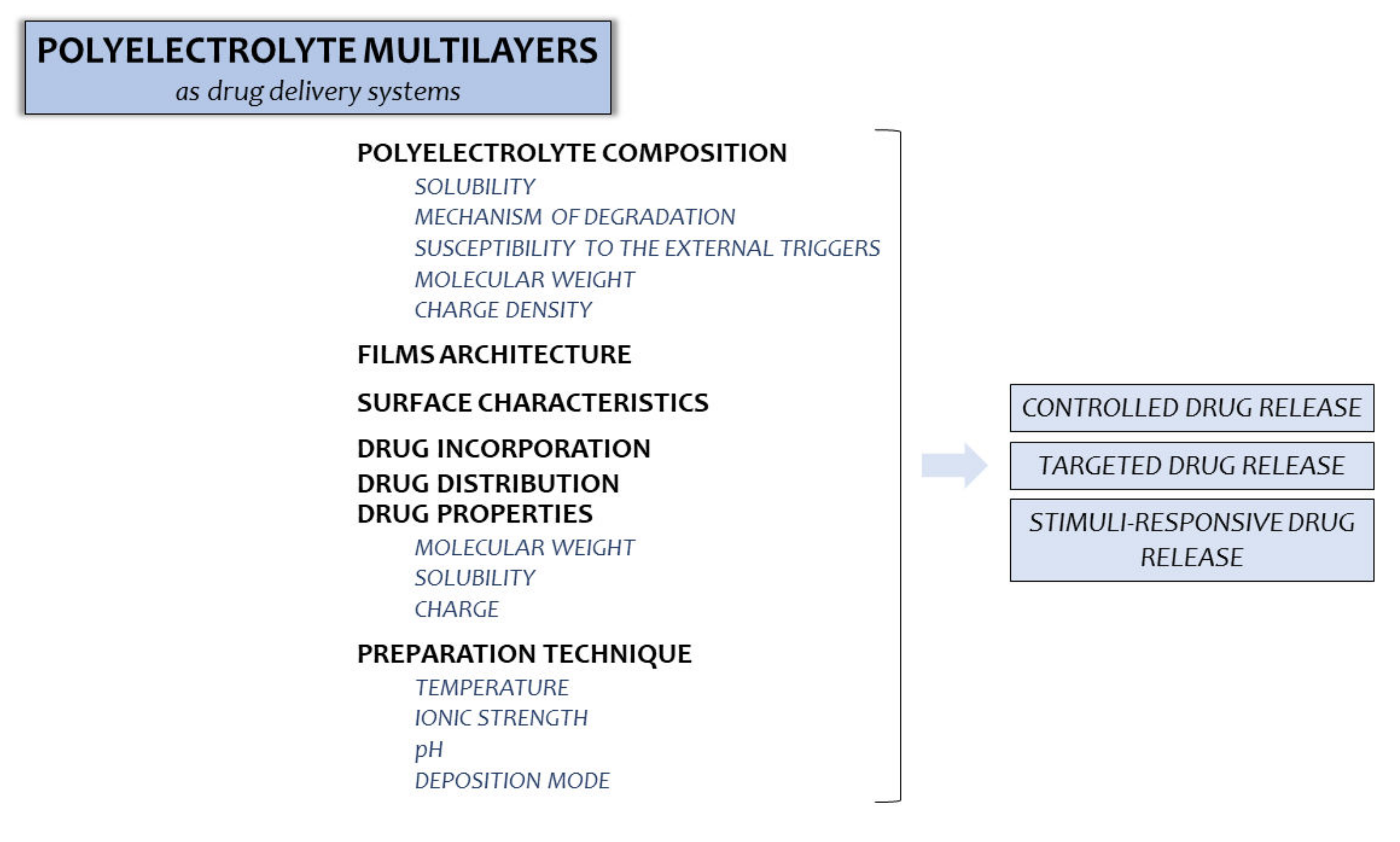
2.1. PEMs as Drug Delivery Systems

| Polycation | Polyanion | Active Substance | Potential Applicability | Reference |
|---|---|---|---|---|
| Chitosan | Sodium alginate | Tamoxifen | Patches, injectable gel-like films | Criado-Gonzalez et al. [35] |
| Hyaluronic acid/sodium hyaluronate | Doxorubicin hydrochloride, Fluorescein isothiocyanate, Ovalbumin | Anticancer rapidly-disintegrating films | Sun et al. [36] | |
| Casein sodium | Benzydamine hydrochloride | Buccal films | Pilicheva et al. [37] | |
| Heparin and dextran sulfate | Transforming GF β1, platelet-derived GF ββ, and insulin-like growth factor 1 | Materials for tissue regeneration | Damanik et al. [38] | |
| Poly(γ-glutamic acid) | Interferon-γ | Systems for gastric cancer treatment | Cardoso et al. [39] | |
| β-cyclodextrin polymer | 4-tert-butylbenzoic acid | Medical devices with antibiotics or antiseptic agents | Martin et al. [40] | |
| Gentamicin | Prevention of perioperative infections | Pérez-Anes et al. [41] | ||
| Sodium salt of carboxymethyl cellulose | Fluorescein isothiocyanate, ovalbumin | Medical devices | Park et al. [42] | |
| Pectin/Xanthan gum/Karaya gum | Tenofovir | Vaginal films | Martín-Illana et al. [33] | |
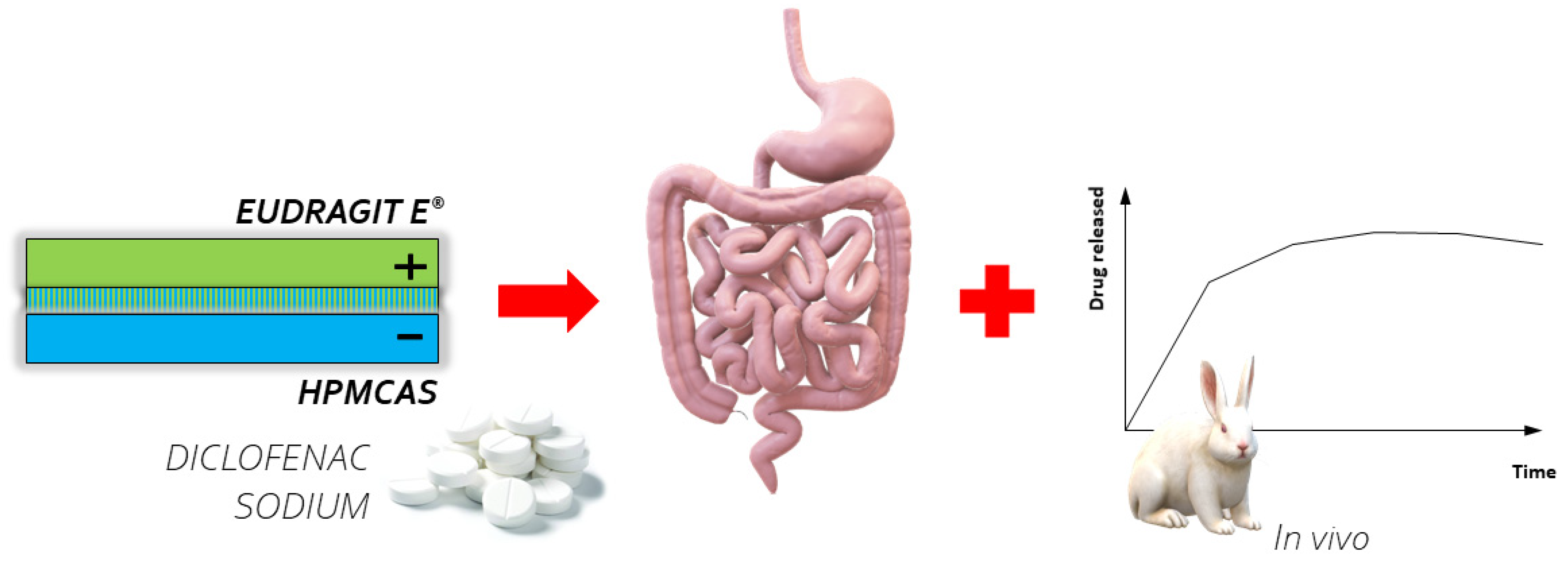
| Eudragit E® | Hypromellose acetate succinate | Diclofenac sodium | Colon-specific tablets | Jeganathan et al. [43] |
| Poly(4-vinylpyridine) | Sodium alginate | Ciprofloxacin hydrochloride | Transdermal systems | Alshhab et al. [31] |
2.1.1. PEMs as Drug Dosage Forms (Films, Patches, Tablets, etc.) for Skin and Mucosal Administration
2.1.2. PEMs as Coatings for Colon-Specific Tablets
2.1.3. PEMs as Platforms for Biomolecules
2.1.4. PEMs as Coatings for Implants with Antimicrobial Activity
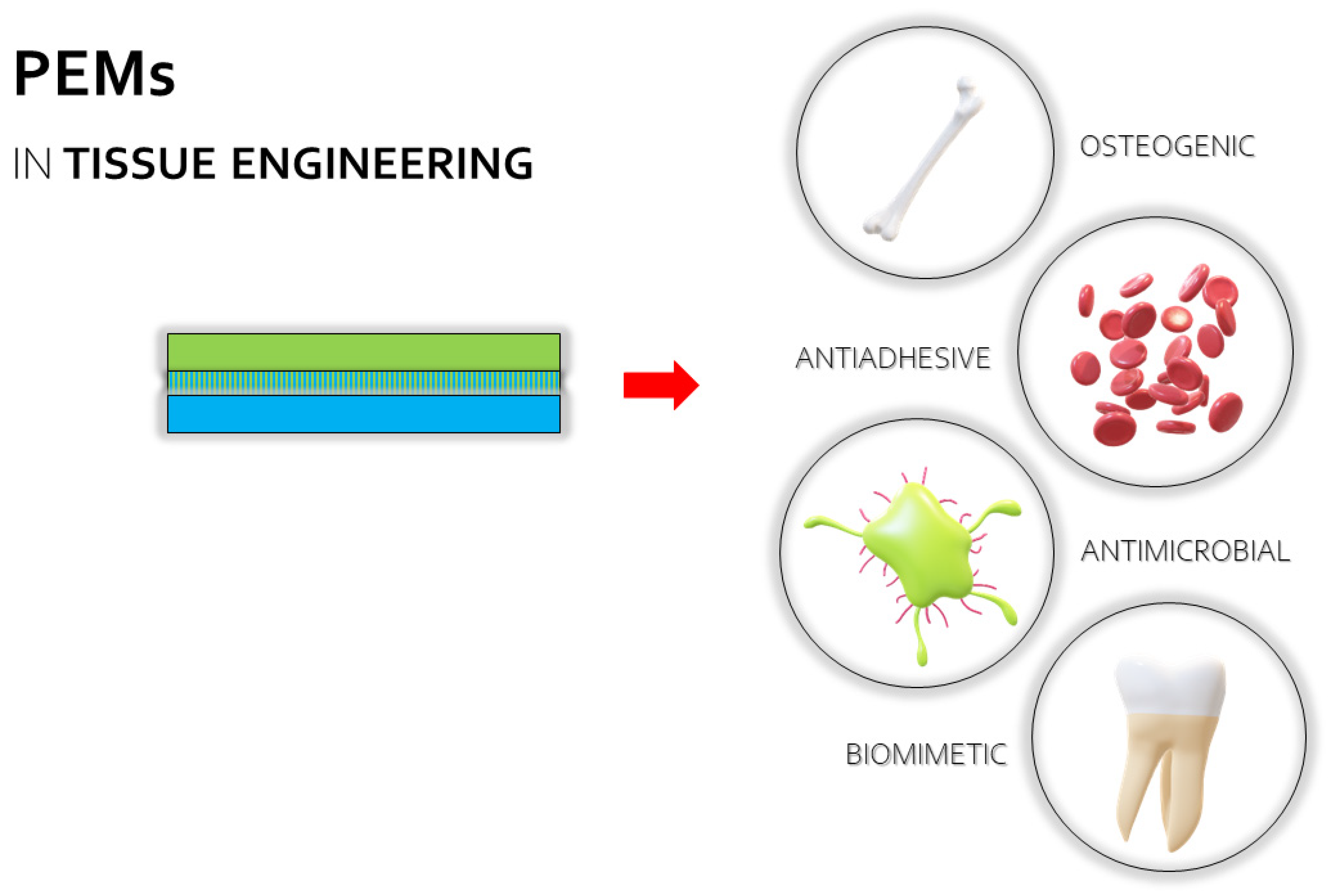
2.2. Utilization of PEMs in Tissue Engineering
2.2.1. Antimicrobial and Antiadhesive Coatings
2.2.2. Regenerative Coatings
2.2.3. Coatings for Coronary Stents
2.2.4. Biomimetic Coatings
2.3. PEMs in Chemical Technology
3. Conclusions
Author Contributions
Funding
Institutional Review Board Statement
Informed Consent Statement
Data Availability Statement
Conflicts of Interest
References
- Mateos-Maroto, A.; Abelenda-Núñez, I.; Ortega, F.; Rubio, R.G.; Guzmán, E. Polyelectrolyte multilayers on soft colloidal nanosurfaces: A new life for the layer-by-layer method. Polymers 2021, 13, 1221. [Google Scholar] [CrossRef] [PubMed]
- Meka, V.S.; Sing, M.K.G.; Pichika, M.R.; Nali, S.R.; Kolapall, V.R.M.; Kesharwani, P. A comprehensive review on polyelectrolyte complexes. Drug Discov. Today 2017, 22, 1697–1706. [Google Scholar] [CrossRef] [PubMed]
- Potaś, J.; Szymańska, E.; Winnicka, K. Challenges in developing of chitosan-based polyelectrolyte complexes as a platform for mucosal and skin drug delivery. Eur. Polym. J. 2020, 140, 110020. [Google Scholar] [CrossRef]
- Vasiliu, S.; Racovita, S.; Popa, M.; Ochiuz, L.; Peptu, C.A. Chitosan-based polyelectrolyte complex hydrogels for biomedical aplications. In Cellulose-Based Superabsorbent Hydrogels. Polymers and Polymeric Composities: A Reference Series; Mondal, M., Ed.; Springer: Cham, Switzerland, 2018. [Google Scholar]
- Luo, Y.; Wang, Q. Recent development of chitosan-based polyelectrolyte complexes with natural polysaccharides for drug delivery. Int. J. Biol. Macromol. 2014, 64, 353–367. [Google Scholar] [CrossRef]
- Gamzazade, A.I.; Nasibov, S.M. Formation and properties of polyelectrolyte complexes of chitosan hydrochloride and sodium dextransulfate. Carbohydr. Polym. 2002, 50, 339–343. [Google Scholar] [CrossRef]
- Magoń, M.S. Layered polyelectrolyte complexes: Physics of formation and molecular properties. J. Phys. Condens. Matter. 2003, 15, 1781. [Google Scholar]
- Montenegro-Nicolini, M.; Morales, J.O. Overview and future potential of buccal mucoadhesive films as drug delivery systems for biologics. AAPS PharmSciTech 2007, 18, 3–14. [Google Scholar] [CrossRef]
- Das, B.P.; Tsianou, M. From polyelectrolyte complexes to polyelectrolyte multilayers: Electrostatic assembly, nanostructure, dynamics, and functional properties. Adv. Colloid Interface Sci. 2017, 244, 71–89. [Google Scholar] [CrossRef]
- Bodurov, I.; Vlaeva, I.; Exner, G.; Uzunova, Y.; Russev, S.; Pilicheva, B.; Viraneva, A.; Yovcheva, T.; Grancharova, T.; Sotirov, S.; et al. Investigation of multilayered polyelectrolyte thin films by means of refractive index measurements, FT-IR spectroscopy and SEM. J. Phys. Conf. 2016, 682, 012026. [Google Scholar] [CrossRef]
- Webber, J.L.; Bradshaw-Hajek, B.H.; Krasowska, M.; Beattie, D.A. Polyelectrolyte multilayer formation on protein layer supports. Colloids Surf. A Physicochem. Eng. 2021, 629, 127470. [Google Scholar] [CrossRef]
- Boudou, T.; Crouzier, T.; Ren, K.; Blin, G.; Picart, C. Multiple functionalities of polyelectrolyte multilayer films: New biomedical applications. Adv Mater. 2010, 22, 441–467. [Google Scholar] [CrossRef]
- Cho, J.; Char, K.; Hong, J.D.; Lee, K.B. Fabrication of highly ordered multilayer films using a spin self-assembly method. Adv. Mater. 2001, 13, 1076–1078. [Google Scholar] [CrossRef]
- Zhao, S.; Caruso, F.; Dähne, L.; Decher, G.; De Geest, B.G.; Fan, J.; Feliu, N.; Gogotsi, Y.; Hammond, P.T.; Hersam, M.C.; et al. The future of layer-by-layer assembly: A tribute to ACS nano associate editor Helmuth Möhwald. ACS Nano 2019, 13, 6151–6169. [Google Scholar] [CrossRef]
- Criado-Gonzalez, M.; Mijangos, C.; Hernández, R. Polyelectrolyte multilayer films based on natural polymers: From fundamentals to bio-applications. Polymers 2021, 13, 2254. [Google Scholar] [CrossRef]
- Petrila, L.M.; Bucatariu, F.; Mihai, M.; Teodosiu, C. Polyelectrolyte multilayers: An overview on fabrication, properties, and biomedical and environmental applications. Materials 2021, 14, 4152. [Google Scholar] [CrossRef]
- Van der Gucht, J.; Spruijt, E.; Lemmers, M.; Stuart, M.A.C. Polyelectrolyte complexes: Bulk phases and colloidal systems. J. Colloid Interface Sci. 2011, 361, 407–422. [Google Scholar] [CrossRef]
- Schlenoff, J.B.; Rmaile, A.H.; Bucur, C.B. Hydration contributions to association in polyelectrolyte multilayers and complexes: Visualizing hydrophobicity. J. Am. Chem. Soc. 2008, 130, 13589–13597. [Google Scholar] [CrossRef]
- von Klitzing, R. Internal structure of polyelectrolyte multilayer assemblies. Phys. Chem. Chem. Phys. 2006, 8, 5012–5033. [Google Scholar] [CrossRef]
- Tsuchida, E. Formation of polyelectrolyte complexes and their structures. J. Macromol. Sci., Part A 1994, 31, 1–15. [Google Scholar] [CrossRef]
- Priftis, D.; Laugel, D.N.; Tirrell, M. Thermodynamic characterization of polypeptide complex coacervation. Langmuir 2012, 28, 15947–15957. [Google Scholar] [CrossRef]
- Izumrudov, V.; Kharlampieva, E.; Sukhishvili, S.A. Salt-induced multilayer growth: Correlation with phase separation in solution. Macromolecules 2004, 37, 8400–8406. [Google Scholar] [CrossRef]
- Wang, Q.; Schlenoff, J.B. The polyelectrolyte complex/coacervate continuum. Macromolecules 2014, 47, 3108–3116. [Google Scholar] [CrossRef]
- Fares, H.M.; Schlenoff, J.B. Diffusion of sites versus polymers in polyelectrolyte complexes and multilayers. J. Am. Chem. Soc. 2017, 139, 14656–14667. [Google Scholar] [CrossRef]
- Titilayo, A.A.; Stanley, D.; Oluwatosin, L.A. Properties of poly-γ-glutamic acid producing-Bacillus species isolated from ogi liquor and lemon-ogi liquor. Front. Microbiol. 2019, 10, 771. [Google Scholar]
- Rowe, R.C.; Sheskey, P.J.; Quinn, M.E. Handbook of Pharmaceutical Excipients; Pharmaceutical Press: Washington, DC, USA , 2009. [Google Scholar]
- Bishnoi, M.; Jain, A.; Hurkat, P.; Jain, S.K. Chondroitin sulphate: A focus on osteoarthritis. Glycoconj. J. 2016, 33, 693–705. [Google Scholar] [CrossRef]
- Luján Medina, G.; Ventura, J.; Lara Ceniceros, A.; Ascacio-Valdés, J.; Boone-Villa, D.; Aguilar, C. Karaya gum: General topics and applications. Macromol. Indian J. 2013, 9, 111–116. [Google Scholar]
- Choi, D.; Hong, J. Layer-by-layer assembly of multilayer films for controlled drug release. Arch. Pharm. Res. 2014, 37, 79–87. [Google Scholar] [CrossRef]
- Ganas, C.; Weiß, A.; Nazarenus, M.; Rösler, S.; Kissel, T.; Rivera Gil, P.; Parak, W.J. Biodegradable capsules as non-viral vectors for in vitro delivery of PEI/siRNA polyplexes for efficient gene silencing. J. Control. Release 2014, 196, 132–138. [Google Scholar] [CrossRef] [PubMed]
- Alshhab, A.; Yilmaz, E. Sodium alginate/poly(4-vinylpyridine) polyelectrolyte multilayer films: Preparation, characterization and ciprofloxacin HCl release. Int. J. Biol. Macromol. 2019, 147, 809–820. [Google Scholar] [CrossRef] [PubMed]
- Timur, S.S.; Yüksel, S.; Akca, G.; Şenel, S. Localized drug delivery with mono and bilayered mucoadhesive films and wafers for oral mucosal infections. Int. J. Pharm. 2019, 559, 102–112. [Google Scholar] [CrossRef] [PubMed]
- Martín-Illana, A.; Chinarro, E.; Cazorla-Luna, R.; Notario-Perez, F.; Veiga-Ochoa, M.D.; Rubio, J.; Tamayo, A. Optimized hydration dynamics in mucoadhesive xanthan-based trilayer vaginal films for the controlled release of tenofovir. Carbohydr. Polym. 2022, 278, 118958. [Google Scholar] [CrossRef]
- Potaś, J.; Szymańska, E.; Wróblewska, M.; Kurowska, I.; Maciejczyk, M.; Basa, A.; Wolska, E.; Wilczewska, A.Z.; Winnicka, K. Multilayer films based on chitosan/pectin polyelectrolyte complexes as novel platforms for buccal administration of clotrimazole. Pharmaceutics 2020, 13, 1588. [Google Scholar] [CrossRef]
- Criado-Gonzalez, M.; Fernandez-Gutierrez, M.; San Roman, J.; Mijangos, C.R. Hernández, Local and controlled release of tamoxifen from multi (layer-by-layer) alginate/chitosan complex systems. Carbohydr Polym. 2019, 206, 428–434. [Google Scholar] [CrossRef]
- Sun, H.; Choi, D.; Heo, J.; Jung, S.Y.; Hong, J. Studies on the drug loading and release profiles of degradable chitosan-based multilayer films for anticancer treatment. Cancers 2020, 12, 593. [Google Scholar] [CrossRef]
- Pilicheva, B.; Uzunova, Y.; Bodurov, I.; Viraneva, A.; Exner, G.; Sotirov, S.; Yovcheva, T.; Marudova, M. Layer-by-layer self-assembly films for buccal drug delivery: The effect of polymer cross-linking. J. Drug Deliv. Sci. Technol. 2020, 59, 101897. [Google Scholar] [CrossRef]
- Damanik, F.F.R.; Brunelli, M.; Pastorino, L.; Ruggiero, C.; van Blitterswijk, C.; Rotmans, J.; Moroni, L. Sustained delivery of growth factors with high loading efficiency in a layer by layer assembly. Biomater. Sci. 2019, 8, 174–188. [Google Scholar] [CrossRef]
- Cardoso, A.P.; Gonçalves, R.M.; Antunes, J.C.; Pinto, M.L.; Pinto, A.T.; Castro, F.; Monteiro, C.; Barbosa, M.A.; Oliveira, M.J. An interferon-amma-delivery system based on chitosan/poly(gamma-glutamic acid) polyelectrolyte complexes modulates macrophage-derived stimulation of cancer cell invasion in vitro. Acta Biomater. 2015, 23, 157–171. [Google Scholar] [CrossRef]
- Martin, A.; Tabary, N.; Leclercq, L.; Junthip, J.; Degoutin, S.; Aubert-Viard, F.; Cazaux, F.; Lyskawa, J.; Janus, L.; Bria, M.; et al. Multilayered textile coating based on a β-cyclodextrin polyelectrolyte for the controlled release of drugs. Carbohydr. Polym. 2013, 93, 718–730. [Google Scholar] [CrossRef]
- Pérez-Anes, A.; Gargouri, M.; Laure, W.; Van Den Berghe, H.; Courcot, E.; Sobocinski, J.; Tabary, N.; Chai, F.; Blach, J.F.; Addad, A.; et al. Bio-inspired titanium drug eluting platforms based on poly-β-cyclodextrin/chitosan layer-by-layer self assembly targeting infections. ACS Appl. Mater. Interfaces 2015, 7, 12882–12893. [Google Scholar] [CrossRef]
- Park, S.; Choi, D.; Jeong, H.; Heo, J.; Hong, J. Drug loading and release behavior depending on the induced porosity of chitosan/cellulose multilayer nanofilms. Mol. Pharm. 2017, 14, 3322–3330. [Google Scholar] [CrossRef]
- Jeganathan, B.; Prakya, V.; Deshmukh, A. Preparation and evaluation of diclofenac sodium tablet coated with polyelectrolyte multilayer film using hypromellose acetate succinate and polymethacrylates for pH-dependent, modified release drug delivery. AAPS PharmSciTech 2016, 17, 578–587. [Google Scholar] [CrossRef] [PubMed][Green Version]
- Ways, T.M.M.; Lau, W.M.; Khutoryanskiy, V.V. Chitosan and its derivatives for application in mucoadhesive drug delivery systems. Polymers 2018, 10, 267. [Google Scholar] [CrossRef] [PubMed]
- Martins, A.F.; Vlcek, J.; Wigmosta, T.; Hedayati, M.; Reynolds, M.M.; Popat, K.C.; Kipper, M.J. Chitosan/iota-carrageenan and chitosan/pectin polyelectrolyte multilayer scaffolds with antiadhesive and bactericidal properties. Appl. Surf. Sci. 2020, 502, 144282. [Google Scholar] [CrossRef]
- Nuraje, N.; Asmatulu, R.; Cohen, R.E.; Rubner, M.F. Durable antifog films from layer-by-layer molecularly blended hydrophilic polysaccharides. Langmuir 2010, 27, 782–791. [Google Scholar] [CrossRef]
- Picart, C.; Lavalle, P.; Hubert, P.; Cuisinier, F.; Decher, G.; Schaaf, P.; Voegel, J.C. Buildup mechanism for poly (L-lysine)/hyaluronic acid films onto a solid surface. Langmuir 2001, 17, 7414–7424. [Google Scholar] [CrossRef]
- Potaś, J.; Szymańska, E.; Basa, A.; Hafner, A.; Winnicka, K. Tragacanth gum/chitosan polyelectrolyte complexesbased hydrogels enriched with xanthan gum as promising materials for buccal application. Materials 2021, 14, 86. [Google Scholar] [CrossRef]
- Kaur, J.; Kaur, G. Optimization of pH conditions and charcterization of polyelectrolyte complexes between gellan gum and cationic guar gum. Polym. Adv. Technol. 2018, 29, 3035–3048. [Google Scholar] [CrossRef]
- Strand, A.; Vähäsalo, L.; Ketola, A.; Salminen, K.; Retulainen, E.; Sundberg, A. In-situ analysis of polyelectrolyte complexes by flow cytometry. Cellulose 2018, 25, 3781–3795. [Google Scholar] [CrossRef]
- Kulkarni, A.; Diehl-Jones, W.; Ghanbar, S.; Liu, S. Layer-by-layer assembly of epidermal growth factors on polyurethane films for wound closure. J. Biomater. Appl. 2014, 29, 278–290. [Google Scholar] [CrossRef]
- Glinel, K.; Déjugnat, C.; Prevot, M.; Schöler, B.; Schönhoff, M.; Klitzing, R.V. Responsive polyelectrolyte multilayers. Colloids Surf., A 2007, 303, 3–13. [Google Scholar] [CrossRef]
- Tan, J.P.; Wang, Q.; Tam, K.C. Control of burst release from nanogels via layer by layer assembly. J. Controlled Release 2008, 128, 248–254. [Google Scholar] [CrossRef]
- Hsu, B.B.; Hagerman, S.R.; Hammond, P.T. Rapid and efficient sprayed multilayer films for controlled drug delivery. J. Appl. Polym. Sci. 2016, 133, 43563–43571. [Google Scholar] [CrossRef]
- Roupie, C.; Labat, B.; Morin-Grognet, S.; Echalard, A.; Ladam, G.; Thebault, P. Dual-functional antibacterial and osteogenic nisin-based layer-by-layer coatings. Mater. Sci. Eng. C 2021, 131, 112479. [Google Scholar] [CrossRef]
- Niaz, T.; Shabbir, S.; Noor, T.; Abbasi, R.; Raza, Z.A.; Imran, M. Polyelectrolyte multicomponent colloidosomes loaded with nisin Z for enhanced antimicrobial activity against foodborne resistant pathogens. Front Microbiol. 2018, 8, 2700. [Google Scholar] [CrossRef]
- Webber, J.L.; Namivandi-Zangeneh, R.; Drozdek, S.; Wilk, K.A.; Boyer, C.; Wong, E.H.H.; Bradshaw-Hajek, B.H.; Krasowska, M.; Beattie, D.A. Incorporation and antimicrobial activity of nisin Z within carrageenan/chitosan multilayers. Sci Rep. 2021, 11, 1690. [Google Scholar] [CrossRef]
- Samuel, R.E.; Shukla, A.; Paik, D.H.; Wang, M.W.; Fang, J.C.; Schmidt, D.J.; Hammond, P.T. Osteoconductive protamine-based polyelectrolyte multilayer functionalized surfaces. Biomaterials 2011, 32, 7491–7502. [Google Scholar] [CrossRef]
- Duchêne, D.; Bochot, A. Thirty years with cyclodextrins. Int. J. Pharm. 2016, 514, 58–72. [Google Scholar] [CrossRef]
- Martel, B.; Ruffin, D.; Weltrowski, M.; Lekchiri, Y.; Morcellet, M. Water-soluble polymers and gels from the polycondensation between cyclodextrins and poly(carboxylic acid)s: A study of the preparation parameters. J. Appl. Polym. Sci. 2005, 97, 433–442. [Google Scholar] [CrossRef]
- Zhou, C.E.; Kan, C.W. Plasma-assisted regenerable chitosan antimicrobial finishing for cotton. Cellulose 2014, 21, 2951–2962. [Google Scholar] [CrossRef]
- Mulders, J.W.; Boerrigter, I.J.; Rollema, H.S.; Siezen, R.J.; de Vos, W.M. Identification and characterization of the lantibiotic nisin Z, a natural nisin variant. Eur. J. Biochem. 1991, 201, 581–584. [Google Scholar] [CrossRef]
- Benhabiles, M.; Salah, R.; Lounici, H.; Drouiche, N.; Goosen, M.; Mameri, N. Antibacterial activity of chitin, chitosan and its oligomers prepared fromshrimp shell waste. Food Hydrocoll. 2012, 29, 48–56. [Google Scholar] [CrossRef]
- Richert, L.; Lavalle, P.; Payan, E.; Shu, X.Z.; Prestwich, G.D.; Stoltz, J.F.; Schaaf, P.; Voegel, J.C.; Picart, C. Layer by layer buildup of polysaccharide films: Physical chemistry and cellular adhesion aspects. Langmuir 2004, 20, 448–458. [Google Scholar] [CrossRef] [PubMed]
- Hernandez-Montelongo, J.; Lucchesi, E.G.; Gonzalez, I.; Macedo, W.; Nascimento, V.F.; Moraes, A.; Beppu, M.; Cotta, M. Hyaluronan/chitosan nanofilms assembled layer-by-layer and their antibacterial effect: A study using Staphylococcus aureus and Pseudomonas aeruginosa. Colloids Surf. B Biointerfaces 2016, 141, 499–506. [Google Scholar] [CrossRef] [PubMed]
- Fu, J.; Ji, J.; Yuan, W.; Shen, J. Construction of anti-adhesive and antibacterial multilayer films via layer-by-layer assembly of heparin and chitosan. Biomaterials 2005, 26, 6684–6692. [Google Scholar] [CrossRef]
- Yoo, D.; Shiratori, S.S.; Rubner, M.F. Controlling bilayer composition and surface wettability of sequentially adsorbed multilayers of weak polyelectrolytes. Macromolecules 1998, 31, 4309–4318. [Google Scholar] [CrossRef]
- Kheilnezhad, B.; Hadjizadeh, A. A review: Progress in preventing tissue adhesions from a biomaterial perspective. Biomater. Sci. 2021, 9, 2850–2873. [Google Scholar] [CrossRef]
- Gao, W.; Li, Z. 21-Nanostructured transition metal oxides and their applications in composites. In Woodhead Publishing Series in Composites Science and Engineering, Physical Properties and Applications of Polymer Nanocomposites; Tjong, S.C., Mai, Y.W., Eds.; Woodhead Publishing: Cambridge, UK, 2010; p. 723. [Google Scholar]
- Barroso García, N.; Guaresti, O.; Perez, L.; Ruiz-Rubio, L.; Gabilondo, N.; Vilas-Vilela, J.L. Self-healable hyaluronic acid/chitosan polyelectrolyte complex hydrogels and multilayers. Eur. Polym. J. 2019, 120, 109268. [Google Scholar] [CrossRef]
- Petrova, V.A.; Chernyakov, D.D.; Poshina, D.N.; Gofman, I.V.; Romanov, D.P.; Mishanin, A.I.; Golovkin, A.S.; Skorik, Y.A. Electrospun bilayer chitosan/hyaluronan material and its compatibility with mesenchymal stem cells. Materials 2019, 12, 2016. [Google Scholar] [CrossRef]
- Nolte, A.; Hossfeld, S.; Schroeppel, B.; Mueller, A.; Stoll, D.; Walker, T.; Wendel, H.P.; Krastev, R. Impact of polyelectrolytes and their corresponding multilayers to human primary endothelial cells. J. Biomater. Appl. 2013, 28, 84–99. [Google Scholar] [CrossRef]
- Park, J.Y.; Park, S.H.; Kim, M.G.; Park, S.H.; Yoo, T.H.; Kim, M.S. Biomimetic scaffolds for bone tissue engineering. In Biomimetic Medical Materials. Advances in Experimental Medicine and Biology; Noh, I., Ed.; Springer: Singapore, 2018; pp. 109–121. [Google Scholar]
- Rudt, A.; Andreeva, T.D.; Krastev, R.; Taneva, S.G. Composite polyelectrolyte multilayers for biofunctionalization of medical devices. Curr. Dir. Biomed. Eng. 2020, 6, 426–429. [Google Scholar] [CrossRef]
- de Lázaro, I.; Vranic, S.; Marson, D.; Rodrigues, A.F.; Buggio, M.; Esteban-Arranz, A.; Mazza, M.; Posocco, P.; Kostarelos, K. Graphene oxide as a 2D platform for complexation and intracellular delivery of siRNA. Nanoscale 2019, 11, 13863–13877. [Google Scholar] [CrossRef]
- Robinson, J.T.; Tabakman, S.M.; Liang, Y.; Wang, H.; Sanchez Casalongue, H.; Vinh, D.; Dai, H. Ultrasmall reduced graphene oxide with high near-infrared absorbance for photothermal therapy. J. Am. Chem. Soc. 2011, 133, 6825–6831. [Google Scholar] [CrossRef]
- Petrova, V.A.; Orekhov, A.D.; Chernyakov, Y.; Baklagina, D.; Romanov, S.; Kononova, A.; Volod’ko, I.; Yermak, V.; Klechkovskaya, Y. Skorik, Preparation and analysis of multilayer composites based on polyelectrolyte complexes. Crystallogr. Rep. 2016, 61, 945–953. [Google Scholar] [CrossRef]
- Kononova, S.V.; Volod’ko, A.V.; Petrova, V.A.; Kruchinina, E.V.; Baklagina, Y.G.; Chusovitin, E.A.; Skorik, Y.A. Pervaporation multilayer membranes based on a polyelectrolyte complex of λ-carrageenan and chitosan. Carbohydr. Polym. 2018, 181, 86–92. [Google Scholar] [CrossRef]
- Kononova, S.V.; Kruchinina, E.V.; Petrova, V.A.; Baklagina, Y.G.; Klechkovskaya, V.V.; Orekhov, A.S.; Vlasova, E.N.; Popova, E.N.; Gubanova, G.N.; Skorik, Y.A. Pervaporation membranes of a simplex type with polyelectrolyte layers of chitosan and sodium hyaluronate. Carbohydr Polym. 2019, 209, 10–19. [Google Scholar] [CrossRef]
- Zhang, L.; Sun, J. Layer-by-layer deposition of polyelectrolyte complexes for the fabrication of foam coatings with high loading capacity. Chem. Commun. 2009, 26, 3901–3903. [Google Scholar] [CrossRef]


| Polyelectrolytes | Physicochemical Characteristics |
|---|---|
| Polycations | |
| Natural origin polycations | |
| Chitosan |
|
| Synthetic polycations | |
| Eudragit E |
|
| Poly(4-vinylpyridine) |
|
| Poly-L-lysine |
|
| Polyanions | |
| Natural origin polyanions | |
| Sodium alginate |
|
| Hyaluronic acid |
|
| Heparin |
|
| Chondroitin sulfate |
|
| Carrageenans |
|
| Tannic acid |
|
| Poly(γ–glutamic acid) |
|
| Xanthan gum |
|
| Karaya gum |
|
| Synthetic polyanions | |
| Hypromellose acetate succinate |
|
| Carboxymethylcellulose sodium |
|
| β-cyclodextrins |
|
Publisher’s Note: MDPI stays neutral with regard to jurisdictional claims in published maps and institutional affiliations. |
© 2022 by the authors. Licensee MDPI, Basel, Switzerland. This article is an open access article distributed under the terms and conditions of the Creative Commons Attribution (CC BY) license (https://creativecommons.org/licenses/by/4.0/).
Share and Cite
Potaś, J.; Winnicka, K. The Potential of Polyelectrolyte Multilayer Films as Drug Delivery Materials. Int. J. Mol. Sci. 2022, 23, 3496. https://doi.org/10.3390/ijms23073496
Potaś J, Winnicka K. The Potential of Polyelectrolyte Multilayer Films as Drug Delivery Materials. International Journal of Molecular Sciences. 2022; 23(7):3496. https://doi.org/10.3390/ijms23073496
Chicago/Turabian StylePotaś, Joanna, and Katarzyna Winnicka. 2022. "The Potential of Polyelectrolyte Multilayer Films as Drug Delivery Materials" International Journal of Molecular Sciences 23, no. 7: 3496. https://doi.org/10.3390/ijms23073496
APA StylePotaś, J., & Winnicka, K. (2022). The Potential of Polyelectrolyte Multilayer Films as Drug Delivery Materials. International Journal of Molecular Sciences, 23(7), 3496. https://doi.org/10.3390/ijms23073496