Article

Knockdown of Tet2 Inhibits the Myogenic Differentiation of Chicken Myoblasts Induced by Ascorbic Acid

Department of Animal Genetics, Breeding and Reproduction, College of Animal Science and Technology, Nanjing Agricultural University, No. 1 Weigang, Nanjing 210095, China
*
Authors to whom correspondence should be addressed.
Int. J. Mol. Sci. 2022, 23(22), 13758; https://doi.org/10.3390/ijms232213758
Submission received: 11 October 2022 / Revised: 1 November 2022 / Accepted: 8 November 2022 / Published: 9 November 2022
(This article belongs to the Special Issue Myoblast and Muscle Cell Genesis and Regeneration)

Abstract

:
Ascorbic acid (also called Vitamin C, VC) strengthens the function of Tets families and directly increases DNA demethylation level to affect myogenic differentiation. However, the precise regulatory mechanism of DNA methylation in chicken myogenesis remains unclear. Results of present study showed that the mRNA expression of MyoD significantly decreased and MyoG and MyHC increased in myoblasts treated with 5 μM 5-azacytidine (5-AZA) and 5 μM VC (p < 0.05). Results also indicated the formation of myotubes was induced by 5-AZA or VC, but this effect was attenuated after knockdown of Tet2. In addition, the protein expression of TET2, DESMIN and MyHC was remarkable increased by the addition of 5-AZA or VC, and the upregulation was inhibited after knockdown of Tet2 (p < 0.05). DNA dot blot and immunofluorescence staining results suggested that the level of 5hmC was significantly increased when treated with 5-AZA or VC, even by Tet2 knockdown (p < 0.05). Moreover, 5-AZA and VC reduced the level of dimethylation of lysine 9 (H3K9me2) and trimethylation of lysine 27 of histone 3 (H3K27me3), and this inhibitory effect was eliminated after Tet2 knockdown (p < 0.05). These data indicated that Tet2 knockdown antagonized the increased levels of 5hmC and H3K27me3 induced by 5-AZA and VC, and eventually reduced myotube formation by modulating the expression of genes involved in myogenic differentiation. This study provides insights that epigenetic regulators play essential roles in mediating the myogenic program of chicken myoblasts.

1. Introduction

Recently, many studies revealed that DNA methylation and histone modification exert essential roles in regulating myogenic differentiation [1,2]. It was reported that the whole genomic DNA methylation decreased during myoblast differentiation [3]. Epigenetic studies increase the knowledge of the molecular mechanism regulating myogenic related-gene expression and myogenesis program [4,5]. Fundamental studies are still required to understand the details of epigenetic regulation in chicken myogenesis.
Myoblasts are myogenic precursor cells that play an important role during skeletal muscle development. The fate of myoblasts is determined by transcription factors including Pax3/Pax7 and the myogenic regulatory factors (MRFs), which work together to establish the epigenetic state in the processes of myofiber formation [6,7]. A series of studies has shown that DNA demethylation exerted a critical role in myogenesis [2,8,9]. The expression of TET1 and TET2 are significantly increased and the DNA methylation level decreases after induction of myoblast differentiation [10]. The Tet1/Tet2 knockout myoblasts showed muscle atrophy and attenuated MyoD expression and myotube formation in mice. Interestingly, knockdown of Tet2 in C2C12 led to increased DNA methylation of gene promoters of MRFs, which correlated with decreased expression of these genes and abrogated myogenic differentiation [10]. Our previous study also indicated that Tet2 affected chicken myoblast differentiation by regulating DNA methylation and histone methylations [8].
Previous study showed that the demethylation of MyoD and MyoG promoter was required for myoblast differentiation [11,12]. MyoG push the differentiation program forward, accompany with epigenetic regulation leading to MRFs expression and myotube fusion. It was reported that the differentiation capacity of C2C12 was reduced by treatment of sodium arsenic, as a result of the increased DNA methylation and decreased expression of MyoG [13]. However, hypermethylation of MyoG decreased sharply after the induction of differentiation in C2C12 [14]. In addition, a decrease of DNA methylation of MyoD promoter occurred during myoblast differentiation [7,15]. Taken together, these findings showed that transcription factors act though epigenetic regulation to genes are required to ensure myogenetic differentiation.
Several molecules have been identified to facilitate myoblast differentiation during the past studies [16,17,18]. Although its mechanism has not been completely revealed, VC was considered to promote myoblast differentiation probably due to mediate DNA demethylation [16]. Studies have also shown that 5-AZA promoted myogenic differentiation by inhibiting DNA methylation and increasing DNA demethylation [18]. Treatment of mouse myoblasts with 5-AZA affected the expression of cell cycle--related genes and myogenic genes to regulate early differentiation [18]. A previous study showed that VC enhanced the activity of Tets enzyme and rapidly increased the level of 5hmC. This effect was inhibited in Tet1 and Tet2 knockout mice, indicating that VC exerted its function by mediating the Tets family [19]. Recent studies found that VC strengthens the function of Tets family and directly increased the level of DNA demethylation to promote myogenic differentiation in mice [10]. A recent study revealed that trimethylation of lysine 9 (H3K9me3) and H3K27me3 are accompanied by chromatin compaction, which is required for myoblast differentiation [8,20]. The demethylation of H3K27me3 was mediated by demethylase KDM6a that opened the chromatin to permit MyoG expression and start to differentiation [21,22]. VC deficiency has been reported to decrease the expression of histone demethylases KDM6b, leading to rapid accumulation of H3K27me3 in cells [9]. Therefore, 5-AZA and VC affected the demethylation of DNA and histone to promote myoblast differentiation. This accumulating evidence corroborated the findings that VC and 5-AZA regulated DNA methylation level and induced myoblast differentiation.
Currently, few data are available about DNA methylation status of epigenetic regulation, so the precise epigenetic mechanisms regulating myogenesis in chickens still needs to be further studied. To address this issue, the present study aimed to explore the relationship with VC and Tet2 in regulation of myogenic differentiation by using a chicken myoblast cell model. Tet2 knockdown myoblasts were treated with 5-AZA and VC to investigate their inductive effects on myoblast differentiation. Thus, this study will provide valuable information on exploration of the epigenetic mechanism of myogenesis.

2. Results

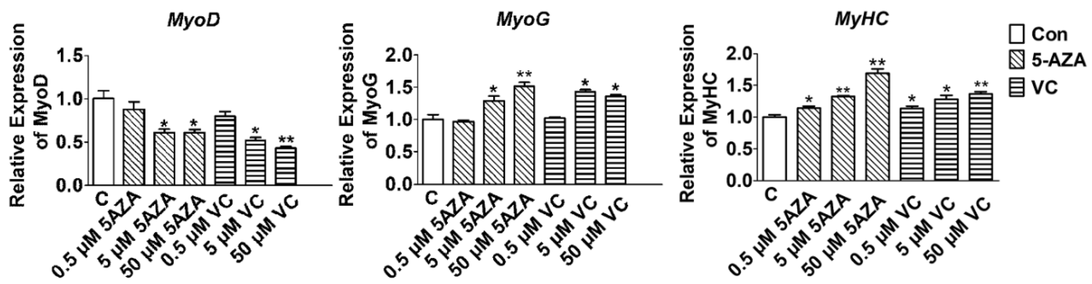
2.1. Effect of 5-AZA and VC on Myogenic Related Gene Expression

In order to determine the effect of decreased Tet2 expression on myoblast differentiation induced by 5-AZA and VC, the optimal concentrations in the regulation of myoblast differentiation were firstly examined. Myoblast differentiation was induced when the density of myoblasts reached 90%. Different concentrations of 5-AZA and VC were added to the differentiation medium, and the change of myogenic factors level was detected after 3 days. Results suggested that 5 and 50 μM 5-AZA remarkably reduced the mRNA expression of MyoD and increased the expression of MyoG and MyHC (p < 0.05) (Figure 1). The expression of MyoD was significantly decreased at 5 μM VC (p < 0.05) and extremely significantly decreased at 50 μM VC (p < 0.01) (Figure 1). However, the expression of MyoG and MyHC was significantly increased after the treatment with 5 μM and 50 μM VC (p < 0.05) (Figure 1). There was no difference in the expression of MyoD and MyoG when the supplemental concentration of was 0.5 μM 5-AZA and 0.5 μM VC (p > 0.05). Therefore, 5 μM 5-AZA and 5 μM VC were selected as the optimal concentrations in the following experiments.

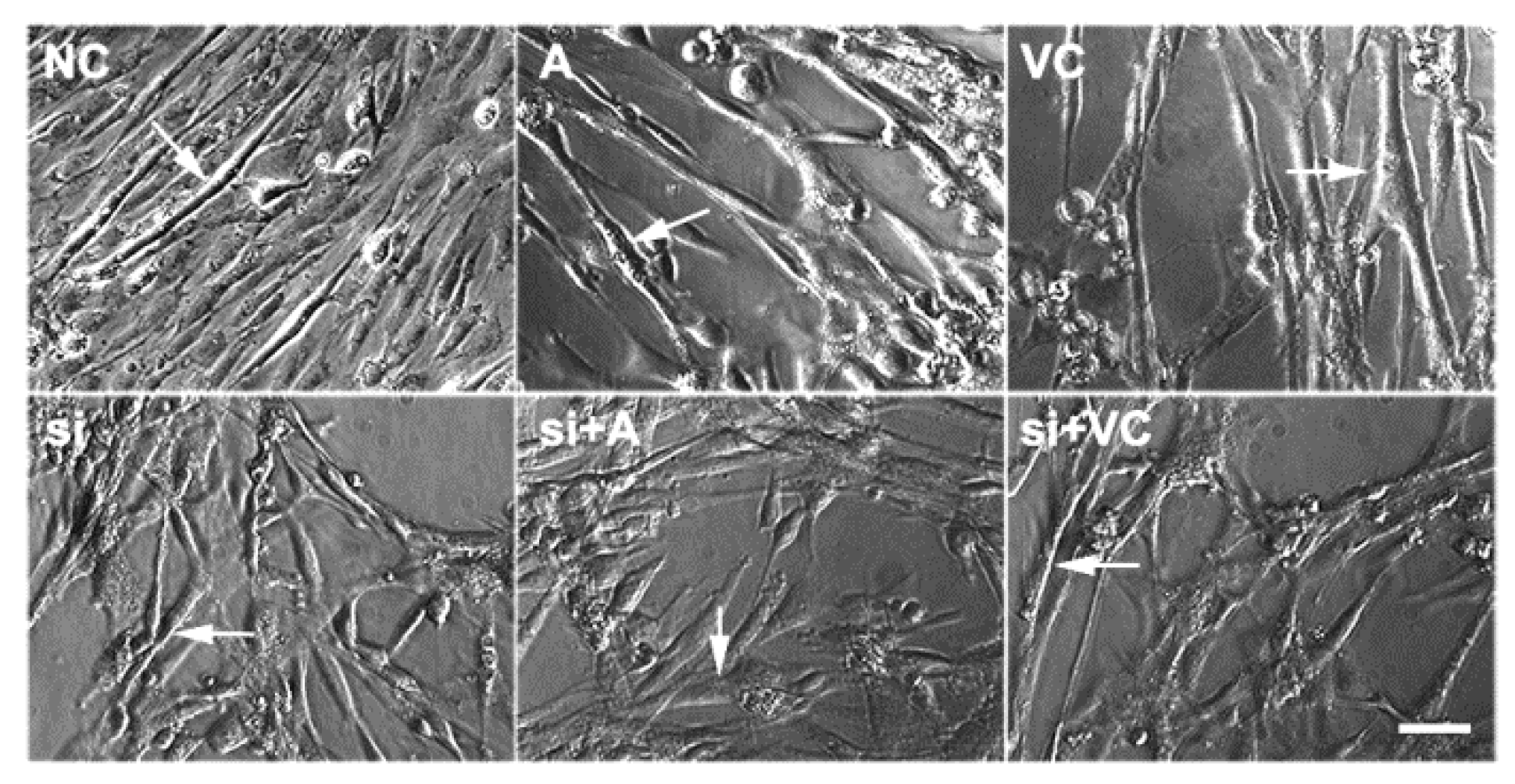
2.2. Effects of 5-AZA and VC on Myoblast Differentiation after Tet2 Knockdown

5-AZA and VC induced myoblast differentiation after Tet2 knockdown was detected. Results showed that the fusion ability of myotubes was enhanced significantly in myoblasts treated with 5-AZA- and VC compared with control, but the formation of myotubes was diminished after Tet2 knockdown (Figure 2). The protein level of TET2 in myoblasts rose rapidly after treatment with 5-AZA and VC (p < 0.01) (Figure 3). It was obviously reduced in the Tet2 knockdown group, but up-regulated in addition of 5-AZA and VC (p < 0.05) (Figure 3). Addition of 5-AZA and VC also significantly increased the protein level of DESMIN and MyHC when compared with the control group (p < 0.01) (Figure 3). Moreover, the up-regulation level of DESMIN by 5-AZA was stronger than that of VC. The level of DESMIN was remarkably decreased after Tet2 knockdown, and this decrease was recovered in addition of 5-AZA and VC (p < 0.05) (Figure 3). The level of MyHC was significantly decreased after Tet2 knockdown, but it quickly returned to the similar lever of control by addition of 5-AZA or VC (p < 0.05) (Figure 3).

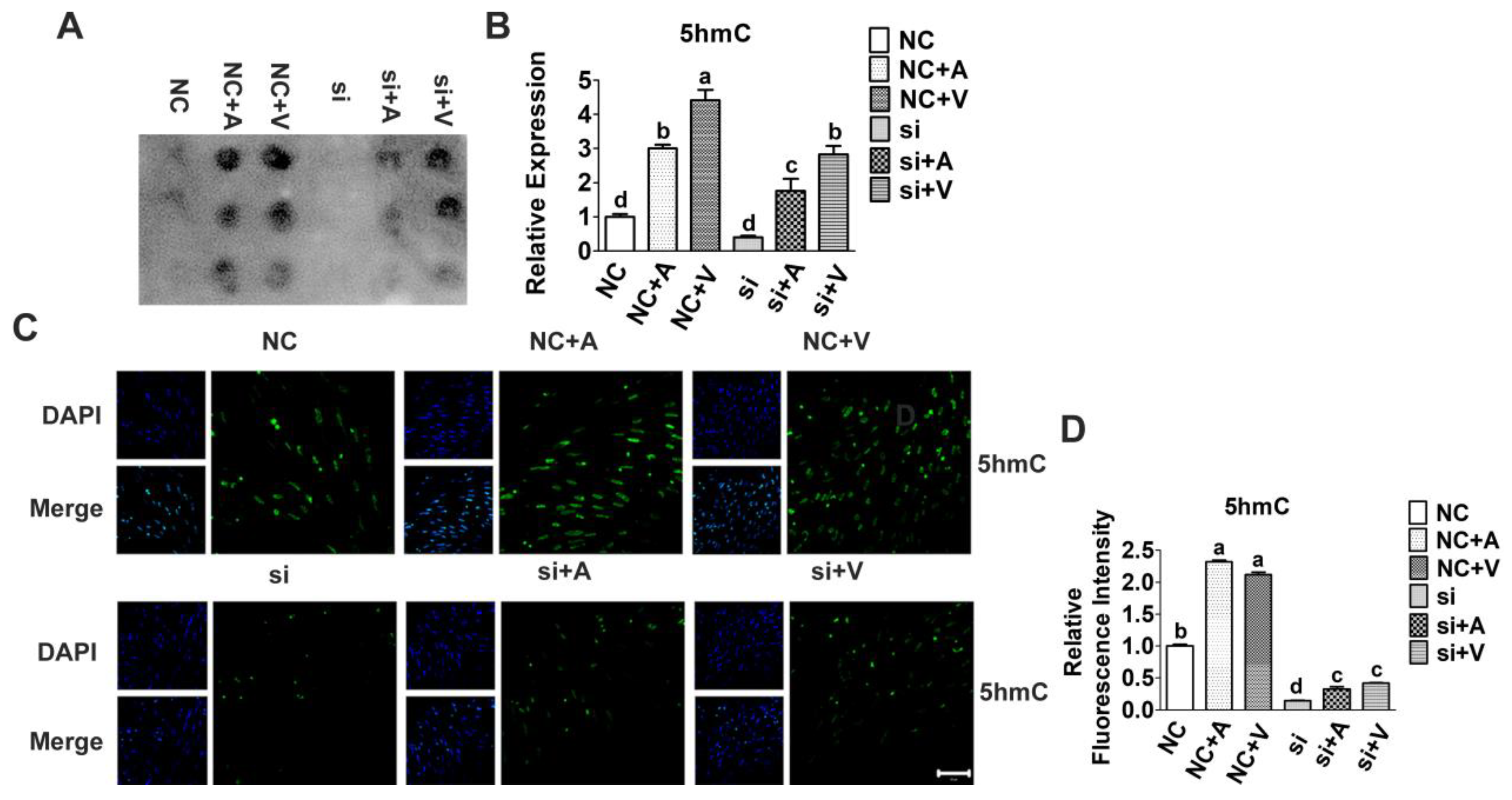
2.3. Effects of 5-AZA and VC on 5hmC Level after Tet2 Knockdown

Our previous study demonstrated that Tet2 knockdown led to a general decrease in 5hmC level and promoted the differentiation of myoblasts. In order to determine whether Tet2 is required for 5-AZA and VC induced myoblast differentiation, the profile of DNA methylation in Tet2 knockdown myoblasts treated with/without of 5-AZA and VC were analyzed. Interestingly, result of a DNA dot blot showed that both 5-AZA and VC significantly increased the level of 5hmC (p < 0.01) (Figure 4A,B), which suggested that they have similar effects on DNA demethylation. Moreover, it was confirmed by the result of immunofluorescence staining that 5-AZA and VC increased the level of 5hmC compared with the control group (p < 0.05) (Figure 4C,D). However, after knockdown of Tet2, the level of 5hmC sharply diminished, although addition of 5-AZA and VC restored its level to some extent (p < 0.05) (Figure 4). These results indicated that 5-AZA and VC promoted the increase of genomic 5hmC level, but this promotion was inhibited after knockdown of Tet2.

2.4. Effects of 5-AZA and VC on H3K9me2 and H3K27me3 Level after Tet2 Knockdown

Our previous study showed that the expression of H3K9me2 and H3K27me3 decreased rapidly after myoblast differentiation, and increased significantly after knockdown of Tet2. To investigate the effect of 5-AZA and VC on the level of H3K9me2 and H3K27me3, their levels were detected by immunofluorescence staining. Result suggested that the level of H3K9me2 and H3K27me3 were significantly decreased after the addition of 5-AZA and VC compared with control (p < 0.05). However, the levels increased rapidly after Tet2 knockdown, and significantly decreased after 5-AZA and VC treatment (p < 0.05) (Figure 5). These results indicated that 5-AZA and VC significantly reduced H3K9me2 and H3K27me3 level, and this inhibition was relieved after Tet2 knockdown.

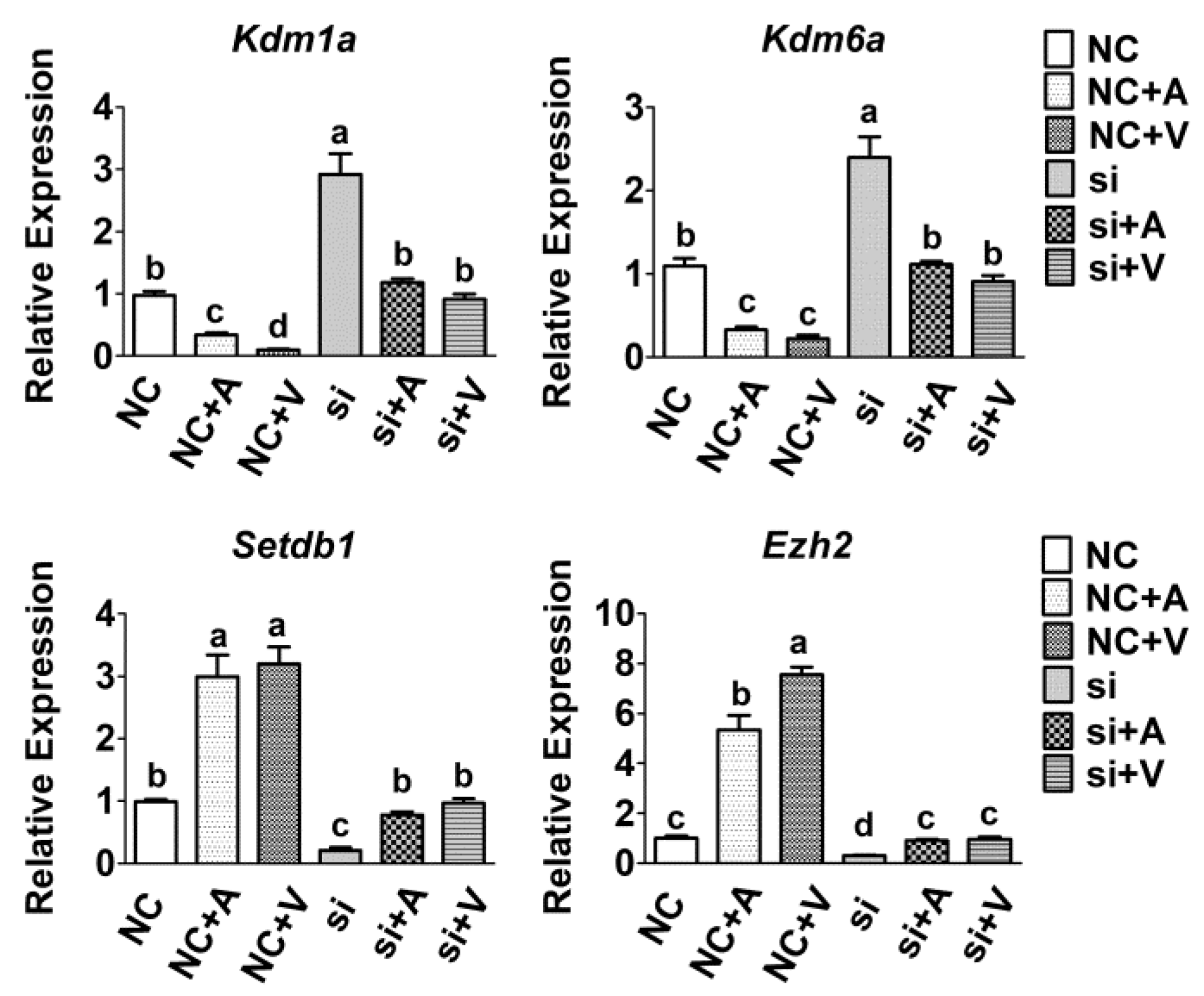
2.5. Effects of 5-AZA and VC on Histone Methyltransferases Expression after Tet2 Knockdown

To investigate the regulation mechanism of Tet2 in the level of H3K9me2 and H3K27me3, the mRNA expression of histone methyltransferases and demethyltransferases was measured by qRT-PCR. Result showed that the mRNA expression of histone methyltransferases (Setdb1 and Ezh2) decreased remarkably by treatment of 5-AZA and VC and increased by knockdown of Tet2 (p < 0.05) (Figure 6). However, Tet2 knockdown induced upregulation was blocked when addition of 5-AZA and VC (p < 0.05) (Figure 6). However, the expression level of histone demethyltransferases (Kdm1a and Kdm6a) was significantly increased when treated with 5-AZA and VC (p < 0.05) (Figure 6). By contrast, their expression was obviously decreased after Tet2 knockdown and recovered by addition of 5-AZA and VC compared to the control group (p < 0.05) (Figure 6).

3. Discussion

DNA methylation of specific genes is required for myogenic differentiation [21,23]. It was reported that DNA methylation modification was also associated with histone modification during myogenic differentiation. They functioned to allowing the maintenance of myogenic-related gene silencing by DNA methylation [24]. The precise role of epigenetic regulators during myogenic differentiation is still not fully understood. Elucidating the relationship between VC and myoblast differentiation would be important to understand the regulatory mechanism of myogenesis. In the present study, to gain insights into mechanisms by which VC contributes to myoblast differentiation, the genome-wide DNA methylation levels were measured after Tet2 knockdown by using chicken myoblasts cell model.
Studies showed that the addition of VC into C2C12 up-regulated the expression of Mrf4 and promoted myoblast differentiation and the formation of multinucleated myotubes [25]. This study found that the mRNA levels of MyoG and MyHC were significantly increased when addition of 5 μM 5-AZA and 5 μM VC. Subsequent results showed that 5-AZA and VC up-regulated the protein level of TET2, MyHC and DESMIN, but the promotion effects were inhibited after Tet2 knockdown. Combined with the results of previous studies, we speculated that 5-AZA and VC up-regulated the expression of Tet2 and 5hmC level to promoted myogenic differentiation.
Epigenetic regulation including DNA methylation and histone modification has been widely studied in muscle development. A series of studies emphasized that the epigenetic states are in dynamic change during the process of myogenesis [3,4]. The decline of Tet2 has been reported to change the status of DNA mythelation and down-regulate myogenic gene expression [8,10]. As demethylation is potently induced in actively differentiating myoblasts, a high level of 5hmC in genomic DNA was observed. Some studies have revealed that VC regulates histone demethylation and modulates related gene expression [26]. This study presented that VC induced genomic DNA demethylation and reduced H3K27me3 level to promote myogenic differentiation by targeting Tet2. The decline of DNA methylation and H3K27me3 induced by VC was increased after Tet2 knockdown, which proved that knockdown Tet2 antagonizes VC. Furthermore, Tet2 knockdown-induced upregulation of histone methyltransferases was attenuated by addition of 5-AZA and VC. The expression levels of histone demethyltransferases was obviously decreased after Tet2 knockdown and recovered by addition of 5-AZA and VC. Therefore, our results using primary-cultured chicken myoblats showed myoblast differentiation was enhanced by addition of VC, while this promotion effect was functionally deteriorated by knockdown of Tet2.
An analogous function of VC with 5-AZA has been observed during myoblast differentiation. As a DNA demethylation reagent, the role of 5-AZA in DNA demethylation has been intensively studied. Previous study found that 5-AZA regulated the cell cycle and MRFs synthesis, and the cell cycle arrest related genes were down-regulated and MyHC expression was down-regulated in the 5-AZA group during differentiation [18]. Studies have found that 5-AZA treatment in Hela cells effectively silenced Dnmt1 transcription and improved the expression of Tet2 and 5hmC level [27]. The decrease expression of Tet2 and Tet3 in hepatocellular carcinoma cells was accompanied by the decrease level of 5hmC, and they were up-regulated in the addition of 5-AZA [28]. These results suggested that 5-AZA played roles in various cell types by promoting the expression of Tets family and up-regulating the level of 5hmC. In the present study, exposure of 5-AZA increased TET2 protein expression, leading to change the genomic methylation state of myoblasts. In addition, VC improved the oxidation of 5mC mediated by Tets family and promoted DNA demethylation in mammals [19]. For example, VC promoted the activity of Tets in ESCs, leading to a rapid increase of 5hmC [19]. Previous studies have found that VC promoted the differentiation of skeletal muscle cells to form myotubes in mice and human, and subsequent studies have found that VC promoted the differentiation of satellite cells [16,25]. To investigate how VC interplays with Tet2, the function of VC in Tet2 knockdown myoblasts was tested in this study. It was found that VC could partially rescue the inhibition of myoblast differentiation in Tet2 knockdown cells, as a result of increasing myotube formation. Moreover, our result demonstrated that the 5hmC level in Tet2 knockdown cells was dramatically increased by VC compared to no VC group, even higher than that in NC group. These results suggested that VC relieved the effect of Tet2 knockdown on reducing the generation of 5hmC, which due to VC enhanced the residual TET2 protein activity caused by incomplete siTtet2.
Collectively, the above results indicated that 5-AZA and VC upregulated expression levels of TET2 and MyHC, and knockdown of Tet2 attenuated the up-regulation of MRFs induced by 5-AZA and VC. 5-AZA and VC exerted remarkable effects on the promotion of DNA demethylation and histone demethylation, but their function was inhibited after Tet2 knockdown. This evidence suggested that 5-AZA and VC promoted DNA and histone demethylation by regulating Tet2 expression to achieve myoblast differentiation (Figure 7). Understanding these facts shed insights on the importance of the epigenetic regulation in myogenic program.

4. Materials and Methods

4.1. Myoblast Cell Culture

All animal experiments were approved by the Animal Care and Use Committee of Nanjing Agricultural University. Primary myoblast cells were isolated from embryonic 9-day chicken skeletal muscle according to a previous study [17]. Myoblasts were maintained and cultured in growth medium (GM) consisting of DMEM, 10% FBS, and a mixture of 1% penicillin-streptomycin (PS). When the cell confluence reached to 90%, culture medium was replaced with differentiation medium (DM) consisting of DMEM, 2% horse serum (HS, Gibco, Carlsbad, CA, USA) and 1% PS. After induction for 3 days, cells were harvested for further analysis.

4.2. The RNA Interference Assays

For RNA interference assays, myoblasts were transfected with Tet2 siRNA or negative control siRNA using Lipofectamine RNAiMAX reagent (Invitrogen, Carlsbad, CA, USA) according to the manufacturer’s protocal as a previous study [17]. The transfected myoblasts were induced differentiation in addition of 5-AZA and VC.

4.3. Quantitative Real-Time PCR (qRT-PCR) Analysis

After culture, total RNA of myoblasts was isolated using Trizol reagent (Invitrogen, Carlsbad, CA, USA) and was reverse transcribed using PrimeScript RT Master Mix (Takara, Dalian, China). Then, qRT-PCR analysis was performed using SYBR Premix ExTaq II (Takara, Dalian, China) with Step-One Real-Time PCR System (Applied Biosystems, Carlsbad, CA, USA). A totol 20 μL reaction mixture contained 2 μL cDNA, 0.5 μL of each primer, 10 μL 2× SYBR Green SuperReal PreMix (ABM Inc., Richmond, BC, Canada), and 7 μL RNase-free H2O. PCR amplification was performed as follows: 94 °C for 30 s, followed by 39 cycles at 94 °C for 15 s, 58–62 °C for 30 s. The amount of each transcript was normalized to that of Gapdh gene. Results are presented as fold-change using the ΔΔCt method. Primer sequences are listed in Table 1.

4.4. Western Blotting

Soluble whole-cell lysates of myoblasts were prepared using lysis buffer containing radioimmunoprecipitation assay (RIPA) buffer supplemented with 0.2 nmol/L PMSF (Complete, Roche, Shanghai, China). The lysates were denatured at 95 °C for 5 min and 10 mg of protein samples were subjected to 10% polyacrylamide gel. Blots were transferred to PVDF membranes and incubated in the primary antibodies, including DESMIN (1:50, DSHB, Iowa City, IA, USA), mouse, TET2 (1:400, Abcam, Fremont, CA, USA), MyHC (1:50, DSHB, Iowa City, IA, USA), α-TUBLIN (1:5000, Novus Biologicals Littleton, CO, USA). HRP-conjugated goat anti-rabbit and anti-mouse IgG antibodies (1:5000, Cell Signaling, Beverly, MA, USA) were used as secondary antibodies, respectively. Then, blots were detected using ECL reagents (Thermo Fisher Scientifc, Waltham, MA, USA). The quantity of detected proteins was normalized to that of α-TUBLIN using ImageJ 1.8 software (National Institutes of Health, Bethesda, MD, USA).

4.5. Immunocytochemistry

Immunofluorescence staining was performed as previously described [8]. Firstly, the myoblasts were fixed with 4% paraformaldehyde and permeabilized with 0.1% Triton X-100 (Nacalai, Nakagyo-ku, Kyoto, Japan). Then, cells were denatured with 2 M HCl for 30 min followed by neutralised with 100 mM Tris-HCl (PH 8). After blocking in 5% goat serum in TBST for 30 min, cells were immunostained with primary antibodies, including 5hmC (1:500, Active Motif, Carlsbad, CA, USA), H3K9me2 and H9K27me3 (5 μg/mL, Abcam, Fremont, CA, USA). FITC-conjugated secondary antibody (1:1000, KPL Inc., Gaithersburg, MD, USA) were used as secondary antibodies. Cell nuclei were counterstained with 4,6-diaminidino-2-phenylindole (DAPI). Fluorescent images were taken immediately under a confocal laser microscope (Zeiss LSM 700, Carl Zeiss AG, Oberkochen, Germany).

4.6. DNA Extraction and Dot Blot Assay

DNA was isolated from cultured myoblasts using a MiniBEST Universal Genomic DNA Extraction Kit (Takara, Dalian, China). Serially diluted 2-fold genomic DNA (300, 150, 75 ng) was spotted onto a nylon membrane (Biosharp, Shanghai, China), following by air-dried and UV-cross linked for 15 min, respectively. After being blocked in 5% skim milk in PBS with 0.1% Tween-20 for 1 h at RT, membranes were incubated with anti-5hmC antibody (1:500; Active Motif, Carlsbad, CA, USA) overnight at 4 ℃. The signal of dots was developed using an ECL reagent (Bio-Rad, Shanghai, China) after incubiation in HRP-conjugated anti-mouse secondary antibody (1:5000; Santa Cruz, Dallas, TX, USA). The signal intensities of dot blot were anylazed by ImageJ 1.8 software.

4.7. Statistical Analysis

Each experiment was replicated three times. Data were analyzed by ANOVA and Duncan’s multiple range tests using SPSS 18.0. All data were presented as the means ± SE. Differences at p < 0.05 were considered to be statistically significant.

Author Contributions

M.Y. conceived the project; M.Y. and Y.L. designed the experiments; K.S., Y.L., H.W., H.C., F.L. and J.Z. performed the experiments; M.Y. and D.Y. analyzed and interpreted the data; Y.L. and M.Y. wrote the manuscript. All authors have read and agreed to the published version of the manuscript.

Funding

This work was supported by the Fundamental Research Funds for the Central Universities (KYZ201724) and the Luhe rural revitalization of industrial development (No. HY0075).

Institutional Review Board Statement

All animal experiments were approved by the Animal Care and Use Committee of Nanjing Agricultural University.

Informed Consent Statement

Not applicable.

Conflicts of Interest

The authors declare no conflict of interest.

References

  1. Massenet, J.; Gardner, E.; Chazaud, B.; Dilworth, F.J. Epigenetic regulation of satellite cell fate during skeletal muscle regeneration. Skelet. Muscle 2021, 11, 4. [Google Scholar] [CrossRef]
  2. Asp, P.; Blum, R.; Vethantham, V.; Parisi, F.; Micsinai, M.; Cheng, J.; Bowman, C.; Kluger, Y.; Dynlacht, B.D. Genome-wide remodeling of the epigenetic landscape during myogenic differentiation. Proc. Natl. Acad. Sci. USA 2011, 108, E149–E158. [Google Scholar] [CrossRef] [Green Version]
  3. Tsumagari, K.; Baribault, C.; Terragni, J.; Varley, K.E.; Gertz, J.; Pradhan, S.; Badoo, M.; Crain, C.M.; Song, L.; Crawford, G.E.; et al. Early de novo DNA methylation and prolonged demethylation in the muscle lineage. Epigenetics 2013, 8, 317–332. [Google Scholar] [CrossRef] [Green Version]
  4. Perdiguero, E.; Sousa-Victor, P.; Ballestar, E.; Munoz-Canoves, P. Epigenetic regulation of myogenesis. Epigenetics 2009, 4, 541–550. [Google Scholar] [CrossRef] [Green Version]
  5. Cai, S.; Zhu, Q.; Guo, C.; Yuan, R.; Zhang, X.; Nie, Y.; Chen, L.; Fang, Y.; Chen, K.; Zhang, J.; et al. MLL1 promotes myogenesis by epigenetically regulating Myf5. Cell Prolif. 2020, 53, e12744. [Google Scholar] [CrossRef] [Green Version]
  6. Berkes, C.A.; Tapscott, S.J. MyoD and the transcriptional control of myogenesis. Semin. Cell. Dev. Biol. 2005, 16, 585–595. [Google Scholar] [CrossRef]
  7. Chao, Z.; Zheng, X.L.; Sun, R.P.; Liu, H.L.; Huang, L.L.; Cao, Z.X.; Deng, C.Y.; Wang, F. Characterization of the Methylation Status of Pax7 and Myogenic Regulator Factors in Cell Myogenic Differentiation. Asian-Australas J. Anim. Sci. 2016, 29, 1037–1043. [Google Scholar] [CrossRef] [Green Version]
  8. Shi, K.; Lu, Y.; Chen, X.; Li, D.; Du, W.; Yu, M. Effects of Ten-Eleven Translocation-2 (Tet2) on myogenic differentiation of chicken myoblasts. Comp. Biochem. Physiol. B Biochem. Mol. Biol. 2021, 252, 110540. [Google Scholar] [CrossRef]
  9. Zhang, T.; Huang, K.; Zhu, Y.; Wang, T.; Shan, Y.; Long, B.; Li, Y.; Chen, Q.; Wang, P.; Zhao, S.; et al. Vitamin C-dependent lysine demethylase 6 (KDM6)-mediated demethylation promotes a chromatin state that supports the endothelial-to-hematopoietic transition. J. Biol. Chem. 2019, 294, 13657–13670. [Google Scholar] [CrossRef]
  10. Zhong, X.; Wang, Q.Q.; Li, J.W.; Zhang, Y.M.; An, X.R.; Hou, J. Ten-Eleven Translocation-2 (Tet2) Is Involved in Myogenic Differentiation of Skeletal Myoblast Cells in Vitro. Sci. Rep. 2017, 7, 43539. [Google Scholar] [CrossRef]
  11. Palacios, D.; Puri, P.L. The epigenetic network regulating muscle development and regeneration. J. Cell. Physiol. 2006, 207, 1–11. [Google Scholar] [CrossRef]
  12. Lucarelli, M.; Fuso, A.; Strom, R.; Scarpa, S. The dynamics of myogenin site-specific demethylation is strongly correlated with its expression and with muscle differentiation. J. Biol. Chem. 2001, 276, 7500–7506. [Google Scholar] [CrossRef] [Green Version]
  13. Steffens, A.A.; Hong, G.M.; Bain, L.J. Sodium arsenite delays the differentiation of C2C12 mouse myoblast cells and alters methylation patterns on the transcription factor myogenin. Toxicol. Appl. Pharmacol. 2011, 250, 154–161. [Google Scholar] [CrossRef] [Green Version]
  14. Fuso, A.; Ferraguti, G.; Grandoni, F.; Ruggeri, R.; Scarpa, S.; Strom, R.; Lucarelli, M. Early demethylation of non-CpG, CpC-rich, elements in the myogenin 5′-flanking region: A priming effect on the spreading of active demethylation. Cell Cycle 2010, 9, 3965–3976. [Google Scholar] [CrossRef] [Green Version]
  15. Blum, R.; Dynlacht, B.D. The role of MyoD1 and histone modifications in the activation of muscle enhancers. Epigenetics 2013, 8, 778–784. [Google Scholar] [CrossRef] [Green Version]
  16. Kim, J.H.; Kim, M.; He, X.B.; Wulansari, N.; Yoon, B.H.; Bae, D.H.; Huh, N.; Kim, Y.S.; Lee, S.H.; Kim, S.Y. Vitamin C Promotes Astrocyte Differentiation Through DNA Hydroxymethylation. Stem. Cells 2018, 36, 1578–1588. [Google Scholar] [CrossRef] [Green Version]
  17. Yu, M.; Wang, H.; Xu, Y.; Yu, D.; Li, D.; Liu, X.; Du, W. Insulin-like growth factor-1 (IGF-1) promotes myoblast proliferation and skeletal muscle growth of embryonic chickens via the PI3K/Akt signalling pathway. Cell Biol. Int. 2015, 39, 910–922. [Google Scholar] [CrossRef]
  18. Montesano, A.; Luzi, L.; Senesi, P.; Terruzzi, I. Modulation of Cell Cycle Progression by 5-Azacytidine Is Associated with Early Myogenesis Induction in Murine Myoblasts. Int. J. Biol. Sci. 2013, 9, 391–402. [Google Scholar] [CrossRef] [Green Version]
  19. Blaschke, K.; Ebata, K.T.; Karimi, M.M.; Zepeda-Martinez, J.A.; Goyal, P.; Mahapatra, S.; Tam, A.; Laird, D.J.; Hirst, M.; Rao, A.; et al. Vitamin C induces Tet-dependent DNA demethylation and a blastocyst-like state in ES cells. Nature 2013, 500, 222–226. [Google Scholar] [CrossRef] [Green Version]
  20. Bhanu, N.V.; Sidoli, S.; Yuan, Z.F.; Molden, R.C.; Garcia, B.A. Regulation of proline-directed kinases and the trans-histone code H3K9me3/H4K20me3 during human myogenesis. J. Biol. Chem. 2019, 294, 8296–8308. [Google Scholar] [CrossRef]
  21. Faralli, H.; Wang, C.C.; Nakka, K.; Benyoucef, A.; Sebastian, S.; Zhuang, L.N.; Chu, A.; Palii, C.G.; Liu, C.Y.; Camellato, B.; et al. UTX demethylase activity is required for satellite cell-mediated muscle regeneration. J. Clin. Investig. 2016, 126, 1555–1565. [Google Scholar] [CrossRef] [Green Version]
  22. Tao, Y.; Neppl, R.L.; Huang, Z.P.; Chen, J.; Tang, R.H.; Cao, R.; Zhang, Y.; Jin, S.W.; Wang, D.Z. The histone methyltransferase Set7/9 promotes myoblast differentiation and myofibril assembly. J. Cell Biol. 2011, 194, 551–565. [Google Scholar] [CrossRef] [Green Version]
  23. Luo, D.; de Morree, A.; Boutet, S.; Quach, N.; Natu, V.; Rustagi, A.; Rando, T.A. Deltex2 represses MyoD expression and inhibits myogenic differentiation by acting as a negative regulator of Jmjd1c. Proc. Natl. Acad. Sci. USA 2017, 114, E3071–E3080. [Google Scholar] [CrossRef] [Green Version]
  24. Agarwal, N.; Hardt, T.; Brero, A.; Nowak, D.; Rothbauer, U.; Becker, A.; Leonhardt, H.; Cardoso, M.C. MeCP2 interacts with HP1 and modulates its heterochromatin association during myogenic differentiation. Nucleic Acids Res. 2007, 35, 5402–5408. [Google Scholar] [CrossRef] [Green Version]
  25. Shima, A.; Pham, J.; Blanco, E.; Barton, E.R.; Sweeney, H.L.; Matsuda, R. IGF-I and vitamin C promote myogenic differentiation of mouse and human skeletal muscle cells at low temperatures. Exp. Cell Res. 2011, 317, 356–366. [Google Scholar] [CrossRef]
  26. Ebata, K.T.; Mesh, K.; Liu, S.; Bilenky, M.; Fekete, A.; Acker, M.G.; Hirst, M.; Garcia, B.A.; Ramalho-Santos, M. Vitamin C induces specific demethylation of H3K9me2 in mouse embryonic stem cells via Kdm3a/b. Epigenet. Chromatin 2017, 10, 36. [Google Scholar] [CrossRef] [Green Version]
  27. Rocha, M.A.; Veronezi, G.M.B.; Felisbino, M.B.; Gatti, M.S.V.; Tamashiro, W.; Mello, M.L.S. Sodium valproate and 5-aza-2′-deoxycytidine differentially modulate DNA demethylation in G1 phase-arrested and proliferative HeLa cells. Sci. Rep. 2019, 9, 18236. [Google Scholar] [CrossRef] [Green Version]
  28. Sajadian, S.O.; Ehnert, S.; Vakilian, H.; Koutsouraki, E.; Damm, G.; Seehofer, D.; Thasler, W.; Dooley, S.; Baharvand, H.; Sipos, B.; et al. Induction of active demethylation and 5hmC formation by 5-azacytidine is TET2 dependent and suggests new treatment strategies against hepatocellular carcinoma. Clin. Epigenet. 2015, 7, 98. [Google Scholar] [CrossRef]
Figure 1. The mRNA expression of myoblast differentiation related genes was analyzed by qRT-PCR. Myoblasts were treated with 5-AZA (0, 0.5, 5 and 50 μM) and VC (0, 0.5, 5, and 50 μM), then the expression of myogenic genes was measured after 3 days. Data are presented as the means ± SEM. p < 0.05 is shown as *; p < 0.01 is shown as **.
Ijms 23 13758 g001
Figure 2. 5-AZA and VC facilitated myoblast differentiation. Myoblasts treated with 5-AZA and VC were induced differentiation, and the morphology of myotubes was detected. Arrows indicate differentiated myotubes; Scale bar: 50 μm.
Ijms 23 13758 g002
Figure 3. Tet2 knockdown inhibited myoblast differentiation induced by 5-AZA and VC. (A): Protein level of TET2, DESMIN and MyHC was analyzed after Tet2 knockdown. (BD): The gray analysis of relative protein expression. Data are presented as the means ± SEM. Different letters indicate significant differences (p < 0.05).
Ijms 23 13758 g003
Figure 4. Level of 5hmC in myoblasts treated by 5-AZA and VC after Tet2 knockdown. The effect of 5-AZA and VC on promoting 5hmC levels after knockdown of Tet2 was measured by DNA dot blot analysis (A,B). (C,D): Immunofluorescence staining for 5hmC (green) on DNA demethylation in myoblasts treated with 5-AZA and VC. The nuclei were counterstained with DAPI (blue). Scale bar = 50 μm. Error bars show mean ± SEM of biological replicates (n = 3). Different letters indicate significant difference (p < 0.05).
Ijms 23 13758 g004
Figure 5. Level of H3K9me2 and H3K27me3 in myoblasts treated by 5-AZA and VC after Tet2 knockdown. Level of H3K9me2 (A,B) and H3K27me3 (C,D) in 5-AZA or VC group was detected by immunofluorescence. (B,D): Relative fluorescence intensity of H3K9me2 (green) and H3K27me3 (green) was analyzed. The nuclei were counterstained with DAPI (blue). Scale bar: 50 μm. Error bars show mean ± SEM of biological replicates (n = 3). Different letters indicate significant difference (p < 0.05).
Ijms 23 13758 g005
Figure 6. Tet2 regulated the mRNA expression of histone de/methyltransferases in myoblasts. The expression of histone methyltransferases and demethyltransferases in myoblasts treated by 5-AZA and VC after Tet2 knockdown was measured by qRT-PCR. Data are presented as the means ± SEM. Different letters indicate significant difference (p < 0.05).
Ijms 23 13758 g006
Figure 7. Model of TET2 in regulating myoblast differentiation.
Ijms 23 13758 g007
Table 1. The primers information.
GeneAccession No.Primer Sequence (5′-3′)Size (bp)
GapdhNM_204305.1F: CTGTTGTTGACCTGACCTGC166
R:TCAAAGGTGGAGGAAATGGCT
MyoDNM_204214F: ATGTCCCATACTGCCTCCAG235
R: GTCTTGGAGCTTGGCTGAAC
MyoGNM_204184F: GGCTTTGGAGGAGAAGGACT184
R: CAGAGTGCTGCGTTTCAGAG
MyHCNM_001044683F: GCTTGAACACACTGCAGGAA236
R: CTTCAGCCCCTCAGCATAAC
Setdb1XM_040690848F: ATCTGAAGGTTGGCATGAGG170
R: GGGGTGGTAGTCGTAAGCAA
Ezh2XM_046912581F: AGCAAAAAGATCGGGAAGGT169
R: GCTCCTGGAAGTTGCTGTTC
Kdm1aXM_417719 F: GCATTTTGCAAAACCTGGAAT219
R: GCAATCACTTCACAGCCTGA
Kdm6aXM_040662413F: ATGGAAACGTGCCTTACCTG151
R: GGACCTGCCAAATGTGAACT
Publisher’s Note: MDPI stays neutral with regard to jurisdictional claims in published maps and institutional affiliations.

