
Metabolomics of Human Semen: A Review of Different Analytical Methods to Unravel Biomarkers for Male Fertility Disorders


Abstract
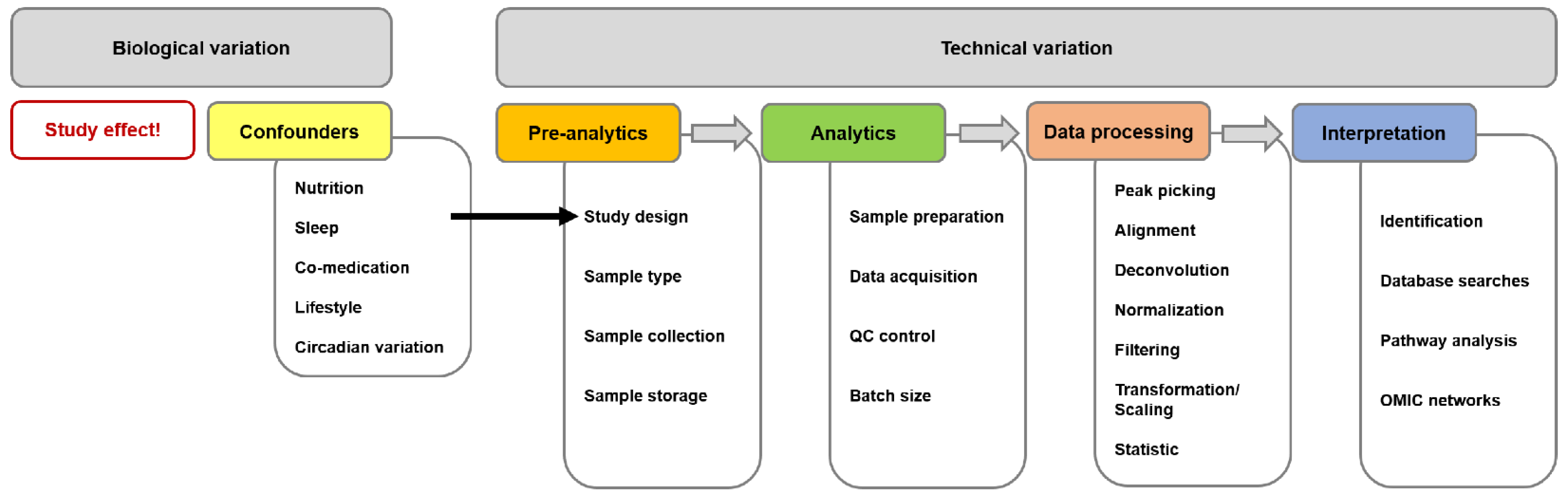
:1. Introduction
2. Analytical Methods Used to Study the Human Semen Metabolome
2.1. Studies Based on Nuclear Magnetic Resonance Spectroscopy

| Ref. | Pathological Condition/ Study Group 1 | Controls/ Study Group 2 | Sample | Discriminatory Metabolites |
|---|---|---|---|---|
| [19] | azoospermia (spermatogenic failure, n = 21) | normozoospermic | SP | ↓ lactate ↓ GPC ↓ citrate |
| obstructive azoospermia (vasectomy, n = 14) | ↓ lactate ↓ GPC | |||
| severe OAT (n = 7) | ||||
| [20] | azoospermia (spermatogenic failure, n = 58) | obstructive azoospermia (vasectomy, n = 17) | SP | ↑ ratio choline/citrate ↑ ratio choline/lactate ↑ ratio GPC/choline |
| azoospermia, normal FSH (spermatogenic failure, n = 9) | obstructive, azoospermia, normal FSH (vasectomy, n = 7) | SP | ↑ ratio choline/lactate ↑ ratio GPC/choline | |
| [21] | idiopathic infertility (normozoospermic, n = 65) | normozoospermic proven fathers (n = 60) | SP | ↓ Ala ↓ citrate ↓ GPC ↑ Phe |
| oligozoospermia (n = 60) | ↓ citrate ↓ GPC ↑ Phe | |||
| [23] | idiopathic infertility (normozoospermic, n = 17) | normozoospermic proven fathers (n = 6) | SP | ↑ fructose ↑ hippurate ↑ 2-hydroxyisovalerate ↑ amino acids Lys, Val |
| oligozoospermia (n = 20) | ↓ guanidoacetate ↓ fructose | |||
| AS (n = 20) | - | |||
| teratozoospermia (n = 20) | - | |||
| azoospermia (n = 20) | ↓ guanidoacetate ↓ fructose | |||
| [27] | OAT (n = 31) | normozoospermic (n = 28) | SP | ↓ amino acids Arg/Lys, Gln, Val, ↓ citrate ↓ choline ↓ creatinine ↓ α-ketoglutarate ↓ lactate ↓ spermine/putrescine ↑ amino acid Tyr |
| [30] | teratozoospermia (n = 14) | normozoospermic proven fathers (n = 15) | SP | ↓ cholesterol ↓ amino acid Glu ↓ taurine ↑ amino acids Ala, Gln, Ile, Leu, Lys, Pro, Thr, Tyr, Val ↑ choline ↑ citrate ↑ D-glucose ↑ lactate ↑ myo-inositol ↑ pyruvate |
| [9] | short-term abstinence (2 h), IVF/ICSI couples, ≥15 × 106 sperm/mL (n = 31) | long-term abstinence (4–7 d), IVF/ICSI couples, ≥15 × 106 sperm/mL (n = 31) | SP | ↓ fructose ↓ acetate ↓ choline ↓ methanol ↓ N-acetylglucosamine ↓ O-acetylglucosamine ↓ uridine ↓ GPC ↑ pyruvate |
2.2. Studies Based on Raman Spectroscopy
2.3. Studies Based on Liquid Chromatography–Mass Spectrometry
| Ref. | Pathological Condition/ Study Group 1 | Controls/ Study Group 2 | Sample | Discriminatory Metabolites |
|---|---|---|---|---|
| [39] | AS (n = 30) | normozoospermic (n = 33) | SP | ↓ PGE2, PGD2, PGF2α ↑ 11,12-DHET ↑ 8,9-EET, 14,15-EET ↑ fatty acid C20:4 ↑ 5-HETE, 15-HETE, 20-HETE ↑ tetranor-PGEM |
| [41] | low-quality semen (n = 23) | high-quality semen (n = 24) | SP | ↓ α-AAA ↓ 5-aminoimidazol-riconucleotide ↓ amino acid Lys ↓ capryloylglycine ↓ carnitines C0, iso-C4, C6-OH, pivaloylcarnitine ↓ fatty acid C22:6 ↓ glutaconate ↓ GPC ↓ imidazole-4-acetaldehyde ↓ lyso-SM(d18:1) ↓ tocotrienol ↑ N-acryloylglycine ↑ carnitine C3-DC ↑ fatty acid C22:4 ↑ G6-P ↑ 11b-hydroxyprogesterone ↑ imidazoleacetate riboside ↑ 3-oxohexanoate ↑ PGB2, PGE2 ↑ PS ↑ dipeptide Trp-Asp ↑ tyramine glucoronide ↑ ubiquinone-2 ↑ uracil |
| [42] | AS (n = 77) | normozoospermic (n = 63) | SP | ↓ amino acids Arg, Met, Phe, Pro, Trp, Tyr, Val ↓ aminobutyrate ↓ citrate, malate, pyruvate, succinate ↓ hypoxanthine, inosine ↓ nucleobases adenine, cytosine ↓ spermine ↑ lactate ↑ Orn |
| [44] | low-quality semen (n = 140) | high-quality semen (n = 220) | SP | ↓ oleamide ↑ adenine, xanthine ↑ amino acids Arg, His ↑ carnitines C0, C2, C3, C6, C16, C18:2 ↑ (S,S)-9,10-dihydroxyoctadecanoate ↑ 7β,12α-dihydroxykaurenolide ↑ 12,13-dihydroxy-9-octadecenoate ↑ epitestosterone, 11b-hydroxyprogesterone ↑ fatty acids C18:1, C22:5, C22:6 ↑ GPC ↑ 6-keto-PGF1 α, 8-iso-15-keto-PGF2α, PGE2 ↑ L-3-phenyllactate, hydroxyphenyllactate |
| [45] | AS (n = 76) | proven fathers (n = 35) | SP | ↓ dipeptides Leu-Pro, Pro-Gly, Glu-Arg ↓ amino acid Val ↑ 2-phosphoglycerate ↑ creatine riboside ↑ isopentenylpyrophosphate ↑ γ-glutamyl-methylselenocysteine |
| AT (n = 32) | ↓ butoconazole ↓ carnitines C0, C3, C6, C22:5, pivaloylcarnitine ↓ dipeptide Gly-Phe ↓ LPC 20:0, LPE 16:0, PE P-16:0 ↓ lithocholate ↓ PGE3 ↑ dethiobiotin ↑ dipeptide Tyr-Glu | |||
| OAT (n = 20) | ↓ dethiobiotin ↓ dipeptides Pro-Gly, Lys-Gly, Val-Ser, Pro-Phe ↓ fatty acids C18:1, C20:4, C20:0(2OH) ↓ 6-methylnicotinamide ↓ methylpyrrolo [1,2-a]pyrazine ↓ nonanol ↓ piperanine ↓ N-oleoylethanolamine ↑ capsiamide ↑ hypoxanthine ↑ amino acid His ↑ lyso-SM(d18:0) ↑ palmitic amide ↑ penmacric acid ↑ spermine | |||
| [46] | men from URSA couples (n = 28) | proven fathers (n = 25) | sperm | ↓ pyroglutamate ↑ cholesterol ↑ 3-phenylbutyrate ↑ hexadecanedioate |
| SP | ↓ guanine ↑ 2-hydroxycaproic acid (2-hydroxyhexanoate) ↑ ascorbate ↑ neopterin ↑ glycocholate ↑ N-oleoylethanolamine ↑ taurodeoxycholate ↑ xanthosine | |||
| [47] | OA (n = 20) | normozoospermic (n = 20) | SP | ↓ amino acids Ala, Asp, Glu, Met, Pro, Trp ↓ biogenic amines alpha-AAA, serotonin, spermine, spermidine ↓ carnitines C0, C3, C5, C5:1, C5-OH, C6 (C4:1-DC) ↓ sphingolipids SM C16:1, SM (OH) C14:1, SM (OH) C16:1 ↓ acyl-acyl phospholipids PC 28:1, PC 34:2, PC 36:2, PC 36:3, PC 36:4, PC 38:0, PC 38:3, PC 38:4, PC 38:5, PC 38:6, PC 40:4, PC 40:5, PC 42:5 ↓ alkyl-acyl phospholipids PC O-34:0, PC O-40:5, PC O-40:6 |
| [51] | AS (n = 12) | normozoospermic (n = 12) | sperm | ↓ LPS 18:1 ↓ DAG 32:0, DAG 34:1, DAG 36:1 ↓ TAG 48:1, TAG 48:0, TAG 50:1, TAG 50:0, TAG 52:2, TAG 52:1 ↑ Cer d18:1/15:0 ↑ CL 66:4, CL 68:6, CL 72:9, CL 74:10, CL 74:9, CL 74:8, CL 74:7, CL 76:10, CL 76:9, CL 78:12, CL 78:11 ↑ total cholesterol, total GM3, LPI, PE ↑ GM3 d18:1/16:0, GM3 d18:1/22:0, GM3 d18:0/22:0, GM3 d18:1/24:1, GM3 d18:1/24:0, GM3 d18:0/24:0 ↑ LPI 18:0 ↑ acyl-acyl phospholipids PE 32:1, PE 34:1, PE 38:6, PE 38:3, PE 40:6, PE 40:5, PG 38:4 ↑ alkenyl-acyl phospholipids PC P-38:3, PC P-40:6, PC P-40:4, PE P-38:6, PE P-38:4, PE P-40:6 |
| [54] | nonobstructive azoospermia (n = 50) | proven fathers (n = 50) | SP | ↓ amino acids Ala, Arg, Asn, Asp, Gly, His, Ile, Leu, Lys, Orn, Pro, Ser, Thr, Trp, Tyr, Val ↓ biogenic amines α-AAA, taurine ↓ Zn2+ |
| OAT (n = 50) | ↓ amino acids Ala, Asn, Asp, His, Leu, Lys, Pro ↓ biogenic amines α-AAA, taurine | |||
| [55] | smoking, normozoospermic (n = 10) | nonsmoking, normozoospermic (n = 10) | sperm | ↓ biogenic amines ADMA, serotonin ↓ carnitines C7-DC, C8, C10, C10:2, C12-DC, C14, C14:1, C14:1-OH, C14:2-OH, C16:1, C16:2, C16:2-OH ↓ ratios ADMA/Arg, total DMA/Arg, Tyr/Phe, spermidine/putrescine, (C16 + C18)/C0 ↑ amino acids Asp, Gln, Gly, Phe, Val ↑ ratios Cit/Arg, spermine/spermidine, C2/C0, (C2 + C3)/C0 |
2.4. Studies Based on Gas Chromatography
| Ref. | Pathological Condition/ Study Group 1 | Controls/ Study Group 2 | Sample | Discriminatory Metabolites |
|---|---|---|---|---|
| [48] | elevated abnormal cells (n = 16) | proven fathers (n = 8) | sperm | ↓ PE ↑ PS, LPS |
| SP | ↑ PA, PS | |||
| total semen | ↓ fatty acids C18:0, C20:5, C22:6 ↑ fatty acids C16:0, C18:3, C24:0 | |||
| [57] | AS (n = 30) | normozoospermic (n = 30) | SP | ↓ Val ↑ benzoate ↑ cholecalciferol ↑ fatty acids C16:0, C18:1, C19:0 ↑ D-pinitol |
| [58] | nonobstructive azoospermia, TESE-negative (n = 11) TESE-positive (n = 9) | normozoospermic (n = 10) | SP | ↑ darwinol ↑ 4,5-dimethoxy-1,2-benzenedicarboxylic acid (4,5-dimethoxyphthalate) ↑ 2,2,4,4,6,6-hexamethyl-1,3,5-trithiane ↑ 2-pyrrolidineacetate ((±)-homoproline) ↑ tartarate |
| [59] | AS (n = 10 pools of 3) | normozoospermic (n = 10 pools of 3) | sperm | ↓ amino acids Cys, Glu, Leu, Trp ↓ amines 2-aminoethanethiol, 2-amino-1-phenylethanol, N-(3-aminopropyl)-morpholine, phenylethylamine ↓ 5-aminovalerate ↓ D,L-dihydrosphingosine ↓ glycerate ↓ cis-gondoate ↓ guanidinosuccinate ↓ lactate ↓ methylheptadecanoate ↓ methylmercaptopurine ↓ monoolein ↓ norvaline ↓ nucleosides guanosine, cytidine ↓ 3-phosphoglycerate ↓ phytosphingosine ↓ picolinate ↓ pipecolinate ↓ α-tocopherol ↓ trans-4-hydroxyproline ↑ benzoate ↑ 2-deoxyerythritol ↑ dithioerythritol ↑ ethanolamine ↑ orotate ↑ zymosterol |
3. Summary
4. Conclusions
Author Contributions
Funding
Institutional Review Board Statement
Data Availability Statement
Acknowledgments
Conflicts of Interest
Abbreviations
References
- Wang, C.; Mbizvo, M.; Festin, M.P.; Björndahl, L.; Toskin, I. Evolution of the WHO “Semen” processing manual from the first (1980) to the sixth edition (2021). Fertil. Steril. 2022, 117, 237–245. [Google Scholar] [CrossRef]
- World Health Organization. WHO Laboratory Manual for the Examination of Human Semen and Sperm-Cervical Mucus Interaction, 4th ed.; Published on behalf of the World Health Organization by Cambridge University Press: Cambridge, UK; New York, NY, USA, 1999; ISBN 0521645999. [Google Scholar]
- World Health Organization. WHO Laboratory Manual for the Examination and Processing of Human Sperm, 5th ed.; World Health Organization: Geneva, Switzerland, 2010; ISBN 978-92-4-154778-9. [Google Scholar]
- Björndahl, L. The usefulness and significance of assessing rapidly progressive spermatozoa. Asian J. Androl. 2010, 12, 33–35. [Google Scholar] [CrossRef]
- Sukcharoen, N.; Keith, J.; Irvine, D.S.; Aitken, R.J. Predicting the fertilizing potential of human sperm suspensions in vitro: Importance of sperm morphology and leukocyte contamination. Fertil. Steril. 1995, 63, 1293–1300. [Google Scholar] [CrossRef]
- World Health Organization. WHO Laboratory Manual for the Examination and Processing of Human Semen, 6th ed.; World Health Organization: Geneva, Switzerland, 2021; ISBN 978-92-4-003079-4. [Google Scholar]
- Deepinder, F.; Chowdary, H.T.; Agarwal, A. Role of metabolomic analysis of biomarkers in the management of male infertility. Expert Rev. Mol. Diagn. 2007, 7, 351–358. [Google Scholar] [CrossRef]
- Lippa, K.A.; Aristizabal-Henao, J.J.; Beger, R.D.; Bowden, J.A.; Broeckling, C.; Beecher, C.; Davis, W.C.; Dunn, W.B.; Flores, R.; Goodacre, R.; et al. Reference materials for MS-based untargeted metabolomics and lipidomics: A review by the metabolomics quality assurance and quality control consortium (mQACC). Metabolomics 2022, 18, 24. [Google Scholar] [CrossRef] [PubMed]
- Alipour, H.; Duus, R.K.; Wimmer, R.; Dardmeh, F.; Du Plessis, S.S.; Jørgensen, N.; Christiansen, O.B.; Hnida, C.; Nielsen, H.I.; van der Horst, G. Seminal plasma metabolomics profiles following long (4–7 days) and short (2 h) sexual abstinence periods. Eur. J. Obstet. Gynecol. Reprod. Biol. 2021, 264, 178–183. [Google Scholar] [CrossRef] [PubMed]
- Grunewald, S.; Fitzl, G.; Springsguth, C. Induction of ultra-morphological features of apoptosis in mature and immature sperm. Asian J. Androl. 2017, 19, 533–537. [Google Scholar] [CrossRef] [PubMed]
- Engel, K.M.; Baumann, S.; Rolle-Kampczyk, U.; Schiller, J.; von Bergen, M.; Grunewald, S. Metabolomic profiling reveals correlations between spermiogram parameters and the metabolites present in human spermatozoa and seminal plasma. PLoS ONE 2019, 14, e0211679. [Google Scholar] [CrossRef] [PubMed]
- Maher, A.D.; Lindon, J.C.; Nicholson, J.K. ¹H NMR-based metabonomics for investigating diabetes. Future Med. Chem. 2009, 1, 737–747. [Google Scholar] [CrossRef]
- Vignoli, A.; Ghini, V.; Meoni, G.; Licari, C.; Takis, P.G.; Tenori, L.; Turano, P.; Luchinat, C. High-throughput metabolomics by 1D NMR. Angew. Chem. Int. Ed. Engl. 2019, 58, 968–994. [Google Scholar] [CrossRef] [PubMed]
- Giraudeau, P.; Silvestre, V.; Akoka, S. Optimizing water suppression for quantitative NMR-based metabolomics: A tutorial review. Metabolomics 2015, 11, 1041–1055. [Google Scholar] [CrossRef]
- Zheng, G.; Price, W.S. Solvent signal suppression in NMR. Prog. Nucl. Magn. Reson. Spectrosc. 2010, 56, 267–288. [Google Scholar] [CrossRef]
- Bliziotis, N.G.; Engelke, U.F.H.; Aspers, R.L.E.G.; Engel, J.; Deinum, J.; Timmers, H.J.L.M.; Wevers, R.A.; Kluijtmans, L.A.J. A comparison of high-throughput plasma NMR protocols for comparative untargeted metabolomics. Metabolomics 2020, 16, 64. [Google Scholar] [CrossRef] [PubMed]
- Beckonert, O.; Keun, H.C.; Ebbels, T.M.D.; Bundy, J.; Holmes, E.; Lindon, J.C.; Nicholson, J.K. Metabolic profiling, metabolomic and metabonomic procedures for NMR spectroscopy of urine, plasma, serum and tissue extracts. Nat. Protoc. 2007, 2, 2692–2703. [Google Scholar] [CrossRef] [PubMed]
- Schiller, J.; Arnhold, J.; Glander, H.-J.; Arnold, K. Lipid analysis of human spermatozoa and seminal plasma by MALDI-TOF mass spectrometry and NMR spectroscopy—Effects of freezing and thawing. Chem. Phys. Lipids 2000, 106, 145–156. [Google Scholar] [CrossRef]
- Hamamah, S.; Seguin, F.; Barthelemy, C.; Akoka, S.; Le Pape, A.; Lansac, J.; Royere, D. 1H nuclear magnetic resonance studies of seminal plasma from fertile and infertile men. J. Reprod. Fertil. 1993, 97, 51–55. [Google Scholar] [CrossRef]
- Hamamah, S.; Seguin, F.; Bujan, L.; Barthelemy, C.; Mieusset, R.; Lansac, J. Quantification by magnetic resonance spectroscopy of metabolites in seminal plasma able to differentiate different forms of azoospermia. Hum. Reprod. 1998, 13, 132–135. [Google Scholar] [CrossRef] [PubMed]
- Gupta, A.; Mahdi, A.A.; Ahmad, M.K.; Shukla, K.K.; Jaiswer, S.P.; Shankhwar, S.N. 1H NMR spectroscopic studies on human seminal plasma: A probative discriminant function analysis classification model. J. Pharm. Biomed. Anal. 2011, 54, 106–113. [Google Scholar] [CrossRef] [PubMed]
- Ando, I.; Hirose, T.; Nemoto, T.; Totsune, K.; Imai, Y.; Takeuchi, K.; Fujiwara, M. Quantification of molecules in (1)H-NMR metabolomics with formate as a concentration standard. J. Toxicol. Sci. 2010, 35, 253–256. [Google Scholar] [CrossRef] [PubMed]
- Jayaraman, V.; Ghosh, S.; Sengupta, A.; Srivastava, S.; Sonawat, H.M.; Narayan, P.K. Identification of biochemical differences between different forms of male infertility by nuclear magnetic resonance (NMR) spectroscopy. J. Assist. Reprod. Genet. 2014, 31, 1195–1204. [Google Scholar] [CrossRef] [PubMed]
- Zhang, X.; Diao, R.; Zhu, X.; Li, Z.; Cai, Z. Metabolic characterization of asthenozoospermia using nontargeted seminal plasma metabolomics. Clin. Chim. Acta 2015, 450, 254–261. [Google Scholar] [CrossRef]
- Ramírez, A.R.; Castro, M.A.; Angulo, C.; Ramió, L.; Rivera, M.M.; Torres, M.; Rigau, T.; Rodríguez-Gil, J.E.; Concha, I.I. The presence and function of dopamine type 2 receptors in boar sperm: A possible role for dopamine in viability, capacitation, and modulation of sperm motility. Biol. Reprod. 2009, 80, 753–761. [Google Scholar] [CrossRef]
- Urra, J.A.; Villaroel-Espíndola, F.; Covarrubias, A.A.; Rodríguez-Gil, J.E.; Ramírez-Reveco, A.; Concha, I.I. Presence and function of dopamine transporter (DAT) in stallion sperm: Dopamine modulates sperm motility and acrosomal integrity. PLoS ONE 2014, 9, e112834. [Google Scholar] [CrossRef]
- Mumcu, A.; Karaer, A.; Dogan, B.; Tuncay, G. Metabolomics analysis of seminal plasma in patients with idiopathic oligoasthenoteratozoospermia using high-resolution NMR spectroscopy. Andrology 2020, 8, 450–456. [Google Scholar] [CrossRef]
- Neto, F.T.L.; Marques, R.A.; de Freitas Cavalcanti Filho, A.; Araujo, L.C.N.; Lima, S.V.C.; Pinto, L.; Silva, R.O. 1H NMR-based metabonomics for infertility diagnosis in men with varicocele. J. Assist. Reprod. Genet. 2020, 37, 2233–2247. [Google Scholar] [CrossRef]
- Murgia, F.; Corda, V.; Serrenti, M.; Usai, V.; Santoru, M.L.; Hurt, K.J.; Passaretti, M.; Monni, M.C.; Atzori, L.; Monni, G. Seminalfluid metabolomic markers of oligozoospermic infertility in humans. Metabolites 2020, 10, 64. [Google Scholar] [CrossRef]
- Mehrparvar, B.; Chashmniam, S.; Nobakht, F.; Amini, M.; Javidi, A.; Minai-Tehrani, A.; Arjmand, B.; Gilany, K. Metabolic profiling of seminal plasma from teratozoospermia patients. J. Pharm. Biomed. Anal. 2020, 178, 112903. [Google Scholar] [CrossRef]
- Hanson, B.M.; Aston, K.I.; Jenkins, T.G.; Carrell, D.T.; Hotaling, J.M. The impact of ejaculatory abstinence on semen analysis parameters: A systematic review. J. Assist. Reprod. Genet. 2018, 35, 213–220. [Google Scholar] [CrossRef]
- Ferro, L.; Gojkovic, Z.; Gorzsás, A.; Funk, C. Statistical methods for rapid quantification of proteins, lipids, and carbohydrates in nordic microalgal species using ATR-FTIR spectroscopy. Molecules 2019, 24, 3237. [Google Scholar] [CrossRef]
- Gilany, K.; Pouracil, R.S.M.; Sadeghi, M.R. Fourier transform infrared spectroscopy: A potential technique for noninvasive detection of spermatogenesis. Avicenna J. Med. Biotechnol. 2014, 6, 47–52. [Google Scholar]
- Gilany, K.; Moazeni-Pourasil, R.S.; Jafarzadeh, N.; Savadi-Shiraz, E. Metabolomics fingerprinting of the human seminal plasma of asthenozoospermic patients. Mol. Reprod. Dev. 2014, 81, 84–86. [Google Scholar] [CrossRef]
- Gilany, K.; Jafarzadeh, N.; Mani-Varnosfaderani, A.; Minai-Tehrani, A.; Sadeghi, M.R.; Darbandi, M.; Darbandi, S.; Amini, M.; Arjmand, B.; Pahlevanzadeh, Z. Metabolic fingerprinting of seminal plasma from non-obstructive azoospermia patients: Positive versus negative sperm retrieval. J. Reprod. Infertil. 2018, 19, 109–114. [Google Scholar] [PubMed]
- Jafarzadeh, N.; Mani-Varnosfaderani, A.; Minai-Tehrani, A.; Savadi-Shiraz, E.; Sadeghi, M.R.; Gilany, K. Metabolomics fingerprinting of seminal plasma from unexplained infertile men: A need for novel diagnostic biomarkers. Mol. Reprod. Dev. 2015, 82, 150. [Google Scholar] [CrossRef] [PubMed]
- Engel, K.M.; Jakop, U.; Müller, K.; Grunewald, S.; Paasch, U.; Schiller, J. MALDI MS analysis to investigate the lipid composition of sperm. CAC 2020, 16, 79–91. [Google Scholar] [CrossRef]
- Chen, X.; Hu, C.; Dai, J.; Chen, L. Metabolomics analysis of seminal plasma in infertile males with kidney-yang deficiency: A preliminary study. Evid. Based Complement. Alternat. Med. 2015, 2015, 892930. [Google Scholar] [CrossRef]
- Yu, L.; Yang, X.; Ma, B.; Ying, H.; Shang, X.; He, B.; Zhang, Q. Abnormal arachidonic acid metabolic network may reduce sperm motility via P38 MAPK. Open Biol. 2019, 9, 180091. [Google Scholar] [CrossRef]
- Szczuko, M.; Kikut, J.; Komorniak, N.; Bilicki, J.; Celewicz, Z.; Ziętek, M. The role of arachidonic and linoleic acid derivatives in pathological pregnancies and the human reproduction process. Int. J. Mol. Sci. 2020, 21, 9628. [Google Scholar] [CrossRef]
- Huang, Q.; Liu, L.; Wu, Y.; Wang, X.; Luo, L.; Nan, B.; Zhang, J.; Tian, M.; Shen, H. Seminal plasma metabolites mediate the associations of multiple environmental pollutants with semen quality in Chinese men. Environ. Int. 2019, 132, 105066. [Google Scholar] [CrossRef]
- Chen, L.; Wen, C.-W.; Deng, M.-J.; Li, P.; Zhang, Z.-D.; Zhou, Z.-H.; Wang, X. Metabolic and transcriptional changes in seminal plasma of asthenozoospermia patients. Biomed. Chromatogr. 2020, 34, e4769. [Google Scholar] [CrossRef]
- Li, L.; Hao, X.; Chen, H.; Wang, L.; Chen, A.; Song, X.; Hu, Z.; Su, Y.; Lin, H.; Fan, P. Metabolomic characterization of semen from asthenozoospermic patients using ultra-high-performance liquid chromatography-tandem quadrupole time-of-flight mass spectrometry. Biomed. Chromatogr. 2020, 34, e4897. [Google Scholar] [CrossRef]
- Wang, Y.-X.; Wu, Y.; Chen, H.-G.; Duan, P.; Wang, L.; Shen, H.-Q.; Lu, W.-Q.; Sun, B.; Wang, Q.; Zhang, B.; et al. Seminal plasma metabolome in relation to semen quality and urinary phthalate metabolites among Chinese adult men. Environ. Int. 2019, 129, 354–363. [Google Scholar] [CrossRef] [PubMed]
- Xu, Y.; Lu, H.; Wang, Y.; Zhang, Z.; Wu, Q. Comprehensive metabolic profiles of seminal plasma with different forms of male infertility and their correlation with sperm parameters. J. Pharm. Biomed. Anal. 2020, 177, 112888. [Google Scholar] [CrossRef] [PubMed]
- Zhang, X.; Wang, H.; Feng, T.; Yang, J.; Huang, Q.; Lu, C.; Guan, Y.; Sun, R.; Chen, M.; Qian, Y. The relationship between semen factors and unexplained recurrent spontaneous abortion. Clin. Chim. Acta 2020, 510, 605–612. [Google Scholar] [CrossRef] [PubMed]
- Boguenet, M.; Bocca, C.; Bouet, P.-E.; Serri, O.; Chupin, S.; Tessier, L.; Blanchet, O.; El Hachem, H.; La Chao de Barca, J.M.; Reynier, P.; et al. Metabolomic signature of the seminal plasma in men with severe oligoasthenospermia. Andrology 2020, 8, 1859–1866. [Google Scholar] [CrossRef]
- Gulaya, N.M.; Margitich, V.M.; Govseeva, N.M.; Klimashevsky, V.M.; Gorpynchenko, I.I.; Boyko, M.I. Phospholipid composition of human sperm and seminal plasma in relation to sperm fertility. Arch. Androl. 2001, 46, 169–175. [Google Scholar] [CrossRef]
- Gallart-Ayala, H.; Teav, T.; Ivanisevic, J. Metabolomics meets lipidomics: Assessing the small molecule component of metabolism. Bioessays 2020, 42, e2000052. [Google Scholar] [CrossRef]
- O’Donnell, V.B.; Ekroos, K.; Liebisch, G.; Wakelam, M. Lipidomics: Current state of the art in a fast moving field. Wiley Interdiscip. Rev. Syst. Biol. Med. 2020, 12, e1466. [Google Scholar] [CrossRef]
- Chen, S.; Wang, M.; Li, L.; Wang, J.; Ma, X.; Zhang, H.; Cai, Y.; Kang, B.; Huang, J.; Li, B. High-coverage targeted lipidomics revealed dramatic lipid compositional changes in asthenozoospermic spermatozoa and inverse correlation of ganglioside GM3 with sperm motility. Reprod. Biol. Endocrinol. 2021, 19, 105. [Google Scholar] [CrossRef]
- Tanaka, N.; Okamoto, E.; Toyosaka, A.; Fujiwara, S. Pathological evaluation of hepatic dearterialization in encapsulated hepatocellular carcinoma. J. Surg. Oncol. 1985, 29, 256–260. [Google Scholar] [CrossRef]
- Liebisch, G.; Fahy, E.; Aoki, J.; Dennis, E.A.; Durand, T.; Ejsing, C.S.; Fedorova, M.; Feussner, I.; Griffiths, W.J.; Köfeler, H.; et al. Update on LIPID MAPS classification, nomenclature, and shorthand notation for MS-derived lipid structures. J. Lipid Res. 2020, 61, 1539–1555. [Google Scholar] [CrossRef]
- Saleem, T.H.; Okasha, M.; Ibrahim, H.M.; Abu El-Hamd, M.; Fayed, H.M.; Hassan, M.H. Biochemical assessments of seminal Plasma Zinc, Testis-Expressed Sequence 101 and free amino acids and their correlations with reproductive hormones in Male Infertility. Biol. Trace Elem. Res. 2021, 199, 1729–1742. [Google Scholar] [CrossRef] [PubMed]
- Engel, K.M.; Baumann, S.; Blaurock, J.; Rolle-Kampczyk, U.; Schiller, J.; von Bergen, M.; Grunewald, S. Differences in the sperm metabolomes of smoking and nonsmoking men. Biol. Reprod. 2021, 105, 1484–1493. [Google Scholar] [CrossRef]
- Fernández-Ochoa, Á.; Leyva-Jiménez, F.J.; de La Luz Cádiz-Gurrea, M.; Pimentel-Moral, S.; Segura-Carretero, A. The role of high-resolution analytical techniques in the development of functional foods. Int. J. Mol. Sci. 2021, 22, 3220. [Google Scholar] [CrossRef]
- Tang, B.; Shang, X.; Qi, H.; Li, J.; Ma, B.; An, G.; Zhang, Q. Metabonomic analysis of fatty acids in seminal plasma between healthy and asthenozoospermic men based on gas chromatography mass spectrometry. Andrologia 2017, 49, e12744. [Google Scholar] [CrossRef] [PubMed]
- Gilany, K.; Mani-Varnosfaderani, A.; Minai-Tehrani, A.; Mirzajani, F.; Ghassempour, A.; Sadeghi, M.R.; Amini, M.; Rezadoost, H. Untargeted metabolomic profiling of seminal plasma in nonobstructive azoospermia men: A noninvasive detection of spermatogenesis. Biomed. Chromatogr. 2017, 31, e3931. [Google Scholar] [CrossRef]
- Zhao, K.; Zhang, J.; Xu, Z.; Xu, Y.; Xu, A.; Chen, W.; Miao, C.; Liu, S.; Wang, Z.; Jia, R. Metabolomic profiling of human spermatozoa in idiopathic asthenozoospermia patients using gas chromatography-mass spectrometry. Biomed Res. Int. 2018, 2018, 8327506. [Google Scholar] [CrossRef]
- Qiao, S.; Wu, W.; Chen, M.; Tang, Q.; Xia, Y.; Jia, W.; Wang, X. Seminal plasma metabolomics approach for the diagnosis of unexplained male infertility. PLoS ONE 2017, 12, e0181115. [Google Scholar] [CrossRef]
- Kavanagh, J.P. Sodium, potassium, calcium, magnesium, zinc, citrate and chloride content of human prostatic and seminal fluid. J. Reprod. Fertil. 1985, 75, 35–41. [Google Scholar] [CrossRef] [PubMed]
- Sharma, U.; Chaudhury, K.; Jagannathan, N.R.; Guha, S.K. A proton NMR study of the effect of a new intravasal injectable male contraceptive RISUG on seminal plasma metabolites. Reproduction 2001, 122, 431–436. [Google Scholar] [CrossRef]
- Wishart, D.S.; Feunang, Y.D.; Marcu, A.; Guo, A.C.; Liang, K.; Vázquez-Fresno, R.; Sajed, T.; Johnson, D.; Li, C.; Karu, N.; et al. HMDB 4.0: The human metabolome database for 2018. Nucleic Acids Res. 2018, 46, D608–D617. [Google Scholar] [CrossRef]
- Long, N.P.; Nghi, T.D.; Kang, Y.P.; Anh, N.H.; Kim, H.M.; Park, S.K.; Kwon, S.W. Toward a standardized strategy of clinical metabolomics for the advancement of precision medicine. Metabolites 2020, 10, 51. [Google Scholar] [CrossRef] [PubMed]
- Steuer, A.E.; Brockbals, L.; Kraemer, T. Untargeted metabolomics approaches to improve casework in clinical and forensic toxicology—“Where are we standing and where are we heading?”. WIREs Forensic Sci. 2022, 4, e1449. [Google Scholar] [CrossRef]
- Baumann, S.; Ceglarek, U.; Fiedler, G.M.; Lembcke, J.; Leichtle, A.; Thiery, J. Standardized approach to proteome profiling of human serum based on magnetic bead separation and matrix-assisted laser desorption/ionization time-of-flight mass spectrometry. Clin. Chem. 2005, 51, 973–980. [Google Scholar] [CrossRef] [PubMed]
- Fiedler, G.M.; Baumann, S.; Leichtle, A.; Oltmann, A.; Kase, J.; Thiery, J.; Ceglarek, U. Standardized peptidome profiling of human urine by magnetic bead separation and matrix-assisted laser desorption/ionization time-of-flight mass spectrometry. Clin. Chem. 2007, 53, 421–428. [Google Scholar] [CrossRef]
- Bruegel, M.; Planert, M.; Baumann, S.; Focke, A.; Bergh, F.T.; Leichtle, A.; Ceglarek, U.; Thiery, J.; Fiedler, G.M. Standardized peptidome profiling of human cerebrospinal fluid by magnetic bead separation and matrix-assisted laser desorption/ionization time-of-flight mass spectrometry. J. Proteomics 2009, 72, 608–615. [Google Scholar] [CrossRef] [PubMed]
- Gertsman, I.; Barshop, B.A. Promises and pitfalls of untargeted metabolomics. J. Inherit. Metab. Dis. 2018, 41, 355–366. [Google Scholar] [CrossRef]
- Dudzik, D.; Barbas-Bernardos, C.; García, A.; Barbas, C. Quality assurance procedures for mass spectrometry untargeted metabolomics. A review. J. Pharm. Biomed. Anal. 2018, 147, 149–173. [Google Scholar] [CrossRef]
- Fiehn, O. Metabolomics by gas chromatography-mass spectrometry: Combined targeted and untargeted profiling. Curr. Protoc. Mol. Biol. 2016, 114, 30–34. [Google Scholar] [CrossRef]
- Wajid, B.; Iqbal, H.; Jamil, M.; Rafique, H.; Anwar, F. MetumpX-a metabolomics support package for untargeted mass spectrometry. Bioinformatics 2020, 36, 1647–1648. [Google Scholar] [CrossRef]
- Lin, Y.; Caldwell, G.W.; Li, Y.; Lang, W.; Masucci, J. Inter-laboratory reproducibility of an untargeted metabolomics GC-MS assay for analysis of human plasma. Sci. Rep. 2020, 10, 10918. [Google Scholar] [CrossRef]
- Blaženović, I.; Kind, T.; Ji, J.; Fiehn, O. Software tools and approaches for compound identification of LC-MS/MS data in metabolomics. Metabolites 2018, 8, 31. [Google Scholar] [CrossRef] [PubMed]
| Technique | Characteristics | Advantages | Disadvantages |
|---|---|---|---|
| Spectroscopic Methods | |||
| Proton nuclear magnetic resonance (1H NMR) |
|
|
|
| Infrared (IR) |
|
|
|
| Raman |
|
|
|
| Chromatography coupled to mass spectrometry (MS) | |||
| High-performance liquid chromatography (HPLC) MS |
|
|
|
| Gas chromatography(GC) MS |
|
|
|
Publisher’s Note: MDPI stays neutral with regard to jurisdictional claims in published maps and institutional affiliations. |
© 2022 by the authors. Licensee MDPI, Basel, Switzerland. This article is an open access article distributed under the terms and conditions of the Creative Commons Attribution (CC BY) license (https://creativecommons.org/licenses/by/4.0/).
Share and Cite
Blaurock, J.; Baumann, S.; Grunewald, S.; Schiller, J.; Engel, K.M. Metabolomics of Human Semen: A Review of Different Analytical Methods to Unravel Biomarkers for Male Fertility Disorders. Int. J. Mol. Sci. 2022, 23, 9031. https://doi.org/10.3390/ijms23169031
Blaurock J, Baumann S, Grunewald S, Schiller J, Engel KM. Metabolomics of Human Semen: A Review of Different Analytical Methods to Unravel Biomarkers for Male Fertility Disorders. International Journal of Molecular Sciences. 2022; 23(16):9031. https://doi.org/10.3390/ijms23169031
Chicago/Turabian StyleBlaurock, Janet, Sven Baumann, Sonja Grunewald, Jürgen Schiller, and Kathrin M. Engel. 2022. "Metabolomics of Human Semen: A Review of Different Analytical Methods to Unravel Biomarkers for Male Fertility Disorders" International Journal of Molecular Sciences 23, no. 16: 9031. https://doi.org/10.3390/ijms23169031
APA StyleBlaurock, J., Baumann, S., Grunewald, S., Schiller, J., & Engel, K. M. (2022). Metabolomics of Human Semen: A Review of Different Analytical Methods to Unravel Biomarkers for Male Fertility Disorders. International Journal of Molecular Sciences, 23(16), 9031. https://doi.org/10.3390/ijms23169031