Review

Role of miRNAs in Human T Cell Leukemia Virus Type 1 Induced T Cell Leukemia: A Literature Review and Bioinformatics Approach

by
Caio Bezerra Machado
1,†,
Leidivan Sousa da Cunha
2,†,
Jersey Heitor da Silva Maués
3,
Flávia Melo Cunha de Pinho Pessoa
1,
Marcelo Braga de Oliveira
4,
Rodrigo Monteiro Ribeiro
5,
Germison Silva Lopes
6,
Manoel Odorico de Moraes Filho
1,
Maria Elisabete Amaral de Moraes
1,
André Salim Khayat
4 and
Caroline Aquino Moreira-Nunes
1,2,4,7,*
1
Department of Medicine, Pharmacogenetics Laboratory, Drug Research and Development Center (NPDM), Federal University of Ceará, Fortaleza 60430-275, CE, Brazil
2
Unichristus University Center, Faculty of Biomedicine, Fortaleza 60430-275, CE, Brazil
3
Hematology and Transfusion Medicine Center, University of Campinas, Campinas 13083-970, SP, Brazil
4
Department of Biological Sciences, Oncology Research Center, Federal University of Pará, Belém 66073-005, PA, Brazil
5
Department of Hematology, Fortaleza General Hospital (HGF), Fortaleza 60150-160, CE, Brazil
6
Department of Hematology, César Cals General Hospital, Fortaleza 60015-152, CE, Brazil
7
Department of Health Sciences, Northeast Biotechnology Network (RENORBIO), Itaperi Campus, Ceará State University, Fortaleza 60740-903, CE, Brazil
*
Author to whom correspondence should be addressed.
These authors contributed equally to this work.
Int. J. Mol. Sci. 2022, 23(10), 5486; https://doi.org/10.3390/ijms23105486
Submission received: 10 April 2022 / Revised: 10 May 2022 / Accepted: 11 May 2022 / Published: 14 May 2022
(This article belongs to the Special Issue Non-coding RNAs in Pathogens and Associated Diseases)

Abstract

:
Human T cell leukemia virus type 1 (HTLV-1) was identified as the first pathogenic human retrovirus and is estimated to infect 5 to 10 million individuals worldwide. Unlike other retroviruses, there is no effective therapy to prevent the onset of the most alarming diseases caused by HTLV-1, and the more severe cases manifest as the malignant phenotype of adult T cell leukemia (ATL). MicroRNA (miRNA) dysfunction is a common feature of leukemogenesis, and it is no different in ATL cases. Therefore, we sought to analyze studies that reported deregulated miRNA expression in HTLV-1 infected cells and patients’ samples to understand how this deregulation could induce malignancy. Through in silico analysis, we identified 12 miRNAs that stood out in the prediction of targets, and we performed functional annotation of the genes linked to these 12 miRNAs that appeared to have a major biological interaction. A total of 90 genes were enriched in 14 KEGG pathways with significant values, including TP53, WNT, MAPK, TGF-β, and Ras signaling pathways. These miRNAs and gene interactions are discussed in further detail for elucidation of how they may act as probable drivers for ATL onset, and while our data provide solid starting points for comprehension of miRNAs’ roles in HTLV-1 infection, continuous effort in oncologic research is still needed to improve our understanding of HTLV-1 induced leukemia.

1. Introduction

In 1979, human T cell leukemia virus type 1 (HTLV-1) was identified as the first pathogenic human retrovirus [1]. It is the cause of adult T cell leukemia (ATL) and is distributed worldwide, although endemic in certain regions of the world, making it a serious global health problem [2,3]. The Southwestern part of Japan, sub-Saharan Africa and South America (in particular Brazil, Colombia, Chile, and Peru), the Caribbean area, Australo-Melanesia, and the Middle East are considered endemic regions, but a study carried out by Gessain and Cassar in 2012 estimated that there are 5 to 10 million individuals infected with HTLV-1 alone. This is, however, a superficial estimation, and the number may be much higher [3].
Brazil is possibly the country with the highest absolute number of HTLV cases in the world, although Japan is the country with the highest prevalence of HTLV-1 infection. Estimates from the Brazilian Ministry of Health indicate that between 700,000 and 2 million people are infected, and although the virus has been among the infectious agents in the country’s blood centers since 1993, there is no specific national policy for HTLV. Most carriers are asymptomatic, and there are more case reports of HTLV-2 infections in people who inject drugs and in the indigenous population [4,5,6,7].
Unlike other retroviruses, such as the Human Immunodeficiency Virus (HIV), there is no effective therapy to prevent the onset of the most alarming diseases caused by HTLV-1 [8]. Outcomes range from hypersensitivity reactions such as arthritis and uveitis to the fatal inflammatory neurologic disorder that is HTLV-1 associated myelopathy/tropical spastic paraparesis (HAM/TSP), HTLV-1 Associated Infectious Dermatitis (IDH), and the malignant phenotype of ATL, an aggressive lymphoproliferative disease which was first described in Japan in 1977 [9].

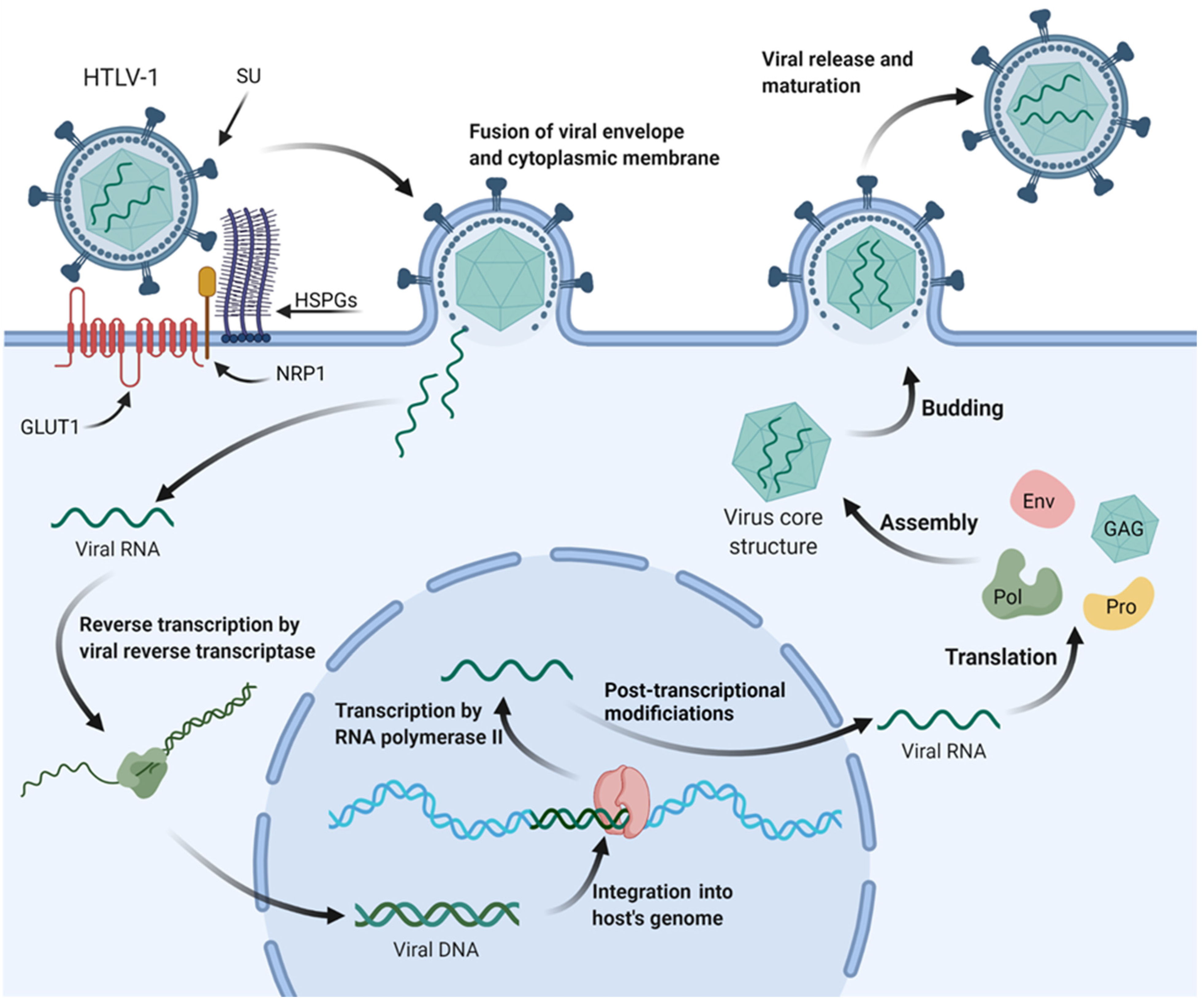
1.1. HTLV-1 Viral Structure and Infection Mechanisms

HTLV-1 is the causative agent of ATL, a malignant tumor of CD4+ T cells. It is a retrovirus of the deltaretrovirus genus, and several subtypes of HTLV were discovered later—HTLV-2, HTLV-3, and HTLV-4. It has a structural organization like all retroviruses; two mature viral glycoproteins derived from a common precursor, an outer surface protein (SU) associated with a transmembrane (TM) protein, which is responsible for anchoring the SU-TM complex on the surface of the infected cell, an envelope consisting of a lipid bilayer of cellular origin, and the matrix protein located next to the membrane [10,11,12,13].
It is an enveloped double-stranded RNA virus, and its genome contains three structural genes: gag, pol, and env, and two regulatory genes, tax and rex. The tax and rex genes regulate the transactivation of viral replication, in addition to regulating the expression of viral proteins and the basic leucine zipper factor (HBZ) of HTLV-1, which are essential for the maintenance of viral persistence and pathogenesis, in addition to helping in the oncogenic process of ATL, stimulation of tumor growth and development [12,13,14,15].
Viral integration into the host’s DNA happens preferably at transcriptionally active regions, specifically near transcription factor binding sites. HTLV-1 uses different strategies to induce neoplastic transformation, but the immunogenic profile of the host is associated with inflammatory responses, promoting or protecting against the development of HAM or ATL. Its replication occurs parallel to the cell during mitosis, and its infection can occur vertically, during childbirth or breastfeeding, or parenterally, which consists of transfusions, transplants, intravenous drug use, and unprotected sexual intercourse [3,9,15,16,17,18].
Furthermore, HTLV infects mainly T lymphocytes but is also capable of infecting monocytes and dendritic cells due to its direct interaction with glucose transporter 1 (GLUT 1), which is ubiquitously expressed on cell surfaces (Figure 1) [18,19,20,21].
After infection and entry into the cell, viral RNA is reverse transcribed into viral DNA or provirus. After that, the virus can integrate into the host cell genome and exploit normal cell physiology. Viral integrase recognizes and binds to viral long terminal repeats (LTRs) in viral DNA, forming a pre-integration complex (PIC). PIC associates with the host enzyme, protein phosphatase 2A, and engages with host DNA in regions of open chromatin. In addition, HTLV-1 increases genomic instability by direct actions on the DNA, and, in turn, genomic instability can also alter the proviral genome that is often mutated or deleted, thus creating an escape from the host’s immune system [22,23,24,25,26].
During infection, there is viral transmission by cell–cell contact due to an organizing center of polarized microtubules at the cell–cell junction and a virological synapse triggered by Tax protein. From there, the HTLV-1 Gag complex, viral RNAs, and enveloped HTLV-1 virions accumulate at the synapse and migrate into the uninfected cell [27,28,29]. Another way in which viral transmission happens is by using the cell itself through proliferation. That is, the virus integrates into the host genome, and after mitosis, it is transmitted to the daughter cell [23,24].

1.2. Leukemogenesis Pathways to Adult T Cell Leukemia

First reported in Japan in 1977, ATL is a lymphoproliferative disease that presents as an oligoclonal or monoclonal expansion of HTLV-1-infected T cells that occurs decades after infection. ATL onset is multifactorial, involving factors related to the virus and the host’s immune and inflammatory responses [10,15].
The disease is classified according to clinical characteristics into acute, chronic, lymphoma-like, and smoldering. Its manifestations include malaise, fever, weight loss, jaundice, skin lesions, adenomegaly, hypercalcemia, elevation of lactate dehydrogenase, and clinical alterations in the number of leukocytes in peripheral blood, with the presence of atypical lymphocytes [10,30,31].
In the acute phase, it is also possible to find lymphocytes with peculiar characteristics called floral cells in the peripheral blood smear, which is a characteristic clinical finding of ATL development. The diagnosis for HTLV-1 infection is performed by ELISA and confirmed by PCR and Western blot, and a worse prognosis may be expected in the acute and lymphoma forms, being more aggressive and with a median survival of one year [24,27,32].
ATL cells’ clonal expansion is promoted by the accessory proteins of HTLV-1 and the Tax protein. Tax utilizes several mechanisms for cellular transformation, including the creation of chromosomal instability, amplification of centrosomes, abrogation of DNA repair, activation of cyclin-dependent kinases and nuclear factor-kB (NF-kB), and AKT signaling, and even silencing of TP53 checkpoints. The maintenance of the ATL transformation occurs through HBZ protein activity, in addition to this protein being essential for the establishment of persistent viral infection [27,28,33,34].

1.3. MicroRNAs

The molecular mechanisms of HTLV-1-mediated transformation and carcinogenesis are still unknown; however, there are studies that correlate HTLV carcinogenesis to microRNAs (miRNAs), which are endogenous 18–25 nucleotide long RNAs that play regulatory roles in cell metabolism, directing messenger RNAs (mRNA) for cleavage or translational repression [35,36]. They are involved in the maintenance of a variety of biological processes, including the cell cycle, post-transcriptional gene expression, and other processes [37,38].
MiRNAs genes can be located within the introns and exons of protein-coding genes or in intergenic regions and are transcribed by RNA Polymerase II and processed by the Drosha and Dicer enzymes [38,39,40]. Deregulation of these miRNAs is a common feature in human tumors and has been described in several types of human cancer, such as lung cancer, colorectal cancer, pancreatic endocrine, and even chronic lymphocytic leukemia [41].
Discovering miRNAs, identifying their targets, and clarifying their functions has been a critical strategy for understanding normal biological processes and their roles in disease development [42]. Furthermore, the miRNA expression pattern can be correlated with cancer, so the miRNA profile can be used as a tool for cancer diagnosis and prognosis [43]. Therefore, several studies demonstrate that the cellular expression of miRNAs is affected in HTLV-1 infected cells [44,45].
For this reason, in this study, we sought to identify in previously published works which miRNAs were altered in ATL; in addition, we performed bioinformatics analysis in order to describe possible pathways of malignant transformation and biological interactions to predict the role of these miRNAs in HTLV-1-mediated leukemogenesis.

2. Literature Review and Bioinformatic Analysis Approach

The results are presented below in Table 1, describing the studies that demonstrated miRNAs’ altered expression after HTLV-1 infection in biological samples. After the literature review process, we used the data generated by the 12 published works presented in Table 1 to investigate the role of those miRNAs in the pathogenic model of ATL induced by HTLV-1.
In this study, a list of 42 miRNAs, pointed out in the studies above, was input to the miRWalk algorithm (version 3.0), and for prediction of individual target genes using TargetScan, (version 7.2), miRDB (version 6.0), and miRTarBase (version 8.0), generating three unique files for further analysis [59,60,61]. TargetScan is a predictor that generates predicted interactions, while miRTarBase and miRDB provide validated interactions. A (p > 0.95) for binding probability [62], preferentially conserved sites within the 3′ UTR (untranslated region) were applied to the data filtering, and preferably all validated interactions were maintained [63].
After that, we simulated miRNA-gene interaction networks that were performed with Cytoscape (version 3.9.1). The data generated by the prediction of the three files obtained with miRWalk were used to construct the three respective miRNA-gene interaction networks. Then, the three networks were merged according to Cytoscape’s manual merge procedure with an intersection operator to obtain a single miRNA-gene interaction network [64].
Lastly, we performed functional annotation of the genes linked to 12 miRNAs that showed a significant biological interaction between all described in the studies, using DAVID (Database for Annotation, Visualization, and Integrated Discovery) [65] to identify the pathways of KEGG (Kyoto Encyclopedia of Genes and Genomes) [66]. All target genes linked to miRNAs (miR-34a-5p, miR-146b-5p, miR-181b-5p, miR-26a-5p, miR-26b-5p, miR-222-3p, miR-155-5p, miR-193a-5p, miR-199a-3p, miR-199b-3p, miR-423-5p, miR-150-5p) were investigated with DAVID using the Refseq identifier of the species (homo sapiens). A total of 90 genes were enriched in 14 KEGG pathways with significant values annotated with (−Log10 p-value) < 0.05. An interactive network was built with Cytoscape to show all target genes enriched in KEGG pathways. The topologies of all networks were constructed with network density < 0.05.
Most miRNAs described in these studies were mentioned only once, and a correlation between authors and cited miRNAs can be found in Supplementary Figure S1. Almost all authors predicted or hypothesized oncogenic-related cellular pathways that might be deregulated due to miRNA erroneous expression [46,47,48,49,50,51,52,53,54,55,56,57,58]; however, our own bioinformatics approach was able to predict pathways through which these miRNAs described previously might induce carcinogenesis.
The results of the bioinformatics analysis are shown in the figure below (Figure 2). The target predictions were integrated through predictive interactions between miRNAs and their target genes using the Cytoscape tool that generated networks of topologies of significant and representative densities [64]. Using 12 miRNAs that stood out in the prediction of targets, it was possible to predict their interactions using miRNA-Target interaction networks.
There was a significant enrichment of miRNA target genes in important KEGG pathways for cancer, such as hsa05200: Pathways in cancer; hsa04310: WNT signaling pathway; hsa04010: MAPK signaling pathway; hsa04350: TGF-β signaling pathway; hsa04014: Ras signaling pathway, all shown at central interaction network in Figure 2, which presented 39 nodes and 80 edges and a density equal to 0.054. A full scope of all identified pathways and targeted gene ontology can be found in Supplementary Table S1.
While the deregulation of any signaling pathways is sure to disrupt cell metabolic homeostasis, some are more relevant for leukemogenesis than others and will be discussed further in this paper. Of importance to note is the presence of TP53 as a trending gene, appearing as part of most of the identified significantly deregulated pathways.

3. TP53 Mutations and Modulation in HTLV-1 Infection

The loss or mutation of the TP53 tumor suppressor gene is a very common genetic lesion in human cancer, so much so that the inheritance of a germline mutation in TP53 generates increased risks of developing neoplasms. The reason for this well-reported carcinogenesis is the tumor suppression functions performed by the protein that is commonly called the guardian of the genome, as it protects DNA integrity [67,68,69].
According to Borrero and El-Deiry [68], the TP53 gene is mutated in about 50% of human cancers, but there is also a biological inactivation of its pathway in the others. It can be altered directly or by targeting the oncoproteins MDM4 regulator of p53 (MDM4) and MDM2 proto-oncogene (MDM2) that regulate TP53 activity, thus being altered indirectly.
In response to stress factors such as DNA damage, hypoxia, UV irradiation, oxidative free radicals, oncogenes, or growth factor deficiencies, the TP53 protein is activated and can modulate target genes that regulate cell cycle arrest, apoptosis, DNA repair, angiogenesis, and metastasis [68,69,70,71].
TP53 importance in carcinogenesis is a well-established concept, and several different miRNAs have been reported to silence TP53 expression directly or indirectly, while TP53 also regulates miRNAs expression, making it clear that this regulation can be associated with malignant changes [68,70]. In ATL cases, inhibition of TP53 activity is associated with HTLV-1 infection due to Tax and HBZ, thus promoting its malignant proliferation [72,73].
Studies by Nascimento et al. [46] and Ruggero et al. [51] both report deregulation of miR-150-5p, which by our analysis demonstrated a direct interaction with TP53 activity. There is, however, controversy when determining miRNA expression levels in HTLV-1 infection, with the former reporting an upregulated expression of miR-150-5p levels in HTLV-1 asymptomatic carriers (ACs) when compared to healthy controls (HC) and ATL patients, and the latter reporting a downregulation of miR-150-5p in HTLV-1 infected cells when compared to HC.
It is important to note that, while the downregulation of a miRNA that interferes with TP53 activity may seem beneficial for proper cell cycle control, the modulation of TP53 and ATL onset may also be correlated with TP53 mutational profile. TP53 status as a tumor suppressor may be easily overridden even by single-nucleotide mutations that are able to induce gain of oncogenic functions (GOF) for TP53 protein and dominant-negative effects (DNE) over wild-type TP53. Oversimplifying the past statements, downregulating TP53 may actually be beneficial for the proper regulation of cell homeostasis if its mutations are inducing an oncogenic-protein phenotype and disrupting its physiological regulating activities [74,75].
This difference in miRNA expression levels may be representative of proliferation and survival advantages that cells obtain in in vivo models during disease development that are not seen when compared to HTLV-1 infected cell lines in vitro. Nascimento et al. [46] described that ATL patients had increased levels of miR-150-5p compared to HC but lower levels when compared to HTLV-1 ACs; this may be what differentiates asymptomatic infections from ATL onset. It is possible to hypothesize that genomic instability driven by HTLV-1 integration into the host’s genome leads to a series of mutations in genes critical for maintaining cell metabolism and, in the case of TP53, overexpression of miRNAs modulating genetic GOFs and DNEs is what keeps leukemogenesis in check [25,26,46].
Furthermore, the reported downregulation of miR-150-5p in HTLV-1 infected cell lines compared to samples of HC by Ruggero et al. [51] only corroborate the idea that TP53 GOFs and DNEs are the main effectors for survival advantage gains in HTLV-1 infections and consequent achievement of malignant phenotypes. Regardless of miRNA levels, though, TP53 mutations and rearrangements are important prognostic factors and major predictors of response to treatment in ATL patients [76,77].

4. Pathways Identified as Deregulated in ATL

Beyond TP53, miR-150-5-p validated targets include other proto-oncogenes, such as c-Myb and the NOTCH3 gene [51]. The c-Myb gene, which encodes transcription factors that regulate cell proliferation, differentiation, and apoptosis, is found to be overexpressed in a wide range of human cancers, and leukemia studies have associated its expression with a worse prognosis and its transcript physically binds to TP53 [78,79].
Another validated target of miR-150-5p is the NOTCH3 pathway, which regulates cell proliferation, differentiation, cell fate determination, and stem/progenitor cell self-renewal in adult and embryonic organs. Elevated expression is increasingly documented in cancers and is correlated with rapid malignant progression, abnormal differentiation, metastasis, and worse prognosis. There is also a known interaction of NOTCH signaling with other major signaling pathways such as WNT and TGF-β [80,81].

4.1. WNT Canonical and Non-Canonical Pathways

The WNT signaling pathway is associated with cell differentiation, polarization, and migration during development and is fundamental in cell growth control. Its main component is the family of WNT proteins that activate cell membrane receptors in a paracrine and autocrine manner. It is based on three signaling pathways: WNT canonical signaling pathway (CP), which is β-catenin-dependent, and two WNT non-CP that are β-catenin-independent: planar cell polarity (PCP) signaling pathway and a WNT/Ca2+ pathway that has Wnt5a as the main effector. This pathway mainly plays a pro-tumor role, and WNT/β-catenin signaling is activated in many types of cancer. Its primary receptors are frizzleds (FZD), which are transmembrane G-coupled proteins. WNT signaling is inhibited by endogenous inhibitors and secreted FZD-related proteins (sFRPs) that interact directly with WNTs [82,83,84].
However, Ma et al. [85] previously reported that ATL cells rely on the WNT non-CP of WNT/Ca2+ for tumorigenesis development while inhibiting the WNT CP, which seems to have inhibitory effects on ATL cell growth through HBZ mediated activity. The miRNA expression profiles identified in our analysis support these observations and corroborate an excessive signaling of Wnt5a.
An overactivation of Wnt5a may be observed in ATL cells mediated by increased expression of c-myb, which is expected to happen at ATL onset after downregulation of miR-150-5p expression [46,51,86]. Also, phospholipase C beta (PLCB) are a family of direct downstream effectors of Wnt5a, and we identified miR-423-5p, which was reported as downregulated in HTLV-1 infected patients by Fayyad–Kazan et al. [47], to directly interact with PLCB1 expression [87]. Therefore, it is possible that HTLV-1 induces WNT non-CP both through increased activation of Wnt5a but also through upregulation of its downstream signaling effectors.
Other miRNAs that we identified as interacting with the WNT pathway, however, are much more associated with HTLV-1 capability of inhibiting the CP. It is worth noting the upregulation of miR-34a-5p in HTLV-1 infected cell lines since we were able to identify its interaction with the transcription factor lymphoid enhancer-binding factor 1 (LEF1), which is a main effector of WNT CP and was already reported to be suppressed by HBZ in HTLV-1 infection [51,85]. In Table 1 also, downregulation of miR-222-3p and miR-146b-5p was associated with ALT onset, and both miRNAs interact directly with inhibitors of WNT CP, those being TLE family member 3 (TLE3) and zinc and ring finger protein 3 (ZNRF3), respectively [46,51,88,89].
This particular profile of respective suppression and overactivation of canonical and non-canonical WNT pathways is not unexpected, even when disregarding miRNA activity since both pathways are activated by different ligands that interact in a reciprocal inhibitory fashion due to their competitive binding with the same FZD membrane receptors [90,91].

4.2. TGF-β Dual Roles in Cancer

Transforming growth factor-β (TGF-β) signaling is essential during embryo development and associated with many important biological processes; however, TGF-β roles in cancer are diverse and dependent on other cell signaling pathways, being considered a tumor suppressor at early tumor stages and an enhancer of malignant phenotypes later in cancer development [92,93,94].
TGF-β achieves its role as a tumor suppressor due to the ability to regulate cell proliferation, differentiation, and immune cell modulation through the activity of its main signal transducers, the SMAD family member (SMAD) proteins. Downstream of TGF-β activation, SMADs interact with cyclin-dependent kinase inhibitors (CDKNs), inhibitors of DNA binding (IDs) proteins and other proliferation drivers to modulate genomic expression, cell cycle, and apoptosis induction [95,96,97].
The oncogenic role of TGF-β, however, is highlighted at later stages of tumor development, where overactivation of TGF-β signaling is associated with increased potential of invasion and metastasis, as well as with downregulation of immune cell activity in the tumor microenvironment [97,98].
SMAD2, zinc finger FYVE-type containing 16 (ZFYVE16), and Sp1 transcription factor (SP1) are signal transducers acting downstream of TGF-β that we identified to be regulated by the activity of miR-155-5p, miR-222-3p, and miR-150-5p, respectively. All of the three previously mentioned miRNAs were reported by Nascimento et al. [46] as upregulated at HTLV-1 infection but downregulated at ATL onset, and this probably points towards the expected panorama of TGF-β inhibition at early stages followed by later overexpression [98].
ZFYVE16 is a direct enhancer of SMAD2 activity, as well as other SMAD members, and was reported by Zhao et al. [99] as an inhibitor of T cell proliferation through modulation of TGF-β signaling. In addition, SP1 is a transcription factor responsible for modulating the activity of a myriad of genes related to oncogenesis and is also an effector for cell cycle arrest through CDKN2B-mediated activity [100,101]. Their inhibition by miRNAs upregulation could mean a proliferation advantage for T cells mediated by HTLV-1 at initial infection.
It is also of note that, after HTLV-1 integration in the host genome, the integrated viral DNA harbors multiple binding sites for SP1 at viral gene promoters. While the roles of all of these binding sites as repressors or enhancers of gene expression are not fully elucidated, SP1 activity seems to be an important regulator for latent HTLV-1 activation in response to cell stressing factors [102,103].
Beyond the above-mentioned interactions, TGF-β inducing factor homologous box 2 (TGIF2) and E2F transcription factor 5 (E2F5), both of which act downstream of TGF-β signaling, have also had an identified interaction in our analyses. Mir-34a-5p, reported by Ruggero et al. [51] as upregulated in HTLV-1 infected cells, acts directly over the expression of both proteins. The transcription factor TGIF2 belongs to the TALE homologous domain protein family, including TGIF, TGIF2, and TGIF2LX/Y, while E2F5 is a transcription factor belonging to the E2F family, which is composed of eight members ranging from E2F1 to E2F8 [104,105].
TGIFs are known antagonists of TGF-β signaling due to their repressor effects over SMAD2/3 activity. By inhibiting the formation of the E1A binding protein p300 (p300)/SP1 transcriptional complex, which acts downstream of SMAD2/3 activity, TGIF2 negatively regulates p300/SP1-mediated cell cycle arrest and thus is implied as a cofactor for carcinogenesis in a variety of tumors [106,107,108,109]. On the other hand, E2F5 acts alongside transcription factor Dp-1 (DP1) and RB transcriptional corepressor like 1 (p107) to repress the activity of the proliferation driver c-myc in a SMAD2/3 dependent way [110,111,112]. Downregulation of both genes by the increased activity of miR-34a-5p in ATL cells possibly defines a profile for overactivation of TGF-β signaling by HTLV-1 during ATL development.
Furthermore, Zhao et al. [113] described TGF-β signaling as essential for successful HTLV-1 persistence of infection as HBZ-mediated activity causes the overactivation of SMAD2/3 response and increased formation of p300 transcriptional complex, leading to modulation of T cell phenotypes and escape from the host’s immune system.

4.3. RAS and MAPK Signaling

Another important interaction of miR-34a-5p was with RAS and MAPK pathways, through RAS-related (RRAS) gene and platelet-derived growth factor receptor (PDGFR), specifically PDGFRA, a classic proto-oncogene encoding receptor tyrosine kinases [114].
Members of the RAS family that contains more than 100 proteins, the RRAS subfamily is composed of only 3 small proteins: RRAS, RRAS2, and MRAS, which are molecular switches that toggle between GTP-bound and GDP-bound conformations. It has been reported that RRAS is involved in several cellular functions, including enhancement of integrin function, regulation of cell adhesion, invasion, and migration, and related to different mechanisms that regulate its oncogenic potential, one of which is epigenetic regulation. The widespread prevalence of RAS mutations in human cancer has been recognized for many years. In cancer, members of the RRAS subfamily have been implicated in anchorage-independent growth, increased invasiveness, stimulation of angiogenesis, and tumorigenicity. In addition, RRASs have been linked to leukemias, and their overexpression can impair tumor-targeted lymphocytes, indirectly promoting tumor growth [115,116,117,118].
MAPK is, in its classical pathway, an extension of RAS signaling through the RAS-RAF-MEK-ERK signaling cascade, which regulates genes that control cell development, differentiation, proliferation, and apoptosis. It plays a central role in human cancer, and many of its elements have been identified as oncogenes, being overactivated in a wide variety of tumors. In addition, aberrant activations of MAPK signaling and its interactions with other cell signaling pathways in the pathogenesis of chronic lymphocytic leukemia have also been described [119,120,121].
Interestingly though, the upregulation of miR-34a-5p reported by Ruggero et al. [51] in HTLV-1 infected cells would possibly point towards a disruption of both of the above-mentioned pathways through inhibition of receptor expression for extracellular growth factors and decreases in RRAS expression and phosphorylation levels. The RAS pathway, however, is activated intracellularly through multiple RAS isoforms, such as KRAS, HRAS, and NRAS, and by many growth and colony-stimulating factors, and the simple inhibition of RRAS isoform and PDGFRA may not be sufficient to inhibit RAS and MAPK activities altogether in ATL leukemogenesis [122,123]. The importance of RAS signaling for ATL cells is even highlighted by the fact that Tax-mediated anti-apoptotic effects are dependent on cell levels of phosphorylated RAS [124,125].
Still adjacent to MAPK signaling is the activity of the tumor necrosis factor (TNF) family and their ambiguous role in tumor development [126,127]. MiR-146b-5p, reported by Ruggero et al. [51] as downregulated in HTLV-1 infected cells, was identified to interact with TNF receptor-associated factor 6 (TRAF6), a transducer of TNF signaling that ultimately leads to increased expression of NF-kB [128]. The role of NF-kB in tumorigenesis is well established, and it is no different in ATL onset, where Tax potential to induce oncogenesis is directly linked to NF-kB at early tumor development, and NF-kB increased activity is even able to be maintained at later stages, regardless of Tax expression profile [129,130,131].
Through a bioinformatic approach, we were able to predict many interactions of miRNAs in important pathways in carcinogenesis that were correlated with HTLV-1 infection, and Figure 3 represents some of these targeted pathways and the miRNAs involved in their regulation.

5. Conclusions

Several studies reveal deregulated miRNAs in HTLV-1 infection, and our bioinformatic analysis was able to predict several of these miRNAs’ interactions with important pathways in tumorigenesis, especially TP53, WNT, RRAS, TGF-β, and MAPK, thus being able to address them as probable drivers for ATL onset. While the data generated by our bioinformatics tools are able to provide solid starting points for further comprehension of miRNAs’ roles in HTLV-1 infection, the continuous effort in oncologic research to clarify still to be elucidated molecular pathways is needed to improve our understanding of HTLV-1 induced leukemia.

Supplementary Materials

The following supporting information can be downloaded at: https://www.mdpi.com/article/10.3390/ijms23105486/s1.

Author Contributions

Invitation received, C.A.M.-N.; Conceptualization, C.B.M., L.S.d.C., J.H.d.S.M. and C.A.M.-N.; Provision of data and subsequent analysis and interpretation, C.B.M., L.S.d.C., F.M.C.d.P.P., M.B.d.O., R.M.R., G.S.L., M.O.d.M.F., M.E.A.d.M., A.S.K. and C.A.M.-N.; Bioinformatics analysis, J.H.d.S.M. and C.A.M.-N.; Writing—original draft preparation, C.B.M., L.S.d.C., J.H.d.S.M. and C.A.M.-N.; Writing—review and editing, C.B.M., L.S.d.C. and C.A.M.-N.; Funding acquisition, A.S.K. and C.A.M.-N. All authors have read and agreed to the published version of the manuscript.

Funding

This study was supported by Brazilian funding agencies: Coordination for the Improvement of Higher Education Personnel (CAPES; to C.B.M), National Council of Technological and Scientific Development (CNPq grant number 404213/2021-9 to C.A.M.-N.; and Productivity in Research PQ to M.O.d.M.F., M.E.A.d.M. and A.S.K.), Cearense Foundation of Scientific and Technological Support (FUNCAP grant number P20-0171-00078.01.00/20 to F.M.C.d.P.P. and M.O.d.M.F. and to C.A.M.-N.) and, we thank PROPESP/UFPA for publication payment.

Institutional Review Board Statement

Not applicable.

Informed Consent Statement

Not applicable.

Data Availability Statement

Not applicable.

Conflicts of Interest

The authors declare no conflict of interest. The funders had no role in the design of the study; in the collection, analyses, or data interpretation; in the writing of the manuscript, or in the decision to publish the results.

References

  1. Tagaya, Y.; Gallo, R.C. The Exceptional Oncogenicity of HTLV-1. Front. Microbiol. 2017, 8, 1425. [Google Scholar] [CrossRef]
  2. Saito, M.; Matsuzaki, T.; Satou, Y.; Yasunaga, J.I.; Saito, K.; Arimura, K.; Matsuoka, M.; Ohara, Y. In Vivo Expression of the HBZ Gene of HTLV-1 Correlates with Proviral Load, Inflammatory Markers and Disease Severity in HTLV-1 Associated Myelopathy/Tropical Spastic Paraparesis (HAM/TSP). Retrovirology 2009, 6, 19. [Google Scholar] [CrossRef] [PubMed] [Green Version]
  3. Gessain, A.; Cassar, O. Epidemiological Aspects and World Distribution of HTLV-1 Infection. Front. Microbiol. 2012, 3, 388. [Google Scholar] [CrossRef] [PubMed] [Green Version]
  4. Rosadas, C.; Menezes, M.L.B.; Galvão-Castro, B.; Assone, T.; Miranda, A.E.; Aragón, M.G.; Caterino-De-araujo, A.; Taylor, G.P.; Ishak, R. Blocking HTLV-1/2 Silent Transmission in Brazil: Current Public Health Policies and Proposal for Additional Strategies. PLoS Negl. Trop. Dis. 2021, 15, e0009717. [Google Scholar] [CrossRef] [PubMed]
  5. Vieira, B.A.; Bidinotto, A.B.; Dartora, W.J.; Pedrotti, L.G.; de Oliveira, V.M.; Wendland, E.M. Prevalence of Human T-Lymphotropic Virus Type 1 and 2 (HTLV-1/-2) Infection in Pregnant Women in Brazil: A Systematic Review and Meta-Analysis. Sci. Rep. 2021, 11, 15367. [Google Scholar] [CrossRef]
  6. Garcia, I.F.d.S.; Hennington, É.A. HTLV: A Stigmatizing Infection? Cad. Saude Publica 2019, 35, e00005419. [Google Scholar] [CrossRef]
  7. Oliveira-Filho, A.B.; Araújo, A.P.S.; Souza, A.P.C.; Gomes, C.M.; Silva-Oliveira, G.C.; Martins, L.C.; Fischer, B.; Machado, L.F.A.; Vallinoto, A.C.R.; Ishak, R.; et al. Human T-Lymphotropic Virus 1 and 2 among People Who Used Illicit Drugs in the State of Pará, Northern Brazil. Sci. Rep. 2019, 9, 14750. [Google Scholar] [CrossRef] [Green Version]
  8. Forlani, G.; Shallak, M.; Accolla, R.S.; Romanelli, M.G. HTLV-1 Infection and Pathogenesis: New Insights from Cellular and Animal Models. Int. J. Mol. Sci. 2021, 22, 8001. [Google Scholar] [CrossRef]
  9. Franchini, G.; Ambinder, R.F.; Barry, M. Viral Disease in Hematology. Hematology 2000, 2000, 409–423. [Google Scholar] [CrossRef]
  10. Delamarre, L.; Rosenberg, A.R.; Pique, C.; Pham, D.; Callebaut, I.; Dokhélar, M.C. The HTLV-I Envelope Glycoproteins: Structure and Functions. J. Acquir. Immune Defic. Syndr. Hum. Retrovirol. 1996, 13, S85–S91. [Google Scholar] [CrossRef]
  11. Mohanty, S.; Harhaj, E.W. Mechanisms of Oncogenesis by HTLV-1 Tax. Pathogens 2020, 9, 543. [Google Scholar] [CrossRef] [PubMed]
  12. Martinez, M.P.; Al-Saleem, J.; Green, P.L. Comparative Virology of HTLV-1 and HTLV-2. Retrovirology 2019, 16, 21. [Google Scholar] [CrossRef] [PubMed]
  13. Eusebio-Ponce, E.; Anguita, E.; Paulino-Ramirez, R.; Candel, F.J. HTLV-1 Infection: An Emerging Risk. Pathogenesis, Epidemiology, Diagnosis and Associated Diseases. Rev. Española Quimioter. 2019, 32, 485. [Google Scholar]
  14. Manns, A.; Hisada, M.; La Grenade, L. Human T-Lymphotropic Virus Type I Infection. Lancet Lond. Engl. 1999, 353, 1951–1958. [Google Scholar] [CrossRef]
  15. Brites, C.; Grassi, M.F.; Quaresma, J.A.S.; Ishak, R.; Vallinoto, A.C.R. Pathogenesis of HTLV-1 Infection and Progression Biomarkers: An Overview. Braz. J. Infect. Dis. 2021, 25, 101594. [Google Scholar] [CrossRef]
  16. Melamed, A.; Laydon, D.J.; Gillet, N.A.; Tanaka, Y.; Taylor, G.P.; Bangham, C.R.M. Genome-Wide Determinants of Proviral Targeting, Clonal Abundance and Expression in Natural HTLV-1 Infection. PLOS Pathog. 2013, 9, e1003271. [Google Scholar] [CrossRef] [Green Version]
  17. Wattel, E.; Cavrois, M.; Gessain, A.; Wain-Hobson, S. Clonal Expansion of Infected Cells: A Way of Life for HTLV-I. J. Acquir. Immune Defic. Syndr. Hum. Retrovirol. 1996, 13, S92–S99. [Google Scholar] [CrossRef]
  18. Roucoux, D.F.; Wang, B.; Smith, D.; Nass, C.C.; Smith, J.; Hutching, S.T.; Newman, B.; Lee, T.-H.; Chafets, D.M.; Murphy, E.L. A Prospective Study of Sexual Transmission of Human T Lymphotropic Virus (HTLV)–I and HTLV-II. J. Infect. Dis. 2005, 191, 1490–1497. [Google Scholar] [CrossRef] [Green Version]
  19. Wu, K.; Bottazzi, M.E.; De La Fuente, C.; Deng, L.; Gitlin, S.D.; Maddukuri, A.; Dadgar, S.; Li, H.; Vertes, A.; Pumfery, A.; et al. Protein Profile of Tax-Associated Complexes. J. Biol. Chem. 2004, 279, 495–508. [Google Scholar] [CrossRef] [Green Version]
  20. Igakura, T.; Stinchcombe, J.C.; Goon, P.K.C.; Taylor, G.P.; Weber, J.N.; Griffiths, G.M.; Tanaka, Y.; Osame, M.; Bangham, C.R.M. Spread of HTLV-I between Lymphocytes by Virus-Induced Polarization of the Cytoskeleton. Science 2003, 299, 1713–1716. [Google Scholar] [CrossRef] [Green Version]
  21. Gross, C.; Thoma-Kress, A.K. Molecular Mechanisms of HTLV-1 Cell-to-Cell Transmission. Viruses 2016, 8, 74. [Google Scholar] [CrossRef] [PubMed]
  22. Maertens, G.N. B′-Protein Phosphatase 2A Is a Functional Binding Partner of Delta-Retroviral Integrase. Nucleic Acids Res. 2016, 44, 364. [Google Scholar] [CrossRef] [PubMed]
  23. Tasayco-Magallanes, E.; Miranda-Ulloa, E.; Romero-Ruiz, S.; Cárdenas-Bustamante, F.; Briceño-Espinoza, R.; Yana-Calatayud, B. Evaluation of Two Brands of Test of ELISA for the Diagnosis of HTLV-1 against Peruvian Samples. Rev. Chil. Infectol. 2020, 37, 780–783. [Google Scholar] [CrossRef]
  24. Cook, L.; Melamed, A.; Yaguchi, H.; Bangham, C.R. The Impact of HTLV-1 on the Cellular Genome. Curr. Opin. Virol. 2017, 26, 125–131. [Google Scholar] [CrossRef] [PubMed]
  25. Giam, C.Z.; Semmes, O.J. HTLV-1 Infection and Adult T-Cell Leukemia/Lymphoma—A Tale of Two Proteins: Tax and HBZ. Viruses 2016, 8, 161. [Google Scholar] [CrossRef]
  26. Ernzen, K.J.; Panfil, A.R. Regulation of HTLV-1 Transformation. Biosci. Rep. 2022, 42, BSR20211921. [Google Scholar] [CrossRef]
  27. Pereira, W.A.; Mesquita, E.M. Vírus Linfotrópico de Células T Humana (HTLV): Doenças Associadas e Dificuldades No Diagnóstico e Tratamento. Rev. Investig. Bioméd. 2016, 8, 92–101. [Google Scholar] [CrossRef]
  28. Matsuoka, M.; Jeang, K.T. Human T-Cell Leukaemia Virus Type 1 (HTLV-1) Infectivity and Cellular Transformation. Nat. Rev. Cancer 2007, 7, 270–280. [Google Scholar] [CrossRef]
  29. Majorovits, E.; Nejmeddine, M.; Tanaka, Y.; Taylor, G.P.; Fuller, S.D.; Bangham, C.R.M. Human T-Lymphotropic Virus-1 Visualized at the Virological Synapse by Electron Tomography. PLoS ONE 2008, 3, e2251. [Google Scholar] [CrossRef] [Green Version]
  30. Yoshida, N.; Weinstock, D.M. Lymphoid Neoplasia: Clinicogenetic Risk Modeling in ATL. Blood 2018, 131, 159. [Google Scholar] [CrossRef] [Green Version]
  31. Shimoyama, M. Diagnostic Criteria and Classification of Clinical Subtypes of Adult T-Cell Leukaemia-Lymphoma. A Report from the Lymphoma Study Group (1984-87). Br. J. Haematol. 1991, 79, 428–437. [Google Scholar] [CrossRef] [PubMed]
  32. Thorstensson, R.; Albert, J.; Andersson, S. Strategies for Diagnosis of HTLV-I and -II. Transfusion 2002, 42, 780–791. [Google Scholar] [CrossRef] [PubMed]
  33. Brand, H.; Alves, J.G.B.; Pedrosa, F.; Lucena-Silva, N. Leucemia de Células T Do Adulto. Rev. Bras. Hematol. Hemoter. 2009, 31, 375–383. [Google Scholar] [CrossRef] [Green Version]
  34. Franchini, G. HTLV-1 and HIV-1 “Accessory” Proteins: A Misleading Misnomer. Mol. Aspects Med. 2010, 31, 331–332. [Google Scholar] [CrossRef] [PubMed]
  35. D’Agostino, D.M.; Zanovello, P.; Watanabe, T.; Ciminale, V. The Metwork in Normal- and HTLV-1-Transformed T Cells. Adv. Cancer Res. 2012, 113, 45–83. [Google Scholar] [CrossRef] [PubMed]
  36. Simonson, B.; Das, S. MicroRNA Therapeutics: The Next Magic Bullet? Mini Rev. Med. Chem. 2015, 15, 467. [Google Scholar] [CrossRef]
  37. Bittencourt, A.L.; Farré, L. Leucemia/Linfoma de Células T Do Adulto. An. Bras. Dermatol. 2008, 83, 351–359. [Google Scholar] [CrossRef] [Green Version]
  38. Lu, T.X.; Rothenberg, M.E. MicroRNA. J. Allergy Clin. Immunol. 2018, 141, 1202–1207. [Google Scholar] [CrossRef] [Green Version]
  39. Garzon, R.; Calin, G.A.; Croce, C.M. MicroRNAs in Cancer. Annu. Rev. Med. 2009, 60, 167–179. [Google Scholar] [CrossRef] [Green Version]
  40. MacFarlane, L.-A.; Murphy, P.R. MicroRNA: Biogenesis, Function and Role in Cancer. Curr. Genom. 2010, 11, 537. [Google Scholar] [CrossRef] [Green Version]
  41. Treiber, T.; Treiber, N.; Meister, G. Regulation of MicroRNA Biogenesis and Its Crosstalk with Other Cellular Pathways. Nat. Rev. Mol. Cell Biol. 2019, 20, 5–20. [Google Scholar] [CrossRef] [PubMed]
  42. Di Leva, G.; Garofalo, M.; Croce, C.M. MicroRNAs in Cancer. Annu. Rev. Pathol. 2014, 9, 287–314. [Google Scholar] [CrossRef] [PubMed] [Green Version]
  43. Liu, B.; Li, J.; Cairns, M.J. Identifying MiRNAs, Targets and Functions. Brief. Bioinform. 2014, 15, 1–19. [Google Scholar] [CrossRef]
  44. Lee, Y.S.; Dutta, A. MicroRNAs in Cancer. Annu. Rev. Pathol. 2009, 4, 199–227. [Google Scholar] [CrossRef] [PubMed]
  45. Moles, R.; Nicot, C. The Emerging Role of MiRNAs in HTLV-1 Infection and ATLL Pathogenesis. Viruses 2015, 7, 4047. [Google Scholar] [CrossRef] [PubMed] [Green Version]
  46. Nascimento, A.; Valadão de Souza, D.R.; Pessôa, R.; Pietrobon, A.J.; Nukui, Y.; Pereira, J.; Casseb, J.; Penalva de Oliveira, A.C.; Loureiro, P.; da Silva Duarte, A.J.; et al. Global Expression of Noncoding RNome Reveals Dysregulation of Small RNAs in Patients with HTLV-1–Associated Adult T-Cell Leukemia: A Pilot Study. Infect. Agent. Cancer 2021, 16, 4. [Google Scholar] [CrossRef]
  47. Fayyad-Kazan, M.; ElDirani, R.; Hamade, E.; El Majzoub, R.; Akl, H.; Bitar, N.; Fayyad-Kazan, H.; Badran, B. Circulating MiR-29c, MiR-30c, MiR-193a-5p and MiR-885-5p: Novel Potential Biomarkers for HTLV-1 Infection Diagnosis. Infect. Genet. Evol. 2019, 74, 103938. [Google Scholar] [CrossRef]
  48. Sharma, V.K.; Raimondi, V.; Ruggero, K.; Pise-Masison, C.A.; Cavallari, I.; Silic-Benussi, M.; Ciminale, V.; D’Agostino, D.M. Expression of MiR-34a in T-Cells Infected by Human T-Lymphotropic Virus 1. Front. Microbiol. 2018, 9, 832. [Google Scholar] [CrossRef] [Green Version]
  49. Moles, R.; Bellon, M.; Nicot, C. STAT1: A Novel Target of MiR-150 and MiR-223 Is Involved in the Proliferation of HTLV-I–Transformed and ATL Cells. Neoplasia 2015, 17, 449–462. [Google Scholar] [CrossRef] [Green Version]
  50. Vernin, C.; Thenoz, M.; Pinatel, C.; Gessain, A.; Gout, O.; Delfau-Larue, M.H.; Nazaret, N.; Legras-Lachuer, C.; Wattel, E.; Mortreux, F. HTLV-1 BZIP Factor HBZ Promotes Cell Proliferation and Genetic Instability by Activating OncomiRs. Cancer Res. 2014, 74, 6082–6093. [Google Scholar] [CrossRef] [Green Version]
  51. Ruggero, K.; Guffanti, A.; Corradin, A.; Sharma, V.K.; De Bellis, G.; Corti, G.; Grassi, A.; Zanovello, P.; Bronte, V.; Ciminale, V.; et al. Small Noncoding RNAs in Cells Transformed by Human T-Cell Leukemia Virus Type 1: A Role for a TRNA Fragment as a Primer for Reverse Transcriptase. J. Virol. 2014, 88, 3612. [Google Scholar] [CrossRef] [PubMed] [Green Version]
  52. Tomita, M. Important Roles of Cellular MicroRNA MiR-155 in Leukemogenesis by Human T-Cell Leukemia Virus Type 1 Infection. ISRN Microbiol. 2012, 2012, 978607. [Google Scholar] [CrossRef] [PubMed] [Green Version]
  53. Rahman, S.; Quann, K.; Pandya, D.; Singh, S.; Khan, Z.K.; Jain, P. HTLV-1 Tax Mediated Downregulation of MiRNAs Associated with Chromatin Remodeling Factors in T Cells with Stably Integrated Viral Promoter. PLoS ONE 2012, 7, e34490. [Google Scholar] [CrossRef] [PubMed]
  54. Yamagishi, M.; Nakano, K.; Miyake, A.; Yamochi, T.; Kagami, Y.; Tsutsumi, A.; Matsuda, Y.; Sato-Otsubo, A.; Muto, S.; Utsunomiya, A.; et al. Polycomb-Mediated Loss of MiR-31 Activates NIK-Dependent NF-ΚB Pathway in Adult T Cell Leukemia and Other Cancers. Cancer Cell 2012, 21, 121–135. [Google Scholar] [CrossRef] [Green Version]
  55. Tomita, M.; Tanaka, Y.; Mori, N. MicroRNA MiR-146a Is Induced by HTLV-1 Tax and Increases the Growth of HTLV-1-Infected T-Cells. Int. J. Cancer 2012, 130, 2300–2309. [Google Scholar] [CrossRef]
  56. Bellon, M.; Lepelletier, Y.; Hermine, O.; Nicot, C. Deregulation of MicroRNA Involved in Hematopoiesis and the Immune Response in HTLV-I Adult T-Cell Leukemia. Blood 2009, 113, 4914–4917. [Google Scholar] [CrossRef] [Green Version]
  57. Pichler, K.; Schneider, G.; Grassmann, R. MicroRNA MiR-146a and Further Oncogenesis-Related Cellular MicroRNAs Are Dysregulated in HTLV-1-Transformed T Lymphocytes. Retrovirology 2008, 5, 100. [Google Scholar] [CrossRef] [Green Version]
  58. Yeung, M.L.; Yasunaga, J.I.; Bennasser, Y.; Dusetti, N.; Harris, D.; Ahmad, N.; Matsuoka, M.; Jeang, K.T. Roles for MicroRNAs, MiR-93 and MiR-130b, and TP53INP1 Tumor Suppressor in Cell Growth Dysregulation by HTLV-1. Cancer Res. 2008, 68, 8976. [Google Scholar] [CrossRef] [Green Version]
  59. Agarwal, V.; Bell, G.W.; Nam, J.W.; Bartel, D.P. Predicting Effective MicroRNA Target Sites in Mammalian MRNAs. Elife 2015, 4, e05005. [Google Scholar] [CrossRef]
  60. Chen, Y.; Wang, X. MiRDB: An Online Database for Prediction of Functional MicroRNA Targets. Nucleic Acids Res. 2020, 48, D127–D131. [Google Scholar] [CrossRef] [Green Version]
  61. Ding, J.; Li, X.; Hu, H. TarPmiR: A New Approach for MicroRNA Target Site Prediction. Bioinformatics 2016, 32, 2768–2775. [Google Scholar] [CrossRef] [PubMed] [Green Version]
  62. Huang, D.W.; Sherman, B.T.; Lempicki, R.A. Systematic and Integrative Analysis of Large Gene Lists Using DAVID Bioinformatics Resources. Nat. Protoc. 2009, 4, 44–57. [Google Scholar] [CrossRef] [PubMed]
  63. Huang, H.Y.; Lin, Y.C.D.; Li, J.; Huang, K.Y.; Shrestha, S.; Hong, H.C.; Tang, Y.; Chen, Y.G.; Jin, C.N.; Yu, Y.; et al. MiRTarBase 2020: Updates to the Experimentally Validated MicroRNA-Target Interaction Database. Nucleic Acids Res. 2020, 48, D148–D154. [Google Scholar] [CrossRef] [PubMed] [Green Version]
  64. Shannon, P.; Markiel, A.; Ozier, O.; Baliga, N.S.; Wang, J.T.; Ramage, D.; Amin, N.; Schwikowski, B.; Ideker, T. Cytoscape: A Software Environment for Integrated Models of Biomolecular Interaction Networks. Genome Res. 2003, 13, 2498–2504. [Google Scholar] [CrossRef] [PubMed]
  65. Kanehisa, M.; Sato, Y.; Furumichi, M.; Morishima, K.; Tanabe, M. New Approach for Understanding Genome Variations in KEGG. Nucleic Acids Res. 2019, 47, D590–D595. [Google Scholar] [CrossRef] [Green Version]
  66. Sticht, C.; De La Torre, C.; Parveen, A.; Gretz, N. MiRWalk: An Online Resource for Prediction of MicroRNA Binding Sites. PLoS ONE 2018, 13, e0206239. [Google Scholar] [CrossRef]
  67. Blagih, J.; Buck, M.D.; Vousden, K.H. P53, Cancer and the Immune Response. J. Cell Sci. 2020, 133, jcs237453. [Google Scholar] [CrossRef] [Green Version]
  68. Borrero, L.J.H.; El-Deiry, W.S. Tumor Suppressor P53: Biology, Signaling Pathways, and Therapeutic Targeting. Biochim. Biophys. Acta. Rev. Cancer 2021, 1876, 188556. [Google Scholar] [CrossRef]
  69. Liu, J.; Zhang, C.; Hu, W.; Feng, Z. Tumor Suppressor P53 and Metabolism. J. Mol. Cell Biol. 2019, 11, 284–292. [Google Scholar] [CrossRef] [Green Version]
  70. Yu, D.; Xu, Z.; Cheng, X.; Qin, J. The Role of MiRNAs in MDMX-P53 Interplay. J. Evid. Based Med. 2021, 14, 152–160. [Google Scholar] [CrossRef]
  71. Slattery, M.L.; Mullany, L.E.; Wolff, R.K.; Sakoda, L.C.; Samowitz, W.S.; Herrick, J.S. The P53-Signaling Pathway and Colorectal Cancer: Interactions between Downstream P53 Target Genes and MiRNAs. Genomics 2019, 111, 762–771. [Google Scholar] [CrossRef] [PubMed]
  72. Fochi, S.; Mutascio, S.; Bertazzoni, U.; Zipeto, D.; Romanelli, M.G. HTLV Deregulation of the NF-ΚB Pathway: An Update on Tax and Antisense Proteins Role. Front. Microbiol. 2018, 9, 285. [Google Scholar] [CrossRef] [PubMed] [Green Version]
  73. Ducasa, N.; Grasso, D.; Benencio, P.; Papademetrio, D.L.; Biglione, M.; Kashanchi, F.; Berini, C.; Garcia, M.N. Autophagy in Human T-Cell Leukemia Virus Type 1 (HTLV-1) Induced Leukemia. Front. Oncol. 2021, 11, 967. [Google Scholar] [CrossRef] [PubMed]
  74. Stein, Y.; Rotter, V.; Aloni-Grinstein, R. Gain-of-Function Mutant P53: All the Roads Lead to Tumorigenesis. Int. J. Mol. Sci. 2019, 20, 6197. [Google Scholar] [CrossRef] [Green Version]
  75. Olivier, M.; Hollstein, M.; Hainaut, P. TP53 Mutations in Human Cancers: Origins, Consequences, and Clinical Use. Cold Spring Harb. Perspect. Biol. 2010, 2, a001008. [Google Scholar] [CrossRef] [Green Version]
  76. Sakamoto, Y.; Ishida, T.; Masaki, A.; Murase, T.; Takeshita, M.; Muto, R.; Iwasaki, H.; Ito, A.; Kusumoto, S.; Nakano, N.; et al. Clinical Significance of TP53 Mutations in Adult T-Cell Leukemia/Lymphoma. Br. J. Haematol. 2021, 195, 571–584. [Google Scholar] [CrossRef]
  77. Cook, L.B.M. Adult T-Cell Leukaemia/Lymphoma and the Trouble with TP53. Br. J. Haematol. 2021, 195, 655–656. [Google Scholar] [CrossRef]
  78. Fry, E.A.; Inoue, K. C-MYB and DMTF1 in Cancer. Cancer Investig. 2019, 37, 46–65. [Google Scholar] [CrossRef]
  79. Dúckaa, M.; Kučeríková, M.; Trčka, F.; Červinka, J.; Biglieri, E.; Šmarda, J.; Borsig, L.; Beneš, P.; Knopfová, L. C-Myb Interferes with Inflammatory IL1α-NF-ΚB Pathway in Breast Cancer Cells. Neoplasia 2021, 23, 326–336. [Google Scholar] [CrossRef]
  80. Inder, S.; O’Rourke, S.; McDermott, N.; Manecksha, R.; Finn, S.; Lynch, T.; Marignol, L. The Notch-3 Receptor: A Molecular Switch to Tumorigenesis? Cancer Treat. Rev. 2017, 60, 69–76. [Google Scholar] [CrossRef]
  81. Zhu, C.; Ho, Y.J.; Salomao, M.A.; Dapito, D.H.; Bartolome, A.; Schwabe, R.F.; Lee, J.S.; Lowe, S.W.; Pajvani, U.B. Notch Activity Characterizes a Common Hepatocellular Carcinoma Subtype with Unique Molecular and Clinicopathologic Features. J. Hepatol. 2021, 74, 613–626. [Google Scholar] [CrossRef] [PubMed]
  82. Taciak, B.; Pruszynska, I.; Kiraga, L.; Bialasek, M.; Krol, M. Wnt Signaling Pathway in Development and Cancer. J. Physiol. Pharmacol. 2018, 69, 185–196. [Google Scholar] [CrossRef]
  83. Nusse, R.; Clevers, H. Wnt/β-Catenin Signaling, Disease, and Emerging Therapeutic Modalities. Cell 2017, 169, 985–999. [Google Scholar] [CrossRef] [PubMed]
  84. Colozza, G.; Koo, B. Ub and Dub of RNF43/ZNRF3 in the WNT Signalling Pathway. EMBO Rep. 2021, 22, e52970. [Google Scholar] [CrossRef] [PubMed]
  85. Ma, G.; Yasunaga, J.; Fan, J.; Yanagawa, S.; Matsuoka, M. HTLV-1 BZIP Factor Dysregulates the Wnt Pathways to Support Proliferation and Migration of Adult T-Cell Leukemia Cells. Oncogene 2013, 32, 4222–4230. [Google Scholar] [CrossRef] [PubMed] [Green Version]
  86. Nakano, K.; Chihara, Y.; Kobayashi, S.; Iwanaga, M.; Utsunomiya, A.; Watanabe, T.; Uchimaru, K. Overexpression of Aberrant Wnt5a and Its Effect on Acquisition of Malignant Phenotypes in Adult T-Cell Leukemia/Lymphoma (ATL) Cells. Sci. Rep. 2021, 11, 4114. [Google Scholar] [CrossRef]
  87. Colli, L.M.; Saggioro, F.; Serafini, L.N.; Camargo, R.C.; Machado, H.R.; Moreira, A.C.; Antonini, S.R.; de Castro, M. Components of the Canonical and Non-Canonical Wnt Pathways Are Not Mis-Expressed in Pituitary Tumors. PLoS ONE 2013, 8, e62424. [Google Scholar] [CrossRef] [Green Version]
  88. Parolia, A.; Cieslik, M.; Chu, S.C.; Xiao, L.; Ouchi, T.; Zhang, Y.; Wang, X.; Vats, P.; Cao, X.; Pitchiaya, S.; et al. Distinct Structural Classes of Activating FOXA1 Alterations in Advanced Prostate Cancer. Nature 2019, 571, 413. [Google Scholar] [CrossRef]
  89. Basham, K.J.; Rodriguez, S.; Turcu, A.F.; Lerario, A.M.; Logan, C.Y.; Rysztak, M.R.; Gomez-Sanchez, C.E.; Breault, D.T.; Koo, B.K.; Clevers, H.; et al. A ZNRF3-Dependent Wnt/β-Catenin Signaling Gradient Is Required for Adrenal Homeostasis. Genes Dev. 2019, 33, 209–220. [Google Scholar] [CrossRef] [Green Version]
  90. Grumolato, L.; Liu, G.; Mong, P.; Mudbhary, R.; Biswas, R.; Arroyave, R.; Vijayakumar, S.; Economides, A.N.; Aaronson, S.A. Canonical and Noncanonical Wnts Use a Common Mechanism to Activate Completely Unrelated Coreceptors. Genes Dev. 2010, 24, 2517. [Google Scholar] [CrossRef] [Green Version]
  91. Flores-Hernández, E.; Velázquez, D.M.; Castañeda-Patlán, M.C.; Fuentes-García, G.; Fonseca-Camarillo, G.; Yamamoto-Furusho, J.K.; Romero-Avila, M.T.; García-Sáinz, J.A.; Robles-Flores, M. Canonical and Non-Canonical Wnt Signaling Are Simultaneously Activated by Wnts in Colon Cancer Cells. Cell. Signal. 2020, 72, 109636. [Google Scholar] [CrossRef] [PubMed]
  92. Zhao, M.; Mishra, L.; Deng, C.X. The Role of TGF-β/SMAD4 Signaling in Cancer. Int. J. Biol. Sci. 2018, 14, 111–123. [Google Scholar] [CrossRef] [PubMed] [Green Version]
  93. Teicher, B.A. TGFβ-Directed Therapeutics: 2020. Pharmacol. Ther. 2021, 217, 107666. [Google Scholar] [CrossRef] [PubMed]
  94. Xie, F.; Ling, L.; Van Dam, H.; Zhou, F.; Zhang, L. TGF-β Signaling in Cancer Metastasis. Acta Biochim. Biophys. Sin. Shanghai 2018, 50, 121–132. [Google Scholar] [CrossRef] [Green Version]
  95. Bertero, A.; Brown, S.; Madrigal, P.; Osnato, A.; Ortmann, D.; Yiangou, L.; Kadiwala, J.; Hubner, N.C.; De Los Mozos, I.R.; Sadée, C.; et al. The SMAD2/3 Interactome Reveals That TGFβ Controls m 6 A MRNA Methylation in Pluripotency. Nature 2018, 555, 256–259. [Google Scholar] [CrossRef]
  96. Tzavlaki, K.; Moustakas, A. TGF-β Signaling. Biomolecules 2020, 10, 487. [Google Scholar] [CrossRef] [Green Version]
  97. Morikawa, M.; Derynck, R.; Miyazono, K. TGF-β and the TGF-β Family: Context-Dependent Roles in Cell and Tissue Physiology. Cold Spring Harb. Perspect. Biol. 2016, 8, a021873. [Google Scholar] [CrossRef] [Green Version]
  98. MaruYama, T.; Chen, W.J.; Shibata, H. TGF-β and Cancer Immunotherapy. Biol. Pharm. Bull. 2022, 45, 155–161. [Google Scholar] [CrossRef]
  99. Zhao, X.; Li, D.; Qiu, Q.; Jiao, B.; Zhang, R.; Liu, P.; Ren, R. Zfyve16 Regulates the Proliferation of B-Lymphoid Cells. Front. Med. 2018, 12, 559–565. [Google Scholar] [CrossRef]
  100. Beishline, K.; Azizkhan-Clifford, J. Sp1 and the ‘Hallmarks of Cancer’. FEBS J. 2015, 282, 224–258. [Google Scholar] [CrossRef]
  101. Xia, Y.; Liu, Y.; Yang, C.; Simeone, D.M.; Sun, T.T.; DeGraff, D.J.; Tang, M.S.; Zhang, Y.; Wu, X.R. Dominant Role of CDKN2B/P15INK4B of 9p21.3 Tumor Suppressor Hub in Inhibition of Cell-Cycle and Glycolysis. Nat. Commun. 2021, 12, 2047. [Google Scholar] [CrossRef] [PubMed]
  102. Fauquenoy, S.; Robette, G.; Kula, A.; Vanhulle, C.; Bouchat, S.; Delacourt, N.; Rodari, A.; Marban, C.; Schwartz, C.; Burny, A.; et al. Repression of Human T-Lymphotropic Virus Type 1 Long Terminal Repeat Sense Transcription by Sp1 Recruitment to Novel Sp1 Binding Sites. Sci. Rep. 2017, 7, srep43221. [Google Scholar] [CrossRef] [PubMed] [Green Version]
  103. Jabareen, A.; Suleman, M.; Abu-Jaafar, A.; Huleihel, M. Different Molecular Mechanisms of HTLV-1 and HIV LTR Activation by TPA. Biochem. Biophys. Res. Commun. 2018, 500, 538–543. [Google Scholar] [CrossRef] [PubMed]
  104. Jiang, J.; Wu, R.H.; Zhou, H.L.; Li, Z.M.; Kou, D.; Deng, Z.; Dong, M.; Chen, L.H. TGIF2 Promotes Cervical Cancer Metastasis by Negatively Regulating FCMR. Eur. Rev. Med. Pharmacol. Sci. 2020, 24, 5953–5962. [Google Scholar] [CrossRef] [PubMed]
  105. Inagaki, Y.; Wu, D.; Fujiwara, K.; Ishizuka, Y.; Oguni, A.; Tokunaga, T.; Takayama, T.; Soma, M.; Fukuda, N.; Ozaki, T.; et al. Knockdown of E2F5 Induces Cell Death via the TP53-dependent Pathway in Breast Cancer Cells Carrying Wild-type TP53. Oncol. Rep. 2020, 44, 2241–2252. [Google Scholar] [CrossRef] [PubMed]
  106. Lo, R.S.; Wotton, D.; Massagué, J. Epidermal Growth Factor Signaling via Ras Controls the Smad Transcriptional Co-Repressor TGIF. EMBO J. 2001, 20, 128–136. [Google Scholar] [CrossRef] [Green Version]
  107. Jin, Z.; Zhou, S.; Ye, H.; Jiang, S.; Yu, K.; Ma, Y. The Mechanism of SP1/P300 Complex Promotes Proliferation of Multiple Myeloma Cells through Regulating IQGAP1 Transcription. Biomed. Pharmacother. 2019, 119, 109434. [Google Scholar] [CrossRef]
  108. Wang, Y.; Wang, H.; Gao, H.; Xu, B.; Zhai, W.; Li, J.; Zhang, C. Elevated Expression of TGIF Is Involved in Lung Carcinogenesis. Tumour Biol. 2015, 36, 9223–9231. [Google Scholar] [CrossRef]
  109. Wang, Y.; Shi, L.; Li, J.; Li, L.; Wang, H.; Yang, H. Long-Term Cadmium Exposure Promoted Breast Cancer Cell Migration and Invasion by up-Regulating TGIF. Ecotoxicol. Environ. Saf. 2019, 175, 110–117. [Google Scholar] [CrossRef]
  110. Chen, C.R.; Kang, Y.; Siegel, P.M.; Massagué, J. E2F4/5 and P107 as Smad Cofactors Linking the TGFbeta Receptor to c-Myc Repression. Cell 2002, 110, 19–32. [Google Scholar] [CrossRef] [Green Version]
  111. Thurlings, I.; De Bruin, A. E2F Transcription Factors Control the Roller Coaster Ride of Cell Cycle Gene Expression. Methods Mol. Biol. 2016, 1342, 71–88. [Google Scholar] [CrossRef] [PubMed]
  112. Kent, L.N.; Leone, G. The Broken Cycle: E2F Dysfunction in Cancer. Nat. Rev. Cancer 2019, 19, 326–338. [Google Scholar] [CrossRef] [PubMed]
  113. Zhao, T.; Satou, Y.; Sugata, K.; Miyazato, P.; Green, P.L.; Imamura, T.; Matsuoka, M. HTLV-1 BZIP Factor Enhances TGF-β Signaling through P300 Coactivator. Blood 2011, 118, 1865–1876. [Google Scholar] [CrossRef] [PubMed]
  114. Guérit, E.; Arts, F.; Dachy, G.; Boulouadnine, B.; Demoulin, J.B. PDGF Receptor Mutations in Human Diseases. Cell. Mol. Life Sci. 2021, 78, 3867–3881. [Google Scholar] [CrossRef]
  115. Liu, W.N.; Yan, M.; Chan, A.M. A Thirty-Year Quest for a Role of R-Ras in Cancer: From an Oncogene to a Multitasking GTPase. Cancer Lett. 2017, 403, 59–65. [Google Scholar] [CrossRef]
  116. Xiao, R.; Shi, L.; Yang, T.; Zhang, M.; Wang, H.; Mai, S. Identification of RRAS Gene Related to Nasopharyngeal Carcinoma Based on Pathway and Network-Based Analyses. Transl. Cancer Res. 2019, 8, 664–675. [Google Scholar] [CrossRef]
  117. Weber, S.M.; Carroll, S.L. The Role of R-Ras Proteins in Normal and Pathologic Migration and Morphologic Change. Am. J. Pathol. 2021, 191, 1499–1510. [Google Scholar] [CrossRef]
  118. Simanshu, D.K.; Nissley, D.V.; McCormick, F. RAS Proteins and Their Regulators in Human Disease. Cell 2017, 170, 17–33. [Google Scholar] [CrossRef] [Green Version]
  119. Dillon, M.; Lopez, A.; Lin, E.; Sales, D.; Perets, R.; Jain, P. Progress on Ras/MAPK Signaling Research and Targeting in Blood and Solid Cancers. Cancers 2021, 13, 5059. [Google Scholar] [CrossRef]
  120. Lee, S.; Rauch, J.; Kolch, W. Targeting MAPK Signaling in Cancer: Mechanisms of Drug Resistance and Sensitivity. Int. J. Mol. Sci. 2020, 21, 1102. [Google Scholar] [CrossRef] [Green Version]
  121. Drosten, M.; Barbacid, M. Targeting the MAPK Pathway in KRAS-Driven Tumors. Cancer Cell 2020, 37, 543–550. [Google Scholar] [CrossRef] [PubMed]
  122. Liu, P.; Wang, Y.; Li, X. Targeting the Untargetable KRAS in Cancer Therapy. Acta Pharm. Sin. B 2019, 9, 871. [Google Scholar] [CrossRef] [PubMed]
  123. Chen, S.; Li, F.; Xu, D.; Hou, K.; Fang, W.; Li, Y. The Function of RAS Mutation in Cancer and Advances in Its Drug Research. Curr. Pharm. Des. 2019, 25, 1105–1114. [Google Scholar] [CrossRef] [PubMed]
  124. Stoppa, G.; Rumiato, E.; Saggioro, D. Ras Signaling Contributes to Survival of Human T-Cell Leukemia/Lymphoma Virus Type 1 (HTLV-1) Tax-Positive T-Cells. Apoptosis 2012, 17, 219. [Google Scholar] [CrossRef] [PubMed] [Green Version]
  125. Vajente, N.; Trevisan, R.; Saggioro, D. HTLV-1 Tax Protein Cooperates with Ras in Protecting Cells from Apoptosis. Apoptosis 2009, 14, 153–163. [Google Scholar] [CrossRef]
  126. Chen, A.Y.; Wolchok, J.D.; Bass, A.R. TNF in the Era of Immune Checkpoint Inhibitors: Friend or Foe? Nat. Rev. Rheumatol. 2021, 17, 213. [Google Scholar] [CrossRef]
  127. Montfort, A.; Colacios, C.; Levade, T.; Andrieu-Abadie, N.; Meyer, N.; Ségui, B. The TNF Paradox in Cancer Progression and Immunotherapy. Front. Immunol. 2019, 10, 1818. [Google Scholar] [CrossRef] [Green Version]
  128. Li, J.; Liu, N.; Tang, L.; Yan, B.; Chen, X.; Zhang, J.; Peng, C. The Relationship between TRAF6 and Tumors. Cancer Cell Int. 2020, 20, 429. [Google Scholar] [CrossRef]
  129. Taniguchi, K.; Karin, M. NF-ΚB, Inflammation, Immunity and Cancer: Coming of Age. Nat. Rev. Immunol. 2018, 18, 309–324. [Google Scholar] [CrossRef]
  130. Rauch, D.A.; Ratner, L. Targeting HTLV-1 Activation of NFκB in Mouse Models and ATLL Patients. Viruses 2011, 3, 886. [Google Scholar] [CrossRef] [Green Version]
  131. Harhaj, E.W.; Giam, C.Z. NF-ΚB Signaling Mechanisms in HTLV-1-Induced Adult T-Cell Leukemia/Lymphoma. FEBS J. 2018, 285, 3324. [Google Scholar] [CrossRef] [PubMed] [Green Version]
Figure 1. HTLV-1 mechanisms of cell infection and replication. Surface subunit (SU) of HTLV-1 glycoproteins interacts with heparan sulfate proteoglycans (HSPGs) of the targeted cell’s cytoplasmic membrane. A complex is then formed between the viral envelope, HSPGs, neuropilin-1 (NRP1), and glucose transporter-1 (GLUT1). The envelope fuses with the cytoplasmic membrane, and the viral RNA is released on the cytoplasm, where it will be reverse transcribed and carried to the nucleus as viral DNA for integration into the host’s genome. The provirus is then transcribed by RNA polymerase II of the cell’s transcriptional machinery, and after post-transcriptional modifications, the mature viral mRNA is transported back to the cytoplasm. Translation of viral mRNA and alternatively spliced mRNAs generate the proteins necessary for viral assembly inside the host cell, such as envelope glycoprotein (Env), polymerase (Pol), protease (Pro), and structural proteins (GAG). These proteins, alongside two copies of viral RNA, migrate to the budding site and are released from the cell’s surface to further mature into infectious viral particles following protease-dependent activity.
Ijms 23 05486 g001
Figure 2. Cytoscape analysis network. A miRNA-gene interaction network was generated from a manual procedure of the intersection of individual networks made from target prediction data generated with miRWalk. It was possible to build all networks of all miRNAs: miR-34a-5p, miR-146b-5p, miR-181b-5p, miR-26a-5p, miR-26b-5p, miR-222-3p, miR-155-5p, miR-193a-5p, miR-199a-3p, miR-199b-3p, miR-423-5p, miR-150-5p. The target genes of these 12 miRNAs were enriched with KEGG and normalized to Log2 (p-value). At the center, we propose a wider interaction network with the target genes of these miRNAs with their respective pathways, where many of these pathways are related to cancer.
Ijms 23 05486 g002
Figure 3. MiRNAs interactions on cellular pathways modulated by HTLV-1 infection driving ATL onset. MiR-34a-5p is an upregulated and extremely relevant miRNA in HTLV-1 cell interactions. Most other miRNAs appear to be downregulated and have a wide range of modulation activity over pathway inhibitors and enhancers. MiR-150-5p interacts directly with TP53 activity and may regulate its activities after oncogenic gains of function. ATL cells also present a preference for WNT non-canonical pathway activation, with downregulation of canonical pathway activity through miRNA modulation and Wnt5a competitive binding. Furthermore, HTLV-1 infected cells also differently express miRNAs involved in the regulation of many other important cell pathways, such as TGF-β, RAS, and MAPK.
Ijms 23 05486 g003
Table 1. Studies describing altered expression of miRNAs after HTLV-1 infection.
miRNAType of Samples AnalyzedProposed Cellular PathwaysExpression LevelsReference
miR-199a-3pSamples of ATLL patients and asymptomatic HTLV-1 carriersNRDownregulated when comparing ATL patients to asymptomatic HTLV-1 carriers, but upregulated when comparing ATL patients to healthy controls[46]
miR-26a-5pPredicted to target ABHD2, HMGA1, EP400, CDK8, ZNF608, KPNA6, and ZSWIM6
miR-199b-3pNR
miR-150-5pPredicted to target ADIPOR2, SP1, ZEB1, EGR2, and CBL
let-7d-3pNR
miR-155-5pPredicted to target MORC3, TRIM32, SMAD2, and TP53INP1
miR-26b-5pPredicted to target CREBZF, USP3, KPNA6, and RAP2C
miR-222-3pPredicted to target PANK3, TLE3, ZFYVE16, PHACTR4, and SUN2
miR-181b-5pPredicted to target ZNF780B, HEPHL1, ZNF268, ZBTB4, PTBP3, NR6A1, PBX3, CAPRIN2, PHC3, C2orf69, INO80D, CPOX, KPNA1, TNPO1, PTEN, GSKIP, ARF6, and MPP5
miR-30e-3pNR
miR-127Samples obtained from HTLV-1 infected patients at the time of diagnosisNRDownregulated in HTLV-1 infected patients[47]
miR-136
miR-142-3p
miR-221
miR-423-5p
let-7bUpregulated in HTLV-1 infected patients
miR-29c
miR-30c
miR-193a-5p
miR-885-5p
miR-34aC91PL, MT-2, HUT-102, C8166, ATL-2, and ED40515(−); Samples from ATLL patientsA transcriptional target of p53, NF-kB, Tap73, and ELK; Targets many cell proliferation and survival pathways such as MYC, MYCN, MET, CCDN1, CDK6, BCL2, and NOTCH1; May modulate expression of tumor suppressor genesUpregulated both in cell lines, except ED40515(−), and in patient samples when compared to PBMC of healthy donors[48]
miR-150MT-4, MT-2, C8166, C91PL, Jurkat, MT-1, ATL-T, ED-40515(−), ALT-25, ATL-43T, LMY1, and ATL-55T; Samples from ATLL patientsInhibition of STAT1 expression and suppression of STAT1-dependent genesDownregulated in HTLV-1 infected and ATL-like cell lines[49]
miR-223
miR-17CD4+ and CD8+ T cells from HTLV-1 infected individuals and healthy donorsUpregulated in an HBZ-dependent manner; Trigger cell proliferation and genomic instability through inhibition of OBFC2A-hSSB2 pathwayUpregulated in CD4+ infected clones when compared to uninfected CD4+ clones[50]
miR-21
miR-23bUpregulated in an HBZ-dependent manner
miR-27b
miR-34a-5pC91PL and MT-2Regulator of cell proliferation and survival in a p53-dependent manner; Its upregulation in other virus-associated malignancies suggests diverse cellular effects depending on context Upregulated in HTLV-1 infected cell lines[51]
miR-150-5pTarget oncogenes c-Myb and NOTCH-3; Antiproliferative and proapoptotic effects on B-lymphoma, T-ALL, and NK cell linesDownregulated in HTLV-1 infected cell lines
miR-146b-5pPotential activity over TRAF6, IRAK1, FADD, and CXCR4
miR-155MT-2, MT-4, C5/MJ, SLB-1, HUT-102, MT-1, and ED-40515(−), Jurkat, MOLT-4, CCRF-CEM, and JPX-9Upregulation of miR-155 by Tax through activation of NF-kB and AP-1; Potential inhibition of transcriptional repressors BACH1 and HIVEP2Upregulated in HTLV-1 infected cell lines[52]
miR-149Jurkat and MT-2Act upon histone acetyltransferases p300 and p/CAF, regulating chromatin remodelingDownregulated in HTLV-1 infected cell lines when compared to Jurkat[53]
miR-873
miR-31Samples of ATLL patientsRegulated by Polycomb proteins activity; Inhibits NF-kB-inducing kinase (NIK)Downregulated in ATL samples when compared to healthy donors[54]
miR-146aMT-2, MT-4, C5/MJ, SLB-1, MT-1, ED-40515(−), HUT-102, Jurkat, MOLT-4, CCRF-CEM, and JPX-9Upregulation of miR-146a by Tax through activation of NF-kB; Enhances cell growth through undetermined mechanisms; Able to target TRAF6 and IRAK1Upregulated in HTLV-1 infected cells when compared to non-infected cell lines[55]
miR-181aC8166, MT-2, MT-4, HUT102, LAF, MUO4; Samples from ATLL patientsFavors B cell differentiation and regulates T cell receptor signalingDownregulated in HTLV-1 infected cell lines and ATL patient samples[56]
miR-132Involved in innate immunity
miR-125aInvolved in innate immunity and regulation of regulatory T cells functions
miR-155Upregulated through NF-kB and JNK pathways; Regulates dendritic and T cell interactions as well as T helper cells differentiationUpregulated in HTLV-1 infected cell lines and ATL patient samples
miR-142-3pInduces differentiation towards T cell lymphopoiesis
miR-150Regulates differentiation of B and T cell lineagesDownregulated in HTLV-1 infected cell lines, but upregulated in ATL patient samples
miR-223Induces differentiation towards T cell lymphopoiesis
miR-142-5pInduces differentiation towards T cell lymphopoiesisUpregulated ATL patient samples
miR-146bInvolved in innate immunityDownregulated ATL patient samples
miR-223Jurkat, HuT-78, CEM, HuT-102, StEd, ATL-3, PaBe, JuanaW, Champ, C91-PL, MT-2, Abgho, Nilu, Eva, Xpos, and TesiNRDownregulated in HTLV-1 infected and ATL-derived cell lines[57]
miR-21Predicted binding sites to a cohort of regulatory genes through in silico analysisUpregulated in HTLV-1 infected and ATL-derived cell lines
miR-24
miR-155
miR-146aUpregulation of miR-146a by Tax through activation of NF-kB; Predicted binding sites to a cohort of regulatory genes through in silico analysis
miR-93MT-1. ATL55T, ATL-2, ATL48T, TLOM1, ED, 43T, MT-4; Samples from ATLL patientsInhibition of tumor suppressor TP53INP1Upregulated in HTLV-1 infected cell lines and ATL patient samples[58]
miR-130b
miRNA: microRNA; HTLV-1: Human T cell leukemia virus type 1; ATL: Adult T cell leukemia/lymphoma; NR: Not reported; PBMC: Peripheral blood mononuclear cells.
Publisher’s Note: MDPI stays neutral with regard to jurisdictional claims in published maps and institutional affiliations.

