
Less Carcinogenic Chlorinated Estrogens Applicable to Hormone Replacement Therapy


Abstract
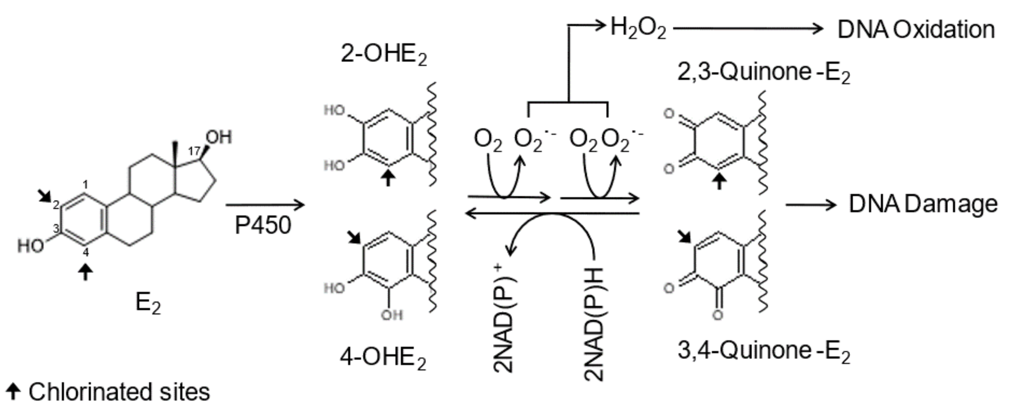
1. Introduction
2. Results
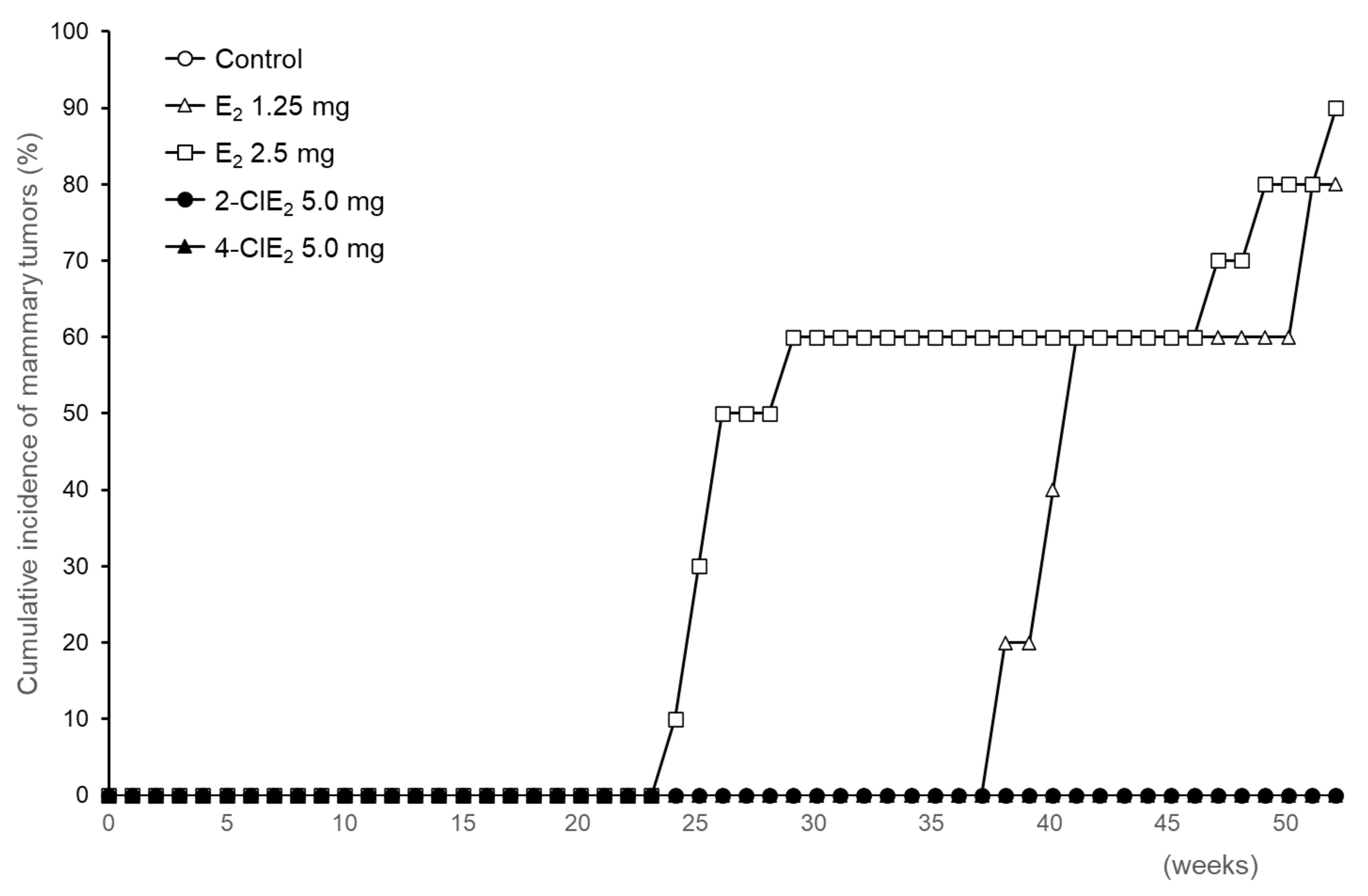
2.1. Evidence of Mammary Tumors
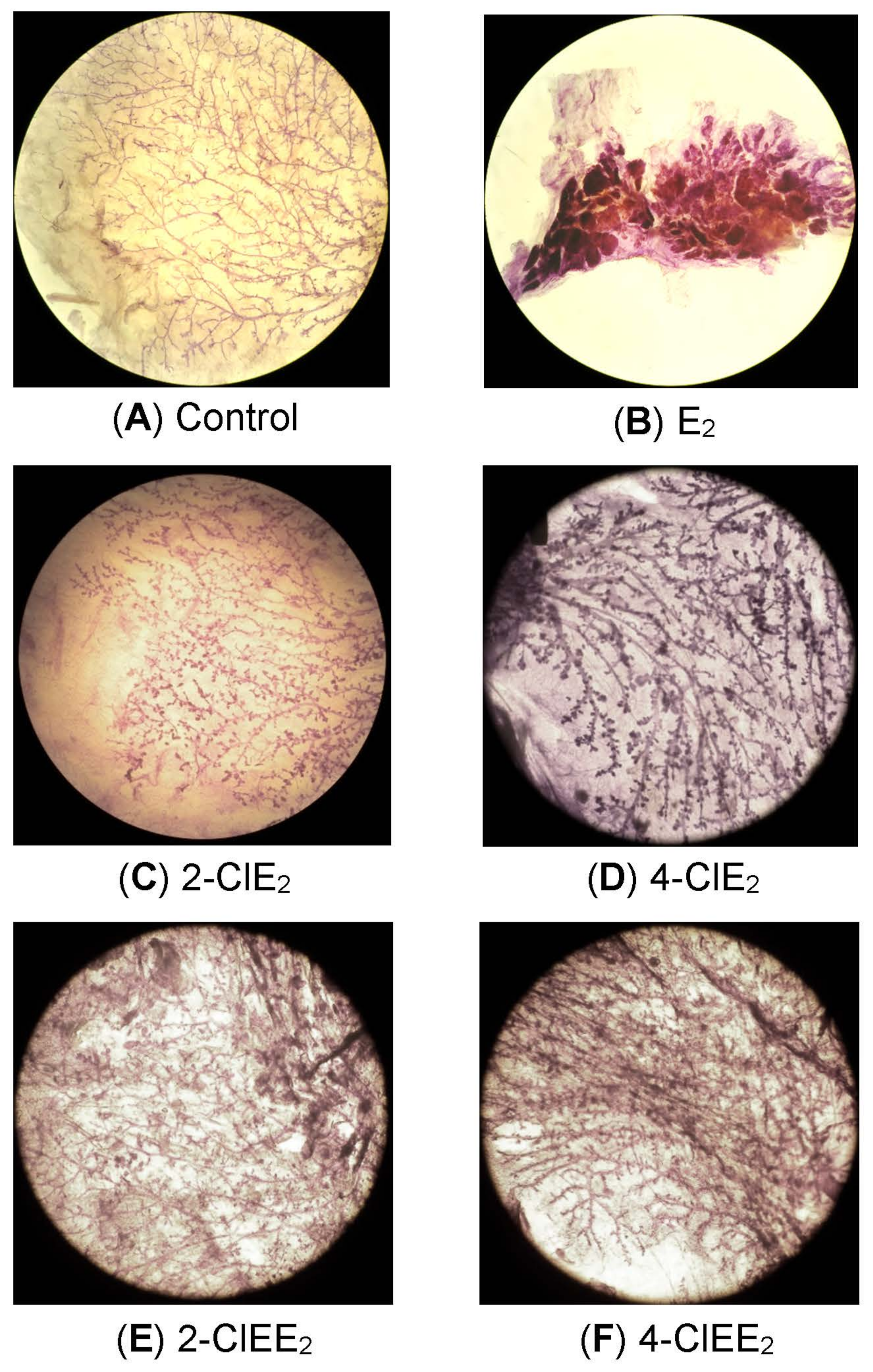
2.2. Histological Examination of Mammary Glands
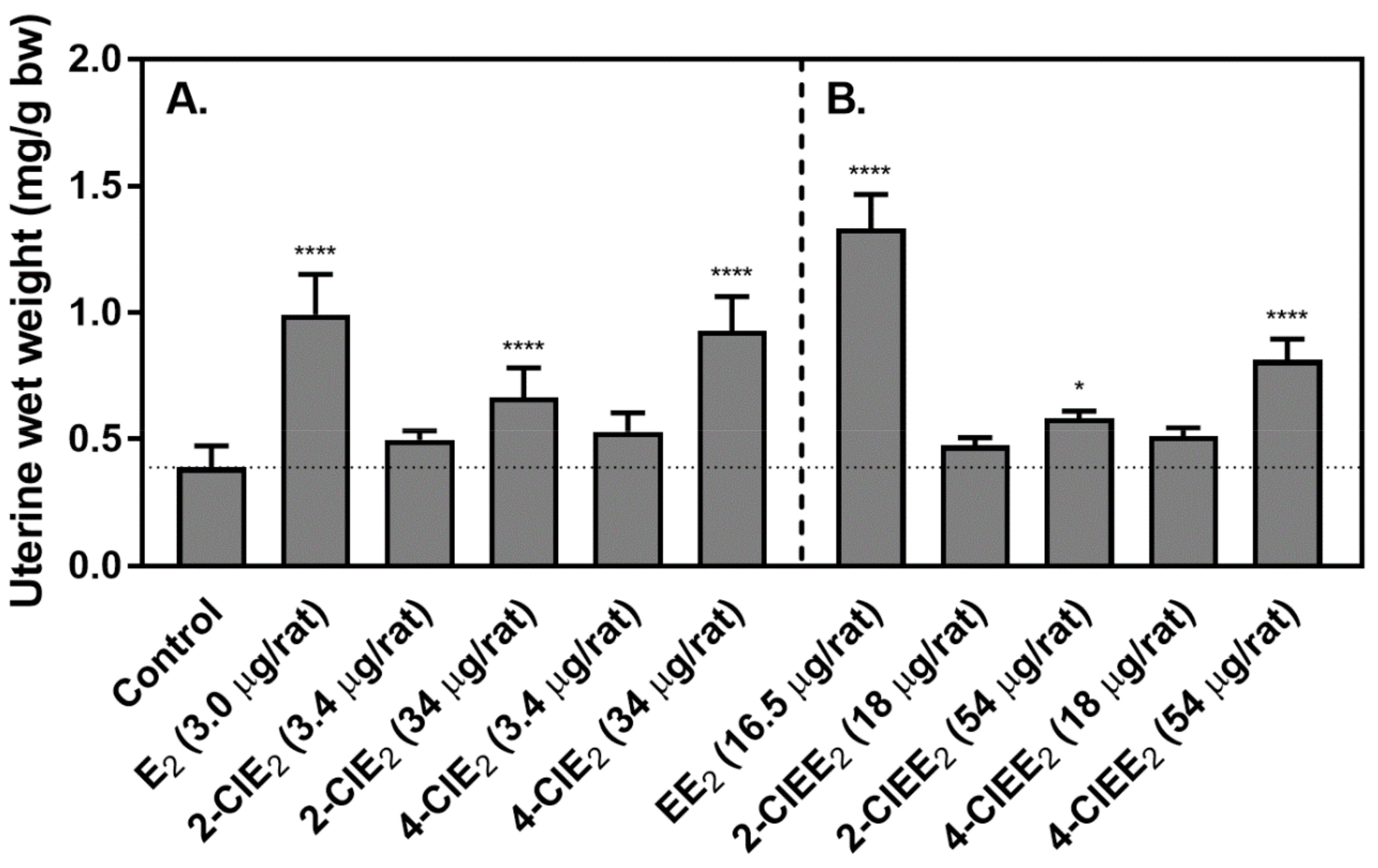
2.3. Uterotrophic Activity of Chlorinated Estrogens
3. Discussion
4. Materials and Methods
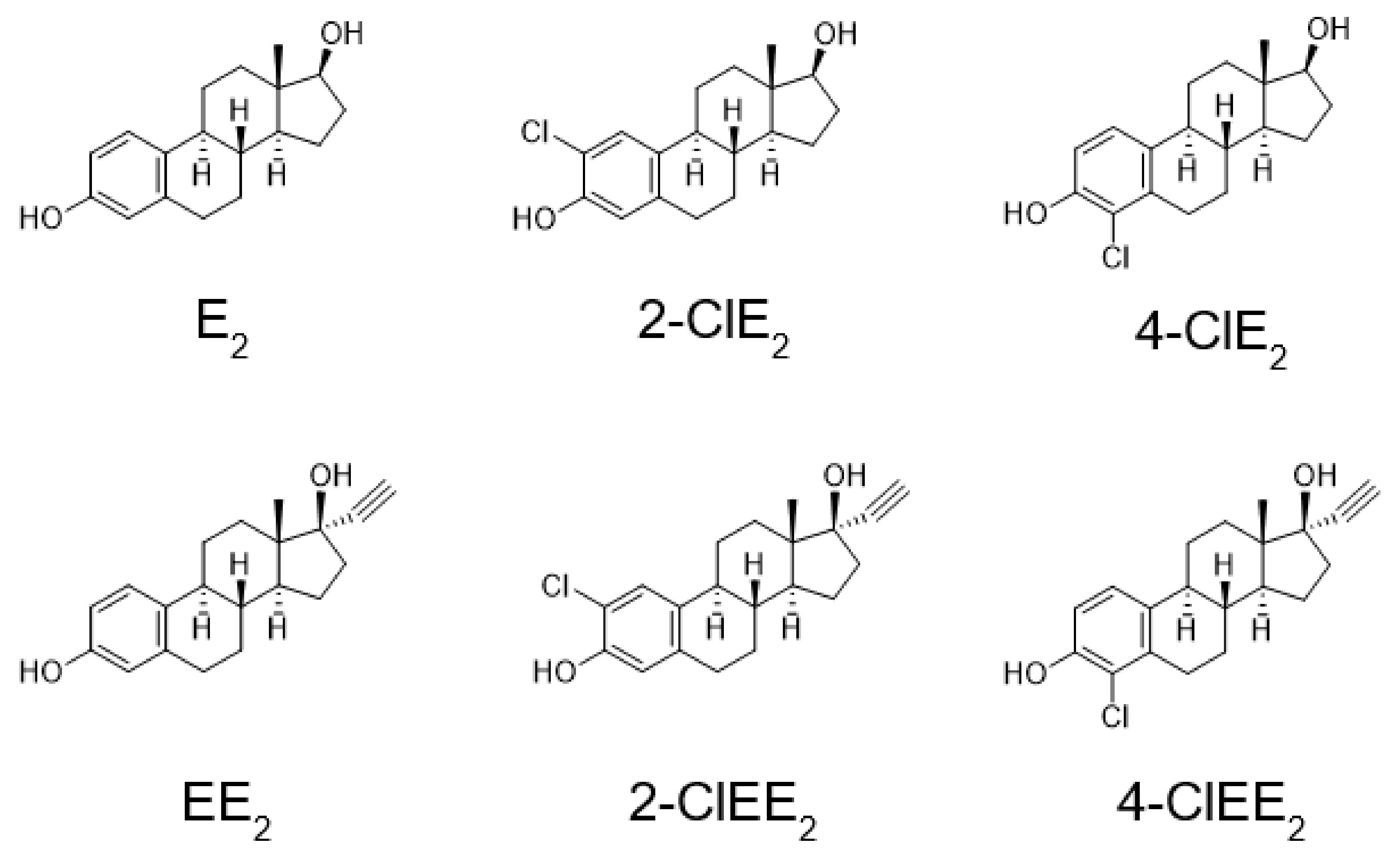
4.1. Chemicals
4.2. Synthesis of Chlorinated Estrogens
4.3. Tumorigenesis of Chlorinated Estrogens
4.4. Mammary Whole-Mount Preparation and Morphometric Analysis
4.5. Determination of Uterotrophic Potential
Author Contributions
Funding
Institutional Review Board Statement
Informed Consent Statement
Data Availability Statement
Acknowledgments
Conflicts of Interest
Abbreviations
| E2 | 17β-estradiol |
| 2-OHE | 2-hydroxyestrogen |
| 4-OHE | 4-hydroxyestrogen |
| 2-ClE2 | 2-chloro-17β-estradiol |
| 4-ClE2 | 4-chloro-17β-estradiol |
| EE2 | 17α-ethinylestradiol |
| 2-ClEE2 | 2-chloro-17α-ethinylestradiol |
| 4-ClEE2 | 4-chloro-17α-ethinylestradiol |
| 2-FE2 | 2-fluoro-17β-estradiol |
| 4-FE2 | 4-fluoro-17β-estradiol |
| ACI | August Copenhagen Irish |
| HRT | Hormone replacement therapy |
| OVX | Ovariectomized |
| bw | Body weight |
| RIA | Radioimmunoassay |
References
- Grodstein, F.; Stampfer, M.J.; Colditz, G.A.; Willett, W.C.; Manson, J.E.; Joffe, M.; Rosner, B.; Fuchs, C.; Hankinson, S.E.; Hunter, D.L.; et al. Postmenopausal hormone therapy and mortality. N. Engl. J. Med. 1997, 336, 1769–1775. [Google Scholar] [CrossRef] [PubMed]
- Bolton, J.L.; Pisha, E.; Zhang, F.; Qiu, S. Role of quinoids in estrogen carcinogenesis. Chem. Res. Toxicol. 1998, 11, 1113–1127. [Google Scholar] [CrossRef] [PubMed]
- Colditz, G.A.; Hankinson, S.E.; Hunter, D.J.; Willett, W.C.; Manson, J.E.; Stampfer, M.J.; Hennekens, C.H.; Rosner, B.; Speizer, F.E. The use of estrogens and progestins and the risk of breast cancer in postmenopausal women. N. Engl. J. Med. 1995, 332, 1589–1593. [Google Scholar] [CrossRef] [PubMed]
- Chen, C.L.; Weiss, N.S.; Newcomb, P.; Barlow, W.; White, E. Hormone replacement therapy in relation to breast cancer. JAMA 2002, 287, 734–741. [Google Scholar] [CrossRef] [PubMed]
- Lacey, J.V., Jr.; Mink, P.J.; Lubin, J.H.; Sherman, M.E.; Troisi, R.; Hartge, P.; Schatzkin, A.; Schairer, C. Menopausal hormone replacement therapy and risk of ovarian cancer. JAMA 2002, 288, 334–341. [Google Scholar] [CrossRef] [PubMed]
- Beral, V. Million Women Study Collaborators. Breast cancer and hormone-replacement therapy in the Million Women Study. Lancet 2003, 362, 419–427. [Google Scholar] [CrossRef]
- Grady, D.; Gebretsadik, T.; Kerlikowske, K.; Emster, V.; Petitti, D. Hormone replacement therapy and endometrial cancer risk: A meta-analysis. Obstet. Gynecol. 1995, 85, 304–313. [Google Scholar] [CrossRef]
- Steinberg, K.K.; Smith, S.J.; Thacker, S.B.; Stroup, D.F. Breast cancer risk and duration of estrogen use: The role of study design in meta-analysis. Epidemiology 1994, 5, 415–421. [Google Scholar] [CrossRef]
- Liehr, J.G. Genotoxic effects of estrogens. Mutat. Res. 1990, 238, 269–276. [Google Scholar] [CrossRef]
- Fishman, J.; Osborne, M.P.; Telang, N.T. The role of estrogen in mammary carcinogenesis. Ann. N. Y. Acad. Sci. 1995, 768, 91–100. [Google Scholar] [CrossRef]
- Hernandez, L.G.; Steeg, H.; Luijten, M.; Benthem, J. Mechanisms of non-genotoxic carcinogens and importance of a weight of evidence approach. Mutat. Res. 2009, 682, 94–109. [Google Scholar] [CrossRef]
- Greenberg, E.R.; Barnes, A.B.; Resseguie, Z.; Barrett, J.A.; Burnside, S.; Lanza, L.L.; Neff, R.K.; Stevens, M.; Young, R.H.; Colton, T. Breast cancer in mothers given diethylstilbestrol in pregnancy. N. Engl. J. Med. 1984, 311, 1393–1398. [Google Scholar] [CrossRef]
- Liehr, J.G.; Avitts, T.A.; Randerath, E.; Randerath, K. Estrogen-induced endogenous DNA adduction: Possible mechanism of hormonal cancer. Proc. Nalt. Acad. Sci. USA 1986, 83, 5301–5305. [Google Scholar] [CrossRef]
- Gladek, A.; Liehr, J.G. Mechanism of genotoxicity of diethylstilbestrol in vivo. J. Biol. Chem. 1989, 264, 16847–16852. [Google Scholar] [CrossRef]
- Preston-Martin, S.; Pike, M.C.; Ross, R.K.; Jones, P.A.; Henderson, B.E. Increased cell division as a cause of human cancer. Cancer Res. 1990, 50, 7415–7421. [Google Scholar]
- Martucci, C.P.; Fishman, J. P450 enzymes of estrogen metabolism. Pharmac. Ther. 1993, 57, 237–257. [Google Scholar] [CrossRef]
- Dwivedy, I.; Devanesan, P.; Cremonesi, P.; Rogan, E.; Cavalieri, E. Synthesis and characterization of estrogen 2,3- and 3,4-quinones. Comparison of DNA adducts formed by the quinones versus horseradish peroxidase-activated catechol estrogens. Chem. Res. Toxicol. 1992, 5, 828–833. [Google Scholar] [CrossRef]
- Hayashi, N.; Hasegawa, K.; Komine, A.; Tanaka, Y.; McLachian, J.A.; Barrett, J.C.; Tsutsui, T. Estrogen-induced cell transformation and DNA adduct formation in cultured Syrian hamster embryo cells. Mol. Carcinog. 1996, 16, 149–156. [Google Scholar] [CrossRef]
- Stack, D.E.; Byun, J.; Gross, M.L.; Rogan, E.G.; Cavalieri, E.L. Molecular characteristics of catechol estrogen quinone in reactions with deoxyribonucleosides. Chem. Res. Toxicol. 1996, 9, 851–859. [Google Scholar] [CrossRef]
- Terashima, I.; Suzuki, N.; Shibutani, S. Mutagenic specificity of 2-hydroxyestrogen quinone-derived DNA adducts in mammalian cells. Biochemistry 2001, 40, 166–172. [Google Scholar] [CrossRef]
- Li, K.M.; Todorovic, R.; Devanesan, P.; Higginbotham, S.; Köfeler, H.; Ramanathan, R.; Gross, M.L.; Rogan, E.G.; Cavalieri, E.L. Metabolism and DNA binding studies of 4-hydroxyestradiol and estradiol-3,4-quinone in vitro and in female ACI rat mammary gland in vivo. Carcinogenesis 2004, 25, 289–297. [Google Scholar] [CrossRef] [PubMed]
- Takeshita, M.; Eisenberg, W. Mechanism of mutation on DNA templates containing synthetic abasic sites: Study with a double strand vector. Nucleic Acids Res. 1994, 22, 1897–1902. [Google Scholar] [CrossRef][Green Version]
- Han, X.; Liehr, J.G. 8-Hydroxylation of guanine bases in kidney and liver DNA of hamsters treated with estradiol: Role of free radicals in estrogen-induced carcinogenesis. Cancer Res. 1994, 54, 5515–5517. [Google Scholar] [PubMed]
- Malins, D.C.; Holmes, E.H.; Polissar, N.L.; Gunselman, S.J. The etiology of breast cancer. Characteristic alteration in hydroxyl radical-induced DNA base lesions during oncogenesis with potential for evaluating incidence risk. Cancer 1993, 71, 3036–3043. [Google Scholar] [CrossRef]
- Turan, V.K.; Sanchez, R.I.; Li, J.J.; Li, S.A.; Reuhl, K.R.; Thomas, P.E.; Conney, A.H.; Gallo, M.A.; Kauffman, F.C.; Mesia-Vela, S. The effects of steroidal estrogens in ACI rat mammary carcinogenesis: 17β-estradiol, 2-hydroxyestradiol, 4-hydroxyestradiol, 16α-hydroxyestradiol, and 4-hydroxyestrone. J. Endocrinol. 2004, 183, 91–99. [Google Scholar] [CrossRef] [PubMed]
- Shull, J.D.; Spady, T.J.; Snyder, M.C.; Johansson, S.L.; Pennington, K.L. Ovary-intact but not ovariectomized female ACI rats treated with 17β-estradiol rapidly develop mammary carcinoma. Carcinogenesis 1997, 18, 1595–1601. [Google Scholar] [CrossRef] [PubMed][Green Version]
- Okamoto, Y.; Jinno, H.; Itoh, S.; Shibutani, S. Carcinogenic potential of fluorinated estrogens in mammary tumorigenesis. Toxicol. Lett. 2020, 318, 99–103. [Google Scholar] [CrossRef]
- Holtzman, S.; Stone, J.P.; Shellabarger, C.J. Synergism of estrogens and X-rays in mammary carcinogenesis in female ACI rats. J. Natl. Cancer Inst. 1981, 67, 455–459. [Google Scholar]
- Kuhl, H. Pharmacology of estrogens and progestogens: Influence of different routes of administration. Climacteric 2005, 8 (Suppl. 1), 3–63. [Google Scholar] [CrossRef]
- Stanczyk, F.Z.; Archer, D.F.; Bhavnani, B.R. Ethinyl estradiol and 17β-estradiol in combined oral contraceptives: Pharmacokinetics, pharmacodynamics and risk assessment. Contraception 2013, 87, 706–727. [Google Scholar] [CrossRef]
- Okamoto, Y.; Liu, X.; Suzuki, N.; Okamoto, K.; Kim, H.J.; Laxmi, Y.R.S.; Sayama, K.; Shibutani, S. Equine estrogen-induced mammary tumors in rats. Toxicol. Lett. 2010, 193, 224–228. [Google Scholar] [CrossRef]
- Hu, J.; Cheng, S.; Aizawa, T.; Terao, Y.; Kunikanw, S. Products of aqueous chlorination of 17β-estradiol and their estrogenic activities. Envirion. Sci. Technol. 2003, 37, 5665–5670. [Google Scholar] [CrossRef]
- Nakamura, H.; Shiozawa, T.; Terao, Y.; Shiraishi, F.; Fukazawa, H. By-products produced by the reaction of estrogens with hypochlorous acid and their estrogen activities. J. Heath Sci. 2006, 52, 124–131. [Google Scholar] [CrossRef]
- Holtzman, S. Retinyl acetate inhibits estrogen-induced mammary carcinogenesis in female ACI rats. Carcinogenesis 1988, 9, 305–307. [Google Scholar] [CrossRef]
- Roberts, D.J.; Caserio, M.C. Basic Principles of Organic Chemistry; W. A. Benjamin Inc.: New York, NY, USA, 1965; Volume 77. [Google Scholar]
- Ashburn, S.P.; Han, X.; Liehr, J.G. Microsomal hydroxylation of 2- and 4-fluoroestradiol to catechol metabolites and their conversion to methyl ethers: Catechol estrogens as possible mediators of hormonal carcinogenesis. Mol. Pharmacol. 1993, 43, 534–541. [Google Scholar]
- Page, P.C.B.; Hussain, F.; Maggs, J.L.; Morgan, P.; Park, B.K. Efficient regioselective A-ring functionalization of oestrogens. Tetrahedron 1990, 46, 2059–2068. [Google Scholar] [CrossRef]
- Mukawa, F. 10β-Chloro-17β-hydroxyestra-1,4-dien-3-one and its related compounds. J. Chem. Soc. Perkin Trans. 1988, 1, 457–460. [Google Scholar] [CrossRef]


| Compound | Dose (mg/pellet) | Cumulative Incidence (No. of Rats) a | Percentage of Rats with Tumors (%) |
|---|---|---|---|
| Control | - | 0/6 | 0 |
| E2 | 1.25 | 4/5 | 80 |
| 2.5 | 9/10 | 90 | |
| 5.0 | - b | - b | |
| 2-ClE2 | 2.5 | 0/6 | 0 |
| 5.0 | 0/6 | 0 | |
| 4-ClE2 | 2.5 | 0/6 | 0 |
| 5.0 | 0/6 | 0 | |
| 2-ClEE2 | 2.5 | 0/5 | 0 |
| 5.0 | 0/6 | 0 | |
| 4-ClEE2 | 2.5 | 0/5 | 0 |
| 5.0 | 0/5 | 0 |
Publisher’s Note: MDPI stays neutral with regard to jurisdictional claims in published maps and institutional affiliations. |
© 2021 by the authors. Licensee MDPI, Basel, Switzerland. This article is an open access article distributed under the terms and conditions of the Creative Commons Attribution (CC BY) license (https://creativecommons.org/licenses/by/4.0/).
Share and Cite
Okamoto, Y.; Jinno, H.; Itoh, S.; Shibutani, S. Less Carcinogenic Chlorinated Estrogens Applicable to Hormone Replacement Therapy. Int. J. Mol. Sci. 2021, 22, 7222. https://doi.org/10.3390/ijms22137222
Okamoto Y, Jinno H, Itoh S, Shibutani S. Less Carcinogenic Chlorinated Estrogens Applicable to Hormone Replacement Therapy. International Journal of Molecular Sciences. 2021; 22(13):7222. https://doi.org/10.3390/ijms22137222
Chicago/Turabian StyleOkamoto, Yoshinori, Hideto Jinno, Shinji Itoh, and Shinya Shibutani. 2021. "Less Carcinogenic Chlorinated Estrogens Applicable to Hormone Replacement Therapy" International Journal of Molecular Sciences 22, no. 13: 7222. https://doi.org/10.3390/ijms22137222
APA StyleOkamoto, Y., Jinno, H., Itoh, S., & Shibutani, S. (2021). Less Carcinogenic Chlorinated Estrogens Applicable to Hormone Replacement Therapy. International Journal of Molecular Sciences, 22(13), 7222. https://doi.org/10.3390/ijms22137222