Review

Mechanisms that Link Chronological Aging to Cellular Quiescence in Budding Yeast

by
Karamat Mohammad
,
Jennifer Anne Baratang Junio
,
Tala Tafakori
,
Emmanuel Orfanos
and
Vladimir I. Titorenko
*
Department of Biology, Concordia University, Montreal, QC H4B 1R6, Canada
*
Author to whom correspondence should be addressed.
Int. J. Mol. Sci. 2020, 21(13), 4717; https://doi.org/10.3390/ijms21134717
Submission received: 10 May 2020 / Revised: 27 June 2020 / Accepted: 30 June 2020 / Published: 2 July 2020

Abstract

:
After Saccharomyces cerevisiae cells cultured in a medium with glucose consume glucose, the sub-populations of quiescent and non-quiescent cells develop in the budding yeast culture. An age-related chronology of quiescent and non-quiescent yeast cells within this culture is discussed here. We also describe various hallmarks of quiescent and non-quiescent yeast cells. A complex aging-associated program underlies cellular quiescence in budding yeast. This quiescence program includes a cascade of consecutive cellular events orchestrated by an intricate signaling network. We examine here how caloric restriction, a low-calorie diet that extends lifespan and healthspan in yeast and other eukaryotes, influences the cellular quiescence program in S. cerevisiae. One of the main objectives of this review is to stimulate an exploration of the mechanisms that link cellular quiescence to chronological aging of budding yeast. Yeast chronological aging is defined by the length of time during which a yeast cell remains viable after its growth and division are arrested, and it becomes quiescent. We propose a hypothesis on how caloric restriction can slow chronological aging of S. cerevisiae by altering the chronology and properties of quiescent cells. Our hypothesis posits that caloric restriction delays yeast chronological aging by targeting four different processes within quiescent cells.

Graphical Abstract

1. Introduction

When Saccharomyces cerevisiae cells are cultured under aerobic conditions in a nutrient-rich liquid medium with 2% glucose, they are not limited in calorie supply [1,2,3,4,5,6]. They exist under so-called non-caloric restriction (non-CR) conditions [1,2,3,4,5,6]. After these cells consume glucose as a sole exogenous carbon source, they enter a diauxic shift period [3,4]. At the time of the diauxic shift, yeast cells decelerate the growth and switch the mode of their metabolism from aerobic alcoholic fermentation to aerobic ethanol catabolism and mitochondrial respiration [3,4]. During the diauxic shift, some S. cerevisiae cells in the culture arrest their cell-division cycle at the nutrient-dependent checkpoint “START A” in the late G1 phase [7,8,9,10,11]. At the time of such cell cycle arrest, the budding yeast culture begins to accumulate the sub-populations of quiescent (Q) and non-quiescent (NQ) cells [7,8,9,10,11]. The Q and NQ cells in yeast cultures under non-CR conditions differ from each other in physical, morphological, reproductive, biochemical, and physiological properties [7,8,9,10,11]. A signaling network that integrates a distinct set of the nutrient-sensing signaling pathways and protein kinases orchestrates the development of properties characteristic of Q cells [4,9]. After S. cerevisiae cells cultured under non-CR conditions consume ethanol as a carbon source, they enter the stationary (ST) phase of culturing and the process of their chronological aging begins [3,4,5,6]. The chronological aging of S. cerevisiae is assessed by measuring the percentage of yeast cells that in liquid cultures remain viable at different time points following the entry of a cell population into the non-proliferative ST phase of culturing [5,6]. Here, we compare the properties of Q and NQ cells cultured under non-CR conditions. Noteworthy, the pace of yeast chronological aging and the longevity of chronologically aging yeast under non-CR conditions depend on the cell entry into and advancement through a quiescence program. S. cerevisiae cells that are not limited in calorie supply enter this cellular quiescence program during the diauxic shift and advance through it during the ST phase of culturing [7,8,9,10,11,12]. As any programmed biological event, this cellular quiescence program (1) is a genetically defined, regulated process, (2) can be accelerated or decelerated by genetic manipulations that alter the abundancies and/or activities of only specific proteins, (3) integrates a cascade of consecutive cellular events that follow each other in a particular order and are regulated by a specific signaling network, (4) is initiated in response to certain stimuli (e.g., nutrient deprivation or chronological aging), and (5) provides a particular benefit for the development, survival, and/or stress resistance of a cell population [7,8,9,10,11,12,13,14,15,16,17].
The chronological aging of S. cerevisiae can be slowed down, and its longevity can be extended by CR [1,2], a low-calorie dietary regimen without malnutrition that prolongs lifespan and postpones the onset of age-related pathologies in other yeast species, nematodes, fruit flies, fishes, dogs, rodents, and primates [18,19,20]. The effects of CR on chronological aging of S. cerevisiae are usually investigated in budding yeast cultured in a nutrient-rich or nutrient-limited synthetic minimal medium initially containing 0.2% or 0.5% glucose [2,5,6]. In contrast to a nutrient-limited synthetic minimal medium, a nutrient-rich medium has plenty of amino acids, nucleotides, vitamins, and other nutrients [21,22,23]. Therefore, the use of a nutrient-rich medium with 0.2% or 0.5% glucose for chronological aging studies under CR conditions provides several important advantages as compared to the use of a minimal synthetic medium [2].
We previously purified the Q and NQ cell populations from budding yeast cultured in a nutrient-rich medium under CR or non-CR conditions [24,25]. We recovered these cell populations at different stages of the chronological aging process and compared their properties [24,25]. Here, we discuss how CR slows the conversion of Q cells into NQ cells because this low-calorie diet alters the specific properties of Q cells. We also examine the evidence that the ability of CR to alter these properties of Q cells is responsible for the CR-dependent delay of chronological aging in budding yeast.

2. Traits of Q and NQ Cells Found in Yeast Populations Cultured Under Non-CR Conditions

Those cells in a budding yeast population cultured under non-CR conditions that undergo cell-cycle arrest enter a non-proliferative state called G0 [7,8,9,10,11,12]. They form the Q cell sub-population [7,8,9,10,11,12]. In contrast, those cells in the budding yeast population not limited in calorie supply that does not arrest their cell cycle give rise to at least three sub-populations of NQ cells [7,8,9,10,11,12]. The Q and NQ cell sub-populations differ from each other in many traits. These traits are discussed below and schematically depicted in Figure 1.

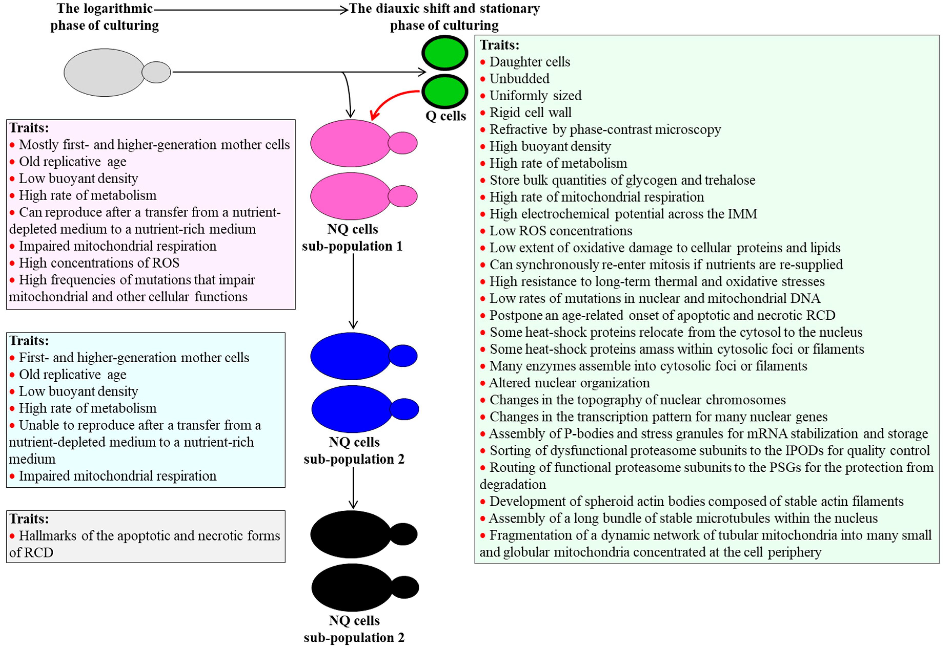
2.1. Traits of Q Cells under Non-CR Conditions

Q cells of S. cerevisiae cultured under non-CR conditions are mostly unbudded and uniformly sized daughter cells that are surrounded by a thick cell wall, refract light when viewed through a phase-contrast microscope, and have a high buoyant density (Figure 1) [7,8,10,11,12,24]. These cells display a high rate of metabolism, amass glycogen and trehalose, have actively respiring mitochondria capable of maintaining a high electrochemical potential across the inner membrane, exhibit low concentrations of reactive oxygen species (ROS), and sustain a low extent of oxidative damage to cellular proteins and lipids (Figure 1) [7,8,9,10,11,12,24]. Q cells of S. cerevisiae cultured under non-CR conditions can synchronously re-enter mitosis and reproduce after being transferred from a nutrient-depleted liquid medium to a surface of a nutrient-rich solid medium or a fresh liquid medium (Figure 1) [7,8,10,11,12,24]. These cells exhibit low susceptibility to chronic (long-term) thermal and oxidative stresses, rarely undergo mutations that affect mitochondrial and other cellular functions, and postpone an age-related onset of the apoptotic and necrotic forms of regulated cell death (RCD) (Figure 1) [7,8,9,10,11,12,24].
Recent findings revealed many other traits characteristic of Q cells in S. cerevisiae cultures not limited in calorie supply. Because these traits have been comprehensively discussed elsewhere [17], we outline them below only briefly. Q cells of budding yeast cultured under non-CR conditions have some heat-shock proteins relocated from the cytosol to the nucleus; they amass other heat-shock proteins and many metabolic and non-metabolic enzymes within multiple cytosolic foci or filaments (Figure 1) [17,26,27,28,29,30,31,32]. These cells also display altered nuclear organization, global changes in the topography of nuclear chromosomes, and substantial alterations in the transcription pattern for many nuclear genes (Figure 1) [17,33,34,35,36,37,38,39,40,41]. Q cells of S. cerevisiae under non-CR conditions enhance their ability to sustain cellular proteostasis by employing the following three mechanisms: (1) by assembling P-bodies and stress granules for mRNA stabilization and storage (respectively) in the cytosol (Figure 1) [32,42,43,44], (2) by routing dysfunctional proteasome subunits from the nucleus to the so-called insoluble protein deposits (IPODs) for quality control (Figure 1) [45], and (3) by sorting functional proteasome subunits from the nucleus to the so-called proteasome storage granules (PSGs) for the protection from degradation (Figure 1) [46,47,48,49]. Q cells of budding yeast under non-CR conditions re-arrange their cytoskeleton. These cells dismantle an elaborate network of dynamic actin cables and develop spheroid actin bodies, which consist of stable actin filaments (Figure 1) [50]. Q cells also disassemble a cytosolic array of dynamic microtubules and build a long bundle of stable microtubules in the nucleus (Figure 1) [34]. Q cells of S. cerevisiae under non-CR conditions undergo a fragmentation of their dynamic network of tubular mitochondria into many small and globular mitochondria concentrated at the cell periphery (Figure 1) [51].
Although mechanisms underlying the regulated establishment of many traits characteristic of Q cells under non-CR conditions remain unknown, some of these traits are essential contributors to the abilities of Q cells to exit quiescence, re-enter the cell cycle, and resume proliferation [9,10,11,12,17,24,26,29,34,47,48,49,50]. Moreover, some of these traits (but not all of them) also contribute to longevity assurance in chronologically aging budding yeast [9,10,11,12,24,34,39,47,48,49,50]. Mechanisms that link these essential traits of Q cells to their exit from quiescence, proliferation, and chronological aging are presently unexplored.

2.2. Traits of NQ Cells under Non-CR Conditions

Three distinct sub-populations of NQ cells exist in an S. cerevisiae culture under non-CR conditions [7,8,9,10,11,12].
Most NQ cells present in the sub-population 1 are first- and higher-generation mother cells, although some daughter cells can also be found there (Figure 1) [7,8,9,10,11,12]. Most NQ cells in the sub-population 1 have one or more bud scars on their surface [7,8,9,10,11,12]. Because each bud scar is formed on the surface of a mother cell when it buds off a new daughter cell [52,53,54], the NQ cells in the sub-population 1 are mainly replicatively older than the daughter Q cells (Figure 1) [7,8,9,10,11,12,24,55,56,57]. Akin to Q cells, the NQ cells in the sub-population 1 are metabolically active (Figure 1) [7,8,9,10,11,12,24]. These NQ cells can reproduce after being transferred from a nutrient-depleted medium to a nutrient-rich medium (Figure 1) [7,8,9,10,11,12,24]. Unlike Q cells, the NQ cells in the sub-population 1 have a low buoyant density and display impaired mitochondrial respiration, high concentrations of ROS, and elevated frequencies of mutations that impair mitochondrial and other cellular functions (Figure 1) [7,8,9,10,11,12,24].
NQ cells in the sub-population 2 are first- and higher-generation mother cells of low buoyant density that are replicatively old (Figure 1) [7,8,9,10,11,12]. These NQ cells exhibit a high rate of metabolism, like NQ cells in sub-population 1 (Figure 1) [7,8,9,10,11,12]. Unlike NQ cells in the sub-population 1, NQ cells in the sub-population 2 cannot reproduce if transferred from a nutrient-depleted medium to a nutrient-rich medium (Figure 1) [7,8,9,10,11,12]. It is conceivable, therefore, that NQ cells in the sub-population 2 may originate from NQ cells in the sub-population 1 (Figure 1) [7,8,9,10,11,12].
Most NQ cells in the sub-population 3 are reproductively incompetent cells displaying the characteristic traits of the apoptotic and necrotic forms of RCD (Figure 1) [7,8,9,10,11,12]. These cells are believed to be descendants of NQ cells in sub-population 2 (Figure 1) [7,8,9,10,11,12].
It is feasible that in chronologically aging S. cerevisiae cultures under non-CR conditions the pathway of a stepwise transformation of NQ cells in the sub-population 1 into NQ cells in the sub-population 3 is sustained via an age-related conversion of Q cells into NQ cells that compose the sub-population 1 (Figure 1, red arrow) [7,8,9,10,11,12,24]. Mechanisms regulating such age-related conversion of Q cells of the sub-population 1 of NQ cells remain unknown.

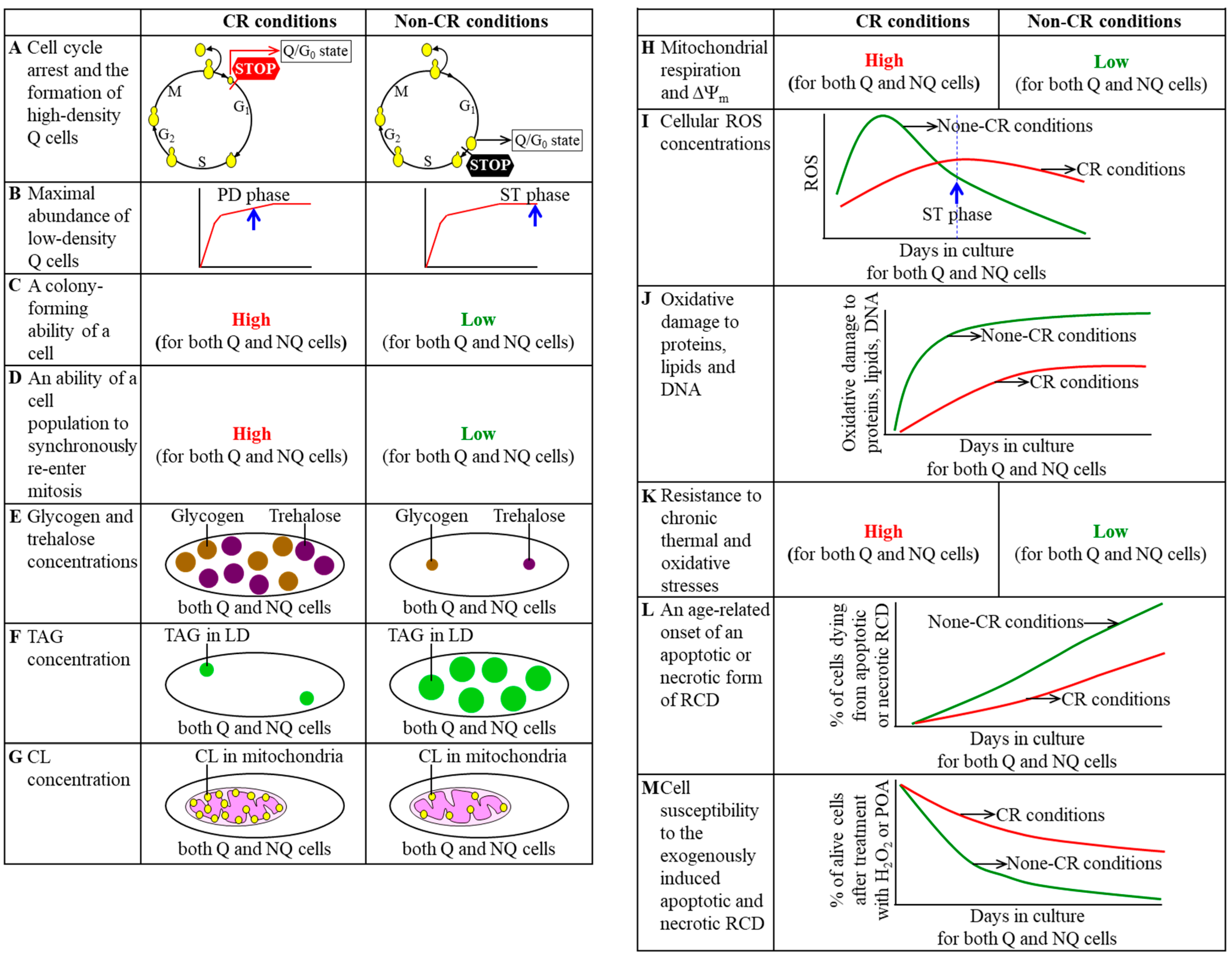
3. CR Diet Alters an Age-Related Chronology and Properties of Q and NQ Cells Found in Yeast Populations

In a recent study, the Q and NQ cell sub-populations were purified from differently aged S. cerevisiae populations cultured in a nutrient-rich medium under CR or non-CR conditions [24]. A comparative analysis of these cell sub-populations has shown that the CR diet revises an age-related chronology of Q and NQ cells and alters several traits characteristic of these cells [24]. The effects of CR on the chronology and traits of Q and NQ cells are discussed below and schematically depicted in Figure 2.
CR creates a sub-population of high-density Q cells by arresting the cell-division cycle at a different checkpoint in the G1 phase than the one responsible for the formation of high-density Q cells under non-CR conditions [24,25]. After cells consume glucose under non-CR conditions, the formation of the high-density Q cell sub-population and entry into the G0 state occur in late G1 [7,8,9,10,11]. In contrast, after cells consume glucose under CR conditions, the formation of the high-density Q cell sub-population and entry into the G0 state happen at a checkpoint in early G1 (Figure 2A) [24].
CR accelerates an age-related buildup of low-density Q cell sub-population in chronologically aging yeast cultures [24,25]. Indeed, the abundance of low-density Q cells in non-CR yeast cultures reaches a plateau in the ST growth phase, while the abundance of low-density Q cells in CR yeast cultures attains a steady-state level already in the post-diauxic (PD) growth phase (Figure 2B) [24].
Both Q and NQ cells formed under CR conditions retain their abilities to reproduce for a longer period of their chronological lifespans than age-matched Q and NQ cells formed under non-CR conditions [24,25]. This chronological extension by the CR diet was observed for two different aspects of the reproductive ability, including (1) the ability of a cell to form a colony after being transferred from a nutrient-depleted liquid medium to a surface of a nutrient-rich solid medium (Figure 2C) [24], and (2) the ability of a cell population to synchronously re-enter the mitotic cell cycle after being transferred from a nutrient-depleted liquid medium to a nutrient-rich liquid medium (Figure 2D) [24].
Glycogen and trehalose are the two major glucose storage molecules in S. cerevisiae [3,4,58]. Trehalose in budding yeast also contributes to the protection of the entire cell and its protein constituents from various stresses, maintenance of cellular proteostasis, and mitotic division of Q cells under non-CR conditions [59,60,61,62,63,64,65,66,67]. A substantial rise in the concentrations of glycogen and trehalose is a characteristic trait of Q and NQ cell sub-populations developed in yeast cultures that are limited in calorie supply (Figure 2E) [24,25]. Indeed, CR significantly increases the concentrations of glycogen and trehalose in both Q and NQ cells beginning of the PD phase of culturing (Figure 2E) [24]. Of note, glycogen and trehalose concentrations within Q cells formed in yeast cultures under CR conditions considerably exceed those within age-matched NQ cells developed in these cultures [24].
After the neutral lipids triacylglycerols (TAG) are synthesized in the endoplasmic reticulum (ER), they are stored in lipid droplets (LD) as the source of free (non-esterified) fatty acids for energy metabolism and phospholipid biosynthesis [68,69,70,71,72,73,74]. One of the characteristic traits of Q and NQ cell sub-populations formed in S. cerevisiae cultured under CR conditions is a considerable decline in TAG concentration, as compared to age-matched Q and NQ cell sub-populations developed in this yeast cultured under non-CR conditions (Figure 2F) [24,25]. Such a decline in TAG concentration is observed through the entire chronological lifespan [24]. Notably, there is no significant difference between age-matched Q and NQ cells regarding TAG concentration in these cells at any stage of the chronological aging process in S. cerevisiae [24].
Cardiolipins (CL) are signature lipids of the inner mitochondrial membrane essential for the maintenance of proper morphology and functionality of yeast mitochondria [68,75,76,77,78,79]. Mitochondria of both Q and NQ cell sub-populations in S. cerevisiae limited in calorie supply exhibit an increase in CL concentration beginning of the PD growth phase (Figure 2G) [24,25]. The increase in CL concentration is a characteristic trait of mitochondria present in Q and NQ cells under CR conditions, as compared to mitochondria found in Q and NQ cells (respectively) under non-CR conditions [24,25]. However, CL concentration in mitochondria of Q cells does not significantly differ from those in mitochondria of age-matched NQ cells regardless of the extent of calorie supply [24].
The rate of mitochondrial respiration and electrochemical potential across the inner mitochondrial membrane (ΔΨm) are essential contributors to longevity assurance in chronologically aging S. cerevisiae [2,6,80,81,82,83,84,85,86]. CR substantially increases the rate of mitochondrial respiration and ΔΨm in both Q and NQ cells through the chronological lifespan of budding yeast (Figure 2H) [24,25]. The increase in these key aspects of mitochondrial functionality is one of the characteristic traits of Q and NQ cells limited in calorie supply, in comparison with Q and NQ cells (respectively) not limited in calorie supply [24,25]. Of note, the rate of respiration and ΔΨm in mitochondria of Q cells cultured under CR conditions exceed those in mitochondria of age-matched NQ cells in these cultures, especially during the ST growth phase [24].
An age-related dynamic of changes in cellular ROS concentrations within Q and NQ cells under CR conditions differs from the one within each of these cell sub-populations under non-CR conditions. Early in chronological lifespan, before the entry of cell culture into the ST growth phase, ROS concentrations within Q and NQ cells under CR conditions are significantly lower than those within age-matched Q and NQ cells (respectively) under non-CR conditions (Figure 2I) [24,25]. In contrast, late in chronological lifespan, before the entry of cell culture into the ST growth phase, ROS concentrations within Q and NQ cells under CR conditions are substantially higher than those within age-matched Q and NQ cells (respectively) under non-CR conditions (Figure 2I) [24,25]. The distinct age-related dynamic of changes in cellular ROS concentrations is a characteristic trait of Q and NQ cells of S. cerevisiae that are cultured under CR conditions. Of note, ROS are generated mostly as by-products of mitochondrial respiration [3,87] and play essential roles in regulating longevity of chronologically aging S. cerevisiae [2,6,84,85,88,89,90,91,92,93].
Age-related accumulation of oxidatively damaged cellular macromolecules contributes to the process of chronological aging in budding yeast [6,84,85,88,93,94,95]. One of the characteristic traits of Q and NQ cells in S. cerevisiae cultured under CR conditions is a significant decline in the extent of ROS-inflicted and age-related oxidative damage to proteins, lipids, nuclear DNA (nDNA), and mitochondrial DNA (mtDNA) (Figure 2J) [24,25]. A degree of such a CR-dependent decline in oxidative macromolecular damage varies for different macromolecules and stages of the chronological aging process [24]. Furthermore, the concentrations of all these oxidatively damaged macromolecules within Q cells limited in calorie supply are lower than within calorically restricted NQ cells of the same chronological age [24].
One of the characteristic traits of Q and NQ cells of S. cerevisiae cultured under CR conditions is a significant rise in the resistance to chronic (long-term) thermal and oxidative stresses, as compared to age-matched Q and NQ cells of this yeast cultured under non-CR conditions (Figure 2K) [24,25]. CR promotes the resistance of Q cells to both types of chronic stresses late in chronological lifespan, in the ST growth phase [24]. The stimulatory effect of CR on the tolerance of NQ cells to these stresses is observed early in chronological lifespan, in the PD growth phase [24]. Of note, the longevity of chronologically aging yeast can be extended by the interventions that enhance cell tolerance to chronic thermal and oxidative stresses [2,6,84,88,94,96,97,98,99,100].
Apoptotic and/or necrotic forms of RCD are hallmark processes taking place in chronologically “old” S. cerevisiae cells not limited in calorie supply [6,101,102,103,104,105,106,107,108,109,110,111,112]. CR postpones an age-related onset of apoptotic and necrotic RCD in both Q and NQ cell sub-populations (Figure 2L) [24,25]. In Q cells limited in calorie supply, a significant delay of the onsets of both forms of RCD occurs late in chronological lifespan in the ST growth phase [24]. In NQ cells of yeast cultures under CR conditions, the onsets of both forms of RCD are prolonged earlier in chronological lifespan, already when these cultures enter the PD growth phase [24]. Notably, the percentage of cells exhibiting characteristic traits of an apoptotic or necrotic form of RCD in NQ cell sub-populations from CR cultures exceeds that in age-matched Q cell sub-populations from these cultures [24].
CR not only delays an age-related onset of apoptotic and necrotic RCD in Q and NQ cells. This low-calorie diet also makes both cell sub-populations less susceptible to the exogenously induced apoptotic and necrotic forms of RCD (Figure 2M) [24,25]. The apoptotic form of RCD can be induced by a short-term treatment of yeast with exogenous hydrogen peroxide [102,105,110,113,114]. In contrast, the so-called “liponecrotic” form of RCD can occur in response to brief exposure of yeast to exogenous palmitoleic acid [109,110,111,112]. Q cells cultured under CR conditions become less susceptible to both forms of exogenously induced RCD than Q cells under non-CR conditions late in chronological lifespan, after the culture enters the ST growth phase [24]. Yet, NQ cells cultured under CR conditions exhibit lower susceptibility to both forms of exogenously induced RCD than NQ cells under non-CR conditions early life, after entry of the culture into the PD phase of culturing [24].

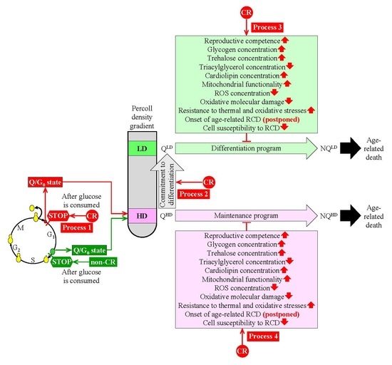
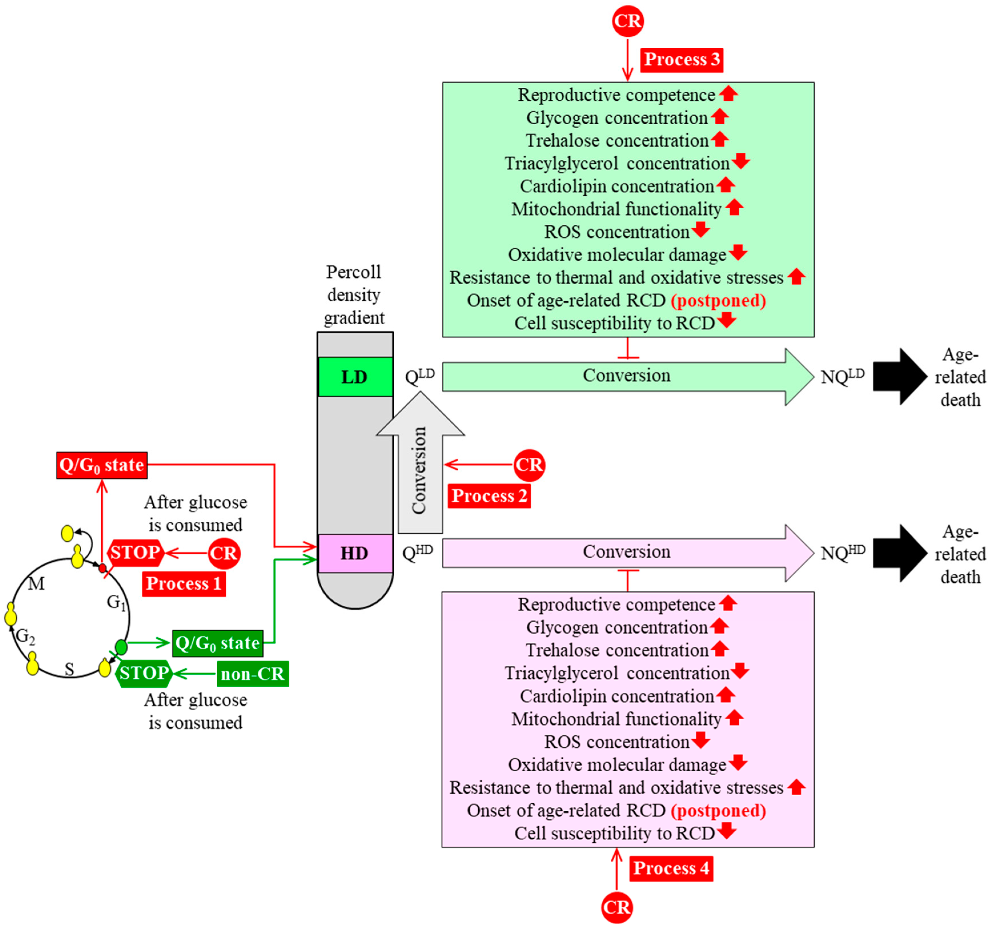
4. A Hypothesis: The CR Diet Slows Yeast Chronological Aging Because It Alters an Age-Related Chronology and Certain Properties of Q Cells

We analyzed changes in an age-related chronology and various properties of Q and NQ cells that were purified from yeast populations cultured under CR or non-CR conditions. Our analysis suggests a hypothesis on how CR can slow yeast chronological aging by altering the chronology and properties of Q cells. This hypothesis is discussed below and schematically depicted in Figure 3.
Our hypothesis posits that CR slows yeast chronological aging by targeting the following four processes within Q cells.
First, because under CR conditions the cell cycle is arrested earlier in G1 than it is under non-CR conditions, high-density Q (QHD) cells in CR cultures are significantly smaller than QHD cells in non-CR cultures (Figure 3, process 1) [24]. Future studies will need to examine if the ability of CR to create small QHD cells by arresting the cell cycle at a checkpoint in early G1 contributes to the ability of CR to delay yeast chronological aging. It is conceivable that some of the pro-longevity properties of QHD cells formed under CR conditions (such as an improved reproductive ability and/or other properties named in Figure 3) are due to the ability of CR to arrest the cell cycle in early G1, thus creating small QHD cells.
Second, CR speeds up an age-related conversion of QHD cells into (QLD) cells in chronologically aging yeast cultures (Figure 3, process 2) [24,25]. A challenge for the future is to examine if the ability of CR to promote such conversion is an essential contributor to the CR-dependent delay of yeast chronological aging. It is plausible that a stimulating effect of CR on the QHD-into-QLD cell conversion contributes to the pro-longevity properties of QLD cells formed under CR conditions. These properties of QLD cells might include their enhanced reproductive potential and/or other traits characteristic of QLD cells (Figure 3).
Third, a conversion of long-lived QLD cells into short-lived non-quiescent cells of low density (which we call NQLD) under CR conditions occurs slower than under non-CR conditions (Figure 3, process 3) [24]. In our hypothesis, the ability of CR to decelerate the QLD-into-NQLD cell conversion might contribute to the CR-dependent delay of yeast chronological aging because it allows QLD cells to maintain the pro-longevity cellular traits. These pro-longevity traits of QLD cells include an enhanced reproductive competence, increased glycogen and trehalose concentrations, a declined TAG concentration, an elevated concentration of CL, improved functionality of mitochondria, a reduced concentration of ROS, a decline in oxidative damage to macromolecules, postponed onsets of apoptotic and necrotic modes of RCD, and decreased susceptibilities to apoptotic and liponecrotic forms of RCD (Figure 3, process 3) [24].
Fourth, QHD cells formed under CR conditions maintain quiescence longer than QHD cells developed under non-CR conditions (Figure 3, process 4) [24]. Thus, CR slows the conversion of long-lived QHD cells into short-lived non-quiescent cells of high density (which we call NQHD). According to our hypothesis, the CR-dependent deceleration of the QHD-into-NQHD cell conversion might contribute to the delay of yeast chronological aging by this low-calorie diet because it allows QHD cells to sustain a pro-longevity cellular pattern. Such pro-longevity cellular pattern is maintained under CR conditions longer than under non-CR conditions because CR has similar effects on the pro-longevity cellular traits of QHD cells as the ones described above for QLD cells (Figure 3, process 4) [24].

5. Conclusions

In this review, we discussed mechanisms linking chronological aging to a program of cellular quiescence in the yeast S. cerevisiae. Our discussion indicates that the pace of yeast chronological aging depends on a complex program underlying cellular quiescence entry, maintenance, and exit. A CR diet remodels the cellular quiescence program, and such remodeling could contribute to the CR-dependent delay of yeast chronological aging. A challenge for the future is to define mechanisms by which the CR-driven remodeling of the cellular quiescence program could be linked to the CR-dependent delay of yeast chronological aging. Because the mechanisms of cellular aging and cellular quiescence have been conserved in the evolution, addressing these challenges in the future will increase our understanding of how the knowledge-based targeting of the cellular quiescence program can be used for delaying cellular and organismal aging and for postponing the onset of aging-associated diseases.

Author Contributions

K.M., J.A.B.J., T.T., E.O. and V.I.T. wrote the text. V.I.T. prepared the figures. All authors have read and agreed to the published version of the manuscript.

Funding

This research was supported by a grant from the NSERC of Canada to Vladimir I. Titorenko. The Concordia University Graduate Fellowship Awards supported Karamat Mohammad and Emmanuel Orfanos.

Acknowledgments

We are grateful to current and former members of the Titorenko laboratory for discussions.

Conflicts of Interest

The authors declare no conflict of interest.

References

  1. Sinclair, D.A. Toward a unified theory of caloric restriction and longevity regulation. Mech. Ageing Dev. 2005, 126, 987–1002. [Google Scholar] [CrossRef] [PubMed]
  2. Goldberg, A.A.; Bourque, S.D.; Kyryakov, P.; Gregg, C.; Boukh-Viner, T.; Beach, A.; Burstein, M.T.; Machkalyan, G.; Richard, V.; Rampersad, S.; et al. Effect of calorie restriction on the metabolic history of chronologically aging yeast. Exp. Gerontol. 2009, 44, 555–571. [Google Scholar] [CrossRef] [PubMed]
  3. Fraenkel, D.G. Yeast Intermediary Metabolism; Cold Spring Harbor Laboratory Press: Cold Spring Harbor, NY, USA, 2011; ISBN 978-0-87969-797-6. [Google Scholar]
  4. Broach, J.R. Nutritional control of growth and development in yeast. Genetics 2012, 192, 73–105. [Google Scholar] [CrossRef] [PubMed] [Green Version]
  5. Longo, V.D.; Shadel, G.S.; Kaeberlein, M.; Kennedy, B. Replicative and chronological aging in Saccharomyces cerevisiae. Cell Metab. 2012, 16, 18–31. [Google Scholar] [CrossRef] [Green Version]
  6. Arlia-Ciommo, A.; Leonov, A.; Piano, A.; Svistkova, V.; Titorenko, V.I. Cell-autonomous mechanisms of chronological aging in the yeast Saccharomyces cerevisiae. Microb. Cell 2014, 1, 163–178. [Google Scholar] [CrossRef] [Green Version]
  7. Allen, C.; Büttner, S.; Aragon, A.D.; Thomas, J.A.; Meirelles, O.; Jaetao, J.E.; Benn, D.; Ruby, S.W.; Veenhuis, M.; Madeo, F.; et al. Isolation of quiescent and nonquiescent cells from yeast stationary-phase cultures. J. Cell Biol. 2006, 174, 89–100. [Google Scholar] [CrossRef] [Green Version]
  8. Davidson, G.S.; Joe, R.M.; Roy, S.; Meirelles, O.; Allen, C.P.; Wilson, M.R.; Tapia, P.H.; Manzanilla, E.E.; Dodson, A.E.; Chakraborty, S.; et al. The proteomics of quiescent and nonquiescent cell differentiation in yeast stationary-phase cultures. Mol. Biol. Cell 2011, 22, 988–998. [Google Scholar] [CrossRef]
  9. De Virgilio, C. The essence of yeast quiescence. FEMS Microbiol. Rev. 2012, 36, 306–339. [Google Scholar] [CrossRef] [Green Version]
  10. Werner-Washburne, M.; Roy, S.; Davidson, G.S. Aging and the survival of quiescent and non-quiescent cells in yeast stationary-phase cultures. Subcell. Biochem. 2012, 57, 123–143. [Google Scholar]
  11. Miles, S.; Li, L.; Davison, J.; Breeden, L.L. Xbp1 directs global repression of budding yeast transcription during the transition to quiescence and is important for the longevity and reversibility of the quiescent state. PLoS Genet. 2013, 9, e1003854. [Google Scholar] [CrossRef] [Green Version]
  12. Aragon, A.D.; Rodriguez, A.L.; Meirelles, O.; Roy, S.; Davidson, G.S.; Tapia, P.H.; Allen, C.; Joe, R.; Benn, D.; Werner-Washburne, M. Characterization of differentiated quiescent and nonquiescent cells in yeast stationary-phase cultures. Mol. Biol. Cell 2008, 19, 1271–1280. [Google Scholar] [CrossRef] [PubMed] [Green Version]
  13. Smets, B.; Ghillebert, R.; De Snijder, P.; Binda, M.; Swinnen, E.; De Virgilio, C.; Winderickx, J. Life in the midst of scarcity: Adaptations to nutrient availability in Saccharomyces cerevisiae. Curr. Genet. 2010, 56, 1–32. [Google Scholar] [CrossRef] [PubMed] [Green Version]
  14. Peters, T.W.; Rardin, M.J.; Czerwieniec, G.; Evani, U.S.; Reis-Rodrigues, P.; Lithgow, G.J.; Mooney, S.D.; Gibson, B.W.; Hughes, R.E. Tor1 regulates protein solubility in Saccharomyces cerevisiae. Mol. Biol. Cell 2012, 23, 4679–4688. [Google Scholar] [CrossRef] [PubMed]
  15. Conrad, M.; Schothorst, J.; Kankipati, H.N.; Van Zeebroeck, G.; Rubio-Texeira, M.; Thevelein, J.M. Nutrient sensing and signaling in the yeast Saccharomyces cerevisiae. FEMS Microbiol. Rev. 2014, 38, 254–299. [Google Scholar] [CrossRef] [Green Version]
  16. Swinnen, E.; Ghillebert, R.; Wilms, T.; Winderickx, J. Molecular mechanisms linking the evolutionary conserved TORC1-Sch9 nutrient signalling branch to lifespan regulation in Saccharomyces cerevisiae. FEMS Yeast Res. 2014, 14, 17–32. [Google Scholar] [CrossRef] [Green Version]
  17. Sagot, I.; Laporte, D. The cell biology of quiescent yeast—A diversity of individual scenarios. J. Cell Sci. 2019, 132, jcs213025. [Google Scholar] [CrossRef] [Green Version]
  18. Fontana, L.; Partridge, L.; Longo, V.D. Extending healthy life span—From yeast to humans. Science 2010, 328, 321–326. [Google Scholar] [CrossRef] [Green Version]
  19. De Cabo, R.; Carmona-Gutierrez, D.; Bernier, M.; Hall, M.N.; Madeo, F. The search for antiaging interventions: From elixirs to fasting regimens. Cell 2014, 157, 1515–1526. [Google Scholar] [CrossRef] [Green Version]
  20. Madeo, F.; Carmona-Gutierrez, D.; Hofer, S.J.; Kroemer, G. Caloric restriction mimetics against age-associated disease: Targets, mechanisms, and therapeutic potential. Cell Metab. 2019, 29, 592–610. [Google Scholar] [CrossRef] [Green Version]
  21. Weissman, J.; Guthrie, C.; Fink, G.R. Guide to Yeast Genetics: Functional Genomics, Proteomics, and Other Systems Analysis; Academic Press: Burlington, NJ, USA, 2010; ISBN 9780123751713. [Google Scholar]
  22. Botstein, D.; Fink, G.R. Yeast: An experimental organism for 21st Century biology. Genetics 2011, 189, 695–704. [Google Scholar] [CrossRef] [PubMed] [Green Version]
  23. Feldmann, H. (Ed.) Yeast: Molecular and Cell Biology; Wiley-Blackwell: Weinheim, Germany, 2012; ISBN 978-3-527-33252-6. [Google Scholar]
  24. Leonov, A.; Feldman, R.; Piano, A.; Arlia-Ciommo, A.; Lutchman, V.; Ahmadi, M.; Elsaser, S.; Fakim, H.; Heshmati-Moghaddam, M.; Hussain, A.; et al. Caloric restriction extends yeast chronological lifespan via a mechanism linking cellular aging to cell cycle regulation, maintenance of a quiescent state, entry into a non-quiescent state and survival in the non-quiescent state. Oncotarget 2017, 8, 69328–69350. [Google Scholar] [CrossRef] [PubMed] [Green Version]
  25. Mohammad, K.; Titorenko, V.I. Yeast chronological aging is linked to cell cycle regulation. Cell Cycle 2018, 17, 1035–1036. [Google Scholar] [CrossRef] [PubMed] [Green Version]
  26. Chughtai, Z.S.; Rassadi, R.; Matusiewicz, N.; Stochaj, U. Starvation promotes nuclear accumulation of the hsp70 Ssa4p in yeast cells. J. Biol. Chem. 2001, 276, 20261–20266. [Google Scholar] [CrossRef] [PubMed] [Green Version]
  27. Narayanaswamy, R.; Levy, M.; Tsechansky, M.; Stovall, G.M.; O’Connell, J.D.; Mirrielees, J.; Ellington, A.D.; Marcotte, E.M. Widespread reorganization of metabolic enzymes into reversible assemblies upon nutrient starvation. Proc. Natl. Acad. Sci. USA 2009, 106, 10147–10152. [Google Scholar] [CrossRef] [Green Version]
  28. Noree, C.; Sato, B.K.; Broyer, R.M.; Wilhelm, J.E. Identification of novel filament-forming proteins in Saccharomyces cerevisiae and Drosophila melanogaster. J. Cell Biol. 2010, 190, 541–551. [Google Scholar] [CrossRef] [PubMed] [Green Version]
  29. Tapia, H.; Morano, K.A. Hsp90 nuclear accumulation in quiescence is linked to chaperone function and spore development in yeast. Mol. Biol. Cell 2010, 21, 63–72. [Google Scholar] [CrossRef] [Green Version]
  30. Liu, I.C.; Chiu, S.W.; Lee, H.Y.; Leu, J.Y. The histone deacetylase Hos2 forms an Hsp42-dependent cytoplasmic granule in quiescent yeast cells. Mol. Biol. Cell 2012, 23, 1231–1242. [Google Scholar] [CrossRef]
  31. Noree, C.; Monfort, E.; Shiau, A.K.; Wilhelm, J.E. Common regulatory control of CTP synthase enzyme activity and filament formation. Mol. Biol. Cell 2014, 25, 2282–2290. [Google Scholar] [CrossRef]
  32. Shah, K.H.; Nostramo, R.; Zhang, B.; Varia, S.N.; Klett, B.M.; Herman, P.K. Protein kinases are associated with multiple, distinct cytoplasmic granules in quiescent yeast cells. Genetics 2014, 198, 1495–1512. [Google Scholar] [CrossRef]
  33. Taddei, A.; Schober, H.; Gasser, S.M. The budding yeast nucleus. Cold Spring Harb. Perspect. Biol. 2010, 2, a000612. [Google Scholar] [CrossRef]
  34. Laporte, D.; Courtout, F.; Salin, B.; Ceschin, J.; Sagot, I. An array of nuclear microtubules reorganizes the budding yeast nucleus during quiescence. J. Cell Biol. 2013, 203, 585–594. [Google Scholar] [CrossRef] [Green Version]
  35. Laporte, D.; Sagot, I. Microtubules move the nucleus to quiescence. Nucl. Austin Tex. 2014, 5, 113–118. [Google Scholar] [CrossRef] [Green Version]
  36. Guidi, M.; Ruault, M.; Marbouty, M.; Loïodice, I.; Cournac, A.; Billaudeau, C.; Hocher, A.; Mozziconacci, J.; Koszul, R.; Taddei, A. Spatial reorganization of telomeres in long-lived quiescent cells. Genome Biol. 2015, 16, 206. [Google Scholar] [CrossRef] [Green Version]
  37. McKnight, J.N.; Boerma, J.W.; Breeden, L.L.; Tsukiyama, T. Global promoter targeting of a conserved lysine deacetylase for transcriptional shutoff during quiescence entry. Mol. Cell 2015, 59, 732–743. [Google Scholar] [CrossRef] [PubMed] [Green Version]
  38. Rutledge, M.T.; Russo, M.; Belton, J.M.; Dekker, J.; Broach, J.R. The yeast genome undergoes significant topological reorganization in quiescence. Nucleic Acids Res. 2015, 43, 8299–8313. [Google Scholar] [CrossRef] [Green Version]
  39. Laporte, D.; Courtout, F.; Tollis, S.; Sagot, I. Quiescent Saccharomyces cerevisiae forms telomere hyperclusters at the nuclear membrane vicinity through a multifaceted mechanism involving Esc1, the Sir complex, and chromatin condensation. Mol. Biol. Cell 2016, 27, 1875–1884. [Google Scholar] [CrossRef]
  40. Miles, S.; Breeden, L. A common strategy for initiating the transition from proliferation to quiescence. Curr. Genet. 2017, 63, 179–186. [Google Scholar] [CrossRef] [Green Version]
  41. Roche, B.; Arcangioli, B.; Martienssen, R. Transcriptional reprogramming in cellular quiescence. RNA Biol. 2017, 14, 843–853. [Google Scholar] [CrossRef] [Green Version]
  42. Ramachandran, V.; Shah, K.H.; Herman, P.K. The cAMP-dependent protein kinase signaling pathway is a key regulator of P body foci formation. Mol. Cell 2011, 43, 973–981. [Google Scholar] [CrossRef] [Green Version]
  43. Shah, K.H.; Zhang, B.; Ramachandran, V.; Herman, P.K. Processing body and stress granule assembly occur by independent and differentially regulated pathways in Saccharomyces cerevisiae. Genetics 2013, 193, 109–123. [Google Scholar] [CrossRef] [Green Version]
  44. Riback, J.A.; Katanski, C.D.; Kear-Scott, J.L.; Pilipenko, E.V.; Rojek, A.E.; Sosnick, T.R.; Drummond, D.A. Stress-triggered phase separation is an adaptive, evolutionarily tuned response. Cell 2017, 168, 1028–1040. [Google Scholar] [CrossRef] [PubMed]
  45. Peters, L.Z.; Karmon, O.; Miodownik, S.; Ben-Aroya, S. Proteasome storage granules are transiently associated with the insoluble protein deposit in Saccharomyces cerevisiae. J. Cell Sci. 2016, 129, 1190–1197. [Google Scholar] [CrossRef] [PubMed] [Green Version]
  46. Laporte, D.; Salin, B.; Daignan-Fornier, B.; Sagot, I. Reversible cytoplasmic localization of the proteasome in quiescent yeast cells. J. Cell Biol. 2008, 181, 737–745. [Google Scholar] [CrossRef] [PubMed] [Green Version]
  47. Weberruss, M.H.; Savulescu, A.F.; Jando, J.; Bissinger, T.; Harel, A.; Glickman, M.H.; Enenkel, C. Blm10 facilitates nuclear import of proteasome core particles. EMBO J. 2013, 32, 2697–2707. [Google Scholar] [CrossRef] [Green Version]
  48. Gu, Z.C.; Wu, E.; Sailer, C.; Jando, J.; Styles, E.; Eisenkolb, I.; Kuschel, M.; Bitschar, K.; Wang, X.; Huang, L.; et al. Ubiquitin orchestrates proteasome dynamics between proliferation and quiescence in yeast. Mol. Biol. Cell 2017, 28, 2479–2491. [Google Scholar] [CrossRef]
  49. Marshall, R.S.; Vierstra, R.D. Proteasome storage granules protect proteasomes from autophagic degradation upon carbon starvation. eLife 2018, 7, e34532. [Google Scholar] [CrossRef]
  50. Sagot, I.; Pinson, B.; Salin, B.; Daignan-Fornier, B. Actin bodies in yeast quiescent cells: An immediately available actin reserve? Mol. Biol. Cell 2006, 17, 4645–4655. [Google Scholar] [CrossRef] [Green Version]
  51. Laporte, D.; Gouleme, L.; Jimenez, L.; Khemiri, I.; Sagot, I. Mitochondria reorganization upon proliferation arrest predicts individual yeast cell fate. eLife 2018, 7, e35685. [Google Scholar] [CrossRef]
  52. Hartwell, L.H.; Unger, M.W. Unequal division in Saccharomyces cerevisiae and its implications for the control of cell division. J. Cell Biol. 1977, 75, 422–435. [Google Scholar] [CrossRef] [Green Version]
  53. Powell, C.D.; Quain, D.E.; Smart, K.A. Chitin scar breaks in aged Saccharomyces cerevisiae. Microbiology 2003, 149, 3129–3137. [Google Scholar] [CrossRef] [Green Version]
  54. Cabib, E.; Arroyo, J. How carbohydrates sculpt cells: Chemical control of morphogenesis in the yeast cell wall. Nat. Rev. Microbiol. 2013, 11, 648–655. [Google Scholar] [CrossRef]
  55. Barton, A.A. Some aspects of cell division in Saccharomyces cerevisiae. J. Gen. Microbiol. 1950, 4, 84–86. [Google Scholar] [CrossRef] [PubMed] [Green Version]
  56. Egilmez, N.K.; Chen, J.B.; Jazwinski, S.M. Preparation and partial characterization of old yeast cells. J. Gerontol. 1990, 45, B9–B17. [Google Scholar] [CrossRef] [PubMed]
  57. Sinclair, D.; Mills, K.; Guarente, L. Aging in Saccharomyces cerevisiae. Annu. Rev. Microbiol. 1998, 52, 533–560. [Google Scholar] [CrossRef] [PubMed]
  58. François, J.; Parrou, J.L. Reserve carbohydrates metabolism in the yeast Saccharomyces cerevisiae. FEMS Microbiol. Rev. 2001, 25, 125–145. [Google Scholar] [CrossRef] [Green Version]
  59. Singer, M.A.; Lindquist, S. Multiple effects of trehalose on protein folding in vitro and in vivo. Mol. Cell 1998, 1, 639–648. [Google Scholar] [CrossRef]
  60. Singer, M.A.; Lindquist, S. Thermotolerance in Saccharomyces cerevisiae: The Yin and Yang of trehalose. Trends Biotechnol. 1998, 16, 460–468. [Google Scholar] [CrossRef]
  61. Benaroudj, N.; Lee, D.H.; Goldberg, A.L. Trehalose accumulation during cellular stress protects cells and cellular proteins from damage by oxygen radicals. J. Biol. Chem. 2001, 276, 24261–24267. [Google Scholar] [CrossRef] [Green Version]
  62. Gancedo, C.; Flores, C.L. The importance of a functional trehalose biosynthetic pathway for the life of yeasts and fungi. FEMS Yeast Res. 2004, 4, 351–359. [Google Scholar] [CrossRef] [Green Version]
  63. Shi, L.; Sutter, B.M.; Ye, X.; Tu, B.P. Trehalose is a key determinant of the quiescent metabolic state that fuels cell cycle progression upon return to growth. Mol. Biol. Cell 2010, 21, 1982–1990. [Google Scholar] [CrossRef] [Green Version]
  64. Trevisol, E.T.; Panek, A.D.; Mannarino, S.C.; Eleutherio, E.C. The effect of trehalose on the fermentation performance of aged cells of Saccharomyces cerevisiae. Appl. Microbiol. Biotechnol. 2011, 90, 697–704. [Google Scholar] [CrossRef] [PubMed]
  65. Kyryakov, P.; Beach, A.; Richard, V.R.; Burstein, M.T.; Leonov, A.; Levy, S.; Titorenko, V.I. Caloric restriction extends yeast chronological lifespan by altering a pattern of age-related changes in trehalose concentration. Front. Physiol. 2012, 3, 256. [Google Scholar] [CrossRef] [PubMed] [Green Version]
  66. Eleutherio, E.; Panek, A.; De Mesquita, J.F.; Trevisol, E.; Magalhães, R. Revisiting yeast trehalose metabolism. Curr. Genet. 2015, 61, 263–274. [Google Scholar] [CrossRef]
  67. Babazadeh, R.; Lahtvee, P.J.; Adiels, C.B.; Goksör, M.; Nielsen, J.B.; Hohmann, S. The yeast osmostress response is carbon source dependent. Sci. Rep. 2017, 7, 990. [Google Scholar] [CrossRef] [PubMed] [Green Version]
  68. Mitrofanova, D.; Dakik, P.; McAuley, M.; Medkour, Y.; Mohammad, K.; Titorenko, V.I. Lipid metabolism and transport define the longevity of the yeast Saccharomyces cerevisiae. Front. Biosci. (Landmark Ed.) 2018, 23, 1166–1194. [Google Scholar] [PubMed] [Green Version]
  69. Walther, T.C.; Farese, R.V., Jr. Lipid droplets and cellular lipid metabolism. Annu. Rev. Biochem. 2012, 81, 687–714. [Google Scholar] [CrossRef] [Green Version]
  70. Kohlwein, S.D.; Veenhuis, M.; van der Klei, I.J. Lipid droplets and peroxisomes: Key players in cellular lipid homeostasis or a matter of fat—Store ‘em up or burn ‘em down. Genetics 2013, 193, 1–50. [Google Scholar] [CrossRef] [Green Version]
  71. Pol, A.; Gross, S.P.; Parton, R.G. Review: Biogenesis of the multifunctional lipid droplet: Lipids, proteins, and sites. J. Cell Biol. 2014, 204, 635–646. [Google Scholar] [CrossRef] [Green Version]
  72. Gao, Q.; Goodman, J.M. The lipid droplet—A well-connected organelle. Front. Cell. Dev. Biol. 2015, 3, 49. [Google Scholar] [CrossRef] [Green Version]
  73. Walther, T.C.; Chung, J.; Farese, R.V., Jr. Lipid droplet biogenesis. Annu. Rev. Cell. Dev. Biol. 2017, 33, 491–510. [Google Scholar] [CrossRef] [Green Version]
  74. Jackson, C.L. Lipid droplet biogenesis. Curr. Opin. Cell Biol. 2019, 59, 88–96. [Google Scholar] [CrossRef]
  75. Horvath, S.E.; Daum, G. Lipids of mitochondria. Prog. Lipid Res. 2013, 52, 590–614. [Google Scholar] [CrossRef]
  76. Baile, M.G.; Lu, Y.W.; Claypool, S.M. The topology and regulation of cardiolipin biosynthesis and remodeling in yeast. Chem. Phys. Lipids 2014, 179, 25–31. [Google Scholar] [CrossRef] [Green Version]
  77. Mårtensson, C.U.; Doan, K.N.; Becker, T. Effects of lipids on mitochondrial functions. Biochim. Biophys. Acta 2017, 1862, 102–113. [Google Scholar] [CrossRef]
  78. Schlame, M.; Greenberg, M.L. Biosynthesis, remodeling and turnover of mitochondrial cardiolipin. Biochim. Biophys. Acta Mol. Cell Biol. Lipids 2017, 1862, 3–7. [Google Scholar] [CrossRef] [Green Version]
  79. Tatsuta, T.; Langer, T. Intramitochondrial phospholipid trafficking. Biochim. Biophys. Acta 2017, 1862, 81–89. [Google Scholar] [CrossRef]
  80. Bonawitz, N.D.; Chatenay-Lapointe, M.; Pan, Y.; Shadel, G.S. Reduced TOR signaling extends chronological life span via increased respiration and upregulation of mitochondrial gene expression. Cell Metab. 2007, 5, 265–277. [Google Scholar] [CrossRef] [PubMed] [Green Version]
  81. Pan, Y.; Shadel, G.S. Extension of chronological life span by reduced TOR signaling requires down-regulation of Sch9p and involves increased mitochondrial OXPHOS complex density. Aging (Albany N. Y.) 2009, 1, 131–145. [Google Scholar] [CrossRef] [PubMed] [Green Version]
  82. Breitenbach, M.; Laun, P.; Dickinson, J.R.; Klocker, A.; Rinnerthaler, M.; Dawes, I.W.; Aung-Htut, M.T.; Breitenbach-Koller, L.; Caballero, A.; Nyström, T.; et al. The role of mitochondria in the aging processes of yeast. Subcell. Biochem. 2012, 57, 55–78. [Google Scholar]
  83. Ocampo, A.; Liu, J.; Schroeder, E.A.; Shadel, G.S.; Barrientos, A. Mitochondrial respiratory thresholds regulate yeast chronological life span and its extension by caloric restriction. Cell Metab. 2012, 16, 55–67. [Google Scholar] [CrossRef] [Green Version]
  84. Leonov, A.; Titorenko, V.I. A network of interorganellar communications underlies cellular aging. IUBMB Life 2013, 65, 665–674. [Google Scholar] [CrossRef] [PubMed] [Green Version]
  85. Beach, A.; Leonov, A.; Arlia-Ciommo, A.; Svistkova, V.; Lutchman, V.; Titorenko, V.I. Mechanisms by which different functional states of mitochondria define yeast longevity. Int. J. Mol. Sci. 2015, 16, 5528–5554. [Google Scholar] [CrossRef] [Green Version]
  86. Ruetenik, A.; Barrientos, A. Dietary restriction, mitochondrial function and aging: From yeast to humans. Biochim. Biophys. Acta 2015, 1847, 1434–1447. [Google Scholar] [CrossRef] [Green Version]
  87. Giorgio, M.; Trinei, M.; Migliaccio, E.; Pelicci, P.G. Hydrogen peroxide: A metabolic by-product or a common mediator of ageing signals? Nat. Rev. Mol. Cell Biol. 2007, 8, 722–728. [Google Scholar] [CrossRef] [PubMed]
  88. Dakik, P.; Medkour, Y.; Mohammad, K.; Titorenko, V.I. Mechanisms through which some mitochondria-generated metabolites act as second messengers that are essential contributors to the aging process in eukaryotes across phyla. Front. Physiol. 2019, 10, 461. [Google Scholar] [CrossRef] [Green Version]
  89. Pan, Y. Mitochondria, reactive oxygen species, and chronological aging: A message from yeast. Exp. Gerontol. 2011, 46, 847–852. [Google Scholar] [CrossRef]
  90. Pan, Y.; Schroeder, E.A.; Ocampo, A.; Barrientos, A.; Shadel, G.S. Regulation of yeast chronological life span by TORC1 via adaptive mitochondrial ROS signaling. Cell Metab. 2011, 13, 668–678. [Google Scholar] [CrossRef] [Green Version]
  91. Shadel, G.S. Live longer on MARS: A yeast paradigm of mitochondrial adaptive ROS signaling in aging. Microb. Cell 2014, 1, 140–144. [Google Scholar] [CrossRef] [Green Version]
  92. Guaragnella, N.; Coyne, L.P.; Chen, X.J.; Giannattasio, S. Mitochondria-cytosol-nucleus crosstalk: Learning from Saccharomyces cerevisiae. FEMS Yeast Res. 2018, 18, foy088. [Google Scholar] [CrossRef]
  93. Ruetenik, A.; Barrientos, A. Exploiting Post-Mitotic Yeast Cultures to Model Neurodegeneration. Front. Mol. Neurosci. 2018, 11, 400. [Google Scholar] [CrossRef]
  94. Gladyshev, V.N. The origin of aging: Imperfectness-driven non-random damage defines the aging process and control of lifespan. Trends Genet. 2013, 29, 506–512. [Google Scholar] [CrossRef] [Green Version]
  95. Ogrodnik, M.; Salmonowicz, H.; Gladyshev, V.N. Integrating cellular senescence with the concept of damage accumulation in aging: Relevance for clearance of senescent cells. Aging Cell 2019, 18, e12841. [Google Scholar] [CrossRef] [Green Version]
  96. Calabrese, V.; Cornelius, C.; Cuzzocrea, S.; Iavicoli, I.; Rizzarelli, E.; Calabrese, E.J. Hormesis, cellular stress response and vitagenes as critical determinants in aging and longevity. Mol. Asp. Med. 2011, 32, 279–304. [Google Scholar] [CrossRef]
  97. Calabrese, V.; Cornelius, C.; Dinkova-Kostova, A.T.; Iavicoli, I.; Di Paola, R.; Koverech, A.; Cuzzocrea, S.; Rizzarelli, E.; Calabrese, E.J. Cellular stress responses, hormetic phytochemicals and vitagenes in aging and longevity. Biochim. Biophys. Acta 2012, 1822, 753–783. [Google Scholar] [CrossRef] [Green Version]
  98. Gladyshev, V.N. The free radical theory of aging is dead. Long live the damage theory! Antioxid. Redox Signal. 2014, 20, 727–731. [Google Scholar] [CrossRef]
  99. Dakik, P.; Titorenko, V.I. Communications between Mitochondria, the Nucleus, Vacuoles, Peroxisomes, the Endoplasmic Reticulum, the Plasma Membrane, Lipid Droplets, and the Cytosol during Yeast Chronological Aging. Front. Genet. 2016, 7, 177. [Google Scholar] [CrossRef] [Green Version]
  100. Gomez-Perez, A.; Kyryakov, P.; Burstein, M.T.; Asbah, N.; Noohi, F.; Iouk, T.; Titorenko, V.I. Empirical validation of a hypothesis of the hormetic selective forces driving the evolution of longevity regulation mechanisms. Front. Genet. 2016, 7, 216. [Google Scholar] [CrossRef] [Green Version]
  101. Herker, E.; Jungwirth, H.; Lehmann, K.A.; Maldener, C.; Fröhlich, K.U.; Wissing, S.; Büttner, S.; Fehr, M.; Sigrist, S.; Madeo, F. Chronological aging leads to apoptosis in yeast. J. Cell Biol. 2004, 164, 501–507. [Google Scholar] [CrossRef] [PubMed] [Green Version]
  102. Büttner, S.; Eisenberg, T.; Herker, E.; Carmona-Gutierrez, D.; Kroemer, G.; Madeo, F. Why yeast cells can undergo apoptosis: Death in times of peace, love, and war. J. Cell Biol. 2006, 175, 521–525. [Google Scholar] [CrossRef]
  103. Fabrizio, P.; Longo, V.D. Chronological aging-induced apoptosis in yeast. Biochim. Biophys. Acta 2008, 1783, 1280–1285. [Google Scholar] [CrossRef] [Green Version]
  104. Eisenberg, T.; Knauer, H.; Schauer, A.; Büttner, S.; Ruckenstuhl, C.; Carmona-Gutierrez, D.; Ring, J.; Schroeder, S.; Magnes, C.; Antonacci, L.; et al. Induction of autophagy by spermidine promotes longevity. Nat. Cell Biol. 2009, 11, 1305–1314. [Google Scholar] [CrossRef]
  105. Carmona-Gutierrez, D.; Eisenberg, T.; Büttner, S.; Meisinger, C.; Kroemer, G.; Madeo, F. Apoptosis in yeast: Triggers, pathways, subroutines. Cell Death Differ. 2010, 17, 763–773. [Google Scholar] [CrossRef] [Green Version]
  106. Eisenberg, T.; Carmona-Gutierrez, D.; Büttner, S.; Tavernarakis, N.; Madeo, F. Necrosis in yeast. Apoptosis 2010, 15, 257–268. [Google Scholar] [CrossRef]
  107. Laun, P.; Büttner, S.; Rinnerthaler, M.; Burhans, W.C.; Breitenbach, M. Yeast aging and apoptosis. Subcell. Biochem. 2012, 57, 207–232. [Google Scholar]
  108. Eisenberg, T.; Büttner, S. Lipids and cell death in yeast. FEMS Yeast Res. 2014, 14, 179–197. [Google Scholar] [CrossRef] [Green Version]
  109. Richard, V.R.; Beach, A.; Piano, A.; Leonov, A.; Feldman, R.; Burstein, M.T.; Kyryakov, P.; Gomez-Perez, A.; Arlia-Ciommo, A.; Baptista, S.; et al. Mechanism of liponecrosis, a distinct mode of programmed cell death. Cell Cycle 2014, 13, 3707–3726. [Google Scholar] [CrossRef] [Green Version]
  110. Sheibani, S.; Richard, V.R.; Beach, A.; Leonov, A.; Feldman, R.; Mattie, S.; Khelghatybana, L.; Piano, A.; Greenwood, M.; Vali, H.; et al. Macromitophagy, neutral lipids synthesis and peroxisomal fatty acid oxidation protect yeast from “liponecrosis,” a previously unknown form of programmed cell death. Cell Cycle 2014, 13, 138–147. [Google Scholar] [CrossRef] [Green Version]
  111. Arlia-Ciommo, A.; Svistkova, V.; Mohtashami, S.; Titorenko, V.I. A novel approach to the discovery of anti-tumor pharmaceuticals: Searching for activators of liponecrosis. Oncotarget 2016, 7, 5204–5225. [Google Scholar] [CrossRef] [Green Version]
  112. Falcone, C.; Mazzoni, C. External and internal triggers of cell death in yeast. Cell. Mol. Life Sci. 2016, 73, 2237–2250. [Google Scholar] [CrossRef]
  113. Fannjiang, Y.; Cheng, W.C.; Lee, S.J.; Qi, B.; Pevsner, J.; McCaffery, J.M.; Hill, R.B.; Basañez, G.; Hardwick, J.M. Mitochondrial fission proteins regulate programmed cell death in yeast. Genes Dev. 2004, 18, 2785–2797. [Google Scholar] [CrossRef] [Green Version]
  114. Pereira, C.; Silva, R.D.; Saraiva, L.; Johansson, B.; Sousa, M.J.; Côrte-Real, M. Mitochondria-dependent apoptosis in yeast. Biochim. Biophys. Acta 2008, 1783, 1286–1302. [Google Scholar] [CrossRef] [PubMed] [Green Version]
Figure 1. At the time of the diauxic shift, a Saccharomyces cerevisiae culture under non-caloric restriction (CR) conditions begins to accumulate a sub-population of quiescent (Q) cells and a sub-population 1 of non-quiescent (NQ) cells. NQ cells of the sub-population 1 undergo a stepwise conversion into NQ cells of the sub-population 3. The Q and NQ cell sub-populations differ from each other in many distinct traits. In chronologically aging S. cerevisiae cultures, Q cells undergo conversion into NQ cells of the sub-population 1 in an age-related manner. See the text for more details. Abbreviations: IMM, the inner mitochondrial membrane; IPODs, insoluble protein deposits; PSGs, proteasome storage granules; RCD, regulated cell death; ROS, reactive oxygen species.
Ijms 21 04717 g001
Figure 2. The caloric restriction (CR) diet alters an age-related chronology and specific traits of quiescent (Q) and non-quiescent (NQ) cell sub-populations. These aspects of an age-related chronology and these specific traits are named in different panels of the figure. They include the following: cell cycle arrest and the formation of high-density Q cells (A), maximal abundance of low-density Q cells (B), a colony-forming ability of a cell (C), an ability of a cell population to synchronously re-enter mitosis (D), glycogen and trehalose concentrations (E), triacylglycerol (TAG) concentration (F), cardiolipin (CL) concentration (G), mitochondrial respiration and the electrochemical potential across the inner mitochondrial membrane (ΔΨm) (H), cellular reactive oxygen species (ROS) concentrations (I), oxidative damage to proteins, lipids and DNA (J), resistance to chronic thermal and oxidative stresses (K), an age-related onset of an apoptotic or necrotic form of regulated cell death (RCD) (L) and cell susceptibility to the exogenously induced apoptotic and necrotic RCD (M). See the text for more details. Other abbreviations: CL, cardiolipins; LD, lipid droplets; POA, palmitoleic acid.
Ijms 21 04717 g002
Figure 3. A hypothesis on how caloric restriction (CR) can slow chronological aging of budding yeast by altering the chronology and properties of quiescent (Q) cells. The hypothesis postulates that CR delays yeast chronological aging by targeting four processes within Q cells. See the text for more details. Other abbreviations: HD, high-density cells; LD, low-density cells; QHD, quiescent cell of high density; QLD, quiescent cell of low density; NQHD, non-quiescent cell of high density; NQLD, non-quiescent cell of low density; RCD, regulated cell death; ROS, reactive oxygen species. 🡱 Increased by CR; 🡳 Decreased by CR; ˧ Slows the conversion of QLD cells into NQLD cells or the QHD-into-NQHD cell conversion.
Ijms 21 04717 g003

