Abstract
Background: Animal and clinical studies have demonstrated that the loudness dependence of auditory evoked potentials (LDAEP) is inversely related to central serotonergic activity, with a high LDAEP reflecting weak serotonergic neurotransmission and vice versa, though the findings in humans have been less consistent. In addition, a high pretreatment LDAEP appears to predict a favorable response to antidepressant treatments that augment the actions of serotonin. The aim of this study was to test whether the baseline LDAEP is correlated with response to long-term maintenance treatment in patients with major depressive disorder (MDD). Methods: Scalp N1, P2 and N1/P2 LDAEP and standardized low resolution brain electromagnetic tomography-localized N1, P2, and N1/P2 LDAEP were evaluated in 41 MDD patients before and after they received antidepressant treatment (escitalopram (n = 32, 10.0 ± 4.0 mg/day), sertraline (n = 7, 78.6 ± 26.7 mg/day), and paroxetine controlled-release formulation (n = 2, 18.8 ± 8.8 mg/day)) for more than 12 weeks. A treatment response was defined as a reduction in the Beck Depression Inventory (BDI) score of >50% between baseline and follow-up. Results: The responders had higher baseline scalp P2 and N1/P2 LDAEP than nonresponders (p = 0.017; p = 0.036). In addition, changes in total BDI score between baseline and follow-up were larger in subjects with a high baseline N1/P2 LDAEP than those with a low baseline N1/P2 LDAEP (p = 0.009). There were significantly more responders in the high-LDAEP group than in the low-LDAEP group (p = 0.041). Conclusions: The findings of this study reveal that a high baseline LDAEP is associated with a clinical response to long-term antidepressant treatment.
1. Introduction
Major depressive disorder (MDD) is a common psychiatric disorder. MDD is a condition characterized by single or recurrent major depressive episodes with personal suffering and significant social and functional impairment []. Many antidepressant agents have been introduced and used for treating MDD, but remission rates for MDD remain low [,,]. To improve treatment efficiency for MDD, many investigators have tried to find a marker to predict a response to antidepressant treatment [,,,,]. Although the findings have been controversial, they have proposed various parameters of genetics, proteomics, metabolics, neuroendocrinology, neuroimaging, and neurophysiology as potential (if not promising) candidate markers to predict a treatment response []. Among them, it has been suggested that the event-related potentials from an electroencephalogram (EEG) could be a useful marker to predict a antidepressant response because it is noninvasive and easy to apply and can measure central serotonergic activity [,].
For some time, the loudness dependence of auditory evoked potentials (LDAEP), which is measured using the event-related potentials associated with auditory processing, has been used as a noninvasive method for measuring central serotonergic activity []. Some animal and clinical studies have revealed that the LDAEP is a reliable marker of central serotonergic activity in psychiatric disorders including MDD [,,]. It has been shown that the LDAEP is inversely related to central serotonergic activity, with a high LDAEP reflecting weak serotonergic neurotransmission and vice versa []. However, the animal studies consistently demonstrated such an inverse relationship between LDAEP and central serotonergic activity, while the studies in human showed less consistent findings. In animal studies, the microinjection into the dorsal raphe nucleus or systemic administration of a serotonin agonist or antagonist led to a decrease or increase, respectively, in the intensity dependence of the auditory evoked potential (AEP) [,,]. However, research on humans examining AEP after treatment with tryptophan depletion, which reduces central serotonin levels, yielded variable results in intensity-dependent N1/P2 amplitudes or LDAEP slopes, including unaltered [,], increased [], and decreased values [,]. Moreover, some studies, although few, have explored the relationship between LDAEP and other neurotransmitters, including dopamine and glutamate, though findings are insufficient to reach conclusions about their relationship [,]. One recent review has concluded that the LDAEP has a lack of sensitivity and specificity to acute changes in serotonergic neurotransmission, but that LDAEP can be a potential predictor of antidepressant treatment response [].
Several clinical studies have examined the relationship between the LDAEP and response to antidepressant treatment in MDD []. An earlier study found that MDD patients with a high pretreatment LDAEP had a significantly greater amelioration of their depressive symptoms after four weeks of treatment with a selective serotonin reuptake inhibitor (SSRI) compared to those with a low pretreatment LDAEP []. This suggests that MDD patients with low serotonergic activity had a more favorable response to the serotonin agonist than those with high serotonergic activity. Similar results have been reported in subsequent studies [,,]. In addition, most of the relevant studies have demonstrated a response to four weeks of acute treatment. However, a recent study did not find anything with baseline scalp LDAEP, though it did with baseline source LDAEP analysis for long-term treatment (12 weeks) [].
The aim of the present study was to test the hypothesis that the pretreatment cortical and source LDAEP in MDD patients is correlated with the response to long-term maintenance treatment (i.e., more than 12 weeks) with SSRIs. The LDAEP was examined in MDD patients before and after they were treated with SSRIs. The pre- and post-treatment LDAEP values were compared between those who responded to the long-term treatment (i.e., responders) and those who did not (nonresponders). In addition, changes in the severity of depression over the treatment period were compared between MDD patients with high and low pretreatment LDAEP.
2. Results and Discussion
The total Beck Depression Inventory (BDI) score for the entire cohort decreased significantly between baseline and follow-up (t = 6.981, p < 0.01), but there were no significant changes in the N1, P2, and N1/P2 LDAEP at the Cz electrode (t = 1.265, degree of freedom (df) = 40, p = 0.213; t = 0.617, df = 40, p = 0.541; t = −0.548, df = 40, p = 0.587, respectively) from baseline to follow-up. Table 1 presents the comparison of pretreatment and post-treatment LDAEP between groups according to sex, the number of episodes, hypnotic medication, and smoking. There were no significant differences in the baseline N1, P2, and N1/P2 LDAEP between males and females. The baseline N1, P2, and N1/P2 LDAEP had no significant differences between first- and recurrent-episode MDD patients. In addition, the pre- and post-treatment LDAEP values did not differ significantly between subjects with and without hypnotic medication or between those who did or did not smoke.
2.1. Responders vs. Nonresponders; Remitters vs. Nonremitters
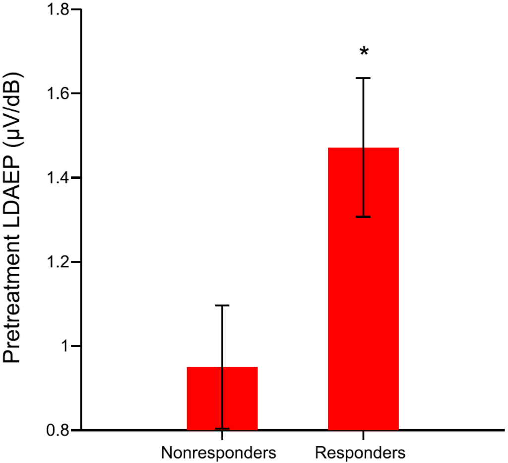
The demographic data, clinical variables, and LDAEP are presented according to treatment response in Table 2. The pretreatment N1 LDAEP, N1 standardized low resolution brain electromagnetic tomography (sLORETA)-LDAEP, P2 sLORETA-LDAEP, and N1/P2 sLORETA-LDAEP did not significantly differ between the responders and nonresponders (t = 0.249, df = 39, p = 0.805; t = 0.155, df = 39, p = 0.878; t = −0.611, df = 39, p = 0.545; t = 0.822, df = 39, p = 0.922, respectively; Table 2). However, the responders had higher pretreatment cortical P2 and N1/P2 LDAEP than nonresponders (t = −2.498, df = 37.02, p = 0.017; t = −2.176, df = 39, p = 0.036, respectively; Figure 1). In addition, those LDAEP values differed significantly between the responders and nonresponders when sex was considered covariate in analysis (F(1, 40) = 4.105, p = 0.050; F(1, 40) = 4.198, p = 0.047, respectively).

Table 1.
Comparison of pretreatment and post-treatment loudness dependence of auditory evoked potentials (LDAEP) between groups according to sex, the number of episodes, hypnotic medication, and smoking.
| Sex | Male (n = 7) | Female (n = 34) | p |
|---|---|---|---|
| Pretreatment N1 | −0.42 ± 0.70 | −0.39 ± 0.50 | 0.919 |
| P2 | 0.65 ± 0.63 | 0.92 ± 0.74 | 0.370 |
| N1/P2 | 1.06 ± 1.09 | 1.31 ± 0.72 | 0.449 |
| Post-treatment N1 | −0.50 ± 0.56 | −0.53 ± 0.58 | 0.909 |
| P2 | 0.72 ± 1.11 | 0.81 ± 0.83 | 0.805 |
| N1/P2 | 1.22 ± 1.10 | 1.34 ± 0.85 | 0.744 |
| Recurrence | First-episode MDD (n = 15) | Recurrent-episode MDD (n = 26) | p |
| Pretreatment N1 | −0.55 ± 0.66 | −0.31 ± 0.42 | 0.174 |
| P2 | 0.79 ± 0.68 | 0.92 ± 0.75 | 0.601 |
| N1/P2 | 1.34 ± 0.89 | 1.23 ± 0.73 | 0.681 |
| Post-treatment N1 | −0.39 ± 0.74 | −0.60 ± 0.45 | 0.344 |
| P2 | 0.92 ± 0.83 | 0.72 ± 0.89 | 0.474 |
| N1/P2 | 1.32 ± 0.97 | 1.32 ± 0.85 | 0.996 |
| Hypnotics | No hypnotic medication (n = 20) | Hypnotic medication MDD (n = 21) | p |
| Pretreatment N1 | −0.42 ± 0.62 | −0.38 ± 0.43 | 0.827 |
| P2 | 1.06 ± 0.75 | 0.69 ± 0.66 | 0.100 |
| N1/P2 | 1.48 ± 0.91 | 1.07 ± 0.59 | 0.103 |
| Post-treatment N1 | −0.53 ± 0.56 | −0.51 ± 0.59 | 0.912 |
| P2 | 1.06 ± 1.01 | 0.55 ± 0.64 | 0.059 |
| N1/P2 | 1.59 ± 0.94 | 1.06 ± 0.76 | 0.053 |
| Smoking | No smoking (n = 31) | Smoking (n = 10) | p |
| Pretreatment N1 | −0.46 ± 0.49 | −0.20 ± 0.62 | 0.164 |
| P2 | 0.90 ± 0.73 | 0.78 ± 0.70 | 0.649 |
| N1/P2 | 1.37 ± 0.70 | 0.97 ± 0.97 | 0.171 |
| Post-treatment N1 | −0.49 ± 0.59 | −0.63 ± 0.51 | 0.514 |
| P2 | 0.88 ± 0.88 | 0.53 ± 0.81 | 0.268 |
| N1/P2 | 1.37 ± 0.85 | 1.16 ± 1.01 | 0.509 |
Data are mean ± SD or n values; BDI, Beck depression inventory; LDAEP, loudness dependence of auditory evoked potentials.

Table 2.
Comparison of demographic data, clinical variables, and LDAEP between nonresponder and responder groups among major depressive disorder (MDD) patients.
| Variable | Nonresponders (n = 16) | Responders (n = 25) | p |
|---|---|---|---|
| Age (years) | 43.0 ± 17.8 | 38.4 ± 13.4 | 0.387 |
| Sex (male/female) | 4/12 | 3/22 | 0.401 |
| First-/Recurrent-episode | 3/13 | 12/13 | 0.058 |
| Nonsmoker/smoker | 10/6 | 21/4 | 0.150 |
| Pretreatment LDAEP (µV/dB) | |||
| Post-treatment LDAEP (µV/dB) | |||
| Baseline BDI score | 28.9 ± 9.8 | 32.4 ± 13.8 | 0.394 |
| Post-treatment BDI score | 25.6 ± 9.5 | 5.5 ± 4.8 | <0.01 ** |
Data are mean ± SD or n values; BDI, Beck depression inventory; LDAEP, loudness dependence of auditory evoked potentials; * Statistically significant difference at p < 0.05; ** Statistically significant difference at p < 0.01.

Figure 1.
The pretreatment N1/P2 loudness dependence of auditory evoked potentials (LDAEP) of responders and nonresponders (responders were defined as those with a reduction in the Beck Depression Inventory (BDI) score of >50% between baseline and follow-up) among major depressive disorder (MDD) patients (t = −2.176, p = 0.036). The data are presented as mean and standard error values. * Statistically significant difference at p < 0.05.
Although the pretreatment total BDI scores there did not differ between these two groups (t = −0.862, df = 39, p = 0.394), a significant difference was found in the post-treatment total BDI scores (t = 7.900, df = 19.91, p < 0.01). In addition, there were no significant changes in the N1, P2, and N1/P2 LDAEP (as assessed using the paired t-test) between baseline and follow-up in either the responders (p > 0.05) or nonresponders (p > 0.05).
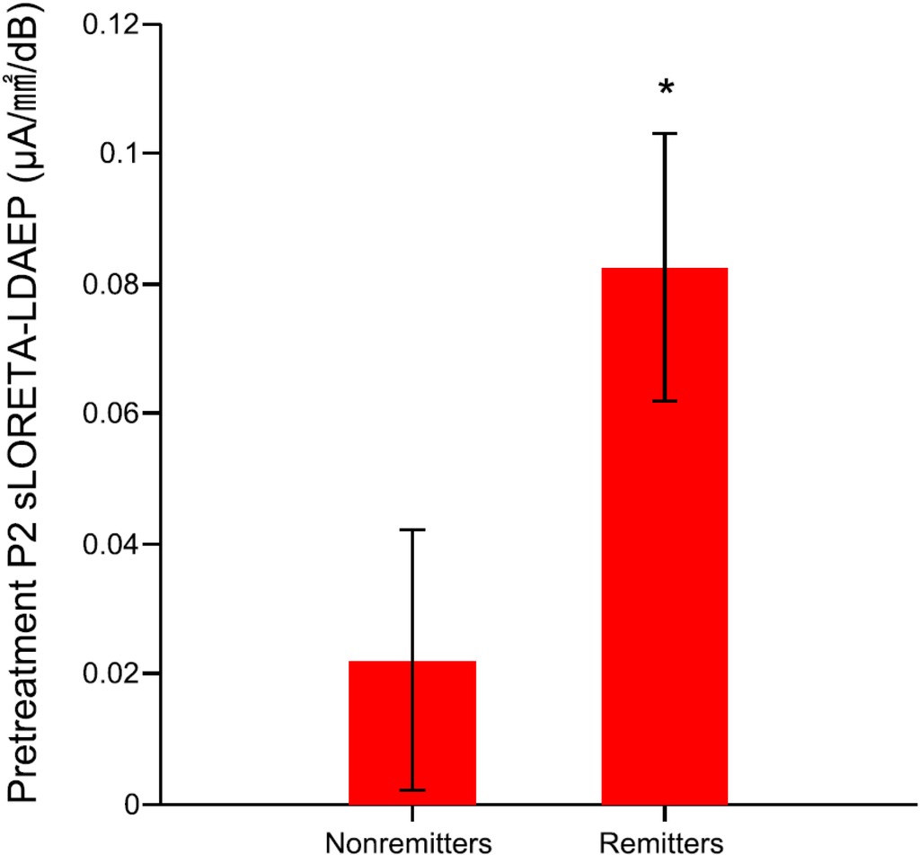
Furthermore, the remitters had higher pretreatment cortical P2 LDAEP and left P2 sLORETA-LDAEP than nonremitters (t = −2.095, df = 31.52, p = 0.044; t = −2.095, df = 39, p = 0.043, respectively; Figure 2).

Figure 2.
The pretreatment left P2 standardized low resolution brain electromagnetic tomography (sLORETA)-LDAEP of remitters and nonremitters (remitters were defined as those with <10 points in the post-treatment BDI score) among MDD patients (t = −2.095, p = 0.043). The data are presented as mean and standard error values. * Statistically significant difference at p < 0.05.
2.2. Low vs. High Pretreatment Loudness Dependence of Auditory Evoked Potentials (LDAEP)
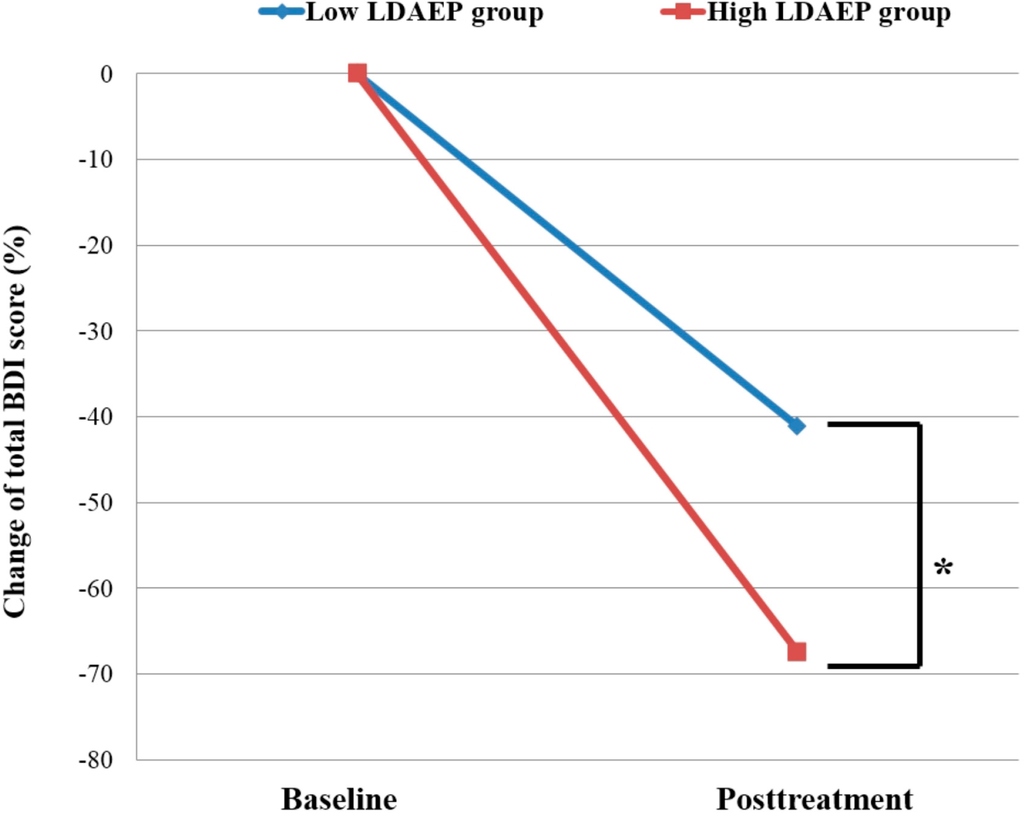
Table 3 gives the characteristics of the patients dichotomized according to their pretreatment N1/P2 LDAEP. There was no significant difference in the pretreatment total BDI score between the low- and high-LDAEP groups (t = −1.260, df = 39, p = 0.215), while a significant difference was shown in the post-treatment total BDI scores (t = 2.47, df = 39, p = 0.018). Moreover, the change in BDI score between baseline and follow-up differed significantly between the two groups (t = −2.741, df = 39, p = 0.009; Figure 3). There was a significantly higher rate for responders in the high-LDAEP group (76.2%) than in the low-LDAEP group (45%) (χ2 = 4.188, p = 0.041). The odds ratio of baseline low N1/P2 LDAEP was 1.91 (95% confidential interval (CI), 1.02–3.58) and the odds ratio of high N1/P2 LDAEP was 0.49 (95% CI, 0.22–1.07) for treatment nonresponse. In addition, these showed the significant effect of BDI change differences between the baseline low- and high-LDAEP groups (Cohen’s d = 0.8).

Table 3.
Comparison of demographic data, clinical variables, and the LDAEP between MDD patients with low and high pretreatment LDAEP (dichotomized at the median into low vs. high).
| Variable | Low LDAEP Group (n = 20) | High LDAEP Group (n = 21) | p |
|---|---|---|---|
| Age (years) | 44.2 ± 14.6 | 36.4 ± 15.1 | 0.102 |
| Sex (male/female) | 3/17 | 4/17 | 1.000 |
| First-/Recurrent-episode | 5/15 | 10/11 | 0.133 |
| Nonsmoker/smoker | 12/8 | 19/2 | 0.032 * |
| Pretreatment LDAEP | |||
| Post-treatment LDAEP | |||
| Pretreatment BDI score | 28.6 ± 10.6 | 33.4 ± 13.7 | 0.215 |
| Post-treatment BDI score | 17.9 ± 13.0 | 9.1 ± 9.6 | 0.018 * |
| BDI change (%) | 37.2 ± 40.9 | 70.2 ± 36.2 | 0.009 ** |
| Responder/nonresponder | 9/11 | 16/5 | 0.041 * |
Data are mean ± SD or n values; BDI, Beck depression inventory; BDI change, change of BDI score from baseline to post-treatment; LDAEP, loudness dependence of auditory evoked potentials; * Statistically significant difference at p < 0.05; ** Statistically significant difference at p < 0.01.

Figure 3.
Changes in the total BDI score between baseline and follow-up in MDD patients dichotomized according to their pretreatment LDAEP (i.e., low or high). The change in total BDI score differed significantly between the low- and high-LDAEP groups (t = −2.741, p = 0.009). BDI change, change in BDI score from baseline to follow-up. * Statistically significant difference at p < 0.01.
2.3. Discussion
The present study examined the relationship between the pretreatment LDAEP and the treatment response to antidepressant monotherapy in MDD patients who had received maintenance antidepressant medication for more than 12 weeks. The findings showed that the pretreatment scalp P2 and N1/P2 LDAEP was significantly higher in MDD patients with a treatment response (i.e., responders) than in those without a treatment response (i.e., nonresponders). Thus, the MDD treatment responders may have a tendency toward relatively low central serotonergic activity. Comparison of the low- and high-LDAEP groups (based on the median N1/P2 LDAEP) revealed that the latter included a higher proportion of responders. Also, when considering the odds ratio of baseline N1/P2 LDAEP for treatment nonresponse, the low LDAEP group had a risk of presenting treatment non-response that was 1.9 times higher. These findings are consistent with previous evidence of an association between a high pretreatment LDAEP in MDD patients, indicating a low central serotonergic activity, and a favorable response to antidepressant agents, SSRIs with a serotonin-enhancing effect [,,,]. The findings of several studies suggest that a high pretreatment LDAEP can predict a favorable response to short-term (i.e., 4 weeks) treatment with an SSRI in MDD patients [,,]. However, the present findings and a recent study by Jaworska et al. [] have observed an association between LDAEP and a treatment response to long-term (at least 12 weeks) maintenance treatment []. The present study revealed that higher cortical P2 and N1/P2 LDAEP were associated with treatment responders, while Jaworska and colleagues [] reported that higher N1 LORETA-LDAEP was associated with responders. Intriguingly, the present study also revealed that higher cortical P2 LDAEP and left P2 sLORETA-LDAEP were associated with treatment remitters.
In addition, some studies have found a high pretreatment LDAEP to be significantly greater in responders to treatment with bupropion or lithium—both of which may affect serotonin activity—than in nonresponders [,]. In contrast, other studies have shown that a low pretreatment LDAEP is associated with response to the norepinephrine reuptake inhibitor (NRI) [,,]. Taken together, variables of LDAEP could be associated with response or nonresponse to antidepressant treatment both for short- and long-term durations. It is necessary to verify in future studies whether the pretreatment LDAEP can predict a response to both acute and chronic antidepressant treatment via augmenting serotonin and other neurotransmitters.
Another finding from the present study was the lack of a significant alteration in the LDAEP between baseline and follow-up after maintenance antidepressant therapy. This finding indicates that treatment with serotonin-enhancing agents for more than 12 weeks did not lead to a change in the LDAEP. Previous studies have found no change in the LDAEP following 24 days or 4 weeks of treatment with SSRIs [,]. Studies exploring the LDAEP after administering an SSRI in healthy adults have also produced conflicting results: some have found no changes in the LDAEP after a single administration [,,] or after 24-day administration [] of SSRIs, while other have shown a decrease in the LDAEP after a single trial [] or after 24-day administration [] of SSRIs. In addition, some studies have reported the association between altered LDAEP and polymorphisms of the serotonin transporter gene [,]. Taken together, these findings suggest that the LDAEP might remain stable in MDD patients before and after SSRI administration.
2.4. Study Limitations
This study was subject to several limitations. First, the sample was relatively small; Second, this study did not include a control condition with either a placebo or another non-serotonergic antidepressant drug; Third, the severity of depression was only assessed using the BDI score. It is therefore necessary for future studies with a large sample to assess the cortical and source LDAEP in order to determine which LDAEP variable is a more sensitive marker for predicting a treatment response.
3. Experimental Section
3.1. Subjects and Study Design
In total, 41 outpatients (7 males and 34 females; 40.2 ± 15.2 years old, mean ± SD) who met the Diagnostic and Statistical Manual of Mental Disorders (DSM)-IV-text revision criteria for MDD were recruited from Ilsan Paik Hospital. Patients were excluded if they had any major mental disorders including anxiety disorder on axis I or II of the DSM-IV, or major medical and neurological disorders. Individuals with hearing impairment were excluded. Patients were either medication-naïve or medication-free for at least eight weeks when entering this study. They all were Korean and of the same ethnicity. Of these, 37% (15/41) of subjects had first-episode MDD, and 63% (26/41) had recurrent-episode MDD. In addition, 24% (10/41) were current smokers. Depressive symptoms were assessed using the Beck Depression Inventory (BDI) at baseline. The pretreatment LDAEP was calculated by measuring the event-related potentials induced by auditory stimuli prior to beginning antidepressants. The MDD patients were treated with the following antidepressants: escitalopram (n = 32, 10.0 ± 4.0 mg/day), sertraline (n = 7, 78.6 ± 26.7 mg/day), and paroxetine controlled-release (CR) formulation (n = 2, 18.8 ± 8.8 mg/day). Among them, 21 patients took hypnotic agents including alprazolam (n = 13, 0.25–0.5 mg/day), clonazepam (n = 7, 0.25–0.5 mg/day), and zolpidem (n = 2, 5–10 mg/day). The post-treatment BDI score and LDAEP were reevaluated in all patients when they had taken antidepressant medications for more than 12 weeks, and their dosage of antidepressants remained the same in the last 4 weeks. The mean duration of antidepressant treatment was 14.1 ± 2.1 weeks (range, 12.9–21.1 weeks).
The SSRI medication could have an influence on results of the LDAEP, and then the baseline LDAEP was measured before antidepressant treatment. Moreover, this study did not enroll patients who had taken any psychotropic agent within 8 weeks before the baseline assessment of LDAEP in our study, except for hypnotic drugs such as alprazolam, clonazepam, and zolpidem.
The subjects were stratified according to their treatment response into responders and nonresponders by comparing their pre- and post-treatment BDI scores; responders were defined as those with a reduction in BDI score of >50% between baseline and follow-up. They were also dichotomized according to their median pretreatment N1/P2 LDAEP into a low or high pretreatment LDAEP group (low- and high-LDAEP groups, respectively). In addition, they were also stratified according to their treatment remission into remitters and nonremitters by comparing their pre- and post-treatment BDI scores; remitters were defined as those with <10s point in the post-treatment Beck Depression Inventory (BDI) score.
The study protocol was approved by the ethics committee of Inje University, Ilsan Paik Hospital, and written informed consent to participate was obtained from all subjects at study entry (IB-3-1105-014, in June 2011).
3.2. Electroencephalogram (EEG) Methods
All of the subjects were seated in a comfortable chair in a sound-attenuated room. They were asked to keep their eyes open during the entire testing with their eyes fixated in the pointer on a monitor. The auditory processing consisted of 1000 stimuli with an interstimulus interval between 500 and 900 ms. It was presented in a randomized fashion. Tones of 1000 Hz and 80 ms duration (with 10 ms rise and fall times) were generated by E-Prime software (Psychology Software Tools, Pittsburgh, PA, USA) and presented at five intensities (55, 65, 75, 85, and 95 dB SPL) via headphones (MDR-D777, Sony, Tokyo, Japan). They were presented in a randomized fashion. EEG data were recorded from 64 scalp sites using silver/silver-chloride electrodes according to the international 10–20 system (impedance < 10 kV), using an Auditory Neuroscan NuAmp amplifier (Compumedics USA, El Paso, TX, USA). Data were collected at a sampling rate of 1000 Hz, using a bandpass filter of 0.5–100 Hz. In addition, four electrodes were used to measure both horizontal and vertical electrooculograms.
Data were reanalyzed using Scan 4.3 software with a bandpass filter of 1–30 Hz, and ocular contamination was removed using standard blink-correction algorithms []. Event-related potential sweeps with artifacts exceeding 70 mV were rejected at all electrode sites. Rejection rate was <5% per each intensity. For each intensity and for each subject, the N1 peak (negative-most amplitude between 80 and 130 ms after the stimulus) and P2 peak (positive-most peak between 130 and 230 ms after the stimulus) were then determined at the Cz electrode. The peak-to-peak N1/P2 amplitudes were calculated for the five stimulus intensities, and the LDAEP was calculated as the slope of the linear-regression curve.
The Cz electrode was chosen because previous studies have shown this to be a reliable site at which the amplitude is larger than at other electrode sites [,,]. The dipole source analysis for the measurement of LDAEP has been used in some studies [,], producing results similar to those obtained when using cortical analysis []. Moreover, many LDAEP studies have been conducted based on cortical analysis [,,,].
3.3. Source LDAEP Analysis
On the basis of the averaged, scalp-recorded electric potential, standardized low-resolution brain electromagnetic tomography (sLORETA) was used to estimate current density []. The sLORETA technique estimates the standardized source current density by using the realistic 3-shell head model, on the basis of the Montreal Neurological Institute (MNI) 152 template provided by the Brain Imaging Center of the MNI, under the assumption that the activity at any single neuron should be highly synchronized to the activity of its closest neighbors []. The solution space is restricted to the cortical gray matter and hippocampus of the head model and partitioned into 6239 voxels at a spatial resolution of 5 mm. Anatomical labels, such as Brodmann areas (BAs), are provided by the use of an appropriate transformation from MNI to Talairach space []. The loudness dependence of the source activity (source LDAEP) was determined by calculating current source densities for each subject and each sound pressure level. Two electrodes (M1, M2) were not used in the sLORETA analysis because these electrode locations are not supported by the sLORETA software. The calculated standardized current density was averaged between 60 and 240 ms post-stimulus from the primary auditory cortex (BA41), in accordance with a previous study [,]. We calculated the 3 values of current density for the left, right, and averaged data from both hemispheres over the voxels that fall under the primary auditory cortex. The source LDAEP was calculated as the slope of the linear regression of current density of BA41 for the 5 stimulus intensities.
3.4. Statistical Analysis
Our data including pre- and post-treatment N1, P2, and N1/P2 LDAEP, and BDI scores showed a Gaussian distribution according to the Kolmogorov-Smimov test (data not shown). The demographic data, clinical variables, and LDAEP values were compared between two groups using Student’s t-test, paired t-test, and the chi-square. Pearson’s correlation coefficients were calculated to examine the relationships between LDAEP and clinical variables. Sex was considered to be a significant covariate when analyzing the N1, P2, N1/P2 LDAEP values, because the previous studies have reported a significant effect in sex on the LDAEP [,]. A general linear model was used while controlling for sex as covariate. All tests were two-tailed, and group differences were tested at the p < 0.05 level. The statistical packages used for analysis were SAS 9.3 and SALT 2.5.
4. Conclusions
In conclusion, the present findings have revealed an apparent association between a high pretreatment LDAEP and a clinical response to long-term treatment with an antidepressant with serotonin-augmenting effects. The pretreatment LDAEP could thus be a useful marker to predict a potential treatment response among MDD patients. More studies with larger samples should be performed to clarify the relationship between the LDAEP and the treatment response in MDD.
Acknowledgments
This study was supported by a 2013 Inje University research grant. They offered financial support to the authors.
Author Contributions
All authorship credit was based on substantial contributions to conception and design, acquisition of data, or analysis and interpretation of data (Young-Min Park, Bun-Hee Lee, Seung-Hwan Lee, Miseon Shim); drafting the article or revising it critically for important intellectual content (Young-Min Park, Bun-Hee Lee); and final approval of the version to be published (Young-Min Park).
Conflicts of Interest
The authors declare no conflict of interest.
References
- Lopez, A.D.; Mathers, C.D.; Ezzati, M.; Jamison, D.T.; Murray, C.J. Global and regional burden of disease and risk factors, 2001: Systematic analysis of population health data. Lancet 2006, 367, 1747–1757. [Google Scholar] [CrossRef] [PubMed]
- Blier, P.; Gobbi, G.; Turcotte, J.E.; de Montigny, C.; Boucher, N.; Hebert, C.; Debonnel, G. Mirtazapine and paroxetine in major depression: A comparison of monotherapy vs. their combination from treatment initiation. Eur. Neuropsychopharmacol. 2009, 19, 457–465. [Google Scholar] [CrossRef] [PubMed]
- Blier, P.; Ward, H.E.; Tremblay, P.; Laberge, L.; Hebert, C.; Bergeron, R. Combination of antidepressant medications from treatment initiation for major depressive disorder: A double-blind randomized study. Am. J. Psychiatry 2010, 167, 281–288. [Google Scholar] [CrossRef] [PubMed]
- Wang, H.R.; Bahk, W.M.; Park, Y.M.; Lee, H.B.; Song, H.R.; Jeong, J.H.; Seo, J.S.; Lim, E.S.; Hong, J.W.; Kim, W.; et al. Korean medication algorithm for depressive disorder: Comparisons with other treatment guidelines. Psychiatry Investig. 2014, 11, 1–11. [Google Scholar] [CrossRef]
- Fabbri, C.; Porcelli, S.; Serretti, A. From pharmacogenetics to pharmacogenomics: The way toward the personalization of antidepressant treatment. Can. J. Psychiatry 2014, 59, 62–75. [Google Scholar] [PubMed]
- Labermaier, C.; Masana, M.; Muller, M.B. Biomarkers predicting antidepressant treatment response: How can we advance the field? Dis. Markers 2013, 35, 23–31. [Google Scholar] [CrossRef] [PubMed]
- Yoshimura, R.; Kishi, T.; Hori, H.; Katsuki, A.; Sugita-Ikenouchi, A.; Umene-Nakano, W.; Atake, K.; Iwata, N.; Nakamura, J. Serum levels of brain-derived neurotrophic factor at 4 weeks and response to treatment with ssris. Psychiatry Investig. 2014, 11, 84–88. [Google Scholar] [CrossRef] [PubMed]
- Paavonen, V.; Kampman, O.; Illi, A.; Viikki, M.; Setala-Soikkeli, E.; Leinonen, E. A cluster model of temperament as an indicator of antidepressant response and symptom severity in major depression. Psychiatry Investig. 2014, 11, 18–23. [Google Scholar] [CrossRef] [PubMed]
- Pae, C.U. Serotonin receptor 2c-759c/t polymorphism and weight change or treatment response to mirtazapine in korean depressive patients. Psychiatry Investig. 2014, 11, 342–343. [Google Scholar] [CrossRef] [PubMed]
- O’Neill, B.V.; Croft, R.J.; Nathan, P.J. The loudness dependence of the auditory evoked potential (LDAEP) as an in vivo biomarker of central serotonergic function in humans: Rationale, evaluation and review of findings. Hum. Psychopharmacol. 2008, 23, 355–370. [Google Scholar] [CrossRef] [PubMed]
- Hegerl, U.; Juckel, G. Intensity dependence of auditory evoked potentials as an indicator of central serotonergic neurotransmission: A new hypothesis. Biol. Psychiatry 1993, 33, 173–187. [Google Scholar] [CrossRef] [PubMed]
- Hegerl, U.; Gallinat, J.; Juckel, G. Event-related potentials. Do they reflect central serotonergic neurotransmission and do they predict clinical response to serotonin agonists? J. Affect. Disord. 2001, 62, 93–100. [Google Scholar] [CrossRef] [PubMed]
- Juckel, G.; Molnar, M.; Hegerl, U.; Csepe, V.; Karmos, G. Auditory-evoked potentials as indicator of brain serotonergic activity—First evidence in behaving cats. Biol. Psychiatry 1997, 41, 1181–1195. [Google Scholar] [CrossRef] [PubMed]
- Wutzler, A.; Winter, C.; Kitzrow, W.; Uhl, I.; Wolf, R.J.; Heinz, A.; Juckel, G. Loudness dependence of auditory evoked potentials as indicator of central serotonergic neurotransmission: Simultaneous electrophysiological recordings and in vivo microdialysis in the rat primary auditory cortex. Neuropsychopharmacology 2008, 33, 3176–3181. [Google Scholar] [CrossRef] [PubMed]
- Park, Y.M.; Lee, S.H.; Kim, S.; Bae, S.M. The loudness dependence of the auditory evoked potential (LDAEP) in schizophrenia, bipolar disorder, major depressive disorder, anxiety disorder, and healthy controls. Prog. Neuropsychopharmacol. Biol. Psychiatry 2010, 34, 313–316. [Google Scholar] [CrossRef] [PubMed]
- Mulert, C.; Jager, L.; Propp, S.; Karch, S.; Stormann, S.; Pogarell, O.; Moller, H.J.; Juckel, G.; Hegerl, U. Sound level dependence of the primary auditory cortex: Simultaneous measurement with 61-channel EEG and FMRI. Neuroimage 2005, 28, 49–58. [Google Scholar] [CrossRef] [PubMed]
- Juckel, G.; Hegerl, U.; Molnar, M.; Csepe, V.; Karmos, G. Auditory evoked potentials reflect serotonergic neuronal activity—A study in behaving cats administered drugs acting on 5-HT1A autoreceptors in the dorsal raphe nucleus. Neuropsychopharmacology 1999, 21, 710–716. [Google Scholar] [CrossRef] [PubMed]
- O’Neill, B.V.; Guille, V.; Croft, R.J.; Leung, S.; Scholes, K.E.; Phan, K.L.; Nathan, P.J. Effects of selective and combined serotonin and dopamine depletion on the loudness dependence of the auditory evoked potential (LDAEP) in humans. Hum. Psychopharmacol. 2008, 23, 301–312. [Google Scholar] [CrossRef] [PubMed]
- Massey, A.E.; Marsh, V.R.; McAllister-Williams, R.H. Lack of effect of tryptophan depletion on the loudness dependency of auditory event related potentials in healthy volunteers. Biol. Psychol. 2004, 65, 137–145. [Google Scholar] [CrossRef] [PubMed]
- Norra, C.; Becker, S.; Brocheler, A.; Kawohl, W.; Kunert, H.J.; Buchner, H. Loudness dependence of evoked dipole source activity during acute serotonin challenge in females. Hum. Psychopharmacol. 2008, 23, 31–42. [Google Scholar] [CrossRef] [PubMed]
- Dierks, T.; Barta, S.; Demisch, L.; Schmeck, K.; Englert, E.; Kewitz, A.; Maurer, K.; Poustka, F. Intensity dependence of auditory evoked potentials (AEPS) as biological marker for cerebral serotonin levels: Effects of tryptophan depletion in healthy subjects. Psychopharmacology (Berlin) 1999, 146, 101–107. [Google Scholar] [CrossRef]
- Kahkonen, S.; Jaaskelainen, I.P.; Pennanen, S.; Liesivuori, J.; Ahveninen, J. Acute tryptophan depletion decreases intensity dependence of auditory evoked magnetic N1/P2 dipole source activity. Psychopharmacology (Berlin) 2002, 164, 221–227. [Google Scholar] [CrossRef]
- O’Neill, B.V.; Croft, R.J.; Leung, S.; Oliver, C.; Phan, K.L.; Nathan, P.J. High-dose glycine inhibits the loudness dependence of the auditory evoked potential (LDAEP) in healthy humans. Psychopharmacology (Berlin) 2007, 195, 85–93. [Google Scholar] [CrossRef]
- Leuchter, A.F.; Cook, I.A.; Hunter, A.; Korb, A. Use of clinical neurophysiology for the selection of medication in the treatment of major depressive disorder: The state of the evidence. Clin. EEG Neurosci. 2009, 40, 78–83. [Google Scholar] [CrossRef] [PubMed]
- Gallinat, J.; Bottlender, R.; Juckel, G.; Munke-Puchner, A.; Stotz, G.; Kuss, H.J.; Mavrogiorgou, P.; Hegerl, U. The loudness dependency of the auditory evoked N1/P2-component as a predictor of the acute ssri response in depression. Psychopharmacology (Berlin) 2000, 148, 404–411. [Google Scholar] [CrossRef]
- Mulert, C.; Juckel, G.; Augustin, H.; Hegerl, U. Comparison between the analysis of the loudness dependency of the auditory N1/P2 component with loreta and dipole source analysis in the prediction of treatment response to the selective serotonin reuptake inhibitor citalopram in major depression. Clin. Neurophysiol. 2002, 113, 1566–1572. [Google Scholar] [CrossRef] [PubMed]
- Lee, T.W.; Yu, Y.W.; Chen, T.J.; Tsai, S.J. Loudness dependence of the auditory evoked potential and response to antidepressants in chinese patients with major depression. J. Psychiatry Neurosci. 2005, 30, 202–205. [Google Scholar] [PubMed]
- Jaworska, N.; Blondeau, C.; Tessier, P.; Norris, S.; Fusee, W.; Blier, P.; Knott, V. Response prediction to antidepressants using scalp and source-localized loudness dependence of auditory evoked potential (LDAEP) slopes. Prog. Neuropsychopharmacol. Biol. Psychiatry 2013, 44, 100–107. [Google Scholar] [CrossRef] [PubMed]
- Juckel, G.; Pogarell, O.; Augustin, H.; Mulert, C.; Muller-Siecheneder, F.; Frodl, T.; Mavrogiorgou, P.; Hegerl, U. Differential prediction of first clinical response to serotonergic and noradrenergic antidepressants using the loudness dependence of auditory evoked potentials in patients with major depressive disorder. J. Clin. Psychiatry 2007, 68, 1206–1212. [Google Scholar] [CrossRef] [PubMed]
- Paige, S.R.; Hendricks, S.E.; Fitzpatrick, D.F.; Balogh, S.; Burke, W.J. Amplitude/intensity functions of auditory event-related potentials predict responsiveness to bupropion in major depressive disorder. Psychopharmacol. Bull. 1995, 31, 243–248. [Google Scholar] [PubMed]
- Juckel, G.; Mavrogiorgou, P.; Bredemeier, S.; Gallinat, J.; Frodl, T.; Schulz, C.; Moller, H.J.; Hegerl, U. Loudness dependence of primary auditory-cortex-evoked activity as predictor of therapeutic outcome to prophylactic lithium treatment in affective disorders--a retrospective study. Pharmacopsychiatry 2004, 37, 46–51. [Google Scholar] [PubMed]
- Linka, T.; Muller, B.W.; Bender, S.; Sartory, G.; Gastpar, M. The intensity dependence of auditory evoked ERP components predicts responsiveness to reboxetine treatment in major depression. Pharmacopsychiatry 2005, 38, 139–143. [Google Scholar] [CrossRef] [PubMed]
- Mulert, C.; Juckel, G.; Brunnmeier, M.; Karch, S.; Leicht, G.; Mergl, R.; Moller, H.J.; Hegerl, U.; Pogarell, O. Prediction of treatment response in major depression: Integration of concepts. J. Affect. Disord. 2007, 98, 215–225. [Google Scholar] [CrossRef] [PubMed]
- Linka, T.; Sartory, G.; Wiltfang, J.; Muller, B.W. Treatment effects of serotonergic and noradrenergic antidepressants on the intensity dependence of auditory erp components in major depression. Neurosci. Lett. 2009, 463, 26–30. [Google Scholar] [CrossRef] [PubMed]
- Guille, V.; Croft, R.J.; O’Neill, B.V.; Illic, S.; Phan, K.L.; Nathan, P.J. An examination of acute changes in serotonergic neurotransmission using the loudness dependence measure of auditory cortex evoked activity: Effects of citalopram, escitalopram and sertraline. Hum. Psychopharmacol. 2008, 23, 231–241. [Google Scholar] [CrossRef] [PubMed]
- Uhl, I.; Gorynia, I.; Gallinat, J.; Mulert, C.; Wutzler, A.; Heinz, A.; Juckel, G. Is the loudness dependence of auditory evoked potentials modulated by the selective serotonin reuptake inhibitor citalopram in healthy subjects? Hum. Psychopharmacol. 2006, 21, 463–471. [Google Scholar] [CrossRef] [PubMed]
- Oliva, J.; Leung, S.; Croft, R.J.; O’Neill, B.V.; O’Kane, J.; Stout, J.; Phan, K.L.; Nathan, P.J. The loudness dependence auditory evoked potential is insensitive to acute changes in serotonergic and noradrenergic neurotransmission. Hum. Psychopharmacol. 2010, 25, 423–427. [Google Scholar] [CrossRef] [PubMed]
- Nathan, P.J.; Segrave, R.; Phan, K.L.; O’Neill, B.; Croft, R.J. Direct evidence that acutely enhancing serotonin with the selective serotonin reuptake inhibitor citalopram modulates the loudness dependence of the auditory evoked potential (LDAEP) marker of central serotonin function. Hum. Psychopharmacol. 2006, 21, 47–52. [Google Scholar] [CrossRef] [PubMed]
- Simmons, J.G.; Nathan, P.J.; Berger, G.; Allen, N.B. Chronic modulation of serotonergic neurotransmission with sertraline attenuates the loudness dependence of the auditory evoked potential in healthy participants. Psychopharmacology (Berlin) 2011, 217, 101–110. [Google Scholar] [CrossRef]
- Trivedi, M.H.; Rush, A.J.; Wisniewski, S.R.; Nierenberg, A.A.; Warden, D.; Ritz, L.; Norquist, G.; Howland, R.H.; Lebowitz, B.; McGrath, P.J.; et al. Evaluation of outcomes with citalopram for depression using measurement-based care in STAR*D: Implications for clinical practice. Am. J. Psychiatry 2006, 163, 28–40. [Google Scholar] [CrossRef] [PubMed]
- Semlitsch, H.V.; Anderer, P.; Schuster, P.; Presslich, O. A solution for reliable and valid reduction of ocular artifacts, applied to the p300 ERP. Psychophysiology 1986, 23, 695–703. [Google Scholar] [CrossRef] [PubMed]
- Gudlowski, Y.; Ozgurdal, S.; Witthaus, H.; Gallinat, J.; Hauser, M.; Winter, C.; Uhl, I.; Heinz, A.; Juckel, G. Serotonergic dysfunction in the prodromal, first-episode and chronic course of schizophrenia as assessed by the loudness dependence of auditory evoked activity. Schizophr. Res. 2009, 109, 141–147. [Google Scholar] [CrossRef] [PubMed]
- Juckel, G.; Gallinat, J.; Riedel, M.; Sokullu, S.; Schulz, C.; Moller, H.J.; Muller, N.; Hegerl, U. Serotonergic dysfunction in schizophrenia assessed by the loudness dependence measure of primary auditory cortex evoked activity. Schizophr. Res. 2003, 64, 115–124. [Google Scholar] [CrossRef] [PubMed]
- Juckel, G.; Gudlowski, Y.; Muller, D.; Ozgurdal, S.; Brune, M.; Gallinat, J.; Frodl, T.; Witthaus, H.; Uhl, I.; Wutzler, A.; et al. Loudness dependence of the auditory evoked N1/P2 component as an indicator of serotonergic dysfunction in patients with schizophrenia—A replication study. Psychiatry Res. 2008, 158, 79–82. [Google Scholar] [CrossRef] [PubMed]
- Strobel, A.; Debener, S.; Schmidt, D.; Hunnerkopf, R.; Lesch, K.P.; Brocke, B. Allelic variation in serotonin transporter function associated with the intensity dependence of the auditory evoked potential. Am. J. Med. Genet. 2003, 118, 41–47. [Google Scholar] [CrossRef]
- Brocke, B.; Beauducel, A.; John, R.; Debener, S.; Heilemann, H. Sensation seeking and affective disorders: Characteristics in the intensity dependence of acoustic evoked potentials. Neuropsychobiology 2000, 41, 24–30. [Google Scholar] [CrossRef] [PubMed]
- Hensch, T.; Herold, U.; Brocke, B. An electrophysiological endophenotype of hypomanic and hyperthymic personality. J. Affect. Disord. 2007, 101, 13–26. [Google Scholar] [CrossRef] [PubMed]
- Linka, T.; Muller, B.W.; Bender, S.; Sartory, G. The intensity dependence of the auditory evoked N1 component as a predictor of response to citalopram treatment in patients with major depression. Neurosci. Lett. 2004, 367, 375–378. [Google Scholar] [CrossRef] [PubMed]
- Pascual-Marqui, R.D. Standardized low-resolution brain electromagnetic tomography (sLORETA): Technical details. Methods Find. Exp. Clin. Pharmacol. 2002, 24, 5–12. [Google Scholar] [PubMed]
- Fuchs, M.; Kastner, J.; Wagner, M.; Hawes, S.; Ebersole, J.S. A standardized boundary element method volume conductor model. Clin. Neurophysiol. 2002, 113, 702–712. [Google Scholar] [CrossRef] [PubMed]
- Brett, M.; Johnsrude, I.S.; Owen, A.M. The problem of functional localization in the human brain. Nat. Rev. Neurosci. 2002, 3, 243–249. [Google Scholar] [CrossRef] [PubMed]
- Park, Y.M.; Kim, D.W.; Kim, S.; Im, C.H.; Lee, S.H. The loudness dependence of the auditory evoked potential (LDAEP) as a predictor of the response to escitalopram in patients with generalized anxiety disorder. Psychopharmacology (Berlin) 2011, 213, 625–632. [Google Scholar] [CrossRef]
- Jaworska, N.; Blier, P.; Fusee, W.; Knott, V. Scalp- and sLORETA-derived loudness dependence of auditory evoked potentials (LDAEPs) in unmedicated depressed males and females and healthy controls. Clin. Neurophysiol. 2012, 123, 1769–1778. [Google Scholar] [CrossRef] [PubMed]
© 2015 by the authors; licensee MDPI, Basel, Switzerland. This article is an open access article distributed under the terms and conditions of the Creative Commons Attribution license (http://creativecommons.org/licenses/by/4.0/).