Article

New Insights into the Metabolic Profile and Cytotoxic Activity of Kigelia africana Stem Bark

by
Dimitrina Zheleva-Dimitrova
1,*,
Rositsa Mihaylova
2,
Maria Nikolova
2,
Nisha Singh
3 and
Spiro Konstantinov
2
1
Department of Pharmacognosy, Faculty of Pharmacy, Medical University of Sofia, 1000 Sofia, Bulgaria
2
Department of Pharmacology, Pharmacotherapy and Toxicology, Faculty of Pharmacy, Medical University of Sofia, 1000 Sofia, Bulgaria
3
School of Life Sciences, University of KwaZulu-Natal, Durban 4000, South Africa
*
Author to whom correspondence should be addressed.
Molecules 2025, 30(6), 1388; https://doi.org/10.3390/molecules30061388
Submission received: 24 January 2025 / Revised: 11 March 2025 / Accepted: 18 March 2025 / Published: 20 March 2025

Abstract

:
Ultra-high-performance liquid chromatography coupled to Orbitrap high-resolution mass spectrometry (UHPLC-HRMS) was recently employed in many fields to obtain a rapid characterization of plant extracts. Kigelia africana (family Bignoniaceae) is a quintessential African herbal medicinal plant with immense indigenous medicinal and non-medicinal applications. The aim of the present research was to obtain an in-depth metabolite profiling of the K. africana stem bark extract using UHPLC-HRMS and to conduct a preliminary screening of its anticancer activity against a panel of malignant human cell lines of different origin. The UHPLC-HRMS analysis revealed 63 secondary metabolites including phenolic acids, gallo- and ellagitannins, iridoids, naphthoquinones, and anthraquinones. A total of 34 of all annotated compounds are reported for the first time in K. africana stem bark. The studied profile was dominated by trimethylellagic acid, dimethylellagic acid isomers, and ellagic acid. In all tumor models, we established a pronounced inhibition of cell growth in a mostly dose-dependent manner, with IC50 values ranging near and well below (4–30 µg/mL) the lowest treatment concentration of 25 µg/mL. The established cytotoxicity profile of the K. africana extract, highly biased toward malignantly transformed but not normal cells, suggests specific modulation of defined molecular tumor targets. This study revealed K. africana stem bark as a new source of gallo- and ellagitannins, and highlighted the studied herbal drug as an antiproliferative agent with potential pharmaceutical application.

1. Introduction

Kigelia africana (Lam.) Benth. (syn. K. pinnata (Jacq.) DC.) (family Bignoniaceae), commonly known as sausage tree due to the shape of its fruit, is a native of the African continent where it is commonly found in the southern, central, and western regions [1]. K. africana is a large tree, growing to 20 m in height, and widely used for the treatment of a wide range of diseases and conditions in addition to its use as an agroforestry plant. Notably, wild plants, including K. africana, hold significant potential not only for direct therapeutic approaches but also for detoxifying harmful substances (xenobiotics) [2,3]. Ethnopharmacological studies have revealed the therapeutic relevance of various preparations from different K. africana parts to treat a wide range of skin complications, dysentery, constipation, wounds, ulcers, gonorrhoea, rheumatism and abscesses [1].
Overall, more than 150 secondary metabolites including naphthoquinones, iridoids, flavonoids, coumarins, terpenes, terpenoids and steroids have been reported in the K. africana extracts. Iridoids were found to be the major chemical compounds in K. africana. Norbiturnial, 10-deoxyeucommiol, 7-hydroxy-10-deoxyeucommiol, 7-hydroxy eucommic acid, des-p-hydroxybenzoyl kisasaganol, jofuran, together with the iridoid glycosides ajugol, caffeoyl ajugol, catalpol, specioside, verminoside, and minecoside were previously isolated from different parts of K. africana [4,5,6,7,8,9]. Limonoids (kigelianolide, khayanolide B, diacetylkhayanolide E were also reported [10].
An additional class of major metabolites present in K. africana is the naphthoquinones. Lapachol, and its derivative dehydro-α-lapachone, kigelinone, and pinnatal were the first isolated from the root, wood, and fruit respectively [11,12,13]. Phenylethanoid glycosides from K. africana play a major part in its medicinal properties. Decaffeoylacteoside, darensdoside A, verbascoside, isoacteoside, echinacoside, 6-p-coumaroylsucrose, and others were isolated from the fruit of K. africana [14].
Several flavonoids were isolated from the leaves and bark of K. africana. Among them are quercetin, luteolin, 6-hydroxy luteolin, together with their glycosides, isovitexin from the leaves and isoschaftoside from the fruit [14]. Coumarins are among the first phytoconstituents reported for K. africana. 6-Methoxymellein, kigelin, and 3-demethylkigelin were isolated from the root and bark [11], while 6-demethylkigelin was found in the fruit, bark and leaves [15]. Other phenolic compounds found in Kigelia are 4-hydroxycinnamic acid, caffeic acid, caffeic acid methyl ester, ferulic acid, and 3, 4-dimethoxy cinnamic acids, kojic acid, melitolic acid, ethylgallic acid, chlorogenic acid, ellagic acid, and rosmarinic acid [1,16]. Previously, using a non-targeted high-performance liquid chromatography coupled to high-resolution time-of-flight (HPLC-MS/MS TOF), 103 compounds (peptides, phenolic acids, coumarins, naphthoquinones) were identified in the K. africana methanol and aqueous fruit extracts [5]. Compound dereplication was carried out using the METLIN metabolomics database and Forensic Toxicology, using the Find Formula function in the software package. Recently, a rapid evaporative-ionization mass spectrometry (REIMS) coupled with an electroknife led to the identification of 78 biomolecules, including phenols, fatty acids, and phospholipids in the fruits. The compound annotation was performed by Humane Metabolome Database, LipidMaps, and METLIN platforms [17]. More than 235 phytoconstituents were annotated, using UHPLC-TOF-MS in the K. africana fruit ethyl acetate extract, including atypical biologically active compounds, yohimbine, and psilocybin [18]. LC/MS analysis of the root bark extract of K. pinnata revealed the presence of 63 phytochemicals from the groups of terpenoids, flavonoids, phenols, glycosyl diterpenoids, naphthoquinones, and steroids [19].
The phytochemistry, pharmacology, and traditional medicinal applications of K. africana were the subject of literature reviews by Bello et al., 2016, Nabatanzi et al., 2020, and Assanti et al., 2022, which highlighted the presence of antibacterial, antifungal, analgesic, anti-inflammatory, antidiabetic, antiprotozoal antioxidant, and anticancer activities of the plant [1,20,21]. Prominent cytotoxic and antiproliferative properties against several carcinoma cell lines have been reported for K. africana extracts. Bark extracts have shown promising results in treating of melanoma, renal carcinoma [9], and breast cancer tumor models [15]. Furthermore, the root bark exhibited notable activity against KB cells [22]. Momekova et al., 2012 reported significant in vitro cytotoxicity of K. africana methanolic stem bark extract against a panel of human cancer cell lines of both leukemic and epithelial origin (DOHH-2, SKW-3, REH, MCF-7, HL-60, HDMY-Z, K-562) [23]. Potent antiproliferative activity against Caco-2 and HeLa carcinoma cell lines was demonstrated by K. africana methanolic fruit extracts [5]. The methanol, water, and ethyl acetate fruit extracts have also displayed significant antiproliferative activity against Jeg-3 choriocarcinoma cells [5].
Despite numerous studies on K. africana phytochemistry and pharmacology, no comprehensive metabolite profiling has yet been reported for the stem bark extract by means of ultra-high-performance liquid chromatography—Orbitrap high-resolution mass spectrometry (UHPLC-HRMS). Therefore, conducting a thorough investigation of all compounds present in K. africana stem bark is needed. Detailed information on the metabolite profile in the stem bark would provide valuable information on the biological potential of these compounds. Considering previous studies, we assumed an in-depth profiling of the secondary metabolites in K. africana methanol-aqueous extract using UHPLC-HRMS, combined with an assessment of its cytotoxic activity. Herein, the majority of annotated gallo- and ellagitannins were reported for the first time in K. africana.

2. Results and Discussion

2.1. UHPLC-HRMS Profiling of Secondary Metabolites in K. africana Stem Bark Extract

Based on the MS and MS/MS accurate masses, retention times, fragmentation patterns in MS/MS spectra, relative ion abundance, and comparison with reference standards and literature data, a total of 63 specialized natural products were identified or tentatively annotated in K. africana stem bark extract (Table 1).
Identification confidence levels for metabolite profiling were performed according to Sumner et al., 2007 [24] and were as follows: level 1—compounds identified by comparison to the reference standard; level 2—putatively annotated compounds, 3—putatively characterized compound classes. Six compounds were assigned to phenolic acids and flavone. Seven compounds were ascribed as gallotannins. Thirty-one ellagitannins, five iridoids, and fourteen naphthoquinones and anthracene derivatives were also identified in the studied extract. Among all these sixty-three metabolites, one phenolic acid derivative, five gallotannins, nineteen ellagitannins, one iridoid, one naphthoquinone, and seven anthraquinones are reported for the first time in K. africana stem bark.

2.1.1. Phenolic Acids and Flavonoids
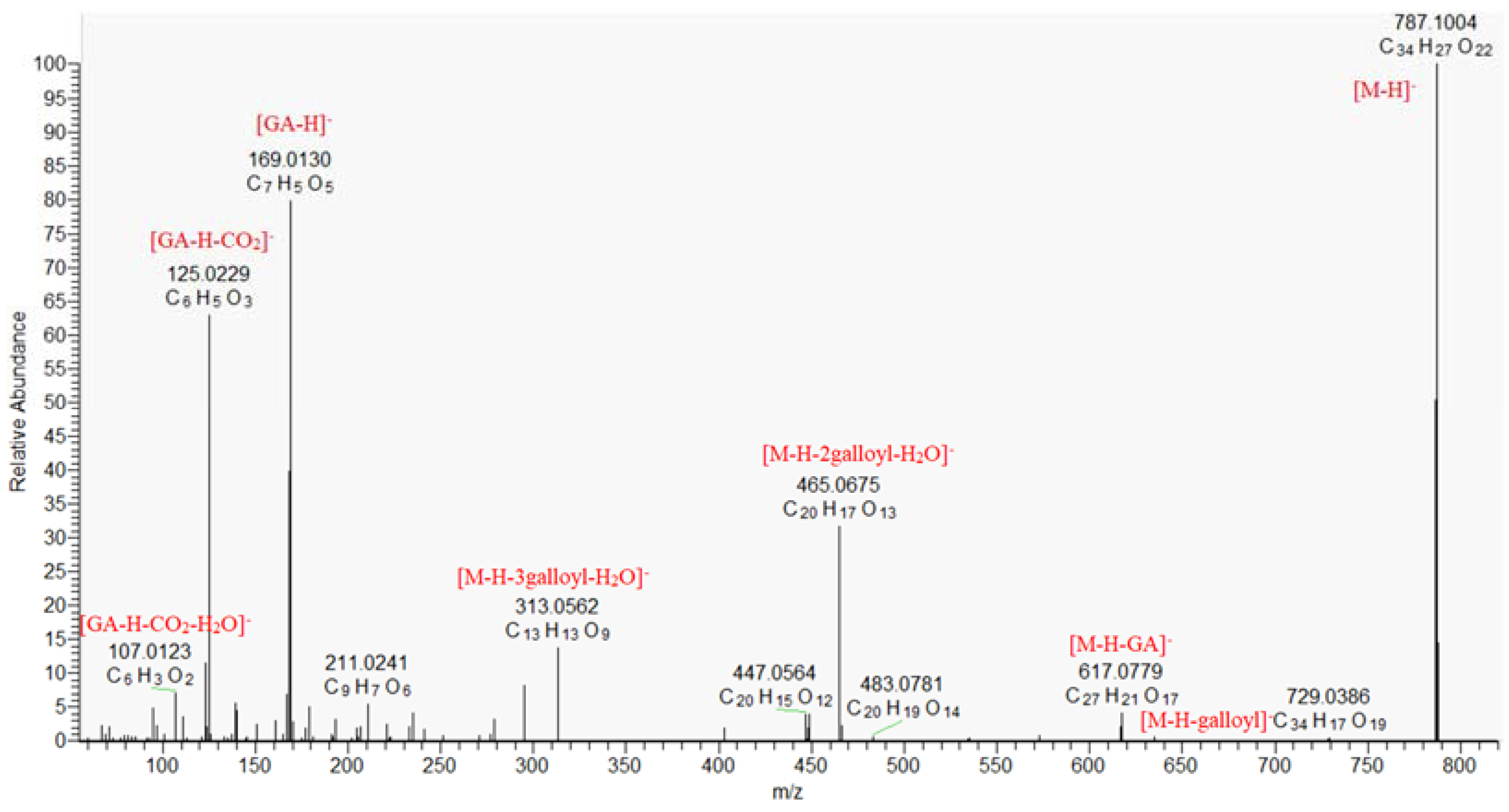
Hydroxybenzoic acids (1 and 3), a pentoside (2), and caffeic acid (4) were identified based on comparison of retention times, exact masses, and fragment spectra with reference standards and literature data (Table 1) [25]. Compound 5 ([M − H] at m/z 247.025, C12H8O6) gave a series of low abundant ions at m/z 219.030 [M − H-CO], 191.034 [M − H-2CO], 163.039 [M − H-3CO], 135.044 [M − H-4CO], and 107.049 [M − H-5CO] (Table 1). Based on the comparison with date from the literature, 5 was annotated as brevifolin [26]. Luteolin (6) was also identified. The identification of the aglycone was determined based on a series of fragment ions at m/z 285.041 [Lu-H], 255.030 [Lu-H-CH2O], 257.042 [Lu-H-CO], 241.051 [Lu-H-CO2], 227.034 [Lu-H-CH2O-CO], 211.039 [Lu-H-H2O-2CO], together with RDA ions 1,3B at m/z 133.029, 1.3A at m/z 151.003 and 0.4A at m/z 107.012 and comparison with authentic standard (Table 1). All compounds except brevifolin were found previously in K. africana [1].

2.1.2. Gallotannins

Herein eight compounds were tentatively identified as gallic acid derivatives. They were esters of gallic acid and polyols, usually hexose. Five gallotannins were identified as monogalloyl hexose (7 and 8), digalloyl hexose (11), trigalloyl hexose (12), and tetragalloyl hexose (13). They showed the characteristic fragment ions in their product ion spectra by consecutive elimination of galloyl and gallate moieties. Tetragalloyl hexose (13), [M − H] at m/z 787.0999 produced a loss of galloyl residue (−152.016 Da) to trigalloyl hexose (at m/z 635.0884), and fragment ions at m/z 617.080 [M − H-GA], 465.067 [M − H-2galloyl-H2O], 313.057 [M − H-3galloyl-H2O] (Table 1, Figure 1). Fragment ions at m/z 271.0466, 169.0145 and 125.0250 were observed as cross-ring fragment ions of a hexose molecule, and deprotonated and decarboxylated ions of the gallic acid moieties, respectively [27]. Compound 9 [M − H] at m/z 169.0142 was identified as gallic acid (GA), based on fragment ions at m/z 125.023 [GA-H-CO2] and 107.012 [GA-H-H2O-CO2], and was further confirmed with an authentic standard [28]. Compound 10 differed from 9 by a CH2 group, demonstrated similar fragmentation pathway and was ascribed to methyl gallate [29].
However, preliminary phytochemical profiling revealed the presence of tannins in the K. africana stem bark, this is the first report on the presence of gallic acid and gallotannins [30]. Gallic acid was previously found in the stem bark of other Bignoniaceae species as Mansoa alliacea (Lam.) AHGentry [31], while tannins were reported for Stereospermum kunthianum Cham [32].

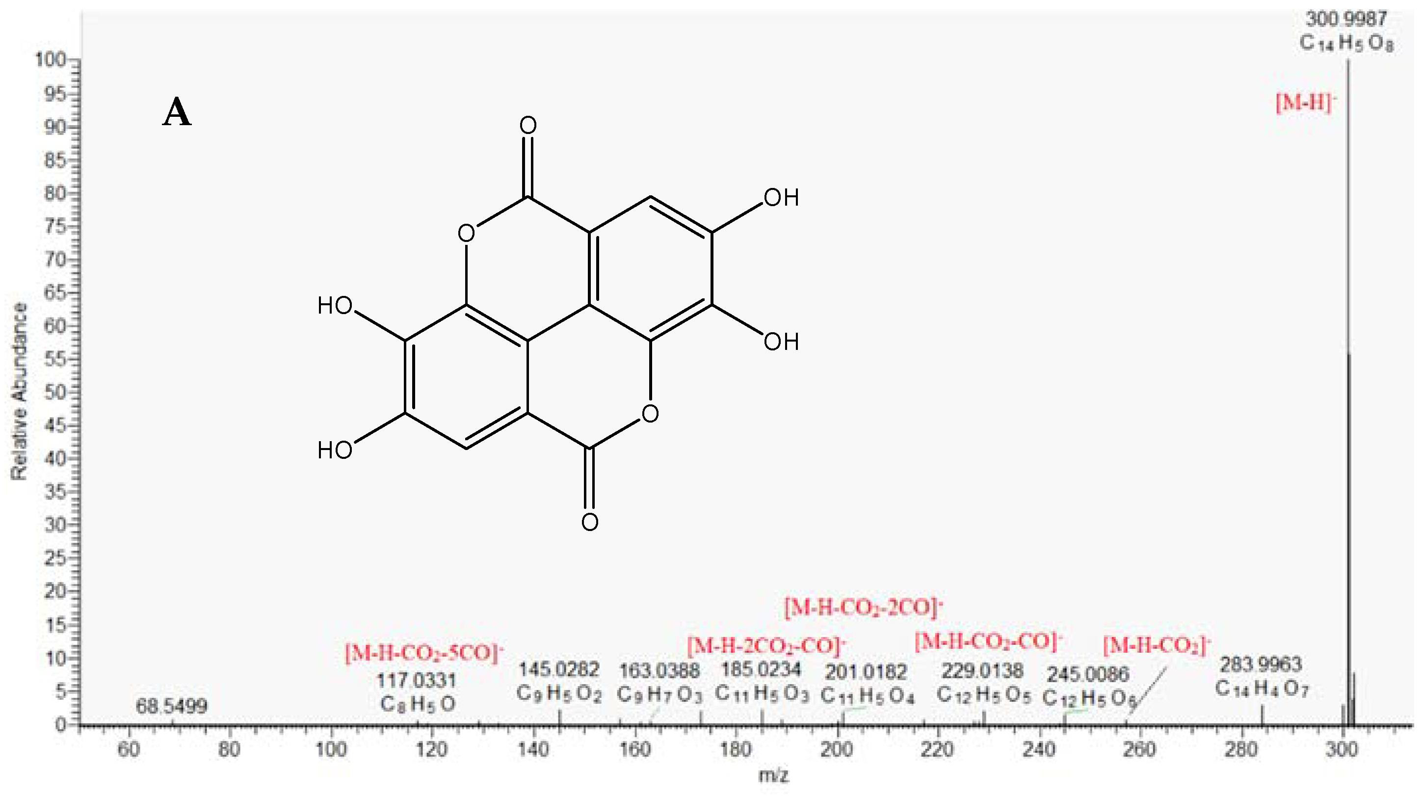
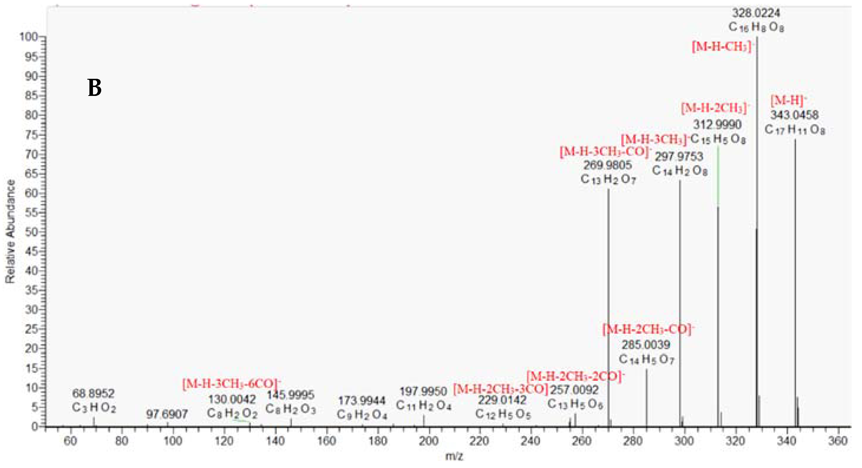
2.1.3. Ellagitannins

Ellagitannins (ETs) are different combinations of gallic acid, hexahydroxydiphenic acid and its dilactone ellagic acid with hexose, existing in a wide range of states and forms such as monomers (i.e., ellagic acid glycosides), oligomers, and complex polymers. Herein, thirty-one compounds were dereplicated as ellagic acid derivatives. Compound 36 (C14H6O8), [M − H] at m/z 300.9990 gave fragment ions at m/z 257.007 [M − H-CO2], 229.013 [M − H-CO2-CO], 201.018 [M − H-CO2-2CO], 185.023 [M − H-2CO2-2CO]. Thus 36 was identified as ellagic acid and was further confirmed by comparison with authentic standard (Table 1). Compounds 38 and 40 (C15H8O8), ([M − H] at m/z 315.0146) differed from 36 by a CH2 group and gave an abundant ion at m/z 299.991 [M − H-CH3]. Thus, 38 and 40 were dereplicated as methylellagic acid [29] (Table 1). Similarly, 4143 and 44 were tentatively identified as dimethyl- and trimethylellagic acid, respectively (Figure 2, Table 1). Compound 22 (C21H10O13) differed from ellagic acid with C7H4O5 moiety and gave a fragment ion at m/z 299.99, corresponding to the loss of deprotonated gallic acid. Therefore, 22 was tentatively dereplicated as valoneic acid dilactone (Table 1). Similarly, 29 was related to valoneic acid dilactone methyl ester.
The fragmentation pathways of 30, 33, and 35 showed neutral mass losses of hexose (162.053 Da), while 34, 37, and 39 demonstrated neutral mass losses of pentose (132.043 Da). Consequently, the aglycone of 30, 33, and 34 was registered at m/z 300.9987, and compounds were ascribed as glycosides of ellagic acid. Compounds 35/37 and 39 were identified as glycosides of methylellagic ([Agl-H] at m/z 315.0145) and dimethylellagic acid ([Agl-H] at m/z at 329.0271), respectively.
MS/MS spectra of compound 14 and 15 revealed fragment ion at m/z 421.04 [M − H-hex-H2O], m/z 300.999 corresponding to a deprotonated ellagic acid, and m/z 257.009 by decarboxylation of the ellagic acid. Hence, compounds were identified as the hexahydroxydiphenic acid (HHDP), esterified with two OH groups of a hexose (HHDP-hexose) (Table 1) [29]. Similarly, compounds 16 and 23, [M − H] at m/z 783.0686 were identified as diHHDP-hexose (pedunculagin) isomers, while 17 was ascribed to castalin/vescalin [29] (Table 1). Compounds 24/26 differed from 14/15 with a galloyl residue and were related to galloyl-HHDP-hexose (Table 1). Compound 32 [M − H] at m/z 600.9896 gave fragment ion at m/z 300.999 [M − H-2galloyl-2H] and demonstrated similar to ellagic acid MS/MS spectrum. Thus, 32 was ascribed as gallagic acid dilactone (terminalin), a condensation between ellagic acid and two gallic acids with lactone structures [29].
Two isobars 18 and 19 [M − H] at m/z 781.0530 were identified as punicalin isomers (α/β-isomers) based on comparison with literature data [29]. They gave fragment ions at m/z 600.990 [M − H-hex-H2O], 448.979 [M − H-hex-H2O-galloyl], and 420.984 [M − H-hex-H2O-galloyl-CO], revealing a hexoside and ether bond of the gallic acid residues. Analogically, 20 was tentatively identified as galloylpunicalin [29].
Three isobars 21, 25, and 28 shared the same [M − 2H]2− at m/z 541.0260, corresponding to the doubly-charged punicalagin isomers ions, respectively [28]. Previously, Falode et al., 2017 found that the content of ellagic acid reach up to 4.01 ± 0.01 mg/g in the K. africana leaves extract [16]. However, this is the first report on the composition of ellagitannins in K. africana stem bark.

2.1.4. Phenylethanoid Glycosides and Iridoids

Three phenylethanoid glycosides (45, 47, and 48), and two iridoid glycosides (46 and 49) were annotated in the studied K. africana stem bark extract. The MS/MS spectra of compounds 47 and 48 revealed similar fragmentation patterns with ions at m/z 461.167 [M − H-caffeoyl], 315.108 [M − H-caffeoyl-dHex], and 153.054 [M − H-caffeoyl-dHex-Hex], together with fragment ions of caffeic acid at m/z 179.033 [CA-H], 161.023 [CA-H-H2O], 135.043 [CA-H-CO2]. Based on the comparison of MS/MS spectra and retention times to literature data, compounds 47/48 were related to verbascoside/acteoside (Table 1) [33]. Analogically, but with an additional glycosyl residue, compound 45 was annotated as echinacoside [14]. The phenylethanoid glycosides were previously found in K. africana fruits.
Compound 46 ([M − H] at m/z 523.1457) gave fragment ions at 363.094 [M − H-hex], 343.083 [M − H-hex-H2O], as well as fragments at m/z 179.033, 161.023, and 135.043, corresponding to the presence of caffeoyl residue. Thus, 46 was dereplicated as verminoside, formerly isolated from K. africana fruits [8].
The MS/MS spectrum of compound 49 ([M − H]- at m/z 481.1715) revealed subsequent losses of hexosyl moiety (−162.053) at m/z 319.119, CH3 group at m/z 304.095, and fragment ions at m/z 138.031 corresponding to the hydroxybenzoic acid (Figure 3, Table 1). Hence, 49 was tentatively annotated as methoxybenzoylajugol, previously isolated from the stem bark of Tabebuia avellanedae, Bignoniaceae [34].

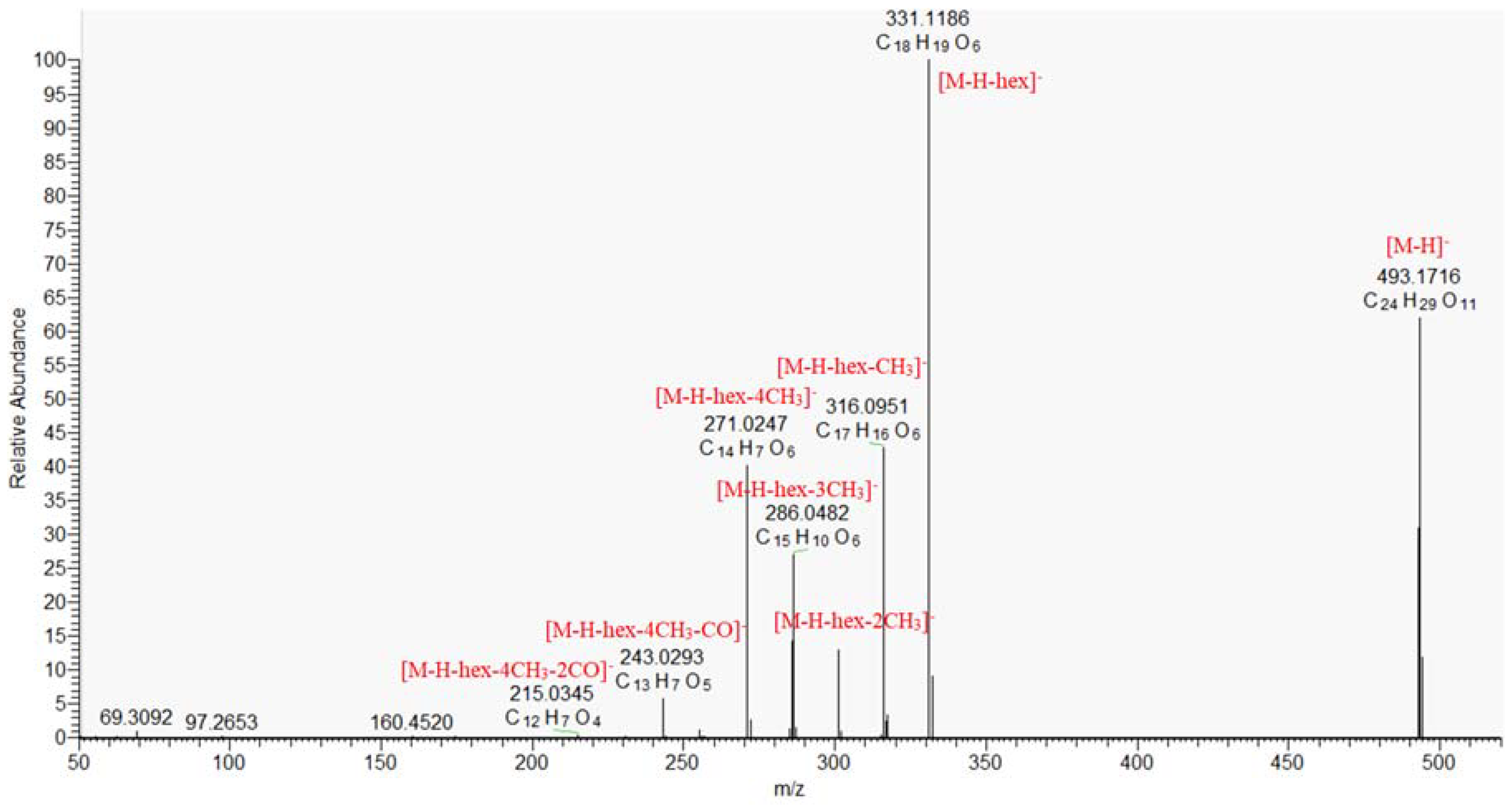
2.1.5. Naphthoquinones and Anthracene Derivatives

Monoterpenoid naphthoquinones (pinnatal, isopinnatal, kigelinol and isokigelinol) are specific for K. africana and other Bignoniaceae species [20]. A key step in the dereplication/annotation of naphto- and anthraquinones was a series of losses of 18 and 28 Da from hydroxyl (OH) and carbonyl (CO) groups on the benzene ring [35], as well as losses of 26 and 44 Da resulted from degradation of the benzene ring (C2H2) and cleavage of the carboxyl group (CO2), respectively [36]. Three isobars 55, 57, and 60 shared the same [M − H] at m/z 337.1082. They gave fragment ions at m/z 309.113 [M − H-CO], 307.097 [M − H-CH2O], 279.066 [M − H-CH2O-C2H2]. Additional indicative ion at m/z 237.055 (C15H9O3) corresponded to the presence of hydroxymethylanthracene-9,10-dione structure. Consequently, 55, 57, and 60 were ascribed to pinnatal and isopinnatal, previously isolated from K. africana root and stem bark [6,37] and sterekunthal A, found in the root bark of Stereospermum kunthianum (Bignoniaceae) [38]. Compounds 59/62 were tentatively identified as kigelinol/isokigelinol based on the deprotonated molecule at m/z 307.0976 (C19H16O4) and fragments at m/z 289.087 [M − H-H2O], 274.063 [M − H-H2O-CH3], and 261.091 [M − H-H2O-CO], typical for naphto- and anthraquinones. In addition, the significant ion at m/z 246.068 [M − H-H2O-CO-CH3] (C17H10O2) corresponded to the presence of condensed structure of anthraquinone and dihydrocyclopentane [39]. Compound 50 ([M − H] at m/z 237.004) gave a series of fragments at m/z 193.013 [M − H-CO2], 149.023 [M − H-2CO2], 121.028 [M − H-2CO2-CO], 93.033 [M − H-3CO2-CO], 77.038 [M − H-3CO2-2CO] and was tentatively annotated as pentahydroxy-1,4-naptoquinone (Table 1). The MS/MS spectrum of 51/54 ([M − H at m/z 285.077) revealed a consecutive loss of two CH3 and a two CO groups at m/z 270.053 [M − H-CH3], 255.029 [M − H-2CH3], 227.034 [M − H-2CH3-CO], and 199.039 [M − H-2CH3-2CO]. Additionally, the fragment at m/z 255.029 (C14H7O5) corresponded to tri-substituted anthraquinone. Hence, 51/54 were related to dimethoxy-hydroxyanthraquinone (Table 1). Similarly, 52, 56, 58/63, and 61 were tentatively ascribed to dehydroanthraquinones substituted with a variety number of methoxy and hydroxy groups (Table 1). The MS/MS spectrum of 53 gave a base peak at m/z 331.119, resulting from the loss of hexose (−162.053 Da) and successive losses of four CH3 groups. Therefore, 53 were annotated as hydroxy-tetramethoxy-dihydroanthraquinone-O-hexoside (Figure 4, Table 1).
All compounds except pinnatal, isopinnatal, kigelinol, and isokigelinol are reported for the first time in K. africana. As indicated by the UHPLC analysis, the studied profile of the K. africana extract was dominated by the following compounds: trimetylellagic acid (44), dimethylellagic acid isomers (41 and 42) (8.82 and 8.72%), ellagic acid (36) (6.27%), hydroxy-tetramethoxy-dihydroanthraquinone-O-hexoside (53) (6.30%), punicalgin α/β/γ (28) (4.42%), and methylellagic acid isomers (38 and 40) (4.22 and 4.20%).

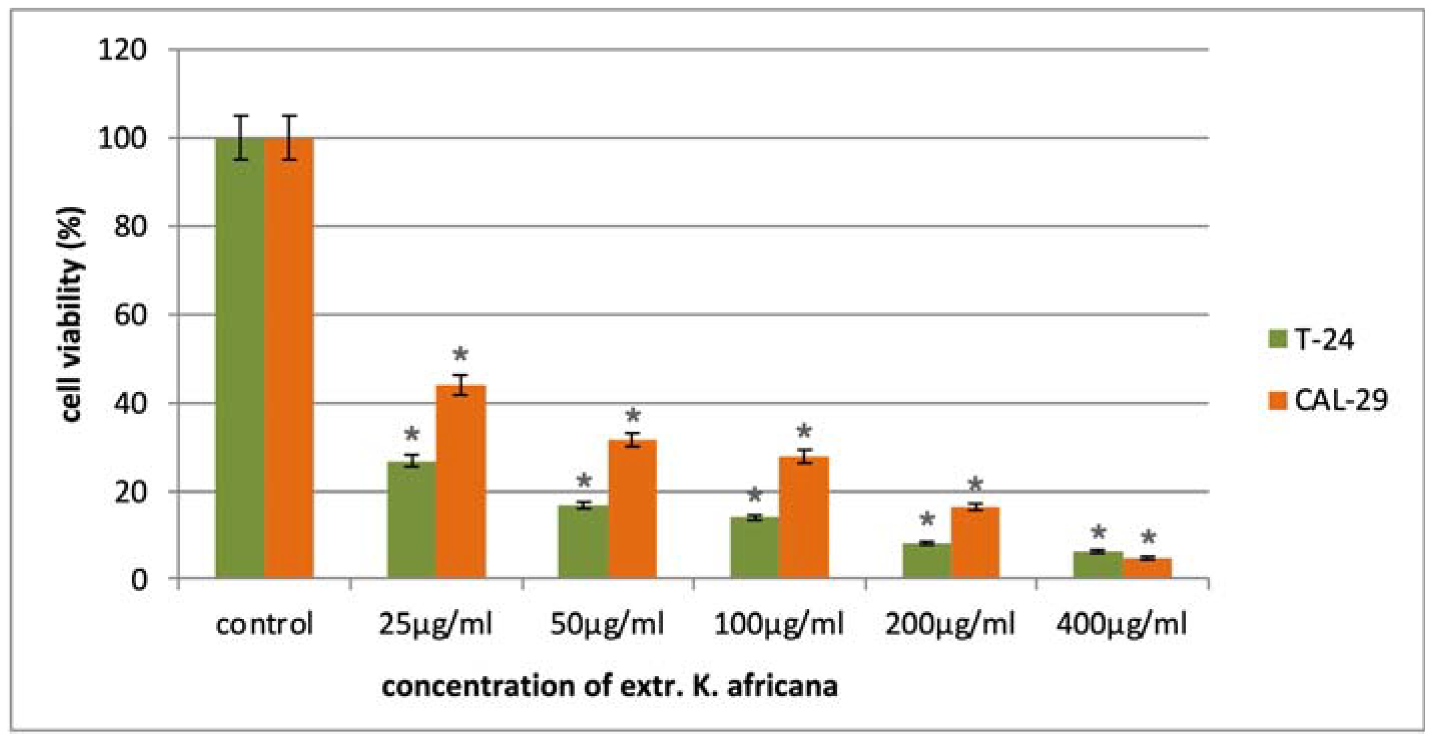
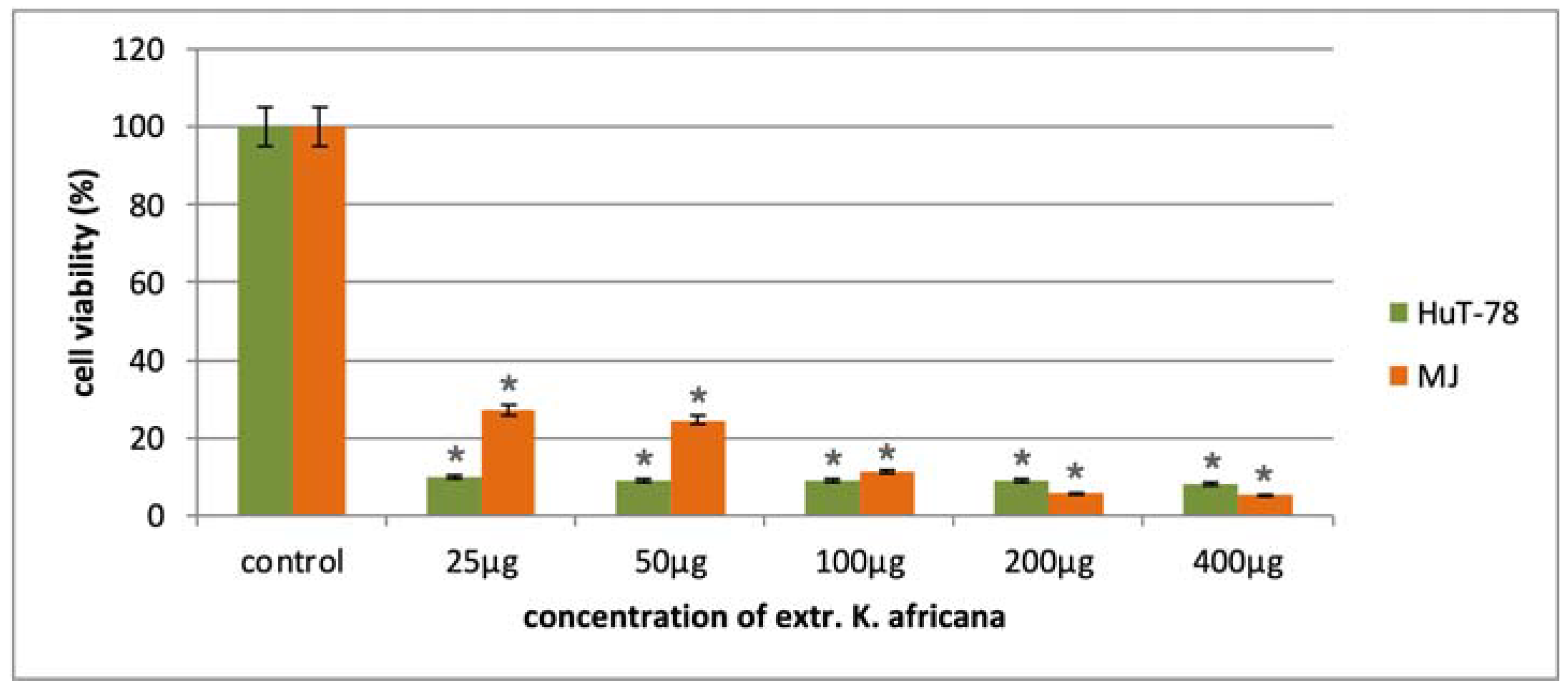
2.2. In Vitro Cytotoxicity of K. africana Stem Bark Extract

The anticancer activity of the K. africana stem bark extract was evaluated in a panel of malignant human cell lines of different origin in the concentration range of 25–400 µg/mL. The antiproliferative effects of the extract were monitored after 72 h exposure to five serial concentrations using a standard MTT-based colorimetric assay for assessing cell viability. The obtained data were fitted to “concentration–effect” curves by means of non-linear regression (GraphPad Prism 8.0 software) and the corresponding equi-effective concentrations (IC50) were calculated. In all tumor models, we established a pronounced inhibition of cell growth in a mostly dose-dependent manner (Figure 5, Figure 6 and Figure 7), with IC50 values ranging near and well below (4–30 µg/mL) the lowest treatment concentration of 25 µg/mL (Table 2). Furthermore, in all malignant models was observed a marked tumor selectivity in the cytostatic effects of the K. africana extract, as indicated by the calculated selectivity indices (SI, the ratio of the IC50 for the normal HEK-293 cells to the IC50 for the correspondent malignant cell line), ranging from about 8 to over 50. A further analysis of the experimental results revealed some variations in EKA performance depending on the origin and type of the screened tumor models.
In the cytotoxicity screenings, the two breast cancer models (TNBC MDA-MB-231 cells and hormone-responsive MCF-7 cell line) showed the lowest chemosensitivity to the Kigelia extract with almost identical IC50 values of about 30 µg/mL and a selectivity index of about 8 (Table 2). In the tested concentration range, both cell lines exhibited very similar cytotoxicity profiles, displaying significant deviation only at the exposure level of 200 µg/mL. At this concentration, the growth-inhibitory effect of the EKA was about seven times stronger against the hormone-sensitive MCF-7 tumor model, as compared to the more resilient TNBC subtype.
In the other pair of epithelial malignancies (T-24 and CAL-29 urothelial carcinomas), comparing cell viability profiles revealed a more defined trend in the mean potency of the tested extract. Accordingly, the anticancer activity of EKA is nearly twice higher against the T-24 tumor model at all but the highest treatment concentrations (25 µg/mL, 50 µg/mL, 100 µg/mL and 200 µg/mL). As a result, more than a two-fold difference was observed in the estimated IC50 values (10.2 µg/mL for T-24 and 24.8 for CAL-29 cells, respectively) and the correspondent SIs (23.3 for the T-24 cells and 9.6 for the CAL-29 model). The lower chemosensitivity displayed by the CAL-29 carcinoma model is most likely due to its high expression of the multidrug resistance (MDR1) transporter, which is known to facilitate the cellular efflux of various xenobiotics, including phytochemicals. In addition, CAL-29 cells originate from a patient with primary lesion of a fatally invasive and much more aggressive metastatic transitional cell carcinoma of the bladder (grade IV, stage T2) [40].
As shown in Figure 7 and Table 2, the antineoplastic activity of the EKA was most pronounced in the two models of cutaneous T-cell lymphoma (CTCL), namely Mycosis fungoides (MJ cell line) and Sézary syndrome (HUT-78 cells), exhibiting IC50 values of 11.50 µg/mL and 4.63 µg/mL, respectively. Accordingly, the estimated selectivity indices for the lymphoma models are of the order of tens (51.6 for the HUT-78 and 20.6 for the MJ cell line). The HUT-78 T-lymphocytes demonstrated a nearly twice higher susceptibility, which is particularly evident at the two lowest treatment concentrations (25 and 50 µg/mL).
The primary data from the conducted cytotoxicity screenings testify to the extremely high potential of the K. africana extract as an antineoplastic remedy of natural origin. Calculated IC50 values for all tumor models ranged in the low µg/mL range, qualifying it as a highly potent plant extract, matching or far exceeding the in vitro activity of some of the most toxic plant sources, including Catharanthus roseus, Annona muricata, and Curcuma longa [41,42,43]. Even more valuable in practical terms, the studied extract showed explicit selectivity in its cytotoxic action towards all malignant cell types, with extremely favorable selectivity indices invariably higher than 7–8 (a SI of at least 2 is considered as merely favorable). Moreover, the observed lack of toxicity towards healthy HEK-293 cells is well in line with the results of our previous study [23], evaluating the in vivo effects of the same extract on Lewis lung carcinoma (LLC) bearing mice at far greater exposure levels. The established cytotoxicity profile of the K. africana extract, highly biased toward malignantly transformed but not normal cells, suggests specific modulation of defined molecular tumor targets, which will be the subject of future investigation, aiming to further elucidate the relationship between the phytochemical composition and antitumor activity of the extract.
Our findings support previous studies of the anticancer activity of the plant towards other tumor cell lines. Arkhipov et al., 2014 [5] reported the significant antiproliferative activity of K. africana fruit extracts against Jeg-3 choriocarcinoma cells. The methanol and water extracts displayed the strongest cytostatic activity, inhibiting Jeg-3 growth to 42% and 46% compared to the untreated control group, respectively [5]. Cytotoxic activity for K. africana fruit extracts has also been reported against a melanoma and two breast cancer cell lines. The same study also identified several metabolites, including demethylkigelin, kigelin, ferulic acid, and 2-(1-hydroxyethyl)-naphtho[2,3-b] furan-4,9-dione as inhibiting cancer cell proliferation. The compound 2-(1-hydroxyethyl)-naphtho[2,3-b] furan-4,9-dione was reported to be a particularly strong anticancer agent [15]. Crude dichloromethane extracts of stem bark and fruit showed cytotoxic activity in vitro against cultured melanoma and other cancer cell lines (G361, StML11a, C32, ACHN, Colo 668, CHO) using the sulphorhodamine B assay. Thin layer chromatography examination of the most active fractions of both stem bark and fruits showed the presence of the same major components which were found to be norviburtinal and beta-sitosterol. Norviburtinal was found to be the most active compound but had little selectivity for melanoma cell lines whilst isopinnatal also showed some cytotoxic activity [44]. Recently, the anticancer activity of 1,8-dihydroanthraquinone derivatives in in vitro and in vivo breast cancer models were comprehensively analyzed. Mechanistic studies have established that these compounds exert their anti-breast cancer activities through a wide array of molecular targets and mechanisms, including the modulation of angiogenesis, apoptotic pathways, autophagy, synthesis and repair genes expression, damage response, cell cycle regulators, EMT markers, epigenetic mechanisms, heat shock response, inflammation, metastasis-related markers, oxidative status, miRNA, protein synthesis, proliferation, or stem-like markers [45]. Therefore, the annotated naphthoquinones and anthracene derivatives (5063) are likely to be at least partly accountable for the established anticancer activity of the studied extract. Similarly to anthracyclines, naphthoquinones feature a conjugated cyclic dione structure that enables them to serve as electron donors and acceptors and produce extremely toxic hydroxyl radicals leading to lipid peroxidation and ferroptosis in breast, prostate and bladder carcinomas, among others [46,47]. Tricyclic anthracene derivatives, on the other hand, bear a planar polycyclic structure and can readily intercalate into the DNA matrix, thus interfering with topoisomerase II activity during DNA replication [48]. Furthermore, along with other chemically related derivatives, anthracenes and naphthoquinones have been recognized as typical substrates of the MDR1 efflux pump, which would explain the lower activity of the extract in the MDR1 positive CAL-29 bladder carcinoma model [49].
However, the dominating compounds in the studied K. africana extract were trimethylellagic acid (44), other ellagic acid derivatives (36, 40, 41, 42), and ellagitannins (28). Wardana et al., 2022 [50] reported the anticancer activity of 3,4,3′-tri-O-methylellagic acid isolated from Syzygium polycephalum. The in vitro evaluation showcased the inhibition activity of compound towards the T47D and HeLa cell lines with EC50 values of 55.35 ± 6.28 μg mL−1 and 12.57 ± 2.22 μg mL−1, respectively. Moreover, the in silico evaluation aimed to elucidate the interaction of tri-O-methylellagic acid with enzymes responsible for cancer regulation at the molecular level by targeting the hindrance of cyclin-dependent kinase 9 (CDK9) and sirtuin 1 (SIRT1) enzymes [50]. Numerous in vitro and in vivo studies in different cancer cell lines revealed the antiproliferative properties of ellagic acid [15,51,52,53,54,55]. The antiproliferative action of ellagic acid could be mediated for its ability to directly inhibit the DNA binding of certain carcinogens, including nitrosamines [56] and polycyclic aromatic hydrocarbons [57]. In addition, de Molina et al., 2014 [58] reported that 4,4′-di-O-methylellagic acid was the most effective compound in the inhibition of colon cancer cell proliferation among other chemically related compounds. 4,4′-di-O-methylellagic acid was highly active against colon cancer cells resistant to the chemotherapeutic agent 5-fluoracil, whereas no effect was observed in non-malignant colon cells [58].

3. Materials and Methods

3.1. Plant Material

K. africana plant material (stem bark) was collected at Durban, South Africa in December 2018 and identified by one of the authors (N.S.) A voucher specimen (1/30.01.2025) was deposited at the Bews Herbarium (NU), School of Life Sciences, of University of KwaZulu-Natal, Durban, South Africa. The plant material was dried at room temperature.

3.2. Sample Extraction

Air-dried grounded stem bark (100 g) was extracted twice with 80% MeOH (1:20 w/v) by sonication (80 kHz, ultra-sound bath Biobase UC-20C, Jinan, China) for 15 min at room temperature. The extracts were concentrated in vacuo and subsequently lyophilized (lyophilizer Biobase BK-FD10P, Jinan, China) to yield crude extracts of 10.20 g.

3.3. Chemicals

Acetonitrile (hypergrade for LC–MS), formic acid (for LC–MS), and methanol (an-alytical grade) were purchased from Chromasolv (Sofia, Bulgaria). The reference standards used for compound identification were obtained from Extrasynthese (Genay, France) for gallic, gentisic, p-hydroxybenzoic acids, and luteolin. Ellagic and caffeic acids were supplied from Phytolab (Vestenbergsgreuth, Bavaria, Germany). MTT was provided from BLD Pharmatech GmbH (Kaiserslautern, Germany).

3.4. UHPLC-HRMS

The UHPLC-HRMS analyses were carried out on a Q Exactive Plus mass spectrometer (ThermoFisher Scientific, Inc., Waltham, MA, USA) equipped with a heated electrospray ionization (HESI-II) probe (ThermoScientific). The equipment was operated in negative and positive ion modes within the m/z range from 100 to 1000. The mass spectrometer parameters were as follows: spray voltage 3.5 kV (+) and 2.5 kV (−); sheath gas flow rate 38; auxiliary gas flow rate 12; spare gas flow rate 0; capillary temperature 320 °C; probe heater temperature 320 °C; S-lens RF level 50; scan mode: full MS (resolution 70,000), and MS/MS (17,500). The chromatographic separation was achieved on a reversed phase column Kromasil EternityXT C18 (1.8 µm, 2.1 × 100 mm) at 40 °C. The UHPLC analyses were run with a mobile phase consisting of 0.1% formic acid in water (A) and 0.1% formic acid in acetonitrile (B). The run time was 33 min. The flow rate was 0.3 mL/min. The gradient elution program was used as follows: 0–1 min, 0–5% B; 1–20 min, 5–30% B; 20–25 min, 30–50% B; 25–30 min, 50–70% B; 30–33 min, 70–95%; 33–34 min 95–5% B. Equilibration time was 4 min [25]. Data were processed by Xcalibur 4.2 (ThermoScientific, Waltham, MA, USA) instrument control/data handling software.

3.5. MTT Cell Viability Assay

The antiproliferative activity of the K. africana stem bark extract was assessed against normal embryonic kidney cells (HEK-293), as well as malignant human cell lines of different origin (hormone-responsive breast carcinoma cell line, MCF-7; triple negative breast carcinoma cells, MDA-MB-231; urothelial bladder carcinoma cell lines, T-24, CAL-29; cutaneous T-cell lymphoma cell lines, HUT-78, MJ). The antineoplastic activity of the extract was measured using a standard MTT-based colorimetric assay for assessing cell viability. According to protocol, exponential-phased cells were harvested by trypsinization and seeded (100 μL/well) in 96-well plates at the appropriate density of at least 1 × 10⁵ cells/mL. After a 24 h incubation, cells were treated with serial dilutions of the tested extract in the concentration range of 25–400 µg/mL. Following a 72 h exposure, filter sterilized MTT substrate solution (5 mg/mL in PBS) was added to each well of the culture plate. A further 2–4 h incubation allowed the reduction of the yellow MTT reagent into purple formazan crystals in metabolically active viable cells, which were dissolved in isopropyl alcohol solution containing 5% formic acid prior to absorbance measurement at 550 nm. Collected absorbance values were blanked against MTT and isopropanol solution and normalized to the mean value of untreated control (100% cell viability).

3.6. Statistical Analysis

The experiments for the evaluation of antiproliferative activity were performed in triplicate and the results were presented as mean and standard deviation. The obtained data were fitted to “concentration-effect” curves by means of non-linear regression (GraphPad Prism 8.0 software) and the corresponding equi-effective concentrations (IC50) were calculated. Statistical analysis of the data was performed via a one-way ANOVA (* p values ≤ 0.01 are considered statistically significant.

4. Conclusions

The study presents a comprehensive metabolite profiling of the K. africana stem bark extract by means of ultra-high-performance liquid chromatography—Orbitrap high-resolution mass spectrometry. A total of 63 secondary metabolites, noticeably including phenolic acids, gallo- and ellagitannins, iridoids, naphthoquinone, and anthraquinones, were dereplicated/annotated. Herein, the majority of annotated gallo- and ellagitannins were reported for the first time in K. africana. In all of the tested in vitro models, the studied stem bark extract showed substantial concentration dependent antineoplastic activity, while exhibiting marked tumor selectivity with SIs ranging between 8 and 51. The strongest inhibitory effect of the extract was found on the growth of non-invasive urinary bladder T-24 cancer cells and CTCL-derived malignant cells with estimated IC50 concentrations of less than 12 µg/mL, while the breast carcinomas and the invasive bladder carcinoma cell line were slightly less responsive. The isolation of the main compounds and their pharmacological investigation as potential candidate with antiproliferative activity may be a subject of further complementary studies.

Author Contributions

Conceptualization, S.K. and N.S.; methodology, D.Z.-D., R.M. and M.N.; software, D.Z.-D. and M.N.; validation, R.M., M.N. and S.K.; formal analysis, D.Z.-D.; investigation, S.K. and N.S.; resources, N.S.; data curation, D.Z.-D., M.N. and R.M.; writing—original draft preparation, D.Z.-D. and R.M.; writing—review and editing, S.K.; visualization, D.Z.-D.; supervision, S.K.; project administration, D.Z.-D.; funding acquisition, D.Z.-D. All authors have read and agreed to the published version of the manuscript.

Funding

This research is financed by the European Union—NextGenerationEU, through the National Recovery and Resilience Plan of the Republic of Bulgaria, project BG-RRP-2.004-0004-C01 “Strategic research and innovation program for development of Medical University—Sofia”.

Institutional Review Board Statement

Not applicable.

Informed Consent Statement

Not applicable.

Data Availability Statement

The original contributions presented in the study are included in the article; further inquiries can be directed to the corresponding author/s.

Conflicts of Interest

The authors declare no conflicts of interest.

References

  1. Bello, I.; Shehu, M.W.; Musa, M.; Zaini Asmawi, M.; Mahmud, R. Kigelia africana (Lam.) Benth. (Sausage Tree): Phytochemistry and Pharmacological Review of a Quintessential African Traditional Medicinal Plant. J. Ethnopharmacol. 2016, 189, 253–276. [Google Scholar] [CrossRef] [PubMed]
  2. Vicidomini, C.; Palumbo, R.; Moccia, M.; Roviello, G.N. Oxidative Processes and Xenobiotic Metabolism in Plants: Mechanisms of Defense and Potential Therapeutic Implications. JoX 2024, 14, 1541–1569. [Google Scholar] [CrossRef] [PubMed]
  3. Falode, J.A.; Akinmoladun, A.C.; Olaleye, M.T. Ameliorative Property of Kigelia Africana Crude and Flavonoid Leaf Extracts on Aluminum-Induced Hepatotoxicity in Albino Rats. Comp. Clin. Pathol. 2019, 28, 1495–1506. [Google Scholar] [CrossRef]
  4. Akunyili, D.N.; Houghton, P.J.; Raman, A. Antimicrobial Activities of the Stembark of Kigelia Pinnata. J. Ethnopharmacol. 1991, 35, 173–177. [Google Scholar] [CrossRef]
  5. Arkhipov, A.; Sirdaarta, J.; Matthews, B.; Cock, I.E. Metabolomic Profiling of Kigelia Africana Extracts with Anti-Cancer Activity by High Resolution Tandem Mass Spectroscopy. Pharmacogn. Commun. 2014, 4, 10–32. [Google Scholar]
  6. Akunyili, D.N.; Houghton, P.J. Meroterpenoids and Naphthaquinones from Kigelia Pinnata. Phytochemistry 1993, 32, 1015–1018. [Google Scholar] [CrossRef]
  7. Khan, M.F.; Dixit, P.; Jaiswal, N.; Tamrakar, A.K.; Srivastava, A.K.; Maurya, R. Chemical Constituents of Kigelia Pinnata Twigs and Their GLUT4 Translocation Modulatory Effect in Skeletal Muscle Cells. Fitoterapia 2012, 83, 125–129. [Google Scholar] [CrossRef]
  8. Gouda, Y.G.; Abdel-baky, A.M.; Darwish, F.M.; Mohamed, K.M.; Kasai, R.; Yamasaki, K. Iridoids from Kigelia Pinnata DC. Fruits. Phytochemistry 2003, 63, 887–892. [Google Scholar] [CrossRef]
  9. Houghton, P.J.; Jâger, A.K. The Sausage Tree (Kigelia pinnata): Ethnobotany and Recent Scientific Work. South Afr. J. Bot. 2002, 68, 14–20. [Google Scholar] [CrossRef]
  10. Jabeen, B.; Riaz, N. Isolation and Characterization of Limonoids from Kigelia Africana. Z. Für Naturforschung B 2013, 68, 1041–1048. [Google Scholar] [CrossRef]
  11. Govindachari, T.R.; Patankar, S.J.; Viswanathan, N. Isolation and Structure of Two New Dihydroisocoumarins from Kigelia pinnata. Phytochemistry 1971, 10, 1603–1606. [Google Scholar] [CrossRef]
  12. Inoue, K.; Inouye, H.; Chen, C.-C. A Naphthoquinone and a Lignan from the Wood of Kigelia pinnata. Phytochemistry 1981, 20, 2271–2276. [Google Scholar] [CrossRef]
  13. Joshi, K.C.; Singh, P.; Taneja, S.; Cox, P.J.; Allan Howie, R.; Thomson, R.H. New Terpenoid Aldehydes from Kigelia pinnata: Crystal Structure of Pinnatal. Tetrahedron 1982, 38, 2703–2708. [Google Scholar] [CrossRef]
  14. Gouda, Y.G.; Abdel-baky, A.M.; Mohamed, K.M.; Darwish, F.M.; Kasai, R.; Yamasaki, K. Phenylpropanoid and Phenylethanoid Derivatives from Kigelia pinnata DC. Fruits. Nat. Prod. Res. 2006, 20, 935–939. [Google Scholar] [CrossRef]
  15. Higgins, C.; Bell, T.; Delbederi, Z.; Feutren-Burton, S.; McClean, B.; O’Dowd, C.; Watters, W.; Armstrong, P.; Waugh, D.; Van Den Berg, H. Growth Inhibitory Activity of Extracted Material and Isolated Compounds from the Fruits of Kigelia pinnata. Planta Med. 2010, 76, 1840–1846. [Google Scholar] [CrossRef] [PubMed]
  16. Falode, J.A.; Akinmoladun, A.C.; Olaleye, M.T.; Akindahunsi, A.A. Sausage Tree (Kigelia africana) Flavonoid Extract Is Neuroprotective in AlCl 3 -Induced Experimental Alzheimer’s Disease. Pathophysiology 2017, 24, 251–259. [Google Scholar] [CrossRef]
  17. Arena, K.; Rigano, F.; Mangraviti, D.; Cacciola, F.; Occhiuto, F.; Dugo, L.; Dugo, P.; Mondello, L. Exploration of Rapid Evaporative-Ionization Mass Spectrometry as a Shotgun Approach for the Comprehensive Characterization of Kigelia africana (Lam) Benth. Fruit. Molecules 2020, 25, 962. [Google Scholar] [CrossRef]
  18. Fagbohun, O.F.; Olawoye, B.; Ademakinwa, A.N.; Oriyomi, O.V.; Fagbohun, O.S.; Fadare, O.A.; Msagati, T.A.M. UHPLC/GC-TOF-MS Metabolomics, MTT Assay, and Molecular Docking Studies Reveal Physostigmine as a New Anticancer Agent from the Ethyl Acetate and Butanol Fractions of Kigelia africana (Lam.) Benth. Fruit Extracts. Biomed. Chromatogr. 2021, 35, e4979. [Google Scholar] [CrossRef] [PubMed]
  19. Swati, D.; Vishwatej, P.; Vinay, S.; Bhupesh, P. LC-MS Analysis of Kigelia Pinnata (JACQ) DC. Root Bark- A Multi-Potent Medicinal Plant. Ayushdhara 2023, 10, 60–67. [Google Scholar] [CrossRef]
  20. Nabatanzi, A.; Nkadimeng, S.M.; Lall, N.; Kabasa, J.D.; McGaw, L.J. Ethnobotany, Phytochemistry and Pharmacological Activity of Kigelia africana (Lam.) Benth. (Bignoniaceae). Plants 2020, 9, 753. [Google Scholar] [CrossRef]
  21. Assanti, G.; Kaur, R.; Nizard, S.; Pollack, E.; Rafferty, B.; Priano, C.; Fernández Romero, J.A.; Koroch, A.R. Biology, Chemistry, and Pharmacological Activity of Kigelia Africana (Bignoniaceae) and Garcinia Kola (Clusiaceae)—A Review. J. Med. Act. Plants 2022, 11, 1–21. [Google Scholar] [CrossRef] [PubMed]
  22. Weiss, C.R.; Moideen, S.V.K.; Croft, S.L.; Houghton, P.J. Activity of Extracts and Isolated Naphthoquinones from Kigelia pinnata against Plasmodium falciparum. J. Nat. Prod. 2000, 63, 1306–1309. [Google Scholar] [CrossRef]
  23. Momekova, D.; Momekov, G.; Pencheva, I.; Konstantinov, S. Antineoplastic Activity of a Methanolic Extract from Kigelia Pinnata DC Stem Bark. J. Cancer Ther. Res. 2012, 1, 17. [Google Scholar] [CrossRef]
  24. Sumner, L.W.; Amberg, A.; Barrett, D.; Beale, M.H.; Beger, R.; Daykin, C.A.; Fan, T.W.-M.; Fiehn, O.; Goodacre, R.; Griffin, J.L.; et al. Proposed Minimum Reporting Standards for Chemical Analysis: Chemical Analysis Working Group (CAWG) Metabolomics Standards Initiative (MSI). Metabolomics 2007, 3, 211–221. [Google Scholar] [CrossRef]
  25. Gevrenova, R.; Zheleva-Dimitrova, D.; Balabanova, V.; Voynikov, Y.; Sinan, K.I.; Mahomoodally, M.F.; Zengin, G. Integrated Phytochemistry, Bio-Functional Potential and Multivariate Analysis of Tanacetum macrophyllum (Waldst. & Kit.) Sch.Bip. and Telekia speciosa (Schreb.) Baumg. (Asteraceae). Ind. Crops Prod. 2020, 155, 112817. [Google Scholar] [CrossRef]
  26. Sinan, K.I.; Zengin, G.; Zheleva-Dimitrova, D.; Etienne, O.K.; Fawzi Mahomoodally, M.; Bouyahya, A.; Lobine, D.; Chiavaroli, A.; Ferrante, C.; Menghini, L.; et al. Qualitative Phytochemical Fingerprint and Network Pharmacology Investigation of Achyranthes Aspera Linn. Extracts. Molecules 2020, 25, 1973. [Google Scholar] [CrossRef]
  27. Hooi Poay, T.; Sui Kiong, L.; Cheng Hock, C. Characterisation of Galloylated Cyanogenic Glucosides and Hydrolysable Tannins from Leaves of Phyllagathis rotundifolia by LC-ESI-MS/MS. Phytochem. Anal. 2011, 22, 516–525. [Google Scholar] [CrossRef] [PubMed]
  28. Mena, P.; Calani, L.; Dall’Asta, C.; Galaverna, G.; García-Viguera, C.; Bruni, R.; Crozier, A.; Del Rio, D. Rapid and Comprehensive Evaluation of (Poly)Phenolic Compounds in Pomegranate (Punica granatum L.) Juice by UHPLC-MSn. Molecules 2012, 17, 14821–14840. [Google Scholar] [CrossRef]
  29. Singh, A.; Bajpai, V.; Kumar, S.; Sharma, K.R.; Kumar, B. Profiling of Gallic and Ellagic Acid Derivatives in Different Plant Parts of Terminalia arjuna by HPLC-ESI-QTOF-MS/MS. Nat. Prod. Commun. 2016, 11, 239–244. [Google Scholar] [CrossRef]
  30. Ss, S.; Sm, D.; Ma, A. Antibacterial and Toxicity Evaluation of Stem Bark Extract of Kigelia africana (Lam.) Benth. Glob. Acad. J. Agri. Biosci. 2022, 4, 86–91. [Google Scholar] [CrossRef]
  31. Pires, F.B.; Dolwitsch, C.B.; Dal Prá, V.; Faccin, H.; Monego, D.L.; Carvalho, L.M.D.; Viana, C.; Lameira, O.; Lima, F.O.; Bressan, L.; et al. Qualitative and Quantitative Analysis of the Phenolic Content of Connarus Var. Angustifolius, Cecropia obtusa, Cecropia palmata and Mansoa alliacea Based on HPLC-DAD and UHPLC-ESI-MS/MS. Rev. Bras. De Farmacogn. 2017, 27, 426–433. [Google Scholar] [CrossRef]
  32. Sarr, A.; Dieng, S.I.M.; Diatta-Badji, K.; Mbaye, A.I.; Diatta, W.; Ka, A.; Fall, A.D.; Bassène, E. Phytochemical Screening and Determination of Polyphenols in the Hydro-Ethanolic Extract of Trunk Bark and Its Fractions of Stereospermum kunthianium Cham (Bignoniaceae). APRJ 2021, 7, 1–9. [Google Scholar] [CrossRef]
  33. Costa, R.; Albergamo, A.; Pellizzeri, V.; Dugo, G. Phytochemical Screening by LC-MS and LC-PDA of Ethanolic Extracts from the Fruits of Kigelia africana (Lam.) Benth. Nat. Prod. Res. 2017, 31, 1397–1402. [Google Scholar] [CrossRef] [PubMed]
  34. Nakano, K.; Maruyama, K.; Murakami, K.; Takaishi, Y.; Tomimatsu, T. Iridoids from Tabebuia Avellanedae. Phytochemistry 1993, 32, 371–373. [Google Scholar] [CrossRef]
  35. Zhan, C.; Xiong, A.; Shen, D.; Yang, L.; Wang, Z. Characterization of the Principal Constituents of Danning Tablets, a Chinese Formula Consisting of Seven Herbs, by an UPLC-DAD-MS/MS Approach. Molecules 2016, 21, 631. [Google Scholar] [CrossRef] [PubMed]
  36. Ye, M.; Han, J.; Chen, H.; Zheng, J.; Guo, D. Analysis of Phenolic Compounds in Rhubarbs Using Liquid Chromatography Coupled with Electrospray Ionization Mass Spectrometry. J. Am. Soc. Mass Spectrom. 2007, 18, 82–91. [Google Scholar] [CrossRef]
  37. Moideen, S.V.K.; Houghton, P.J.; Rock, P.; Croft, S.L.; Aboagye-Nyame, F. Activity of Extracts and Naphthoquinones from Kigelia Pinnata Against Trypanosoma Brucei Brucei and Trypanosoma Brucei Rhodesiense. Planta Med. 1999, 65, 536–540. [Google Scholar] [CrossRef]
  38. Onegi, B.; Kraft, C.; Köhler, I.; Freund, M.; Jenett-Siems, K.; Siems, K.; Beyer, G.; Melzig, M.F.; Bienzle, U.; Eich, E. Antiplasmodial Activity of Naphthoquinones and One Anthraquinone from Stereospermum kunthianum. Phytochemistry 2002, 60, 39–44. [Google Scholar] [CrossRef]
  39. Lazare Sidjui, S.; Zeuko´o Menkem, E.; Toghueo, R.; Noté, O.; Mahiou-Leddet, V.; Herbette, G.; Fekam Boyom, F.; Ollivier, E.; Folefoc, G.N. Secondary Metabolites from Jacaranda mimosifolia and Kigelia africana (Bignoniaceae) and Their Anticandidal Activity. Rec. Nat. Prod. 2014, 8, 307–311. [Google Scholar]
  40. Shkondrov, A.; Krasteva, I.; Ionkova, I.; Popova, P.; Zarev, Y.; Mihaylova, R.; Konstantinov, S. Production of Saponins from in Vitro Cultures of Astragalus glycyphyllos and Their Antineoplastic Activity. Biotechnol. Biotechnol. Equip. 2019, 33, 1413–1418. [Google Scholar] [CrossRef]
  41. Ilango, S.; Sahoo, D.K.; Paital, B.; Kathirvel, K.; Gabriel, J.I.; Subramaniam, K.; Jayachandran, P.; Dash, R.K.; Hati, A.K.; Behera, T.R.; et al. A Review on Annona Muricata and Its Anticancer Activity. Cancers 2022, 14, 4539. [Google Scholar] [CrossRef] [PubMed]
  42. Cosquillo-Rafael, M.F.; Placencia-Medina, M.D.; Miranda-Tomasevich, T.Y.; Moreno-Hinojosa, M.; Retuerto-Figueroa, M.G. Efecto Citotóxico y Genotóxico in Vitro Del Extracto Crudo y Etanólico Del Rizoma de Curcuma longa L. Rev. Peru. Med. Exp. Salud. Publica. 2020, 37, 454–461. [Google Scholar] [CrossRef]
  43. Nisar, A.; Mamat, A.S.; Hatim Mohamed Dzahir, M.I.; Aslam, M.S.; Ahmad, M.S. Antioxidant and Total Phenolic Content of Catharanthus Roseus Using Deep Eutectic Solvent. Recent Adv. Biol. Med. 2017, 03, 7. [Google Scholar] [CrossRef]
  44. Jackson, S.J.; Houghton, P.J.; Retsas, S.; Photiou, A. In Vitro Cytotoxicity of Norviburtinal and Isopinnatal from Kigelia pinnata Against Cancer Cell Lines. Planta Med. 2000, 66, 758–761. [Google Scholar] [CrossRef] [PubMed]
  45. Okon, E.; Gaweł-Bęben, K.; Jarzab, A.; Koch, W.; Kukula-Koch, W.; Wawruszak, A. Therapeutic Potential of 1,8-Dihydroanthraquinone Derivatives for Breast Cancer. IJMS 2023, 24, 15789. [Google Scholar] [CrossRef]
  46. Rahman, M.M.; Islam, M.R.; Akash, S.; Shohag, S.; Ahmed, L.; Supti, F.A.; Rauf, A.; Aljohani, A.S.M.; Al Abdulmonem, W.; Khalil, A.A.; et al. Naphthoquinones and Derivatives as Potential Anticancer Agents: An Updated Review. Chem. Biol. Interact. 2022, 368, 110198. [Google Scholar] [CrossRef]
  47. Wellington, K.W. Understanding Cancer and the Anticancer Activities of Naphthoquinones—A Review. RSC Adv. 2015, 5, 20309–20338. [Google Scholar] [CrossRef]
  48. Malik, M.S.; Alsantali, R.I.; Jassas, R.S.; Alsimaree, A.A.; Syed, R.; Alsharif, M.A.; Kalpana, K.; Morad, M.; Althagafi, I.I.; Ahmed, S.A. Journey of Anthraquinones as Anticancer Agents—A Systematic Review of Recent Literature. RSC Adv. 2021, 11, 35806–35827. [Google Scholar] [CrossRef]
  49. Zhang, X.P.; Ritke, M.K.; Yalowich, J.C.; Slovak, M.L.; Ho, J.P.; Collins, K.I.; Annable, T.; Arceci, R.J.; Durr, F.E.; Greenberger, L.M. P-Glycoprotein Mediates Profound Resistance to Bisantrene. Oncol. Res. 1994, 6, 291–301. [Google Scholar]
  50. Wardana, A.P.; Abdjan, M.I.; Aminah, N.S.; Fahmi, M.Z.; Siswanto, I.; Kristanti, A.N.; Saputra, M.A.; Takaya, Y. 3,4,3′-Tri- O -Methylellagic Acid as an Anticancer Agent: In Vitro and in Silico Studies. RSC Adv. 2022, 12, 29884–29891. [Google Scholar] [CrossRef]
  51. Chung, Y.-C.; Lu, L.-C.; Tsai, M.-H.; Chen, Y.-J.; Chen, Y.-Y.; Yao, S.-P.; Hsu, C.-P. The Inhibitory Effect of Ellagic Acid on Cell Growth of Ovarian Carcinoma Cells. Evid. Based Complement. Altern. Med. 2013, 2013, 306705. [Google Scholar] [CrossRef] [PubMed]
  52. Qiu, Z.; Zhou, B.; Jin, L.; Yu, H.; Liu, L.; Liu, Y.; Qin, C.; Xie, S.; Zhu, F. In Vitro Antioxidant and Antiproliferative Effects of Ellagic Acid and Its Colonic Metabolite, Urolithins, on Human Bladder Cancer T24 Cells. Food Chem. Toxicol. 2013, 59, 428–437. [Google Scholar] [CrossRef]
  53. Vanella, L.; Di Giacomo, C.; Acquaviva, R.; Barbagallo, I.; Li Volti, G.; Cardile, V.; Abraham, N.; Sorrenti, V. Effects of Ellagic Acid on Angiogenic Factors in Prostate Cancer Cells. Cancers 2013, 5, 726–738. [Google Scholar] [CrossRef]
  54. Umesalma, S.; Nagendraprabhu, P.; Sudhandiran, G. Antiproliferative and Apoptotic-Inducing Potential of Ellagic Acid against 1,2-Dimethyl Hydrazine-Induced Colon Tumorigenesis in Wistar Rats. Mol. Cell Biochem. 2014, 388, 157–172. [Google Scholar] [CrossRef] [PubMed]
  55. Zhang, T.; Chen, H.-S.; Wang, L.-F.; Bai, M.-H.; Wang, Y.-C.; Jiang, X.-F.; Liu, M. Ellagic Acid Exerts Anti-Proliferation Effects via Modulation of Tgf-Β/Smad3 Signaling in MCF-7 Breast Cancer Cells. Asian Pac. J. Cancer Prev. 2014, 15, 273–276. [Google Scholar] [CrossRef]
  56. Mandal, S.; Stoner, G.D. Inhibition of N-Nitrosobenzylmethylamine-Induced Esophageal Tumorigenesis in Rats by Ellagic Acid. Carcinogenesis 1990, 11, 55–61. [Google Scholar] [CrossRef] [PubMed]
  57. Teel, R.W.; Babcock, M.S.; Dixit, R.; Stoner, G.D. Ellagic Acid Toxicity and Interaction with Benzo[a]Pyrene and Benzo[a]Pyrene 7,8-Dihydrodiol in Human Bronchial Epithelial Cells. Cell Biol. Toxicol. 1986, 2, 53–62. [Google Scholar] [CrossRef]
  58. Ramírez De Molina, A.; Vargas, T.; Molina, S.; Sánchez, J.; Martínez-Romero, J.; González-Vallinas, M.; Martín-Hernández, R.; Sánchez-Martínez, R.; Gómez De Cedrón, M.; Dávalos, A.; et al. The Ellagic Acid Derivative 4,4′-Di- O -Methylellagic Acid Efficiently Inhibits Colon Cancer Cell Growth through a Mechanism Involving WNT16. J. Pharmacol. Exp. Ther. 2015, 353, 433–444. [Google Scholar] [CrossRef]
Figure 1. MS/MS spectrum of tetragalloyl hexose (13).
Molecules 30 01388 g001
Figure 2. MS/MS spectrum of ellagic acid (36) (A) and trimethylellagic acid (44) (B).
Molecules 30 01388 g002aMolecules 30 01388 g002b
Figure 3. MS/MS spectrum of methoxybenzoylajugol (49).
Molecules 30 01388 g003
Figure 4. MS/MS spectrum of hydroxy-tetramethoxy-dihydroanthraquinone-O-hexoside (53).
Molecules 30 01388 g004
Figure 5. In vitro cytotoxicity of the K. africana extract in the tested concentration range against breast carcinoma cell lines (hormone-responsive MCF-7 breast cancer and triple negative MDA-MB-231 breast cancer). All experiments were run in triplicate and data are expressed as mean ± SD. Statistical analysis of the data was performed via one-way ANOVA (* p-values ≤ 0.01 are considered statistically significant).
Molecules 30 01388 g005
Figure 6. In vitro cytotoxicity of the K. africana extract in the tested concentration range against urothelial bladder carcinoma cell lines T-24 and CAL-29. All experiments were run in triplicate and data are expressed as mean ± SD. Statistical analysis of the data was performed via one-way ANOVA (* p-values ≤ 0.01 are considered statistically significant).
Molecules 30 01388 g006
Figure 7. In vitro cytotoxicity of the K. africana extract in the tested concentration range against cutaneous T-cell lymphoma cell lines HUT-78 and MJ. All experiments were run in triplicate and data are expressed as mean ± SD. Statistical analysis of the data was performed via one-way ANOVA (* p-values ≤ 0.01 are considered statistically significant).
Molecules 30 01388 g007
Table 1. Secondary metabolites in Kigelia africana stem bark methanol-aqueous extract assayed by UHPLC-HRMS.
NoIdentified/Tentatively Annotated CompoundMolecular FormulaExact Mass
[M − H]
Fragmentation Pattern in (-) ESI-MS/MStR
(min)
Δ ppmLevel of Confidence
Phenolic acids, flavonoids and derivatives
1.gentisic acid aC7H6O4153.0193153.0184 (14.5), 109.0279 (100), 81.0328 (1.6)3.26−8.5741
2.dihydroxybenzoic acid O-pentosideC12H14O8285.0616285.0615 (92.9), 153.0180 (42.6), 152.0101 (65.7), 123.0070 (0.7), 108.0200 (100), 85.0280 (0.6)4.18−0.4582
3.p-hydroxybenzoic acid aC7H6O3137.0244137.0232 (100), 119.0121 (0.9), 108.0201 (6.1), 93.0330 (22.6), 81.0329 (2.5)4.66−8.7381
4.caffeic acid aC9H8O4179.0350179.0343 (19.9), 135.0437 (100), 161.1659 (1.1), 107.0486 (1.3)6.73−3.8651
5.brevifolinC12H8O6247.0248247.0242 (100), 219.0292 (2.9), 201.0181 (0.5), 191.0339 (9.7), 173.0230 (3.1), 163.0389 (2.1), 145.0280 (3.7), 135.0435 (1.1), 119.0488 (1.4), 107.0485 (0.2)9.02−2.3932
6.luteolin aC15H10O6285.0405285.0404 (100), 151.0023 (6.3), 133.0282 (21.4), 107.0122 (5.7)16.62−0.3201
Gallotannins
7.galloyl hexose isomer IC13H16O10331.0671331.0670 (100), 271.0670 (1.0), 241.0361 (1.2), 311.0242 (14.3), 169.0129 (40.3), 151.0024 (13.9), 125.0229 (13.8), 107.0123 (4.5)1.27−0.3322
8.galloyl hexose isomer IIC13H16O10331.0671331.0670 (16.3), 271.0458 (100), 241.0343 (1.4), 211.0240 (30.2), 169.0130 (24.9), 125.0229 (19.3)1.38−0.1512
9.gallic acid aC7H6O5169.0142169.0134 (35.79), 125.0229 (100), 123.0071 (0.94), 107.0122 (1.1), 97.0279 (4.11)1.68−5.1861
10.methyl gallateC8H8O5183.0299183.0292 (100), 168.0052 (13.1), 140.0101 (10.9), 127.0020 (2.5), 111.0072 (6.6)5.35−3.7512
11.digalloyl hexoseC20H20O14483.0780483.0786 (100), 331.0675 (3.0), 313.0569 (16.7), 271.0462 (52.6), 211.0220 (14.4), 169.0133 (48.6), 125.0229 (36.4)5.811.2662
12.trigalloyl hexoseC27H24O18635.0879635.0895 (90.0), 483.0808 (0.6), 465.0673 (100), 447.0559 (0.5), 313.0565 (47.6), 295.0457 (5.2), 241.0346 (2.2), 211.0240 (8.6), 193.0130 (4.4), 169.0129 (73.5), 125.0229 (60.9), 107.0122 (9.4)8.220.7762
13.tetragalloyl hexoseC34H28O22787.0999787.1002 (100), 635.0847 (0.7), 617.0803 (6.0), 465.0673 (29.8), 447.0556 (2.7), 403.0667 (0.9), 313.0559 (12.5), 300.9977 (0.5), 271.0448 (0.8), 295.0461 (8.5), 251.0532 (0.5), 211.0234 (5.9), 193.0129 (2.1), 169.0130 (75.6), 125.0229 (65.6)10.920.3492
Ellagitannins
14.HHDP-hexose isomer IC20H18O14481.0624481.0623 (100), 300.9989 (66.3), 275.0197 (36.9), 257.0085 (6.1), 229.0136 (10.6), 201.0185 (6.1), 173.0233 (2.8), 145.0289 (2.5), 123.0074 (1.6)0.83−0.2462
15.HHDP-hexose isomer IIC20H18O14481.0624481.0624 (100), 421.0404 (0.4), 300.9988 (76.5), 275.0196 (40.3), 257.0087 (6.4), 229.0137 (11.7), 201.0184 (5.0), 173.0233 (3.2), 145.0278 (1.9), 123.0069 (1.8)1.150.1282
16.pedunculagin isomer IC34H24O22783.0686783.0694 (68.4), 481.0621 (0.9), 300.9988 (100), 275.0196 (40.5), 249.0402 (5.4), 229.0136 (14.6), 145.0285 (3.5), 123.0068 (0.7)2.940.9642
17.castalin/vescalinC27H20O18631.05768631.0579 (100), 450.9943 (88.2), 432.9821 (4.0), 425.0160 (6.3), 407.0040 (1.1), 379.0106 (2.3), 351.0152 (3.2), 323.0195 (2.8), 295.0253 (2.3), 279.0285 (0.6), 267.0292 (2.0)3.780.3702
18.punicalin α/βC34H22O22781.0530781.0535 (100), 448.9783 (48.3), 420.9829 (5.4), 392.9886 (52.1), 298.9839 (0.8), 600.9926 (0.7), 364.9933 (7.3), 336.9989 (19.7), 309.040 (3.4), 300.9981 (0.6), 265.0138 (3.1), 237.0179 (2.9), 123.0071 (0.6)3.990.6852
19.punicalin α/βC34H22O22781.0530781.0538 (100), 600.9902 (0.8), 448.9786 (53.9), 420.9838 (4.5), 392.9891 (56.9), 364.9934 (5.1), 336.9993 (19.2), 321.0037 (2.5), 281.0090 (3.0), 237.0186 (2.6), 209.0237 (1.3), 166.9978 (0.8), 123.0069 (1.2), 299.9911 (0.9)4.161.0692
20.2-O-galloylpunicalinC41H26O26933.0640933.0644 (91.2), 889.3828 (0.3), 781.0530 (44.4), 721.0327 (2.3), 600.9899 (13.5), 450.9941 (100), 425.0152 (10.4), 300.9986 (32.3), 249.0177 (0.5), 229.0132 (3.2), 145.0280 (6.6)4.180.4682
21.punicalgin α/β/γC24H14O15541.0260541.0261 (100), 450.9945 (3.4), 300.9987 (40.9), 275.0195 (17.0), 229.0139 (7.2), 173.0227 (2.6), 145.0282 (2.3)4.190.1242
22.valoneic acid dilactoneC21H10O13469.0048469.0045 (48.8), 425.0150 (100), 407.0042 (15.1), 379.0101 (5.6), 351.0135 (2.6), 335.0205 (2.4), 307.0253 (1.5), 300.9968 (3.7), 299.9908 (15.9), 251.0348 (1.7), 223.0401 (0.6), 207.0450 (0.9), 172.0160 (0.6), 145.0281 (0.5)4.25−0.8603
23.pedunculagin isomer IC34H24O22783.0686783.0696 (69.6), 481.0630 (0.8), 300.9989 (100), 275.0197 (51.7), 249.0404 (7.8), 229.0138 (11.8), 145.0285 (2.9), 123.0075 (0.9)4.561.2702
24.galloyl-HHDP-hexoseC27H22O18633.0733633.0738 (100), 300.9988 (81.0), 275.0197 (55.6), 257.0090 (8.2), 229.0138 (13.7), 201.0184 (6.8), 169.0132 (4.8), 125.0228 (3.9)4.660.7162
25.punicalgin α/β/γC24H14O15541.0260541.0259 (100), 300.9987 (41.99), 275.0197 (21.3), 229.0133 (5.4), 173.0231 (2.8), 125.0229 (2.1)5.64−0.0982
26.galloyl-HHDP-hexose isomerC27H22O18633.07333633.0737 (100), 300.9988 (75.0), 275.0196 (48.4), 257.0080 (7.7), 229.0136 (12.5), 201.0183 (5.7), 169.0127 (4.5), 125.0229 (4.6), 107.0124 (1.6)6.130.6212
27.tellimagrandin IC34H26O22785.0843785.0850 (95.8), 483.0754 (0.8), 313.0577 (1.6), 300.9987 (100), 275.0496 (35.5), 249.0401 (30.6), 229.0134 (10.6), 169.0129(17.4), 125.0230 (16.6), 107.0120 (2.6)7.370.9232
28.punicalgin α/β/γC24H14O15541.0260541.0259 (100), 450.9940 (43.3), 425.0150 (27.3), 300.9987 (32.9), 229.0137 (10.5), 169.0131 (6.9), 125.0231 (15.3), 173.0233 (6.8)9.08−0.0982
29.methylvaloneic acid dilactoneC22H12O13483.0205483.0204 (100), 450.9944 (57.5), 432.09844 (5.2), 407.0046 (4.1), 379.0081 (2.8), 299.9908 (2.4)9.19−0.3182
30.ellagic acid-O-hexosideC20H16O13463.0518463.0526 (100), 300.9991 (83.3), 299.9922 (32.1), 107.0343 (2.7)9.201.5902
31.3,4,8,9,10-pentahydroxy
dibenzo[b,d]pyran-6-on
C13H8O7275.0197275.0196 (100), 257.0088 (9.6), 247.0242 (1.0), 229.0134 (10.3), 219.0290 (1.4), 203.0339 (3.5), 191.0338 (2.2), 185.0233 (1.9), 173.0233 (2.9), 145.0278 (1.8), 129.0331 (0.3), 101.0382 (0.3)9.22−0.3852
32.gallagic acid dilactone
(terminalin)
C28H10O16600.9896600.9901 (100), 300.9991 (10.9), 298.9830 (22.6), 270.9883 (14.7), 257.0099 (0.4), 242.9931 (6.5), 229.0154 (1.7), 201.0182 (0.7), 185.0231 (0.6)10.160.7372
33.ellagic acid-O-hexoside isomerC20H16O13463.052463.0525 (100), 373.0215 (16.2), 343.0091 (9.9), 315.0135 (12.9), 300.9991 (1.6), 299.9909 (13.2), 285.0038 (2.1)10.991.4612
34.ellagic acid-O-pentosideC19H14O12433.0412433.0414 (100), 300.9987 (77.2), 299.9911 (42.4), 283.9970 (1.2), 243.9996 (1.9), 185.0230 (1.8)10.950.3722
35.methylellagic acid-O-hexosideC21H18O13477.0674477.0674 (100), 315.0157 (76.9), 314.0067 (7.3), 299.9903 (68.0), 270.9878 (8.7)11.36−0.0502
36.ellagic acid aC14H6O8300.9990300.9991 (100), 245.0664 (1.5), 257.0074 (1.1), 229.0131 (4.1), 201.0184 (2.9), 185.0234 (2.3), 145.0281 (2.8), 117.0333 (0.4)11.570.8581
37.methylellagic acid-O-pentosideC20H16O12447.0569447.0572 (100), 315.0145 (80.1), 314.0072 (4.2), 299.9912 (64.5), 298.9819 (12.8), 270.9869 (19.5)13.400.7182
38.methylellagic acidC15H8O8315.0146315.0150 (84.6), 299.9912 (100), 270.9866 (2.3), 242.9920 (1.9), 200.0096 (1.7), 300.9955 (11.5)14.452.4432
39.dimethylellagic acid-O-pentosideC21H18O12461.0725461.0735 (100), 329.0271 (7.9), 328.0230 (54.1), 314.0027 (2.8), 312.9988 (33.9), 297.9762 (30.4), 285.0045 (8.2), 269.9809 (21.9)14.732.0412
40.methylellagic acid isomerC15H8O8315.0146315.0149 (100), 299.9912 (89.1), 242.9920 (1.7), 200.0092 (2.1), 300.9946 (6.3)14.781.8722
41.dimethylellagic acidC16H10O8329.0303329.0305 (100), 314.0070 (76.0), 298.9834 (28.8), 285.0030 (6.9), 270.9883 (49.4), 242.9905 (5.4), 214.9963 (1.4), 315.0101 (7.7), 312.9991 (17.8)18.181.6092
42.dimethylellagic acid isomer IC16H10O8329.0303329.0303 (94.6), 314.0071 (100), 298.9835 (30.6), 270.9986 (54.5), 242.9902 (8.4), 315.0095 (10.4), 312.9980 (3.8)18.581.8832
43.dimethylellagic acid isomer IIC16H10O8329.0303329.0304 (100), 314.0070 (43.7), 315.0094 (3.1), 312.9990 (18.8), 298.9836 (19.9), 285.0052 (8.6), 270.9888 (27.6), 242.9902 (1.7)19.041.9742
44.trimethylellagic acidC17H12O8343.0459343.0461 (98.2), 328.0227 (100), 312.9994 (48.5), 297.9756 (38.1), 285.0042 (9.9), 269.9805 (29.7), 197.9932 (2.4), 145.9990 (0.7)22.900.2322
Phenylethanoid glycosides and iridoids
45.echinacosideC35H46O20785.2510785.2520 (66.4), 623.2211 (8.9), 461.1661 (1.5), 179.0337 (1.9), 161.0231 (100), 133.0280 (43.7), 123.0435 (3.2), 135.0436 (12.3)9.711.3292
46.verminosideC24H28O13523.1457523.1464 (100), 361.0936 (0.9), 343.0828 (0.9), 247.0587 (4.2), 180.0364 (0.9), 179.0337 (18.4), 163.0387 (12.9), 161.0231 (75.3), 135.0437 (24.1), 133.0281 (28.9)11.702.1712
47.verbascoside/acteosideC29H36O15623.1981623.1995 (72.2), 461.1653 (3.9), 315.1112 (1.7), 179.0338 (2.0), 161.0232 (100), 153.0542 (1.1), 135.0438 (6.7), 133.0280 (35.7)11.992.2092
48.verbascoside/acteosideC29H36O15623.1981623.1994 (87.8), 461.1672 (11.1), 315.1119 (2.1), 179.0335 (2.7), 161.0233 (100), 153.0548 (0.3), 135.0436 (16.1), 133.0231 (29.9)13.022.0162
49.methoxybenzoylajugolC23H30O11481.1715481.1724 (50.2), 319.1180 (100), 304.0956 (17.1), 152.0101 (1.1), 138.0309 (34.3), 137.0230 (35.0), 109.0280 (12.4)16.351.7153
Naphthoquinones and anthracene derivatives
50.pentahydroxy-1,4-naptoquinoneC10H6O7237.0041237.0035 (22.3), 193.0132 (100), 149.0229 (91.5), 121.0278 (66.2), 107.0127 (0.6), 93.0329 (2.8), 77.0381 (0.6)4.36−2.5983
51.dimethoxy-hydroxy-dihydroanthraquinoneC16H14O5285.0768285.0767 (42.9), 270.0532 (84.5), 255.0295 (100), 227.0343 (57.9), 199.0392 (4.8)17.66−0.4453
52.dihydroxy-methoxyantraquinoneC15H10O5269.0455269.0457 (100), 254.0199 (6.4), 253.0142 (31.2), 237.0163 (9.9), 226.0241 (9.0), 185.0225 (4.4), 161.0228 (8.7)18.010.7563
53.hydroxy-tetramethoxy-dihydroanthraquinone-O-hexosideC24H30O11493.1715493.1724 (56.0), 331.1190 (100), 316.0954 (50.7), 301.0721 (13.6), 286.0484 (28.8), 271.0250 (48.0), 257.0435 (0.8), 227.0307 (0.5), 243.0296 (4.9)18.211.7953
54.dimethoxy-hydroxy-dihydroanthraquinoneC16H14O5285.0768285.0768 (24.1), 270.0533 (100), 255.0296 (2.4), 227.0341 (36.9), 199.0365 (2.9), 178.9915 (3.3)21.26−0.3403
55.pinnatal/isopinnatal/sterekunthal AC20H18O5337.1082337.1081 (100), 309.1129 (3.0), 307.0972 (2.6), 294.0889 (0.3), 279.0661 (1.2), 251.0345 (6.6), 237.0551 (29.6), 223.0395 (5.3), 209.0604 (1.8), 195.0432 (0.2), 160.0149 (0.4)22.52−0.1692
56.hydroxy-trimethoxy- dihydroanthraquinoneC17H16O5299.0925299.0926 (92.5), 284.0692 (100), 269.0455 (99.8), 254.0221 (40.8), 241.0477 (5.4), 230.0210 (15.0), 226.0266 (67.2), 198.0302 (6.4), 92.9940 (1.7)22.990.3113
57.pinnatal/isopinnatal/sterekunthal AC20H18O5337.1082337.1081 (100), 321.0780 (0.3), 307.0999 (0.3), 295.2039 (0.3), 267.0670 (1.1), 279.0670 (0.2), 251.0348 (0.4), 237.0551 (36.9), 223.0394 (5.9), 209.0600 (1.2), 198.0022 (0.2), 163.0394 (0.2)23.14−0.1692
58.tetramethoxy-hydroxy-dihydroanthraquinoneC18H20O6331.1187331.1190 (100), 316.0953 (95.6), 301.0717 (64.1), 286.0482 (22.6), 271.0248 (76.3), 243.0295 (25.0), 227.0329 (1.6), 215.0324 (3.9), 199.0365 (1.1)23.210.8413
59.kigelinol/isokigelinolC19H16O4307.0976307.0973 (100), 289.0868 (23.9), 274.0633 (9.8), 261.0920 (0.3), 246.0672 (0.2), 237.1321 (0.2)23.42−0.8542
60.pinnatal/isopinnatal/sterekunthal AC20H18O5337.1082337.1082 (100), 309.1133 (7.4), 307.0974 (5.9), 294.0902 (1.0), 279.0660 (1.2), 251.0349 (11.0), 237.0552 (18.4), 223.0395 (7.9), 209.0600 (2.5), 195.0446 (0.2), 173.0235 (0.4), 160.0150 (0.4)23.51−0.1692
61.dimethoxy-dihydroanthraquinoneC16H14O4269.0819269.0819 (56.8), 254.0583 (100), 239.0320 (8.5), 228.9896 (15.3), 211.0394 (65.9), 182.9854 (2.3), 154.9913 (6.9)23.840.0293
62.kigelinol/isokigelinolC19H16O4307.0976307.0974 (100), 289.0869 (25.0), 274.0634 (9.9), 261.0910 (0.3), 246.0679 (0.4)24.00−0.4632
63.tetramethoxy-hydroxy-dihydroanthraquinone isomerC18H20O6331.1187331.1190 (100), 316.0953 (95.6), 301.0717 (64.1), 286.0482 (22.6), 271.0248 (76.3), 243.0295 (25.0), 227.0329 (1.6), 215.0324 (3.9), 199.0365 (1.1)24.120.8413
a—Compared to a reference standard; Level 1—compounds identified by comparison to reference standard; level 2—putatively annotated compounds, 3—putatively characterized compound classes.
Table 2. In vitro cytotoxicity of the K. africana extract against a panel of human malignant cell lines of different origin and normal embryonic kidney cells (IC50 [μg/mL ± SD).
Cell LineMCF-7MDA-MB-231T-24CAL-29HUT-78MJHEK-293
K. africana30.3 ± 5.329.5 ± 4.810.2 ± 1.524.8 ± 3.94.6 ± 1.611.5 ± 2.5237.5 ± 13.6
SI *7.88.023.39.651.620.6
MCF-7: hormone-responsive breast carcinoma cell line; MDA-MB-231: triple negative breast carcinoma cell line; T-24, CAL-29: urothelial bladder carcinoma cell lines; HUT-78, MJ: cutaneous T-cell lymphoma cell lines; HEK-293: normal human embryonic kidney cell line; * SI: selectivity index = IC50, normal cell line/IC50 malignant cell line.
Disclaimer/Publisher’s Note: The statements, opinions and data contained in all publications are solely those of the individual author(s) and contributor(s) and not of MDPI and/or the editor(s). MDPI and/or the editor(s) disclaim responsibility for any injury to people or property resulting from any ideas, methods, instructions or products referred to in the content.

