Abstract
The recent global spread of the SARS-CoV-2 pathogen, which causes COVID-19, and its rapid mutation, requires the fast development of effective preventive and treatment measures. According to WHO reports, over 778 million confirmed cases of COVID-19 have been reported, including approximately 7 million deaths. The androgen-regulated cell-surface serine protease TMPRSS2 interacts with the SARS-CoV-2 spike protein. Therefore, directly inhibiting TMPRSS2 will negatively impact the activation of coronaviruses and, consequently, disease progression. That is why TMPRSS2 is a very important target in current drug discovery. On the other hand, it is known that C60 fullerene (a nearly spherical molecule consisting of 60 carbon atoms) exhibits activity against various protein targets. Here, for the first time, the potential binding of C60 fullerene with TMPRSS2 was investigated using different computer simulation methods, including p2Rank, PCA, gmx_MMPBSA analysis, molecular docking, and molecular dynamics simulations. As a result, four potential binding pockets on the TMPRSS2 surface that could interact with C60 fullerene were identified. Among all “C60 fullerene-TMPRSS2” complexes, one was selected as the most promising binding site based on the results of computational modeling evaluations. This opens up the prospect of creating new anticoronavirus drugs based on these carbon nanoparticles.
1. Introduction
First identified in Wuhan, China, COVID-19 rapidly spread around the world. Triggered by the SARS-CoV-2 pathogen, this pandemic has developed into an unprecedented international health emergency [1,2,3,4]. As of today, COVID-19 has resulted in millions of cases, and this number continues to increase [4,5,6,7,8], because no effective treatment is available. SARS-CoV-2 is part of the Coronaviridae family, one of the largest viral families, which comprises more than 54 species [9,10,11,12,13]. It is a member of the Betacoronavirus genus and is highly similar to the previous SARS-CoV, which emerged in 2002. The sequence similarity between those two viruses is, in some domains, near or more than 80% [14,15,16,17,18,19,20,21]. According to the latest studies for both SARS-CoV and SARS-CoV-2, two protein targets are highly interesting: ACE2 (angiotensin-converting enzyme 2) and TMPRSS2 (transmembrane protease serine type 2) on the host cell surface [22,23,24,25,26,27,28]. The ACE2 protein interacts with the spike protein of coronavirus. This is followed by activation of TMPRSS2, which cleaves the S1/S2 and S2’ sites to initiate fusion of the host cell membrane and the coronavirus envelope [29]. These two protein targets are present in different cells of various human organs, including the kidney, liver, heart, and others [30,31,32,33,34]. That is why we can conclude that, for example, TMPRSS2 is a crucial target for virus entry and spread in the body [28,32,35,36,37,38].
Since TMPRSS2 is less important compared to ACE2 in the human body’s homeostasis, inhibiting it will not have any impact on vital processes in the body [39]. However, TMPRSS2 plays a critical role in activating the SARS-CoV-2 S protein’s activity [40,41]. Therefore, by binding to TMPRSS2, it is possible to prevent the fusion of the SARS-CoV-2 envelope with the cell membrane. Another advantage of this protein is that, as a human protease, targeting it therapeutically lowers the risk of resistance development, unlike targeting viral proteins [42,43,44,45,46]. On this basis, we can consider TMPRSS2 to be one of the most promising anti-SARS-CoV-2 therapeutic targets [47,48]. Therefore, understanding the molecular mechanisms of TMPRSS2’s interaction with viral proteins and identifying novel inhibitors could contribute to the development of effective therapeutic strategies against SARS-CoV-2 and potential future coronavirus outbreaks.
Modern biomedical technologies, which widely use nanoparticles, can contribute to solving many clinical problems, including those that arose as a result of the COVID-19 pandemic [49,50]. That is why nanosized, almost spherical, and biocompatible C60 fullerene [51,52] is of practical interest, due to its inherent “specific” antiviral activity [53,54]. In addition, C60 fullerene is a unique molecular scaffold that is capable of modulating the functions of different proteins [55]. It has been shown that a chemically modified C60 molecule sterically blocks the lipophilic channel of the HIV-1 and HIV-2 proteases [56,57]. It is an effective inhibitor of the NS5B polymerase and NS3/4A protease of the hepatitis C virus [58,59]. In a model study [60], the ability of C60 fullerene to block the protein targets 3CLpro (chymotrypsin-like protease) and RdRp (RNA-dependent RNA polymerase) of the SARS-CoV-2 and thus to inhibit its functional activity was demonstrated. Finally, for the first time, the anticoronavirus activity of water-soluble C60 fullerenes was tested in an in vitro system [61]: as a model, being apathogenic for human coronavirus, a transmissible gastroenteritis virus of swine (TGEV), adapted to the BHK-21 cell culture (kidney cells of a newborn Syrian hamster), was used. The water-soluble C60 fullerenes at the maximum allowable cytotoxic concentration of 37.5 μg/mL reduced the titer of TGEV coronavirus infectious activity by a value of 2.00 TCID50/mL. Moreover, recently, an in vitro study was reported, revealing the potent anti-SARS-CoV-2 activity of water-soluble C60 fullerene derivatives with pendant carboxylic groups: time-of-addition analysis and molecular docking results indicated that the viral protease and/or the spike protein are the most probable targets [62].
Here, for the first time, we have conducted an analysis of the TMPRSS2 protein structure and its potential to interact with C60 fullerene utilizing computational methods. For this purpose, a detailed surface analysis of TMPRSS2 was performed. As a result, four binding pockets were selected and analyzed. Then, C60 fullerene was docked into the selected binding pockets 1–3; the 4th binding pocket was rejected, since it is flat and small. Subsequently, molecular dynamics (MD) simulations were performed using the docking results. The obtained MD trajectories were then subjected to gmx_MMPBSA (molecular mechanics Poisson–Boltzmann surface area) investigation. As a final result, one of the four binding pockets was selected as a promising binding pocket for C60 fullerene interaction on the TMPRSS2 protein surface.
2. Results
2.1. TMPRSS2 Binding Pocket Analysis
In accordance with the p2Rank analysis of TMPRSS2, four binding pockets were selected (Figure 1). The first binding pocket is located at the boundary between the SP (trypsin-like serine peptidase) and SRCR (scavenger receptor cysteine-rich) domains (Figure 1A). In contrast, binding pocket 2 is located within the SP domain only (Figure 1A). In the case of binding pocket 3, this one actually refers to the catalytic binding site, based on data from the literature [63] (Figure 1B). Finally, binding pocket 4 is situated on the opposite side of the SP domain compared to the other selected pockets (Figure 1C).
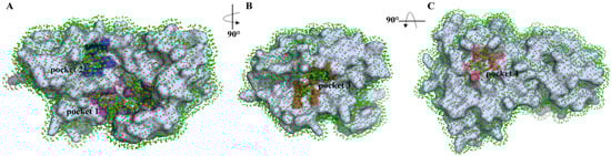
Figure 1.
Ligand binding site visualization predicted by p2Rank. Protein (in gray) is covered by a layer of points (in green) lying on the SAS (solvent accessible surface) of the protein. Each point represents its local chemical neighborhood. Visualization is based on a PyMOL session generated by p2Rank. Binding pocket 1—magenta, 2—blue, 3—brown, 4—pink.
All the detected pockets are more or less small, which is actually optimal for C60 fullerene. Pockets 1–3 are characterized by an irregular shape with a slight indentation. Contrary to them, pocket 4 is flat. The analyzed pockets contain the following amino acids: pocket 1—polar: Ser 121, Tyr 161, Asn 336, Asn 344, Thr 481; non-polar: Ile 118, Pro 129, Trp 132, Trp 168, Pro 335, Ala 423, Val 479, Phe 480, Trp 482; charged: Glu 119, Arg 147, Arg 150, His 334, Arg 486; pocket 2—polar: Gln 374, Thr 407, Asn 476; non-polar: Met 372, Leu 373, Pro 375, Leu 404, Ile 405, Met 424, Ile 425, Cys 426, Ile 456, Met 478; charged: Glu 406; pocket 3—polar: Ser 436, Gln 438, Ser 441, Thr 459; non-polar: Val 280, Cys 281, Leu 302, Cys 437, Gly 439, Trp 461, Gly 462, Gly 464, Cys 465; charged: His 296, Glu 389; pocket 4—polar: Asn 247; non-polar: Ala 266, Trp 267, Trp 380, Trp 453; charged: Glu 260, Lys 401. Also, in p2Rank, we have the ability to characterize their internal parameter sas_points (a value often associated with Solvent Accessible Surface (SAS) Area). Through this, it is possible to evaluate how accessible a region is to solvent molecules [64,65]. According to p2Rank, pockets 1–4 contain 108, 40, 68, and 25 sas_points, respectively.
So, binding pocket 1 is the most solvent-exposed one (sas_points 108) and contains a mix of hydrophobic and polar residues. This pocket has a high potential to create different types of stacking interactions, since it comprises multiple residues like Arg, His, Trp, and Tyr. Binding pocket 2, located near binding pocket 1, is more hydrophobic, but the big advantage here is that this pocket is buried. Thus, C60 fullerene can entirely fill that pocket. Furthermore, it contains a few residues capable of forming stacking interactions with C60 fullerene, namely Met 372, Met 424, Cys 426, and Met 478. Binding pocket 3, like the previous one, is buried. Combined with its volume and the presence of amino acids that can create stacking interactions (Cys 281, His 296, Cys 437, Trp 461, and Cys 465), this pocket is a good candidate for targeting. Finally, because binding pocket 4 is very small and flat, despite the fact that it contains many residues that can create stacking interactions (Trp 267, Trp 380, Lys 401, and Trp 453), we have decided to exclude it from further investigation.
So, within the identified binding pockets, pockets 1, 2, and 3 appear most promising for C60 fullerene targeting because of their shape, residue environment, and volume. Pocket 4 was removed from the investigation.
2.2. “C60 Fullerene-TMPRSS2” Interaction Analysis
Here, our focus was on the investigation of the interaction between C60 fullerene and selected binding pockets (Figure 2). In binding pocket 1, C60 fullerene is tightly bound between the loop and the α-helical region. The C60 fullerene fills only part of binding pocket 1, creating π/π-stacking interactions with Trp 168, Tyr 161, and Trp 132, and cation-π interactions with Arg147, Arg150, and Arg486. It seems that such diverse stacking interactions should stabilize the “C60 fullerene–TMPRSS2” complex and influence the investigated protein functions. Binding pocket 2 is mostly hydrophobic, so it is fair to assume that binding inside that pocket will be less favorable compared to binding pocket 1. Here, C60 fullerene is flanked by polar residues including Thr 407, Asn 476, and Glu 406. On the other hand, the binding pocket comprises two Met amino acids that could create stacking interactions with C60 fullerene, thus fixing it in this binding pocket. However, according to the data from the docking simulation, in its current conformation, Met 372 is a bit too far from C60 fullerene to create stacking interactions. However, considering that Met 372 is flexible, it is still possible. In the case of binding pocket 3, C60 fullerene lies above the catalytic binding pocket, shielding it from interaction with any other chemical structures. This pocket is the smallest, including a smaller amount of residues, but almost all of them are charged and are polar amino acids, such as His 296, Lys 342, Lys 390, and Gln 438. All those residues are capable of forming cation-π or stacking interactions with C60 fullerene.
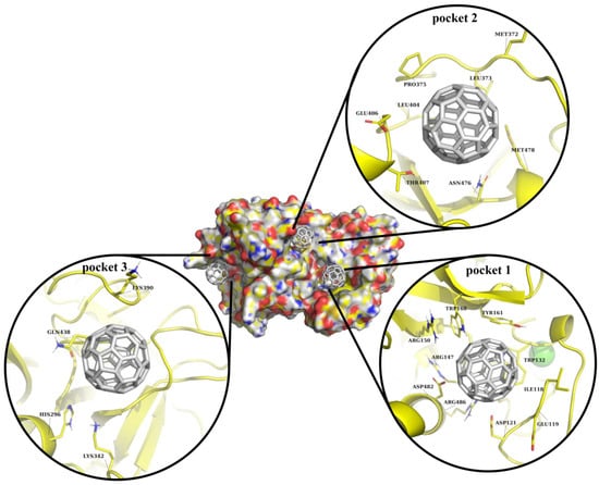
Figure 2.
Visualization of C60 fullerene docking into predicted binding pockets, shown using surface and cartoon representations. In all cases, TMPRSS2 is depicted in yellow and C60 fullerene in gray.
In our opinion, binding with any of those pockets could potentially impact TMPRSS2 functionality. It looks like, based on the molecular docking simulation, pocket 1 potentially represents the most promising TMPRSS2 site for C60 fullerene binding. Binding pockets 2 and 3 seem to be less attractive, since the first one cannot form a sufficient number of interactions with C60 fullerene, and the second one is small. Nevertheless, all this data should be evaluated using more accurate methods, e.g., MD simulation.
2.3. “C60 Fullerene-TMPRSS2” Stability Analysis
In order to achieve greater accuracy, a 1000 ns MD simulation was performed (Figure 3 and Figure 4). The simulation results showed that some predicted complexes remained stable throughout the simulation. The RMSD (root-mean-square deviation) movement in all the complexes stayed within the 1–2 Å range (Figure 4). Interestingly, in some cases, complexes completely lost all interactions in the binding pocket; in others, they established new, potentially stronger interactions.
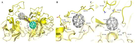
Figure 3.
MD simulation results of TMPRSS2 in complex with C60 fullerene. (A) Binding pocket 1: The initial position of C60 fullerene is shown in gray, and its position after simulation is shown in cyan. (B) Interaction at binding pocket 2. (C) Interaction at binding pocket 3. TMPRSS2 is shown in yellow in all panels, and C60 fullerene in gray, except for the post-simulation pose in A (cyan).
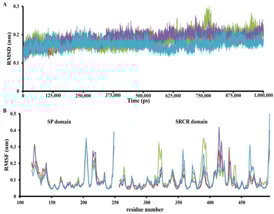
Figure 4.
The RMSD (A) trajectory and RMSF (B) MD trajectory profile of apo TMPRSS2 (red), C60 fullerene interaction in binding pocket 1 (green), binding pocket 2 (violet), and binding pocket 3 (cyan).
The results of the TMPRSS2 MD simulation are presented in Figure 3 and Figure 4. The “C60 fullerene-binding pocket 1” complex showed signs of instability, rapidly losing crucial interactions with TMPRSS2 just after the start of the MD simulation. Here, C60 fullerene eventually completely relocated to the predicted binding pocket 2. As for the interaction insights from binding pocket 2, it was very stable. The C60 fullerene was immersed deeper inside the binding pocket, shifting by 2.6 Å. That displacement caused changes in the pocket conformation and interaction with C60 fullerene. From the close-up view, it was possible to determine different types of interactions, such as hydrophobic/steric contacts and van der Waals forces, which participate in complex stabilization. Interestingly, this effect was mostly achieved by hydrophobic/steric interactions with C60 fullerene, including its contacts with Leu 373, Pro 375, Thr 407, Glu 406, Leu 404, and Asn 476. In terms of stacking, there were stable interactions with Met 478 and an additional one with Met 372. Met 372 is located at the edge of the pocket and can move freely. As a result, Met 372 changed its position and covered C60 fullerene, holding it in place. The C60 fullerene binding with pocket 3 was characterized by huge changes in the binding model. It was observed that C60 fullerene shifted by 3.2 to 7.8 Å inside the catalytic binding pocket, causing various residues to change. For example, Lys 390 and 342 dramatically changed their conformations and created cation-π interactions with C60 fullerene. In contrast, His 269 slightly changed its conformation and at the same time anchored C60 fullerene in the binding pocket through π-stacking interactions. The most intriguing aspect here is that, as a result of the MD simulation, C60 fullerene completely closed the catalytic binding pocket so no other binding agent could reach it. That would definitely have a significant impact on TMPRSS2 functionality.
Finally, the “C60 fullerene-TMPRSS2” complex was subjected to RMSF (root-mean-square fluctuation) analysis. Overall, the RMSF values remained below 0.4 nm for most residues, suggesting protein stability during the simulation (Figure 4). Nevertheless, for the SP and SRCR domains, several distinct dynamic regions could be observed. The SP domain displayed a few RMSF differences, with one region that behaved differently in each investigated model. This region corresponds to residues 225–235 and is interesting because it is not located near any binding pockets. So, that was probably a distance effect of C60 fullerene binding in different models. However, this is just an assumption and requires deeper investigation. In contrast, the SRCR domain contains multiple parts that demonstrated different fluctuations depending on the binding model: these are residues 310–320, 350–360, 385–405, 410–445, and 460–470.
So, according to the obtained results, C60 fullerene showed dynamic behavior, relocating from binding pocket 1 to binding pocket 2. That is a surprising result, since binding pocket 1 comprises many residues able to create stacking interactions with C60 fullerene. In contrast, binding pocket 2 contains just two Met residues, and most of the other residues are able to create steric/hydrophobic interactions with C60 fullerene. As for binding pocket 3, the C60 fullerene inserted more deeply into the catalytic site and completely blocked one. According to the fluctuation analysis, TMPRSS2 is stable, with some fluctuations in both the SP and SRCR domains.
2.4. PCA Study
The application of principal component analysis (PCA) suggests (Figure 5) that TMPRSS2 alone and in complex with C60 fullerene have noticeably different conformational behaviors. The TMPRSS2 apo form can assume a significant number of different conformations (Figure 5A). It indicates a significant structural flexibility of the TMPRSS2 structure. However, it should be noted that the main structure of the apo TMPRSS2 remained relatively rigid during the simulations; the conformational variations were mainly observed in the loop regions. In contrast, the “C60 fullerene-TMPRSS2” complexes demonstrated a dramatic reduction in conformational space, with only one small conformationally dense part (Figure 5B,C). In accordance with that, we can suggest that binding with C60 fullerene imposes structural constraints on TMPRSS2 and fully stabilizes only one specific conformation of the “C60 fullerene-TMPRSS2” complex.
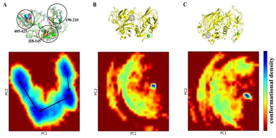
Figure 5.
PCA of TMPRSS2 dynamics and its interaction with C60 fullerene. (A) PCA of TMPRSS2 apo form reveals major conformational changes in regions 118–145, 190–210, and 405–425. With this case as the central example, the following snapshots were selected 1—18,950 (green), 2—350,400 (cyan), 3—708,950 (violet), 4—864,450 (yellow). (B) PCA plot and structure of TMPRSS2 in complex with C60 fullerene at binding pocket 2. (C) PCA plot and structure of TMPRSS2 in complex with C60 at binding pocket 3. Conformational landscapes are shown as 2D density plots along the first two principal components (PC1 and PC2), with the color scale indicating conformational density (blue = high density, red = low density). TMPRSS2 is shown in yellow, and C60 fullerene in gray.
2.5. Binding Affinity Analysis via MMPBSA
For a better understanding of the binding properties between TMPRSS2 and C60 fullerene, a binding energy analysis has been conducted (Table 1). The van der Waals (vdW) interactions have the most impact on the overall binding affinity, with values of −49.56 and −33.28 kcal/mol for pockets 2 and 3, respectively. Those values represent the hydrophobic nature of C60 fullerene, which interacts primarily through dispersion forces with non-polar residues inside the pocket.

Table 1.
The calculated energy contributions of TMPRSS2 binding with C60 fullerene in pockets 2 and 3 (in kcal/mol).
In contrast, the electrostatic interaction (EE), polar solvation (PS), and non-polar solvation (NPS) energies have minimal or unfavorable contributions to complex formation in both binding pockets. So, the EE energy contributes almost nothing to binding (−0.09 and −0.04 kcal/mol), and that is actually expected, because C60 fullerene is neutral, and cannot form classical electrostatic interactions. Similarly, in the case of PS, it is unfavorable for both investigated binding pockets (12.30 and 9.41 kcal/mol), which indicates a significant potential binding penalty during the transfer of the hydrophobic C60 fullerene from solvent into the investigated binding pocket. And finally, the NPS represents only slight compensation compared to the two previously demonstrated energy types (−3.21 and −2.33 kcal/mol). As a result, the binding energy (TOTAL) is significantly more favorable for pocket 2 (−40.56 kcal/mol) compared with pocket 3 (−26.24 kcal/mol); it indicates that binding pocket 2 has better geometric and hydrophobic complementarity to the C60 fullerene surface.
Also, to estimate system efficiencies, a thermodynamic analysis has been performed (Table 2). For binding pocket 2, the enthalpic contribution (∆H) was more favorable (−40.56 kcal/mol) compared to binding pocket 3 (∆H is −26.23 kcal/mol). This indicates stronger interaction in the case of binding pocket 2. As a result, the final Gibbs free energy (∆G) is favorable for binding pocket 2 (−30.71 kcal/mol), despite ∆G (0.45 kcal/mol) for binding pocket 3 indicating very weak or no binding in this pocket with TMPRSS2.
Table 2.
The calculated thermodynamic parameters of C60 fullerene binding in TMPRSS2 selected binding pockets 2 and 3 (in kcal/mol).
Together, these results demonstrate that vdW interactions overwhelmingly determine the binding preference, and that only pocket 2 provides a sufficiently hydrophobic and shape-complementary environment for stable C60 fullerene association with TMPRSS2.
3. Discussion
Recent studies have pointed to TMPRSS2 as one of the key targets that facilitates SARS-CoV-2 entry to the host cells [28,32,35,36,37,38,66]. TMPRSS2 is reported as a multidomain serine protease consisting of an SRCR, trypsin-like SP domains, and a linker region between them [67,68]. The SP domain itself contains the only known well-characterized binding pocket, which includes the catalytic triad His–Asp–Ser [30,42,63,69]. This region, referred to as binding pocket 3, was also identified in our analysis. The obtained results have demonstrated that this pocket is relatively narrow, which explains why it typically interacts with elongated, aromatic ligands such as camostat and nafamostat [47,63,70]. However, our analysis also demonstrates that this pocket is enriched with polar and charged amino acid residues capable of creating π–π and cat-ion–π interactions (e.g., His 269 and Cys 465). So, binding pocket 3 is potentially able to interact with spherical structures like C60 fullerene.
In contrast, the remaining pockets have not been well described in the available literature. According to the available data [39,47,70,71], TMPRSS2 contains secondary pockets adjacent to and distinct from the catalytic cleft. However, many studies are focused on the catalytic binding pocket’s description and its different possible sub-pockets [39,70]. Contrary to this, the authors of [47,71] describe all potential high-affinity binding spots for chemicals (e.g., camostat and nafamostat). However, the binding pocket mapping in each publication is different. For example, the region that we define as pocket 2 is divided into two distinct sub-sites in their models (M-5F8T_site_1 and M-5F8T_site_4) [71]. Overall, previous studies have made only limited attempts to select binding pockets distinct from the catalytic one, and these efforts have primarily concentrated on essentially the same parts of the TMPRSS2 protein, that is, on the interface between the SRCR and SP domains and the SP domain, on the opposite side from the location of the catalytic binding pocket. Thus, our analysis systematically identifies and characterizes these additional binding pockets, expanding the current understanding of TMPRSS2’s ligand-binding landscape beyond the classical catalytic site.
The obtained MD simulation and docking results provide important insights into the interaction between C60 fullerene and TMPRSS2. Although docking initially suggested that the binding pocket 1 was one of the most favorable sites due to its presence of aromatic and charged residues, nevertheless, the long-timescale MD simulation revealed that those potential interactions with C60 fullerene are unstable. That is actually not surprising, because this region is located near flexible parts of the protein (Figure 5), making ligand binding in such a region often transient and unstable [72,73,74,75]. This actually explains why C60 fullerene migrates from pocket 1 into pocket 2 during the MD simulation. It is interesting that binding pocket 2 comprises fewer aromatic residues. However, it has a deeper pocket cavity and is located in a less flexible region of TMPRSS2. A similar picture is observed in the studies of [47,71], where camostat binds in the SRCR and SP domain interface, with a docking energy of −9.95 kcal/mol, but during the MD simulation it demonstrates conformational plasticity of this region. All those data suggest that binding pocket 1 initially appears favorable based on static docking, but dynamic fluctuations and solvent reduce its stability, and as a result, different potential binders lose their ability to interact with one. Therefore, we suggest that the C60 fullerene migration from binding pocket 1 to pocket 2 during MD simulation reflects a fundamental transition from an un-favorable state to a preferred, more stable complex, where the C60 fullerene is buried within a deeper, less mobile cavity.
The gmx_MMPBSA results demonstrate that the van der Waals forces are dominant in the complex formation between C60 fullerene and TMPRSS2 protein. This behavior is fully consistent with the intrinsic physicochemical properties of C60 fullerene, a neutral hydrophobic molecule that interacts mainly through dispersion forces [76,77]. In contrast, classical TMPRSS2 inhibitors such as camostat and nafamostat rely on a combination of electrostatic and hydrogen bonding with the TMPRSS2 catalytic binding pocket [47,70]. So, that is aligned with our results. Based on the gmx_MMPBSA analysis, it is highly unlikely that C60 fullerene would bind to the catalytic binding pocket. Rather, it interacts with the TMPRSS2 only via a non-classical binding mechanism, driven by hydrophobic and dispersion interactions, predominantly. And actually, binding pocket 2 provides a sufficient residue environment and geometric complementarity for C60 fullerene binding, whereas binding pocket 3 (catalytic pocket) is smaller and more polar, which limits its ability to stabilize the C60 fullerene. Overall, compared to the previously studied “TMPRSS2–ligand” systems, where polar interactions are dominant, our results reveal a distinct binding model that is based on pocket geometry and dispersion forces.
4. Materials and Methods
4.1. Hardware and Software
All the computational study was carried out on Ubuntu 24.1 (64-bit) operating systems equipped with 96 GB of RAM and 3 × 3.60 GHz AMD Ryzen™ 5 1600X Six-Core processors, except for the MD simulations. The MD simulations were performed using 3× Gigabyte GeForce RTX 3060 12228MB GPUs. For visualization, PyMOL (v3.1) [78] and Matplotlib (v3.10) [79] tools were used.
4.2. Selection of Protein Structure and Binding Site Determination
Based on the available data, the Protein Data Bank (PDB) [80] contains 30 structures. All the structures were retrieved and analyzed. Initially, all crystallographic water molecules and native ligands were removed from the protein structure. Next, the structure was refined by adding missing hydrogens, correcting amide protonation states, and repairing incomplete side chains using MolProbity 4.5.2 [81,82]. The system was further minimized using the CHARMM36 force field to remove all the strains and steric clashes inside the protein system [83,84,85]. Finally, taking into consideration structure integrity, resolution, host organism (human), and novelty, 9jd1 PDB ID [86] was selected for further investigation.
In the next stage, the binding pockets for C60 fullerene were defined by the p2Rank 2.5 [68] software tool. p2Rank evenly spread the points on the protein’s SAS. Each point served as the center of its own spherical region, which together may represent possible binding pockets for different binders. To predict potential binding pockets, p2Rank ran the following steps:
- 1.
- Generation of spaced points on a protein SAS, based on a fast numerical algorithm [87] implemented in the CDK library [88,89].
- 2.
- Calculation of the location of points on the protein surface.
- 3.
- Prediction of ligand ability of SAS points employing a Random Forest approach [90,91].
- 4.
- Clustering of all the points to create potential binding pockets (the cut-off distance 3 Å).
- 5.
- Ordering predicted binding pockets by the ligand ability score of the points they contain (sum of squared ligand ability scores of all points in the cluster).
4.3. Molecular Docking Study
Both C60 fullerene and TMPRSS2 were prepared before the molecular docking simulation. In the case of TMPRSS2, all the preparation data were retrieved from the previous stage (preparation based on MolProbity). The C60 fullerene coordinates were downloaded from the MSU Fullerene Database. Then, OpenBabel 3.1.0 software was applied to prepare the C60 fullerene for the molecular docking simulation [92]. Specifically, the protonation and geometry of the structure were checked to ensure proper chemical integrity before the simulation. The molecular docking simulations were performed utilizing SwissDock 2024 [93], using cavities 2.0 (AC) algorithm (a detailed description of the docking algorithm is provided in [94,95]). The binding pockets were defined in accordance with p2Rank results.
Prior to running the molecular docking simulation, the central amino acids in each binding pocket were indicated. For each pocket, residues located within a 6.0 Å sphere of the central amino acid were considered part of the binding pocket. Specifically, pocket 1 was centered on Arg 147, pocket 2 on Leu 404, and pocket 3 on Gln 438. In each docking case, the 10 best complexes were selected for analysis stages. In the end, the molecular docking results were estimated using the docking score and visual inspection of all the obtained poses.
4.4. MD Simulation Strategy
To evaluate the stability and key interactions of the obtained complexes after molecular docking, MD simulation was carried out. The calculations were performed using GROMACS 2024.2 [96] with the CHARMM36 force field [83,84,85]. The TMPRSS2 protein was protonated according to the built-in function in GROMACS 2024.2—“-ignh”. The topology for C60 fullerene was generated by SwissParam 2023 [97,98]. The best complexes obtained after molecular docking were used for MD simulation. Each system was placed at the center of a periodic cubic box, which was then filled with TIP3P water molecules. A minimum 10 Å distance was maintained between the nearest atom of the complex and the edge of the simulation box so that the complex could be fully immersed in water and rotate freely. Then, to neutralize the system and mimic the cellular environment (pH = 7), Na+ and Cl− ions were added to bring the ionic concentration to 150 mM. Here, the solvent molecules were randomly replaced with monoatomic ions. Next, the obtained complex was energy minimized, which also relieved any steric clashes. The system was relaxed by applying the steepest descent algorithm (the maximum number of steps was 50,000). Then the equilibration was computed in two stages: NVT (constant volume-temperature simulation) was first equilibrated for 5 ns, followed by NPT (constant pressure-temperature simulation) equilibration for 5 ns. After that, we launched the MD simulation for 1000 ns. All calculations were performed at a temperature of 300 K and at constant atmospheric pressure.
4.5. PCA Study
PCA has been applied to reveal high-amplitude concerted motions within the structure of the TMPRSS2 protein [99,100,101]. The simulations were carried out utilizing eigenvectors derived from the mass-weighted covariance matrix of RNA atomic fluctuations. To generate the covariance matrix, the built-in GROMACS 2024.2 function ‘gmx covar’ was used. Then, we calculated eigenvectors and eigenvalues to define the dimensionality of the essential subspace. Note that a small subset of eigenvectors (<10) characterizes 90% of all motions, revealing the main motions occurring within an atomic system. Cosine content is used as a measure of principal components [102,103]. If the cosine content is close to 1, it indicates significant movement within the TMPRSS2 molecule and renders it unsuitable for PCA. However, most snapshots captured during MD simulation exhibit cosine values close to 0.2, with some approaching 0.5, making them suitable for PCA. Thus, according to the above, by using the ‘gmx analyse’ utility, the FEL (free energy landscape) was constructed utilizing cosine contents <0.2 of the first two projection eigenvectors (defined as PC1 and PC2, respectively). The most prevalent and energetically favorable structures extracted from the FEL’s minimum energy basins were then utilized for subsequent analyses.
4.6. Binding Energy Calculation
The MM/PBSA molecular mechanics [104,105,106,107] was executed to estimate “C60 fullerene-TMPRSS2” complex binding energy using the gmx_MMPBSA v1.6.3 [106] software tool. This is an Amber-based tool [108] that calculates binding energy (∆ETOTAL) based on the following equation
where EvdW—van der Waals energy; EEE—electrostatic energy; EPS and ENPS—polar and non-polar solvation energy, respectively, and also the Gibbs free energy (∆G):
∆ETOTAL = EvdW + EEE + EPS + ENPS,
∆G = ∆H − TΔS.
Here ∆H—enthalpy and T∆S—entropic term.
5. Conclusions
The TMPRSS2 is essential for SARS-CoV-2 activation and entry. That is why this protein represents an important therapeutic target. On the other hand, nanostructures like C60 fullerene have been widely discussed as unconventional molecular scaffolds that are capable of modulating the functions of different proteins. So, understanding whether C60 fullerene is able to interact with TMPRSS2 is significant in the context of a potential alternative antiviral therapy design.
In this study, a comprehensive computational investigation to evaluate the C60 fullerene’s ability to interact with TMPRSS2 was conducted. According to the p2Rank investigation, four binding pockets were identified. Among them, pockets 1–3 have the most promising shape and amino acid residue environment for C60 fullerene targeting, while pocket 4 was unsuitable due to its flat architecture. According to subsequent simulations, the C60 fullerene interaction with binding pocket 1 is not stable, and based on thermodynamic analysis, binding in pocket 3 is very weak or not possible at all. So, among all the evaluated binding pockets, pocket 2 seems to be the most promising one for binding with C60 fullerene.
Furthermore, further simulations have shown that C60 fullerene noticeably restricts the conformational dynamics of TMPRSS2, while the unbound protein has a broader conformational landscape. This suggests that C60 fullerene may fix specific structural states of TMPRSS2, and such binding can potentially affect its biological functions, in particular to block interactions with SARS-CoV-2.
Author Contributions
Conceptualization, supervision, Y.P. and V.H.; writing—original draft, V.H., Z.K. and Y.P.; writing—review and editing, V.H., Z.K., Y.P. and V.G.; methodology, investigation, V.H., V.K. and S.P.; formal analysis, data curation, Z.K., S.V. and U.R.; project administration, V.G. All authors have read and agreed to the published version of the manuscript.
Funding
This research received no external funding.
Institutional Review Board Statement
Not applicable.
Informed Consent Statement
Not applicable.
Data Availability Statement
The data presented in this study are available on request from the corresponding author.
Conflicts of Interest
The authors declare no conflicts of interest.
References
- Wang, W.; Xu, Y.; Gao, R.; Lu, R.; Han, K.; Wu, G.; Tan, W. Detection of SARS-CoV-2 in Different Types of Clinical Specimens. JAMA 2020, 323, 1843–1844. [Google Scholar] [CrossRef]
- Morens, D.M.; Fauci, A.S. Emerging Pandemic Diseases: How We Got to COVID-19. Cell 2020, 182, 1077–1092. [Google Scholar] [CrossRef]
- Zhu, N.; Zhang, D.; Wang, W.; Li, X.; Yang, B.; Song, J.; Zhao, X.; Huang, B.; Shi, W.; Lu, R.; et al. A Novel Coronavirus from Patients with Pneumonia in China, 2019. N. Engl. J. Med. 2020, 382, 727–733. [Google Scholar] [CrossRef] [PubMed]
- Chung, Y.-S.; Lam, C.-Y.; Tan, P.-H.; Tsang, H.-F.; Wong, S.-C.C. Comprehensive Review of COVID-19: Epidemiology, Pathogenesis, Advancement in Diagnostic and Detection Techniques, and Post-Pandemic Treatment Strategies. Int. J. Mol. Sci. 2024, 25, 8155. [Google Scholar] [CrossRef] [PubMed]
- Kim, S.Y.; Yeniova, A.Ö. Global, regional, and national incidence and mortality of COVID-19 in 237 countries and territories, January 2022: A systematic analysis for World Health Organization COVID-19 Dashboard. Life Cycle 2022, 2, e10. [Google Scholar] [CrossRef]
- Available online: https://data.who.int/dashboards/covid19/deaths?n=c (accessed on 26 November 2025).
- Available online: https://covid19.who.int (accessed on 26 November 2025).
- Available online: https://www.elifecycle.org/archive/view_article?pid=lc-2-0-10 (accessed on 26 November 2025).
- Payne, S. Family Coronaviridae. In Viruses; Academic Press: Cambridge, MA, USA, 2017; pp. 149–158. [Google Scholar] [CrossRef]
- Coronaviridae Study Group of the International Committee on Taxonomy of Viruses. The species Severe acute respiratory syndrome-related coronavirus: Classifying 2019-nCoV and naming it SARS-CoV-2. Nat. Microbiol. 2020, 5, 536–544. [Google Scholar] [CrossRef] [PubMed]
- Zhou, Z.; Qiu, Y.; Ge, X. The taxonomy, host range and pathogenicity of coronaviruses and other viruses in the Nidovirales order. Anim. Dis. 2021, 1, 5. [Google Scholar] [CrossRef]
- Woo, P.C.Y.; de Groot, R.J.; Haagmans, B.; Lau, S.K.P.; Neuman, B.W.; Perlman, S.; Sola, I.; van der Hoek, L.; Wong, A.C.P.; Yeh, S.-H. ICTV Virus Taxonomy Profile: Coronaviridae 2023. J. Gen. Virol. 2023, 104, 001843. [Google Scholar] [CrossRef]
- Karnitskaya, Y.; Drobysh, M.; Ramanaviciene, A.; Korniienko, V.; Balevicius, S.; Ramanavicius, A. Investigation of interaction between immobilized SARS-CoV-2 nucleoprotein and monoclonal antibodies by cyclic voltammetry based electrochemical immunosensor. Microchem. J. 2025, 219, 115810. [Google Scholar] [CrossRef]
- Xia, S.; Zhu, Y.; Liu, M.; Lan, Q.; Xu, W.; Wu, Y.; Ying, T.; Liu, S.; Shi, Z.; Jiang, S.; et al. Fusion mechanism of 2019-nCoV and fusion inhibitors targeting HR1 domain in spike protein. Cell. Mol. Immunol. 2020, 17, 765–767. [Google Scholar] [CrossRef]
- Zhou, P.; Yang, X.L.; Wang, X.G.; Hu, B.; Zhang, L.; Zhang, W.; Si, H.R.; Zhu, Y.; Li, B.; Huang, C.L.; et al. A pneumonia outbreak associated with a new coronavirus of probable bat origin. Nature 2020, 579, 270–273. [Google Scholar] [CrossRef]
- Wang, H.; Li, X.; Li, T.; Zhang, S.; Wang, L.; Wu, X.; Liu, J. The genetic sequence, origin, and diagnosis of SARS-CoV-2. Eur. J. Clin. Microbiol. Infect. Dis. 2020, 39, 1629–1635. [Google Scholar] [CrossRef] [PubMed]
- Keshta, A.S.; Mallah, S.I.; Al Zubaidi, K.; Ghorab, O.K.; Keshta, M.S.; Alarabi, D.; Abousaleh, M.A.; Salman, M.T.; Taha, O.E.; Zeidan, A.A.; et al. COVID-19 versus SARS: A comparative review. J. Infect. Public. Health 2021, 14, 967–977. [Google Scholar] [CrossRef] [PubMed]
- Sallard, E.; Halloy, J.; Casane, D.; Decroly, E.; van Helden, J. Tracing the origins of SARS-COV-2 in coronavirus phylogenies: A review. Environ. Chem. Lett. 2021, 19, 769–785. [Google Scholar] [CrossRef] [PubMed]
- Cicaloni, V.; Costanti, F.; Pasqui, A.; Bianchini, M.; Niccolai, N.; Bongini, P. A Bioinformatics Approach to Investigate Structural and Non-Structural Proteins in Human Coronaviruses. Front. Genet. 2022, 13, 891418. [Google Scholar] [CrossRef]
- Rosas-Lemus, M.; Minasov, G.; Brunzelle, J.S.; Taha, T.Y.; Lemak, S.; Yin, S.; Shuvalova, L.; Rosecrans, J.; Khanna, K.; Seifert, H.S.; et al. Torsional twist of the SARS-CoV and SARS-CoV-2 SUD-N and SUD-M domains. Protein Sci. 2025, 34, e70050. [Google Scholar] [CrossRef]
- Hoenigsperger, H.; Sivarajan, R.; Sparrer, K.M. Differences and similarities between innate immune evasion strategies of human coronaviruses. Curr. Opin. Microbiol. 2024, 79, 102466. [Google Scholar] [CrossRef]
- Kuhn, J.H.; Li, W.; Choe, H.; Farzan, M. Angiotensin-converting enzyme 2: A functional receptor for SARS coronavirus. Cell Mol. Life Sci. 2004, 61, 2738–2743. [Google Scholar] [CrossRef]
- Thunders, M.; Delahunt, B. Gene of the month: TMPRSS2 (transmembrane serine protease 2). J. Clin. Pathol. 2020, 73, 773–776. [Google Scholar] [CrossRef]
- Hoffmann, M.; Kleine-Weber, H.; Schroeder, S.; Krüger, N.; Herrler, T.; Erichsen, S.; Schiergens, T.S.; Herrler, G.; Wu, N.H.; Nitsche, A.; et al. SARS-CoV-2 Cell Entry Depends on ACE2 and TMPRSS2 and Is Blocked by a Clinically Proven Protease Inhibitor. Cell 2020, 181, 271–280.е8. [Google Scholar] [CrossRef]
- Baughn, L.B.; Sharma, N.; Elhaik, E.; Sekulic, A.; Bryce, A.H.; Fonseca, R. Targeting TMPRSS2 in SARS-CoV-2 Infection. Mayo Clin. Proc. 2020, 95, 1989–1999. [Google Scholar] [CrossRef] [PubMed]
- Lukassen, S.; Chua, R.L.; Trefzer, T.; Kahn, N.C.; Schneider, M.A.; Muley, T.; Winter, H.; Meister, M.; Veith, C.; Boots, A.W.; et al. SARS-CoV-2 receptor ACE2 and TMPRSS2 are primarily expressed in bronchial transient secretory cells. EMBO J. 2020, 39, e105114. [Google Scholar] [CrossRef] [PubMed]
- Gkogkou, E.; Barnasas, G.; Vougas, K.; Trougakos, I.P. Expression profiling meta-analysis of ACE2 and TMPRSS2, the putative anti-inflammatory receptor and priming protease of SARS-CoV-2 in human cells, and identification of putative modulators. Redox Biol. 2020, 36, 101615. [Google Scholar] [CrossRef]
- Grisard, H.B.D.S.; Schörner, M.A.; Barazzetti, F.H.; Wachter, J.K.; Valmorbida, M.; Wagner, G.; Fongaro, G.; Bazzo, M.L. ACE2 and TMPRSS2 expression in patients before, during, and after SARS-CoV-2 infection. Front. Cell. Infect. Microbiol. 2024, 14, 1355809. [Google Scholar] [CrossRef]
- Shang, J.; Ye, G.; Shi, K.; Wan, Y.; Luo, C.; Aihara, H.; Geng, Q.; Auerbach, A.; Li, F. Structural basis of receptor recognition by SARS-CoV-2. Nature 2020, 581, 221–224. [Google Scholar] [CrossRef]
- Antalis, T.M.; Bugge, T.H.; Wu, Q. Membrane-anchored serine proteases in health and disease. Prog. Mol. Biol. Transl. Sci. 2011, 99, 1–50. [Google Scholar] [CrossRef] [PubMed]
- Ou, X.; Zheng, W.; Shan, Y.; Mu, Z.; Dominguez, S.R.; Holmes, K.V.; Qian, Z. Identification of the Fusion Peptide-Containing Region in Betacoronavirus Spike Glycoproteins. J. Virol. 2016, 90, 5586–5600. [Google Scholar] [CrossRef]
- Dong, M.; Zhang, J.; Ma, X.; Tan, J.; Chen, L.; Liu, S.; Xin, Y.; Zhuang, L. ACE2, TMPRSS2 distribution and extrapulmonary organ injury in patients with COVID-19. Biomed. Pharmacother. 2020, 131, 110678. [Google Scholar] [CrossRef]
- Ren, X.; Wang, S.; Chen, X.; Wei, X.; Li, G.; Ren, S.; Zhang, T.; Zhang, X.; Lu, Z.; You, Z.; et al. Multiple Expression Assessments of ACE2 and TMPRSS2 SARS-CoV-2 Entry Molecules in the Urinary Tract and Their Associations with Clinical Manifestations of COVID-19. Infect. Drug Resist. 2020, 13, 3977–3990. [Google Scholar] [CrossRef]
- Williams, T.L.; Strachan, G.; Macrae, R.G.C.; Kuc, R.E.; Nyimanu, D.; Paterson, A.L.; Sinha, S.; Maguire, J.J.; Davenport, A.P. Differential expression in humans of the viral entry receptor ACE2 compared with the short delta ACE2 isoform lacking SARS-CoV-2 binding sites. Sci. Rep. 2021, 11, 24336. [Google Scholar] [CrossRef]
- Glowacka, I.; Bertram, S.; Müller, M.A.; Allen, P.; Soilleux, E.; Pfefferle, S.; Steffen, I.; Tsegaye, T.S.; He, Y.; Gnirss, K.; et al. Evidence that TMPRSS2 activates the severe acute respiratory syndrome coronavirus spike protein for membrane fusion and reduces viral control by the humoral immune response. J. Virol. 2011, 85, 4122–4134. [Google Scholar] [CrossRef] [PubMed]
- Gierer, S.; Bertram, S.; Kaup, F.; Wrensch, F.; Heurich, A.; Krämer-Kühl, A.; Welsch, K.; Winkler, M.; Meyer, B.; Drosten, C.; et al. The spike protein of the emerging betacoronavirus EMC uses a novel coronavirus receptor for entry, can be activated by TMPRSS2, and is targeted by neutralizing antibodies. J. Virol. 2013, 87, 5502–5511. [Google Scholar] [CrossRef]
- Zhou, Y.; Vedantham, P.; Lu, K.; Agudelo, J.; Carrion, R.; Nunneley, J.W.; Barnard, D.; Pöhlmann, S.; McKerrow, J.H.; Renslo, A.R.; et al. Protease inhibitors targeting coronavirus and filovirus entry. Antivir. Res. 2015, 116, 76–84. [Google Scholar] [CrossRef]
- Chen, V.H.E.; Tay, J.K.; Gurung, R.; Nair, S.; Tay, D.J.W.; Tan, K.S.; Foo, R.S.Y.; Tambyah, P.A. ACE2 and TMPRSS2 gene expression is reduced acutely in SARS-CoV-2 patients but returns to normal with recovery. Sci. Rep. 2025, 15, 12828. [Google Scholar] [CrossRef]
- Idris, M.O.; Yekeen, A.A.; Alakanse, O.S.; Durojaye, O.A. Computer-aided screening for potential TMPRSS2 inhibitors: A combination of pharmacophore modeling, molecular docking and molecular dynamics simulation approaches. J. Biomol. Struct. Dyn. 2021, 39, 5638–5656. [Google Scholar] [CrossRef]
- Stopsack, K.H.; Mucci, L.A.; Antonarakis, E.S.; Nelson, P.S.; Kantoff, P.W. TMPRSS and COVID-19: Serendipity or Opportunity for Intervention? Cancer Discov. 2020, 10, 779–782. [Google Scholar] [CrossRef]
- Felix, F.A.; Filiú, F.M.V.; da Silva, T.A.; Calderaro, D.C.; Tanure, L.A.; de Castro, M.A.A.; Gomez, R.S.; Diniz, M.G.; de Sousa, S.F. SARS-CoV-2 Entry Factors ACE2, TMPRSS2 and FURIN in Salivary Glands From Primary Sjögren’s Disease. J. Oral. Pathol. Med. 2025, 54, 290–297. [Google Scholar] [CrossRef]
- Shen, L.W.; Mao, H.J.; Wu, Y.L.; Tanaka, Y.; Zhang, W. TMPRSS2: A potential target for treatment of influenza virus and coronavirus infections. Biochimie 2017, 142, 1–10. [Google Scholar] [CrossRef] [PubMed]
- Zhou, H.; Fang, Y.; Xu, T.; Ni, W.J.; Shen, A.Z.; Meng, X.M. Potential therapeutic targets and promising drugs for combating SARS-CoV-2. Br. J. Pharmacol. 2020, 177, 3147–3161. [Google Scholar] [CrossRef] [PubMed]
- Keller, C.; Böttcher-Friebertshäuser, E.; Lohoff, M. TMPRSS2, a novel host-directed drug target against SARS-CoV-2. Sig. Transduct. Target. Ther. 2022, 7, 251. [Google Scholar] [CrossRef]
- Nowak, R.; Gazecka, M.; Hoffmann, M.; Kierzek, R.; Pöhlmann, S.; Zmora, P. TMPRSS2-specific antisense oligonucleotides inhibit host cell entry of emerging viruses. Virology 2024, 600, 110218. [Google Scholar] [CrossRef]
- Xia, Q.; Liu, X.; Huang, H. Host proteases: Key regulators in viral infection and therapeutic targeting. Front. Immunol. 2025, 16, 1671173. [Google Scholar] [CrossRef]
- Zhu, H.; Du, W.; Song, M.; Liu, Q.; Herrmann, A.; Huang, Q. Spontaneous binding of potential COVID-19 drugs (Camostat and Nafamostat) to human serine protease TMPRSS2. Comput. Struct. Biotechnol. 2021, 19, 467–476. [Google Scholar] [CrossRef]
- Koyou, H.L.; Salleh, M.N.; Jelemie, C.S.; Badrin, M.J.Q.; Prastiyanto, M.E.; Ramachandran, V. TMPRSS2: A Key Host Factor in SARS-CoV-2 Infection and Potential Therapeutic Target. Medeni. Med. J. 2025, 26, 101–109. [Google Scholar] [CrossRef]
- Nikaeen, G.; Abbaszadeh, S.; Yousefinejad, S. Application of nanomaterials in treatment, anti-infection and detection of coronaviruses. Nanomedicine 2020, 15, 1501–1512. [Google Scholar] [CrossRef]
- Innocenzi, P.; Stagi, L. Carbon-based antiviral nanomaterials: Graphene, C-dots, and fullerenes. A perspective. Chem. Sci. 2020, 11, 6606–6622. [Google Scholar] [CrossRef] [PubMed]
- Dechant, P.P.; Wardman, J.; Keef, T.; Twarock, R. Viruses and fullerenes—Symmetry as a common thread? Acta Crystallogr. A Found. Adv. 2014, 70, 162–167. [Google Scholar] [CrossRef] [PubMed]
- Halenova, T.; Raksha, N.; Savchuk, O.; Ostapchenko, L.; Prylutskyy, Y.I.; Ritter, U.; Scharff, P. Evaluation of the biocompatibility of water-soluble pristine C60 fullerenes in rabbit. BioNanoSci. 2020, 10, 721–730. [Google Scholar] [CrossRef]
- Goodarzi, S.; Da Ros, T.; Conde, J.; Sefat, F.; Mozafari, M. Fullerene: Biomedical engineers get to revisit an old friend. Mater. Today 2017, 20, 460–480. [Google Scholar] [CrossRef]
- Kraevaya, O.A.; Peregudov, A.S.; Godovikov, I.A.; Shchurik, E.V.; Martynenko, V.M.; Shestakov, A.F.; Balzarini, J.; Schols, D.; Troshin, P.A. Direct arylation of C60Cl6 and C70Cl8 with carboxylic acids: A synthetic avenue to water-soluble fullerene derivatives with promising antiviral activity. Chem. Commun. 2020, 56, 1179–1182. [Google Scholar] [CrossRef] [PubMed]
- Di Costanzo, L.; Geremia, S. Atomic Details of Carbon-Based Nanomolecules Interacting with Proteins. Molecules 2020, 25, 3555. [Google Scholar] [CrossRef]
- Sijbesma, R.; Srdanov, G.; Wudl, F.; Castoro, J.A.; Wilkins, C.; Friedman, C.H.; DeCamp, D.L.; Kenyon, G.L. Synthesis of a fullerene derivative for the inhibition of HIV enzymes. J. Am. Chem. Soc. 1993, 115, 6510–6512. [Google Scholar] [CrossRef]
- Marchesan, S.; Da Ros, T.; Spalluto, G.; Balzarini, J.; Prato, M. Anti-HIV properties of cationic fullerene derivatives. Bioorg. Med. Chem. Lett. 2005, 15, 3615–3618. [Google Scholar] [CrossRef] [PubMed]
- Kataoka, H.; Ohe, T.; Takahashi, K.; Nakamura, S.; Mashino, T. Novel fullerene derivatives as dual inhibitors of Hepatitis C virus NS5B polymerase and NS3/4A protease. Bioorg. Med. Chem. Lett. 2016, 26, 4565–4567. [Google Scholar] [CrossRef]
- Nader, K.; Shetta, A.; Saber, S.; Mamdouh, W. The potential of carbon-based nanomaterials in hepatitis C virus treatment: A review of carbon nanotubes, dendrimers and fullerenes. Discov. Nano 2023, 18, 116. [Google Scholar] [CrossRef]
- Hurmach, V.V.; Platonov, M.O.; Prylutska, S.V.; Scharff, P.; Prylutskyy, Y.I.; Ritter, U. C60 fullerene against SARS-CoV-2 coronavirus: An in silico insight. Sci. Rep. 2021, 11, 17748. [Google Scholar] [CrossRef]
- Hurmach, V.; Platonov, M.; Prylutska, S.; Klestova, Z.; Cherepanov, V.; Prylutskyy, Y.I.; Ritter, U. Anticoronavirus activity of water-soluble pristine C60 fullerenes: In vitro and In Silico screenings. In Coronavirus Therapeutics—Volume I; Advances in Experimental Medicine and Biology; Asea, A.A.A., Kaur, P., Eds.; Springer: Cham, Switzerland, 2021; Volume 1352, pp. 159–172. [Google Scholar] [CrossRef]
- Kraevaya, O.A.; Bolshakova, V.S.; Slita, A.V.; Esaulkova, I.L.; Zhilenkov, A.V.; Mikhalsky, M.G.; Sinegubova, E.O.; Voronov, I.I.; Peregudov, A.S.; Shestakov, A.F.; et al. Buckyballs to fight pandemic: Water-soluble fullerene derivatives with pendant carboxylic groups emerge as a new family of promising SARS-CoV-2 inhibitors. Bioorg. Chem. 2025, 154, 108097. [Google Scholar] [CrossRef]
- Fraser, B.J.; Beldar, S.; Seitova, A.; Hutchinson, A.; Mannar, D.; Li, Y.; Kwon, D.; Tan, R.; Wilson, R.P.; Leopold, K.; et al. Structure and activity of human TMPRSS2 protease implicated in SARS-CoV-2 activation. Nat. Chem. Biol. 2022, 18, 963–971. [Google Scholar] [CrossRef]
- Durham, E.; Dorr, B.; Woetzel, N.; Staritzbichler, R.; Meiler, J. Solvent accessible surface area approximations for rapid and accurate protein structure prediction. J. Mol. Model. 2009, 15, 1093–1108. [Google Scholar] [CrossRef] [PubMed]
- Guo, Z.; Li, B.; Cheng, L.T.; Zhou, S.; McCammon, J.A.; Che, J. Identification of protein-ligand binding sites by the level-set variational implicit-solvent approach. J. Chem. Theory Comput. 2015, 11, 753–765. [Google Scholar] [CrossRef] [PubMed]
- Wettstein, L.; Kirchhoff, F.; Münch, J. The Transmembrane Protease TMPRSS2 as a Therapeutic Target for COVID-19 Treatment. Int. J. Mol. Sci. 2022, 23, 1351. [Google Scholar] [CrossRef] [PubMed]
- Zhou, R.; He, L.; Zhang, J.; Zhang, X.; Li, Y.; Zhan, X.; Tao, L. Molecular basis of TMPRSS2 recognition by Paeniclostridium sordellii hemorrhagic toxin. Nat. Commun. 2024, 15, 1976. [Google Scholar] [CrossRef]
- Zhao, Z.; Yang, Q.; Liu, X.; Li, M.; Duan, Y.; Du, M.; Zhou, A.; Liu, H.; He, Y.; Wang, W.; et al. The crystal structure of coronavirus RBD-TMPRSS2 complex provides basis for the discovery of therapeutic antibodies. Nat. Commun. 2025, 16, 6636. [Google Scholar] [CrossRef]
- Shi, Y.-H.; Shen, J.-X.; Tao, Y.; Xia, Y.-L.; Zhang, Z.-B.; Fu, Y.-X.; Zhang, K.-Q.; Liu, S.-Q. Dissecting the Binding Affinity of Anti-SARS-CoV-2 Compounds to Human Transmembrane Protease, Serine 2: A Computational Study. Int. J. Mol. Sci. 2025, 26, 587. [Google Scholar] [CrossRef] [PubMed]
- Ugrani, S. Inhibitor design for TMPRSS2: Insights from computational analysis of its backbone hydrogen bonds using a simple descriptor. Eur. Biophys. J. 2024, 53, 27–46. [Google Scholar] [CrossRef]
- Sgrignani, J.; Cavalli, A. Computational Identification of a Putative Allosteric Binding Pocket in TMPRSS2. Front. Mol. Biosci. 2021, 8, 666626. [Google Scholar] [CrossRef]
- Tuffery, P.; Derreumaux, P. Flexibility and binding affinity in protein-ligand, protein-protein and multi-component protein interactions: Limitations of current computational approaches. J. R. Soc. Interface. 2012, 9, 20–33. [Google Scholar] [CrossRef]
- Amaral, M.; Kokh, D.B.; Bomke, J.; Wegener, A.; Buchstaller, H.P.; Eggenweiler, H.M.; Matias, P.; Sirrenberg, C.; Wade, R.C.; Frech, M. Protein conformational flexibility modulates kinetics and thermodynamics of drug binding. Nat. Commun. 2017, 8, 2276. [Google Scholar] [CrossRef]
- Beglov, D.; Hall, D.R.; Wakefield, A.E.; Luo, L.; Allen, K.N.; Kozakov, D.; Whitty, A.; Vajda, S. Exploring the structural origins of cryptic sites on proteins. Proc. Natl. Acad. Sci. USA 2018, 115, E3416–E3425. [Google Scholar] [CrossRef]
- Vajda, S.; Beglov, D.; Wakefield, A.E.; Egbert, M.; Whitty, A. Cryptic binding sites on proteins: Definition, detection, and druggability. Curr. Opin. Chem. Biol. 2018, 44, 1–8. [Google Scholar] [CrossRef] [PubMed]
- Labille, J.; Masion, A.; Ziarelli, F.; Rose, J.; Brant, J.; Villiéras, F.; Pelletier, M.; Borschneck, D.; Wiesner, M.R.; Bottero, J.Y. Hydration and dispersion of C60 in aqueous systems: The nature of water-fullerene interactions. Langmuir 2009, 25, 11232–11235. [Google Scholar] [CrossRef] [PubMed]
- Marforio, T.D.; Calza, A.; Mattioli, E.J.; Zerbetto, F.; Calvaresi, M. Dissecting the Supramolecular Dispersion of Fullerenes by Proteins/Peptides: Amino Acid Ranking and Driving Forces for Binding to C60. Int. J. Mol. Sci. 2021, 22, 11567. [Google Scholar] [CrossRef]
- Available online: https://www.pymol.org/ (accessed on 26 November 2025).
- Available online: https://matplotlib.org/ (accessed on 26 November 2025).
- Berman, H.M.; Westbrook, J.; Feng, Z.; Gilliland, G.; Bhat, T.N.; Weissig, H.; Shindyalov, I.N.; Bourne, P.E. The Protein Data Bank. Nucleic Acids Res. 2000, 28, 235–242. [Google Scholar] [CrossRef]
- Davis, I.W.; Leaver, F.A.; Chen, V.B.; Block, J.N.; Kapral, G.J.; Wang, X.; Murray, L.W.; Arendall, W.B., 3rd; Snoeyink, J.; Richardson, J.S.; et al. MolProbity: All-atom contacts and structure validation for proteins and nucleic acids. Nucleic Acids Res. 2007, 35, W375–W383. [Google Scholar] [CrossRef]
- Williams, C.J.; Headd, J.J.; Moriarty, N.W.; Prisant, M.G.; Videau, L.L.; Deis, L.N.; Verma, V.; Keedy, D.A.; Hintze, B.J.; Chen, V.B.; et al. MolProbity: More and better reference data for improved all-atom structure validation. Protein Sci. 2018, 27, 293–315. [Google Scholar] [CrossRef]
- Zhu, X.; Lopes, P.E.; Mackerell, A.D., Jr. Recent Developments and Applications of the CHARMM force fields. Wiley Interdiscip. Rev. Comput. Mol. Sci. 2012, 2, 167–185. [Google Scholar] [CrossRef]
- Vanommeslaeghe, K.; MacKerell, A.D., Jr. CHARMM additive and polarizable force fields for biophysics and computer-aided drug design. Biochim. Biophys. Acta. 2015, 1850, 861–871. [Google Scholar] [CrossRef]
- Available online: https://mackerell.umaryland.edu/charmm_ff.shtml (accessed on 26 November 2025).
- Krivák, R.; Hoksza, D. P2Rank: Machine learning based tool for rapid and accurate prediction of ligand binding sites from protein structure. J. Cheminform. 2018, 10, 39. [Google Scholar] [CrossRef]
- Eisenhaber, F.; Lijnzaad, P.; Argos, P.; Chris, S.; Scharf, M. The double cubic lattice method: Efficient approaches to numerical integration of surface area and volume and to dot surface contouring of molecular assemblies. Comp. Chem. 1995, 16, 273–284. [Google Scholar] [CrossRef]
- Steinbeck, C.; Han, Y.; Kuhn, S.; Horlacher, O.; Luttmann, E.; Willighagen, E. The Chemistry Development Kit (CDK): An open-source Java library for Chemo- and Bioinformatics. J. Chem. Inf. Comput. Sci. 2003, 43, 493–500. [Google Scholar] [CrossRef] [PubMed]
- Willighagen, E.L.; Mayfield, J.W.; Alvarsson, J.; Berg, A.; Carlsson, L.; Jeliazkova, N.; Kuhn, S.; Pluskal, T.; Rojas-Chertó, M.; Spjuth, O.; et al. The Chemistry Development Kit (CDK) v2.0: Atom typing, depiction, molecular formulas, and substructure searching. J. Cheminform. 2017, 9, 33. [Google Scholar] [CrossRef]
- Qiu, Z.; Wang, X. Improved prediction of protein ligand-binding sites using random forests. Protein Pept. Lett. 2011, 18, 1212–1218. [Google Scholar] [CrossRef]
- Chen, P.; Huang, J.Z.; Gao, X. LigandRFs: Random forest ensemble to identify ligand-binding residues from sequence information alone. BMC Bioinform. 2014, 15, S4. [Google Scholar] [CrossRef] [PubMed]
- O’Boyle, N.M.; Banck, M.; James, C.A.; Morley, C.; Vandermeersch, T.; Hutchison, G.R. Open Babel: An open chemical toolbox. J. Cheminform. 2011, 3, 33. [Google Scholar] [CrossRef] [PubMed]
- Available online: https://www.swissdock.ch/ (accessed on 26 November 2025).
- Zoete, V.; Schuepbach, T.; Bovigny, C.; Chaskar, P.; Daina, A.; Röhrig, U.F.; Michielin, O. Attracting cavities for docking. Replacing the rough energy landscape of the protein by a smooth attracting landscape. J. Comput. Chem. 2016, 37, 437–447. [Google Scholar] [CrossRef]
- Röhrig, U.F.; Goullieux, M.; Bugnon, M.; Zoete, V. Attracting Cavities 2.0: Improving the Flexibility and Robustness for Small-Molecule Docking. J. Chem. Inf. Model. 2023, 63, 3925–3940. [Google Scholar] [CrossRef]
- Abraham, M.J.; Murtola, T.; Schulz, R.; Páll, S.; Smith, J.C.; Hess, B.; Lindahl, E. GROMACS: High performance molecular simulations through multi-level parallelism from laptops to supercomputers. SoftwareX 2015, 1–2, 19–25. [Google Scholar] [CrossRef]
- Zoete, V.; Cuendet, M.A.; Grosdidier, A.; Michielin, O. SwissParam: A fast force field generation tool for small organic molecules. J. Comput. Chem. 2011, 32, 2359–2368. [Google Scholar] [CrossRef]
- Bugnon, M.; Goullieux, M.; Röhrig, U.F.; Perez, M.A.S.; Daina, A.; Michielin, O.; Zoete, V. SwissParam 2023: A Modern Web-Based Tool for Efficient Small Molecule Parametrization. J. Chem. Inf. Model. 2023, 63, 6469–6475. [Google Scholar] [CrossRef]
- Amadei, A.; Linssen, A.B.; Berendsen, H.J. Essential dynamics of proteins. Proteins 1993, 17, 412–425. [Google Scholar] [CrossRef]
- Stein, S.A.M.; Loccisano, A.E.; Firestine, S.M.; Evanseck, J.D. Principal Components Analysis: A Review of its Application on Molecular Dynamics Data. Annu. Rep. Comput. Chem. 2006, 2, 233–261. [Google Scholar] [CrossRef]
- Burkoff, N.S.; Várnai, C.; Wells, S.A.; Wild, D.L. Exploring the Energy Landscapes of Protein Folding Simulations with Bayesian Computation. Biophys. J. 2012, 102, 878–886. [Google Scholar] [CrossRef]
- Maisuradze, G.G.; Leitner, D.M. Free energy landscape of a biomolecule in dihedral principal component space: Sampling convergence and correspondence between structures and minima. Proteins Struct. Funct. Bioinform. 2007, 67, 569–578. [Google Scholar] [CrossRef]
- Meshulam, L.; Gauthier, J.L.; Brody, C.D.; Tank, D.W.; Bialek, W. Successes and failures of simple statistical physics models for a network of real neurons. arXiv 2021, arXiv:1010.4735. [Google Scholar] [CrossRef]
- Fogolari, F.; Brigo, A.; Molinari, H. Protocol for MM/PBSA molecular dynamics simulations of proteins. Biophys. J. 2003, 85, 159–166. [Google Scholar] [CrossRef] [PubMed]
- Genheden, S.; Ryde, U. The MM/PBSA and MM/GBSA methods to estimate ligand-binding affinities. Expert Opin. Drug Discov. 2015, 10, 449–461. [Google Scholar] [CrossRef]
- Chaudhary, N.; Aparoy, P. Application of per-residue energy decomposition to identify the set of amino acids critical for in silico prediction of COX-2 inhibitory activity. Heliyon 2020, 7, e04944. [Google Scholar] [CrossRef] [PubMed]
- Valdés-Tresanco, M.S.; Valdés-Tresanco, M.E.; Valiente, P.A.; Moreno, E. gmx_MMPBSA: A New Tool to Perform End-State Free Energy Calculations with GROMACS. J. Chem. Theory Comput. 2021, 17, 6281–6291. [Google Scholar] [CrossRef]
- Miller, B.R., 3rd; Dwight McGee, T.D., Jr.; Swails, J.M.; Homeyer, N.; Gohlke, H.; Roitberg, A.E. MMPBSA.py: An Efficient Program for End-State Free Energy Calculations. J. Chem. Theory Comput. 2012, 8, 3314–3321. [Google Scholar] [CrossRef]


Disclaimer/Publisher’s Note: The statements, opinions and data contained in all publications are solely those of the individual author(s) and contributor(s) and not of MDPI and/or the editor(s). MDPI and/or the editor(s) disclaim responsibility for any injury to people or property resulting from any ideas, methods, instructions or products referred to in the content. |
© 2025 by the authors. Licensee MDPI, Basel, Switzerland. This article is an open access article distributed under the terms and conditions of the Creative Commons Attribution (CC BY) license (https://creativecommons.org/licenses/by/4.0/).