
Applications and Recent Advances in 3D Bioprinting Sustainable Scaffolding Techniques

Abstract
1. Introduction
2. Requirements for 3D-Printed Cell Scaffolds
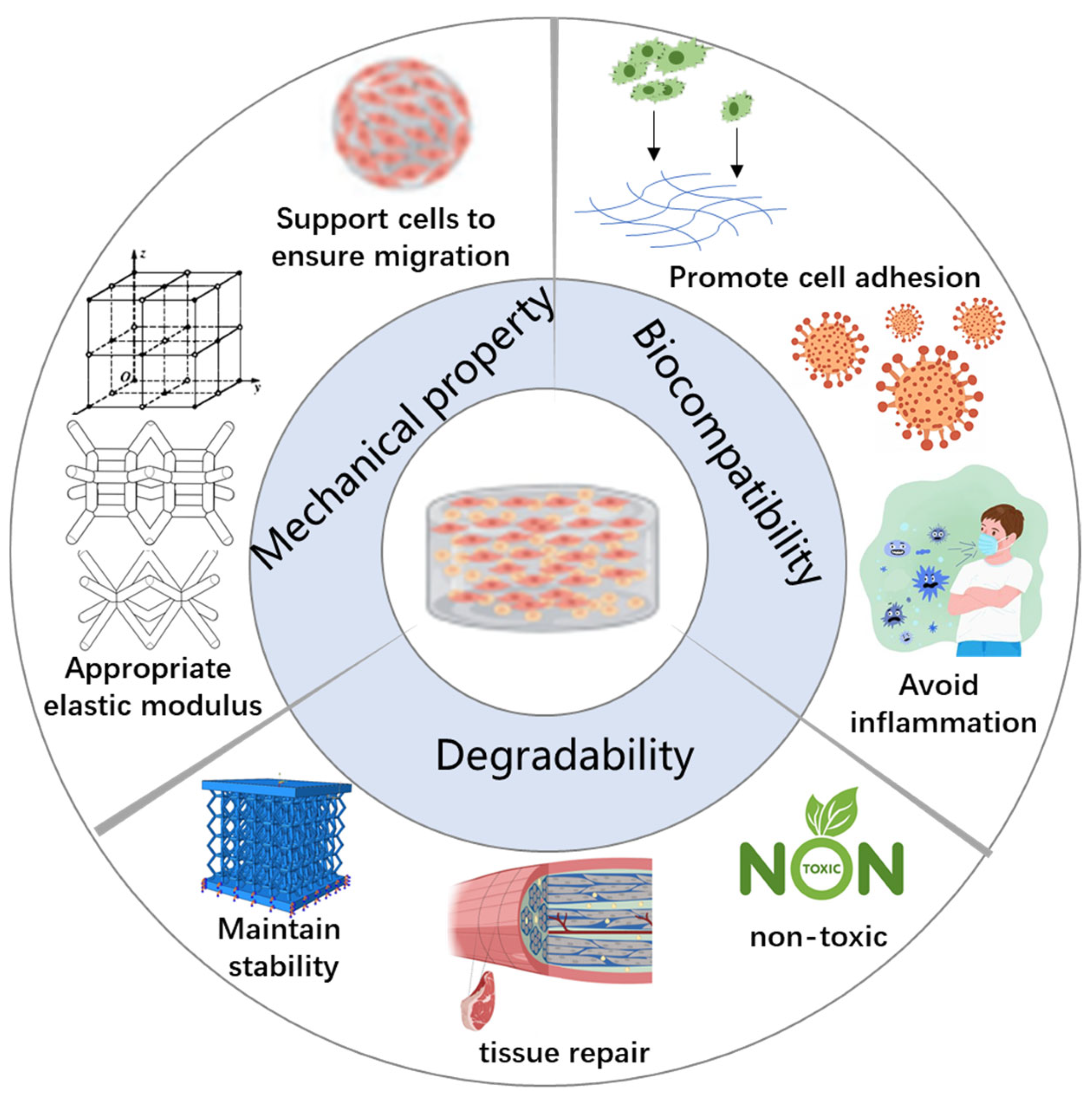
2.1. Key Performance Requirements
2.1.1. Biocompatibility
- Cell adhesion is the initial and crucial step in the interaction between scaffold materials and cells as effective adhesion supports subsequent cell proliferation and differentiation. Key factors influencing cell adhesion include the following: Surface chemical properties: Scaffold surfaces should possess suitable chemical functional groups such as hydroxyl (-OH), carboxyl (-COOH), and amino (-NH2) groups, which facilitate the binding of cell membrane proteins to the scaffold.
- Surface coating: Applying bioactive molecular coatings such as collagen, hyaluronic acid, or laminin can significantly enhance cell adhesion. For example, nanocellulose coatings have been shown to promote both adhesion and mineralization of osteoblasts.
- Topological structure: Micro- and nano-scale surface patterns created via 3D printing—such as nano-patterning or micro-grooves—can effectively regulate cell adhesion behavior. For example, rough surfaces on 3D-printed bone scaffolds have been found to encourage osteoblast adhesion more than smooth surfaces.
2.1.2. Degradability
- Maintaining mechanical stability: The degradation rate should align with the growth rate of new tissue to prevent scaffold collapse before tissue formation is complete [21]. For example, in bone tissue engineering, scaffolds must remain mechanically stable over extended periods to adequately support osteogenesis [22].
- Promoting tissue recovery: Biodegradable scaffolds can serve as slow-release carriers for bioactive factors such as vascular endothelial growth factor (VEGF), bone morphogenetic protein (BMP), anti-inflammatory drugs, or antibacterial agents. These substances are gradually released in tandem with scaffold degradation to enhance tissue regeneration [23].
- Non-toxic degradation products: Many synthetic polymers produce acidic by-products upon degradation; for example, polylactic acid (PLA) breaks down into lactic acid, which can cause local acidification and negatively impact cell viability. Therefore, improving the biocompatibility of degradation products remains a crucial research focus [23].
- Hydrolytic degradation: This process involves water molecules cleaving the chemical bonds within scaffold polymers, leading to gradual depolymerization. Commonly used materials undergoing hydrolysis include PLA and polyglycolic acid (PGA), which are widely applied in bone scaffolds and drug slow-release systems [26]. Notably, the degradation rate of starch-based scaffolds in aqueous environments can be finely controlled by adjusting their cross-linking degree. Hydrolytic degradation is particularly suitable for enzyme-free environments such as bone tissue engineering. However, it may generate acidic degradation products that can alter local pH, necessitating material optimization. Recent research indicates that the degradation rate of PLGA (poly(lactic-co-glycolic acid)) can be tailored by adjusting the PLA/PGA copolymer ratio, enabling control over degradation cycles ranging from weeks to several months.
- Enzymatic degradation: This mechanism involves selective cleavage of material chemical bonds by enzymes such as proteases and lipases present in the body. It is especially applicable to natural biomaterials such as gelatin, hyaluronic acid, chitosan, and alginate [27]. For example, silk protein scaffolds can be enzymatically degraded, with degradation rates adjustable through enzyme concentration. The degradation products are generally non-toxic [28]. However, enzymatic degradation rates can be variable and challenging to control precisely due to fluctuating enzyme levels in vivo. Recent advances have demonstrated that doping alginate scaffolds with proteolytic enzyme-responsive groups allows precise control over degradation, enabling dynamic tissue regeneration.
- Oxidative degradation: This pathway involves scaffold degradation through oxidation by free radicals or peroxides. It is suitable for synthetic polymers such as polyether imide and polycarbonate. Emerging smart biomaterials exploit oxidative environments, such as those present at sites of inflammation, to trigger scaffold degradation. This approach is promising for applications in inflammatory or hypoxic tissue repair. However, degradation products from oxidative processes may induce oxidative stress, requiring further modification of materials to mitigate adverse effects. Recent studies on oxidation-responsive smart scaffolds show accelerated degradation under pathological conditions characterized by high oxidative stress, offering innovative strategies for targeted tissue regeneration at inflammation sites.
2.1.3. Mechanical Property
- High mechanical strength: It should be capable of withstanding mechanical loads during transplantation and providing stable support throughout the tissue healing process.
- Appropriate elastic modulus: Mechanical properties should be tailored to closely mimic the native tissue, thereby promoting optimal cell behavior and tissue integration.
- Dynamic adjustability: It should have the ability to adapt to tissue growth and changing external environmental factors, such as variations in mechanical stress or humidity.
- Controlled degradation characteristics: Sufficient mechanical support should be maintained as the scaffold gradually degrades, ensuring sustained tissue growth and stability over time.
- Physical cross-linking: This approach utilizes temperature, pH changes, or ionic bonding (e.g., Ca2+ and alginate) [37]. This approach is typical for hydrogel scaffolds, offering good biocompatibility and tunability but resulting in relatively low mechanical strength and faster degradation.
- Chemical cross-linking: Agents such as glutaraldehyde, ultraviolet–visible (UV) light, or amide bond formation are employed to improve scaffold stability and durability. Although this method significantly increases mechanical strength, residual chemicals may affect biocompatibility.
- Starch-cellulose composites that enhance mechanical strength while maintaining biodegradability;
- Nano-hydroxyapatite composites, widely used in bone tissue engineering, providing superior mechanical properties and biological activity;
- Nano-SiO2 composites, which improve scaffold durability and optimize porosity;
- Chitosan–gelatin hydrogels, offering enhanced elasticity and biocompatibility for soft tissue engineering applications [38].
2.1.4. Printability
- Rheological properties: The bioink should exhibit shear-thinning behavior, meaning it has low viscosity during extrusion but rapidly recovers high viscosity after deposition to maintain structural integrity. The optimal viscosity range is approximately 1–300 mPa·s; too high viscosity hinders extrusion, while too low viscosity may cause collapse of the printed structure.
- Curing rate: Photocurable printing methods (e.g., SLA) require rapid photosensitive cross-linking, such as methylacryloyl hyaluronic acid curing under UV or blue light [42]. Extrusion bioprinting typically requires rapid curing through temperature changes (e.g., gelatin–alginate gels) or ionic cross-linking (e.g., Ca2+-induced alginate gelation).
- Biocompatibility: Bioinks must be compatible with cellular environments (pH and biochemical conditions), maintain a high cell viability rate (over 85%), and support cell proliferation and differentiation without adverse effects.
- Extrusion-based bioprinting (EBB): This is the most widely used bioprinting technique, requiring materials with good rheological behavior and shape retention.
- Inkjet printing (IJP): This is suitable for low-viscosity materials, enabling high-resolution printing, but demands strict control over surface tension and jetting properties.
- Stereolithography (SLA)/photocurable printing: This is ideal for micro-scale biological structures such as blood vessels and nerve scaffolds, requiring materials with rapid and efficient photosensitive cross-linking capabilities.
3. Analysis of Materials and Properties of Common 3D-Printed Biological Scaffolds
- Polymer materials: This category includes natural polymers such as gelatin, alginate, and chitosan, as well as synthetic polymers such as poly(lactic-co-glycolic acid) (PLGA) and polycaprolactone (PCL).
- Composite materials: These combine the beneficial properties of polymers and bioceramics to enhance the overall scaffold performance.
3.1. Natural Polymers
3.1.1. Gelatin
- Weak mechanical properties: Gelatin scaffolds are prone to collapse and deformation under mechanical stress, making them unsuitable for load-bearing applications such as bone tissue engineering. To enhance mechanical stability, gelatin is often combined with rigid materials such as PCL or nano-hydroxyapatite (nHA).
- Thermal sensitivity: Gelatin melts at temperatures above 37 °C, making it highly sensitive to thermal fluctuations during the printing process. To maintain its structural integrity, it must be printed at low temperatures (typically 4–10 °C).
- Rapid degradation: Gelatin degrades quickly, which can result in scaffold resorption before tissue regeneration is complete. This issue is commonly addressed by chemical cross-linking, copolymerization, or blending with other polymers to regulate its degradation rate.
- Skin Tissue Engineering: Gelatin scaffolds promote the growth of fibroblasts and keratinocytes, accelerating wound healing. Gelatin methacryloyl (GelMA), a photo-cross-linkable derivative of gelatin, has shown high-resolution printability and is widely used for constructing 3D skin scaffolds that mimic complex skin architecture.
- Cartilage Tissue Repair: Gelatin can be combined with hyaluronic acid, chitosan, or nHA to create scaffolds that support chondrocyte proliferation and cartilage matrix synthesis. Gelatin–silk fibroin composite hydrogels are particularly effective in articular cartilage repair.
- Soft Tissue Engineering (e.g., vascular and muscle tissue): Gelatin-based hydrogels serve as scaffolds for blood vessel formation, promoting endothelial cell adhesion and proliferation. In muscle regeneration, gelatin supports the alignment and growth of muscle fibers [55].
- Gelatin/Alginate Composite Scaffolds: Researchers have developed composite scaffolds by blending gelatin with alginate and using freeze-drying and 3D printing techniques. These scaffolds exhibited improved mechanical properties and promoted skin regeneration. In one study, Wang et al. demonstrated that a gelatin–alginate hydrogel significantly accelerated wound healing in animal models.
- GelMA Hydrogels: GelMA offers tunable mechanical strength, temperature sensitivity, and viscoelasticity. Its photocurable nature enables high-precision bioprinting [58]. Optimized GelMA scaffolds have shown excellent biostability in vitro and potential applications in cartilage and nerve tissue engineering [59,60].
- Gelatin/Chitosan and Gelatin/Nanocellulose Composites [61]: Combining gelatin with chitosan improves mechanical integrity and supports chondrocyte differentiation for cartilage repair [62,63]. Gelatin–nanocellulose scaffolds produced via 3D bioprinting have shown enhanced strength, cytocompatibility, and osteogenic potential, making them promising for bone tissue engineering [64,65].
3.1.2. Alginate
- Rapid Ion-Induced Gelation: Alginate quickly forms hydrogels in the presence of divalent cations (e.g., Ca2+, Ba2+, and Sr2+), enabling high-resolution 3D structures with excellent shape fidelity during printing [70].
- Tunable Mechanical and Degradation Properties: Mechanical strength, porosity, and degradation rate can be adjusted by modifying alginate’s molecular weight, altering cross-linker concentrations, or blending with other biomaterials, allowing its application across different tissue engineering demands [73,74].
- Lack of Cell Adhesion Motifs: Native alginate lacks bioactive sequences such as RGD (Arg-Gly-Asp), reducing its ability to support cell adhesion and spreading. It often requires modification or combination with collagen, gelatin, or chitosan for improved cytocompatibility [75].
- Undesirable Degradation Byproducts: Alginate degrades via hydrolysis and enzymatic breakdown, and its by-products may alter the local pH or adversely affect cell function. Optimization of degradation kinetics and by-product clearance is necessary for long-term applications.
- High Brittleness: Ionically cross-linked alginate hydrogels are typically brittle and mechanically weak, limiting their use in load-bearing tissues such as bone. Blending with nanocellulose, synthetic polymers, or chitosan is essential to improve toughness and mechanical stability [66].
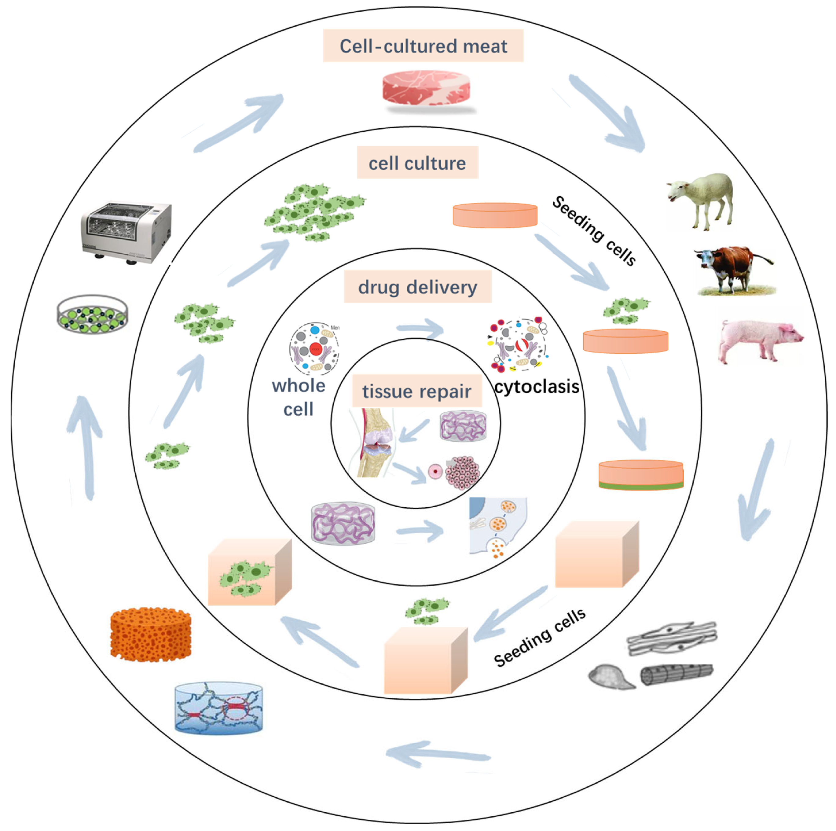
- Cell Encapsulation and Transplantation: Thanks to its mild gelation, alginate is extensively used for cell encapsulation in applications such as islet transplantation and neural tissue engineering. For example, alginate–chitosan microspheres have been shown to enhance liver cell metabolic activity.
- Controlled Drug Delivery Systems: Alginate gels are effective carriers for the sustained release of drugs, such as antibiotics, growth factors, and anticancer agents. Alginate combined with PLGA nanoparticles has been used to construct composite scaffolds for slow-release drug delivery, enhancing both drug stability and therapeutic effect.
- Preparation of alginate/gelatin composite scaffolds: Researchers mixed alginate with gelatin to create a 3D bioprinted model of skin tissue. Following freeze-drying, the composite scaffold exhibited significantly enhanced mechanical properties and showed potential for promoting skin tissue regeneration.
- Application of alginate gel in myocardial infarction repair: Alginate gel has been widely used in treating myocardial infarction due to its extracellular matrix-like structure, ease of modification, and good biocompatibility. In experimental studies, the compound alginate–saline gel demonstrated potential in promoting tissue regeneration.
- Use of alginate scaffolds in articular cartilage repair: Studies have shown that alginate composite hydrogel scaffolds can generate tissue resembling surrounding cartilage in animal models and effectively repair cartilage defects.
3.1.3. Hyaluronic Acid
- Promotion of cell adhesion: HA can bind to receptors on the cell surface; activate signaling pathways; and enhance stem cell proliferation, migration, and differentiation. Its effects on cellular behavior can be modulated by adjusting its molecular weight—high-molecular-weight HA contributes to scaffold stability, while low-molecular-weight HA promotes cell proliferation [83,84].
- Excellent moisturizing performance: HA’s ability to absorb water allows it to form a hydrogel matrix in vivo, creating a hydrated environment conducive to cell growth. This makes it particularly suitable for cartilage and skin tissue engineering as it helps prevent tissue dehydration and maintains microenvironment stability.
- Low mechanical strength: The soft hydrogel structure of HA cannot withstand high mechanical loads, making it unsuitable for load-bearing tissues such as bone. To improve its mechanical properties, HA must be blended with stiffer materials (e.g., nanocellulose).
- Cartilage tissue repair: HA is a major component of the articular cartilage matrix and can promote chondrocyte proliferation while enhancing the performance of cartilage repair scaffolds. Gelatin–HA composite hydrogels have been used in knee cartilage repair to improve cell adhesion and collagen synthesis [80,81,82,83,84,85,86,87,88].
- Cell culture matrix: HA hydrogels serve as effective scaffolds for cell culture, preserving cell viability and activity. They have been used for in vitro cultivation of neural stem cells, embryonic stem cells, and bone marrow mesenchymal stem cells.
4. Advantages of Starch-Based 3D Printing Scaffolds
- Soft tissue scaffolds: Starch hydrogels blended with biomaterials such as hyaluronic acid and gelatin can form highly biocompatible scaffolds, suitable for soft tissue repair applications such as skin regeneration, muscle tissue engineering, and vascular scaffolds [98].
- Drug delivery systems: Starch-based scaffolds can serve as biodegradable carriers for controlled drug release within the body. They help regulate the drug release rate, improve therapeutic efficacy, and function as degradable implants to minimize long-term adverse effects associated with implantable materials [99,100].
4.1. Advantages of Structure and Mechanical Properties
4.2. Degradation Characteristics
- Cross-linking modification: Pure starch degrades rapidly in aqueous environments; however, its degradation rate can be effectively controlled by blending or chemically cross-linking it with other polymers. For example, the cross-linked network formed by copolymerizing starch with PLA slows degradation, making it suitable for long-term support applications such as bone tissue engineering.
- Chemical modification: The degradation behavior of starch is influenced by the introduction of functional groups. Oxidized starch, which contains hydrophilic carboxyl groups, exhibits faster degradation, making it ideal for applications requiring rapid scaffold resorption, such as skin repair. Conversely, acetylated starch incorporates hydrophobic acetyl groups, which reduce the degradation rate and make it appropriate for implants requiring prolonged support.
4.3. Comparison with Other Biomaterials
- Mechanical strength: The mechanical strength of starch-based scaffolds surpasses that of gelatin and hyaluronic acid, making them particularly suitable for high-load tissues such as bone tissue engineering [112,113]. Studies have shown that starch–nanocellulose composite scaffolds significantly enhance structural stability [114]. In contrast, natural polymers such as gelatin and hyaluronic acid degrade easily in wet environments and exhibit poor form retention, whereas starch scaffolds maintain long-term support through cross-linking or composite reinforcement [115].
- Controllable degradation rate: Compared with alginate and hyaluronic acid, the degradation rate of starch-based scaffolds is tunable and can be optimized via cross-linking, chemical modification, or blending with other polymers [103]. For example, certain scaffolds degrade slowly, making them suitable for long-term tissue support, while oxidized starch degrades more rapidly, fitting short-term absorbable implant applications. In contrast, alginate scaffold degradation is less controllable, which may compromise application stability due to varying cellular environments or degrees of cross-linking.
- Cell adhesion: The surface of starch scaffolds can be modified with hydroxyl, carboxyl, or phosphoric acid groups to enhance cell adhesion and demonstrate stronger biocompatibility than alginate scaffolds [105,116]. Research indicates that starch–gelatin composite scaffolds effectively promote osteoblast adhesion and differentiation, outperforming pure alginate or hyaluronic acid scaffolds [117]. Although hyaluronic acid exhibits certain cellular compatibility, its low mechanical strength necessitates combination with other materials to form stable scaffolds [118].
- Three-dimensional printing adaptability: Starch-based scaffolds can be 3D-printed into complex structures, including porous scaffolds, gradient scaffolds, and supportive bone repair constructs. Alginate and hyaluronic acid inherently have low viscosity and weak molding capacity, often requiring blending with other polymers for suitable 3D printing [119]. Combining starch scaffolds with nanocellulose and bioceramics (such as hydroxyapatite) further improves mechanical properties and biological activity, meeting diverse tissue engineering needs [120].
5. Conclusions and Prospect
- Composite material optimization: Integrating nanocellulose, bioceramics, and other polymers to enhance mechanical strength, degradation control, and biological activity, thereby broadening their applicability across tissue engineering.
- Development of intelligent responsive scaffolds: Designing scaffolds with stimuli-responsive features such as pH sensitivity, temperature responsiveness, and controlled release of growth factors to better address diverse tissue repair needs and improve clinical outcomes.
- Optimization of 3D printing processes: Combining techniques such as melt extrusion, bioink printing, and optical curing to create high-precision, high-resolution starch-based scaffolds suitable for personalized medicine and precision tissue engineering.
- Biological functional modification: Employing surface modifications and functional cross-linking to improve cell adhesion, antibacterial properties, and biodegradation stability, thereby enhancing scaffold performance in tissue regeneration.
Author Contributions
Funding
Institutional Review Board Statement
Informed Consent Statement
Data Availability Statement
Conflicts of Interest
References
- Alami, A.H.; Olabi, A.G.; Khuri, S.; Aljaghoub, H.; Alasad, S.; Ramadan, M.; Abdelkareem, M.A. 3D printing in the food industry: Recent progress and role in achieving sustainable development goals. Ain Shams Eng. J. 2024, 15, 102386. [Google Scholar] [CrossRef]
- Engelsma, J.J.; Arora, S.S.; Jain, A.K.; Paulter, N.G. Universal 3D Wearable Fingerprint Targets: Advancing Fingerprint Reader Evaluations. IEEE Trans. Inf. Forensics Secur. 2018, 13, 1564–1578. [Google Scholar] [CrossRef]
- Farzan, A.; Borandeh, S.; Ezazi, N.Z.; Lipponen, S.; Santos, H.A.; Seppälä, J. 3D scaffolding of fast photocurable polyurethane for soft tissue engineering by stereolithography: Influence of materials and geometry on growth of fibroblast cells. Eur. Polym. J. 2020, 139, 109988. [Google Scholar] [CrossRef]
- Qi, Y.J.; Lv, H.Y.; Huang, Q.H.; Pan, G.Y. The Synergetic Effect of 3D Printing and Electrospinning Techniques in the Fabrication of Bone Scaffolds. Ann. Biomed. Eng. 2024, 52, 1518–1533. [Google Scholar] [CrossRef] [PubMed]
- Biazar, E.; Najafi, S.M.; Heidari, K.S.; Yazdankhah, M.; Rafiei, A.; Biazar, D. 3D bio-printing technology for body tissues and organs regeneration. J. Med. Eng. Technol. 2018, 42, 187–202. [Google Scholar] [CrossRef] [PubMed]
- Bhattacharyya, A.; Janarthanan, G.; Noh, I. Nano-biomaterials for designing functional bioinks towards complex tissue and organ regeneration in 3D bioprinting. Addit. Manuf. 2021, 37, 101639. [Google Scholar] [CrossRef]
- Sadeghianmaryan, A.; Ahmadian, N.; Wheatley, S.; Sardroud, H.A.; Nasrollah, S.A.S.; Naseri, E.; Ahmadi, A. Advancements in 3D-printable polysaccharides, proteins, and synthetic polymers for wound dressing and skin scaffolding—A review. Int. J. Biol. Macromol. 2024, 266, 131207. [Google Scholar] [CrossRef]
- Alam, A.; Kim, C.J.; Kim, S.H.; Kumari, S.; Lee, E.Y.; Hwang, Y.H.; Joo, S.T. Scaffolding fundamentals and recent advances in sustainable scaffolding techniques for cultured meat development. Food Res. Int. 2024, 189, 114549. [Google Scholar] [CrossRef]
- Do, A.V.; Khorsand, B.; Geary, S.M.; Salem, A.K. 3D Printing of Scaffolds for Tissue Regeneration Applications. Adv. Healthc. Mater. 2015, 4, 1742–1762. [Google Scholar] [CrossRef]
- Rodrigo-Navarro, A.; Sankaran, S.; Dalby, M.J.; del Campo, A.; Salmeron-Sanchez, M. Engineered living biomaterials. Nat. Rev. Mater. 2021, 6, 1175–1190. [Google Scholar] [CrossRef]
- Chiesa-Estomba, C.M.; Aiastui, A.; González-Fernández, I.; Hernáez-Moya, R.; Rodiño, C.; Delgado, A.; Garces, J.P.; Paredes-Puente, J.; Aldazabal, J.; Altuna, X.; et al. Three-Dimensional Bioprinting Scaffolding for Nasal Cartilage Defects: A Systematic Review. Tissue Eng. Regen. Med. 2021, 18, 343–353. [Google Scholar] [CrossRef]
- Laird, N.Z.; Acri, T.M.; Chakka, J.L.; Quarterman, J.C.; Malkawi, W.I.; Elangovan, S.; Salem, A.K. Applications of nanotechnology in 3D printed tissue engineering scaffolds. Eur. J. Pharm. Biopharm. 2021, 161, 15–28. [Google Scholar] [CrossRef] [PubMed]
- Yadav, L.R.; Chandran, S.V.; Lavanya, K.; Selvamurugan, N. Chitosan-based 3D-printed scaffolds for bone tissue engineering. Int. J. Biol. Macromol. 2021, 183, 1925–1938. [Google Scholar] [CrossRef] [PubMed]
- Maresca, J.A.; DeMel, D.C.; Wagner, G.A.; Haase, C.; Geibel, J.P. Three-Dimensional Bioprinting Applications for Bone Tissue Engineering. Cells 2023, 12, 1230. [Google Scholar] [CrossRef] [PubMed]
- Zaszczyńska, A.; Niemczyk-Soczynska, B.; Sajkiewicz, P. A Comprehensive Review of Electrospun Fibers, 3D-Printed Scaffolds, and Hydrogels for Cancer Therapies. Polymers 2022, 14, 5278. [Google Scholar] [CrossRef]
- Al Sawaftah, N.M.; Pitt, W.G.; Husseini, G.A. Incorporating nanoparticles in 3D printed scaffolds for bone cancer therapy. Bioprinting 2023, 36, e00322. [Google Scholar] [CrossRef]
- Svanström, A.; Rosendahl, J.; Salerno, S.; Leiva, M.C.; Gregersson, P.; Berglin, M.; Bogestål, Y.; Lausmaa, J.; Oko, A.; Chinga-Carrasco, G.; et al. Optimized alginate-based 3D printed scaffolds as a model of patient derived breast cancer microenvironments in drug discovery. Biomed. Mater. 2021, 16, 045046. [Google Scholar] [CrossRef]
- Jung, M.; Ghamrawi, S.; Du, E.Y.; Gooding, J.J.; Kavallaris, M. Advances in 3D Bioprinting for Cancer Biology and Precision Medicine: From Matrix Design to Application. Adv. Healthc. Mater. 2022, 11, 2200690. [Google Scholar] [CrossRef]
- Zhang, X.; Wu, W. Research progress on 3D printing technology and biomaterials for bone reconstruction in maxillofacial regions. J. Int. Stomatol. 2020, 47, 677–685. [Google Scholar] [CrossRef]
- Iravani, S.; Varma, R.S. Cellulose-Based Composites as Scaffolds for Tissue Engineering: Recent Advances. Molecules 2022, 27, 8830. [Google Scholar] [CrossRef]
- Dalal, N.; Challa, R.; Thimukonda, J.J.; Tayalia, P. Gelatin Methacryloyl Based Injectable Cryogels with Tunable Degradability for Cell Delivery. Macromol. Biosci. 2024, 24, 2200562. [Google Scholar] [CrossRef]
- Khan, A.R.; Grewal, N.S.; Jun, Z.; Tawfiq, F.M.O.; Tchier, F.; Zulqarnain, R.; Zhang, H.J. Raising the Bar: Progress in 3D-Printed Hybrid Bone Scaffolds for Clinical Applications: A Review. Cell Transplant. 2024, 33, 1–17. [Google Scholar] [CrossRef]
- Morouço, P.; Azimi, B.; Milazzo, M.; Mokhtari, F.; Fernandes, C.; Reis, D.; Danti, S. Four-Dimensional (Bio-)printing: A Review on Stimuli-Responsive Mechanisms and Their Biomedical Suitability. Appl. Sci. 2020, 10, 9143. [Google Scholar] [CrossRef]
- Koons, G.L.; Kontoyiannis, P.D.; Diaz-Gomez, L.; Elsarrag, S.Z.; Scott, D.W.; Diba, M.; Mikos, A.G. Influence of Polymeric Microparticle Size and Loading Concentration on 3D Printing Accuracy and Degradation Behavior of Composite Scaffolds. 3D Print. Addit. Manuf. 2024, 11, e813–e827. [Google Scholar] [CrossRef]
- Aranci, K.; Uzun, M.; Su, S.N.; Cesur, S.; Ulag, S.; Amin, A.; Guncu, M.M.; Aksu, B.; Kolayli, S.; Ustundag, C.B.; et al. 3D Propolis-Sodium Alginate Scaffolds: Influence on Structural Parameters, Release Mechanisms, Cell Cytotoxicity and Antibacterial Activity. Molecules 2020, 25, 5082. [Google Scholar] [CrossRef]
- Shi, Z.; Liu, W.Y.; Zhai, D.; Xie, J.J.; Zhu, Y.F. Akermanite Scaffolds for Bone Tissue Engineering: 3D Printing Using Polymer Precursor and Scaffold Properties. J. Inorg. Mater. 2023, 38, 763–770. [Google Scholar] [CrossRef]
- Liu, J.Y.; Luo, D.M.; Fu, X.Y.; Yang, T.T.; Hou, R.X.; Li, P.W.; Chen, Y.R.; Zhang, X.Y.; Sun, X.N.; Yue, Y.G.; et al. Preparation, Characterization, and In Vitro Osteogenic Properties of a Novel Glucose-Sensitive 3D-Printed Scaffold Containing Metformin Based on Enzymatic Cascade Reaction. J. Polym. Sci. 2025, 63, 595–609. [Google Scholar] [CrossRef]
- Ye, J.J.; Chu, T.S.; Chu, J.L.; Gao, B.B.; He, B.F. A Versatile Approach for Enzyme Immobilization Using Chemically Modified 3D-Printed Scaffolds. ACS Sustain. Chem. Eng. 2019, 7, 18048–18054. [Google Scholar] [CrossRef]
- Sultan, S.; Thomas, N.; Varghese, M.; Dalvi, Y.; Joy, S.; Hall, S.; Mathew, A.P. The Design of 3D-Printed Polylactic Acid-Bioglass Composite Scaffold: A Potential Implant Material for Bone Tissue Engineering. Molecules 2022, 27, 7214. [Google Scholar] [CrossRef] [PubMed]
- Nacu, I.; Bercea, M.; Niță, L.E.; Peptu, C.A.; Butnaru, M.; Vereștiuc, L. 3D bioprinted scaffolds based on functionalized gelatin for soft tissue engineering. React. Funct. Polym. 2023, 190, 105636. [Google Scholar] [CrossRef]
- Gokyer, S.; Yilgor, E.; Yilgor, I.; Berber, E.; Vrana, E.; Orhan, K.; Monsef, Y.A.; Guvener, O.; Zinnuroglu, M.; Oto, C.; et al. 3D Printed Biodegradable Polyurethaneurea Elastomer Recapitulates Skeletal Muscle Structure and Function. ACS Biomater. Sci. Eng. 2021, 7, 5189–5205. [Google Scholar] [CrossRef]
- Ullah, M.; Bibi, A.; Wahab, A.; Hamayun, S.; Rehman, M.U.; Khan, S.U.; Awan, U.A.; Riaz, N.-U.-A.; Naeem, M.; Saeed, S.; et al. Shaping the Future of Cardiovascular Disease by 3D Printing Applications in Stent Technology and its Clinical Outcomes. Curr. Probl. Cardiol. 2024, 49, 102039. [Google Scholar] [CrossRef]
- Spreda, M.; Hauptmann, N.; Lehner, V.; Biehl, C.; Liefeith, K.; Lips, K.S. Porous 3D Scaffolds Enhance MSC Vitality and Reduce Osteoclast Activity. Molecules 2021, 26, 6258. [Google Scholar] [CrossRef]
- Vaezi, M.; Black, C.; Gibbs, D.M.R.; Oreffo, R.O.C.; Brady, M.; Moshrefi-Torbati, M.; Yang, S.F. Characterization of New PEEK/HA Composites with 3D HA Network Fabricated by Extrusion Freeforming. Molecules 2016, 21, 687. [Google Scholar] [CrossRef]
- Arifin, N.; Sudin, I.; Ngadiman, N.H.A.; Ishak, M.S.A. A Comprehensive Review of Biopolymer Fabrication in Additive Manufacturing Processing for 3D-Tissue-Engineering Scaffolds. Polymers 2022, 14, 2119. [Google Scholar] [CrossRef] [PubMed]
- Xu, C.L.; Molino, B.Z.; Wang, X.J.; Cheng, F.; Xu, W.Y.; Molino, P.; Bacher, M.; Su, D.D.; Rosenau, T.; Willför, S.; et al. 3D printing of nanocellulose hydrogel scaffolds with tunable mechanical strength towards wound healing application. J. Mater. Chem. B 2018, 6, 7066–7075. [Google Scholar] [CrossRef] [PubMed]
- Ardelean, I.L.; Gudovan, D.; Ficai, D.; Ficai, A.; Andronescu, E.; Albu-Kaya, M.G.; Neacsu, P.; Ion, R.N.; Cimpean, A.; Mitran, V. Collagen/hydroxyapatite bone grafts manufactured by homogeneous/heterogeneous 3D printing. Mater. Lett. 2018, 231, 179–182. [Google Scholar] [CrossRef]
- Xu, Y.H.; Zhang, F.Y.; Zhai, W.J.; Cheng, S.J.; Li, J.H.; Wang, Y. Unraveling of Advances in 3D-Printed Polymer-Based Bone Scaffolds. Polymers 2022, 14, 566. [Google Scholar] [CrossRef]
- Panwar, A.; Tan, L.P. Current Status of Bioinks for Micro-Extrusion-Based 3D Bioprinting. Molecules 2016, 21, 685. [Google Scholar] [CrossRef]
- Pudkon, W.; Laomeephol, C.; Damrongsakkul, S.; Kanokpanont, S.; Ratanavaraporn, J. Comparative Study of Silk Fibroin-Based Hydrogels and Their Potential as Material for 3-Dimensional (3D) Printing. Molecules 2021, 26, 3887. [Google Scholar] [CrossRef]
- Naghieh, S.; Chen, X.B. Printability-A key issue in extrusion-based bioprinting. J. Pharm. Anal. 2021, 11, 564–579. [Google Scholar] [CrossRef]
- Galarraga, J.H.; Kwon, M.Y.; Burdick, J.A. 3D bioprinting via an in situ crosslinking technique towards engineering cartilage tissue. Sci. Rep. 2019, 9, 19987. [Google Scholar] [CrossRef]
- Chopin-Doroteo, M.; Mandujano-Tinoco, E.A.; Krötzsch, E. Tailoring of the rheological properties of bioinks to improve bioprinting and bioassembly for tissue replacement. Biochim. Biophys. Acta (BBA)-Gen. Subj. 2021, 1865, 129782. [Google Scholar] [CrossRef]
- Wu, Y.F.; Wen, Y.T.; Salamanca, E.; Aung, L.M.; Chao, Y.Q.; Chen, C.Y.; Sun, Y.S.; Chang, W.J. 3D-bioprinted alginate-based bioink scaffolds with β-tricalcium phosphate for bone regeneration applications. J. Dent. Sci. 2024, 19, 1116–1125. [Google Scholar] [CrossRef]
- Liu, K.; Zhang, Y.F.; Huang, L.; Feng, C.Z.; Li, Y.T.; Zhang, S.Q.; Jin, X.; Jiang, H.J.; Zhu, Q.; Zhang, P. Enhanced printability of high-viscosity chitosan/acrylamide inks via aluminum ions coordination for precision 3D bioprinting of scaffolds. Carbohydr. Polym. 2025, 355, 123359. [Google Scholar] [CrossRef]
- Dzobo, K.; Motaung, K.; Adesida, A. Recent Trends in Decellularized Extracellular Matrix Bioinks for 3D Printing: An Updated Review. Int. J. Mol. Sci. 2019, 20, 4628. [Google Scholar] [CrossRef] [PubMed]
- Choi, Y.; Kim, C.; Kim, H.S.; Moon, C.; Lee, K.Y. 3D Printing of dynamic tissue scaffold by combining self-healing hydrogel and self-healing ferrogel. Colloids Surf. B Biointerfaces 2021, 208, 112108. [Google Scholar] [CrossRef] [PubMed]
- Bashiri, Z.; Amiri, I.; Gholipourmalekabadi, M.; Falak, R.; Asgari, H.; Maki, C.B.; Moghaddaszadeh, A.; Koruji, M. Artificial testis: A testicular tissue extracellular matrix as a potential bio-ink for 3D printing. Biomater. Sci. 2021, 9, 3465–3484. [Google Scholar] [CrossRef]
- Kim, J.W.; Han, Y.S.; Lee, H.M.; Kim, J.K.; Kim, Y.J. Effect of Morphological Characteristics and Biomineralization of 3D-Printed Gelatin/Hyaluronic Acid/Hydroxyapatite Composite Scaffolds on Bone Tissue Regeneration. Int. J. Mol. Sci. 2021, 22, 6794. [Google Scholar] [CrossRef]
- Wang, R.Q.; Deng, S.; Wu, Y.P.; Wei, H.Y.; Jing, G.P.; Zhang, B.S.; Liu, F.Z.; Tian, H.; Chen, X.B.; Tian, W.M. Remodelling 3D printed GelMA-HA corneal scaffolds by cornea stromal cells. Colloid Interface Sci. Commun. 2022, 49, 100632. [Google Scholar] [CrossRef]
- Bahcecioglu, G.; Hasirci, N.; Bilgen, B.; Hasirci, V. Hydrogels of agarose, and methacrylated gelatin and hyaluronic acid are more supportive for in vitro meniscus regeneration than three dimensional printed polycaprolactone scaffolds. Int. J. Biol. Macromol. 2019, 122, 1152–1162. [Google Scholar] [CrossRef]
- Ibañez, R.I.R.; do Amaral, R.; Reis, R.L.; Marques, A.P.; Murphy, C.M.; O’Brien, F.J. 3D-Printed Gelatin Methacrylate Scaffolds with Controlled Architecture and Stiffness Modulate the Fibroblast Phenotype towards Dermal Regeneration. Polymers 2021, 13, 2510. [Google Scholar] [CrossRef] [PubMed]
- Olate-Moya, F.; Rubí-Sans, G.; Engel, E.; Mateos-Timoneda, M.A.; Palza, H. 3D Bioprinting of Biomimetic Alginate/Gelatin/Chondroitin Sulfate Hydrogel Nanocomposites for Intrinsically Chondrogenic Differentiation of Human Mesenchymal Stem Cells. Biomacromolecules 2024, 25, 3312–3324. [Google Scholar] [CrossRef] [PubMed]
- Li, S.Y.; Huang, C.Z.; Liu, H.L.; Han, X.; Wang, Z.C.; Huang, J.; Yan, Y.G.; Wang, Z. A Silk Fibroin Methacryloyl-Modified Hydrogel Promoting Cell Adhesion for Customized 3D Cell-Laden Structures. ACS Appl. Polym. Mater. 2022, 4, 7014–7024. [Google Scholar] [CrossRef]
- Simnska-Stanny, J.S.; Hachemi, F.; Dodi, G.; Cojocaru, F.D.; Gardikiotis, I.; Podstawczyk, D.; Delporte, C.; Jiang, G.H.; Nie, L.; Shavandi, A. Optimizing phenol-modified hyaluronic acid for designing shape-maintaining biofabricated hydrogel scaffolds in soft tissue engineering. Int. J. Biol. Macromol. 2023, 244, 125201. [Google Scholar] [CrossRef]
- Markovic, M.; Van Hoorick, J.; Holzl, K.; Tromayer, M.; Gruber, P.; Nurnberger, S.; Dubruel, P.; Van Vlierberghe, S.; Liska, R.; Ovsianikov, A. Hybrid Tissue Engineering Scaffolds by Combination of Three-Dimensional Printing and Cell Photoencapsulation. J. Nanotechnol. Eng. Med. 2015, 6, 0210011–0210017. [Google Scholar] [CrossRef]
- Xia, Z.H.; Guo, B.; Wu, D.N.; Yang, F.; Ding, Y.D. Advances of natural hydrogel-based vascularization strategies for soft tissue repair. Front. Mater. 2024, 11, 1446035. [Google Scholar] [CrossRef]
- Gao, Q.; Niu, X.F.; Shao, L.; Zhou, L.Y.; Lin, Z.W.; Sun, A.Y.; Fu, J.Z.; Chen, Z.C.; Hu, J.; Liu, Y.D.; et al. 3D printing of complex GelMA-based scaffolds with nanoclay. Biofabrication 2019, 11, 035006. [Google Scholar] [CrossRef]
- Elomaa, L.; Keshi, E.; Sauer, I.M.; Weinhart, M. Development of GelMA/PCL and dECM/PCL resins for 3D printing of acellular in vitro tissue scaffolds by stereolithography. Mater. Sci. Eng. C-Mater. Biol. Appl. 2020, 112, 110958. [Google Scholar] [CrossRef]
- Choi, D.; Qiu, M.F.; Hwang, Y.C.; Oh, W.M.; Koh, J.T.; Park, C.; Lee, B.N. The Effects of 3-Dimensional Bioprinting Calcium Silicate Cement/Methacrylated Gelatin Scaffold on the Proliferation and Differentiation of Human Dental Pulp Stem Cells. Materials 2022, 15, 2170. [Google Scholar] [CrossRef]
- Heidarian, P.; Kouzani, A.Z. A self-healing nanocomposite double network bacterial nanocellulose/gelatin hydrogel for three dimensional printing. Carbohydr. Polym. 2023, 313, 120879. [Google Scholar] [CrossRef]
- Xu, X.D.; Zhou, J.P.; Feng, C.; Jiang, Y.N.; Zhang, Q.; Shi, H.C. 3D printing algorithm of anisotropic biological scaffold with oxidized nanocellulose and gelatin. J. Biomater. Sci. -Polym. Ed. 2019, 30, 1260–1275. [Google Scholar] [CrossRef]
- Cernencu, A.I.; Lungu, A.; Dragusin, D.M.; Stancu, I.C.; Dinescu, S.; Balahura, L.R.; Mereuta, P.; Costache, M.; Iovu, H. 3D Bioprinting of Biosynthetic Nanocellulose-Filled GelMA Inks Highly Reliable for Soft Tissue-Oriented Constructs. Materials 2021, 14, 4891. [Google Scholar] [CrossRef] [PubMed]
- Xu, X.D.; Zhou, J.P.; Jiang, Y.N.; Zhang, Q.; Shi, H.C.; Liu, D.F. 3D printing process of oxidized nanocellulose and gelatin scaffold. J. Biomater. Sci. -Polym. Ed. 2018, 29, 1498–1513. [Google Scholar] [CrossRef] [PubMed]
- Jiang, Y.N.; Zhou, J.P.; Yang, Z.; Liu, D.F.; Xv, X.D.; Zhao, G.Q.; Shi, H.C.; Zhang, Q. Dialdehyde cellulose nanocrystal/gelatin hydrogel optimized for 3D printing applications. J. Mater. Sci. 2018, 53, 11883–11900. [Google Scholar] [CrossRef]
- Khoshnood, N.; Zamanian, A.; Abbasi, M. The potential impact of polyethylenimine on biological behavior of 3D-printed alginate scaffolds. Int. J. Biol. Macromol. 2021, 178, 19–28. [Google Scholar] [CrossRef]
- Abouzeid, R.E.; Khiari, R.; Salama, A.; Diab, M.; Beneventi, D.; Dufresne, A. In situ mineralization of nano-hydroxyapatite on bifunctional cellulose nanofiber/polyvinyl alcohol/sodium alginate hydrogel using 3D printing. Int. J. Biol. Macromol. 2020, 160, 538–547. [Google Scholar] [CrossRef]
- Lin, Z.Y.; Xie, W.K.; Cui, Z.H.; Huang, J.A.; Cao, H.; Li, Y. 3D printed alginate/gelatin-based porous hydrogel scaffolds to improve diabetic wound healing. Giant 2023, 16, 100185. [Google Scholar] [CrossRef]
- Kim, M.H.; Lee, Y.W.; Jung, W.K.; Oh, J.; Nam, S.Y. Enhanced rheological behaviors of alginate hydrogels with carrageenan for extrusion-based bioprinting. J. Mech. Behav. Biomed. Mater. 2019, 98, 187–194. [Google Scholar] [CrossRef]
- Feng, L.I.U.; Renji, Z.; Yongnian, Y.A.N.; Haixia, L.I.U. Rapid prototyping of gelatin/sodium alginate tissue engineering scaffolds. J. Tsinghua Univ. (Sci. Technol.) 2006, 46, 1357–1360. [Google Scholar]
- Fu, S.Y.; Du, X.Y.; Zhu, M.; Tian, Z.F.; Wei, D.X.; Zhu, Y.F. 3D printing of layered mesoporous bioactive glass/sodium alginate-sodium alginate scaffolds with controllable dual-drug release behaviors. Biomed. Mater. 2019, 14, 065011. [Google Scholar] [CrossRef]
- Wang, Z.Y.; Liu, C.Y.; Chen, B.R.; Luo, Y.X. Magnetically-driven drug and cell on demand release system using 3D printed alginate based hollow fiber scaffolds. Int. J. Biol. Macromol. 2021, 168, 38–45. [Google Scholar] [CrossRef]
- Alruwaili, M.; Lopez, J.A.; McCarthy, K.; Reynaud, E.G.; Rodriguez, B.J. Liquid-phase 3D bioprinting of gelatin alginate hydrogels: Influence of printing parameters on hydrogel line width and layer height. Bio-Des. Manuf. 2019, 2, 172–180. [Google Scholar] [CrossRef]
- Sümbelli, Y.; Diltemiz, S.E.; Say, M.G.; Ünlüer, Ö.; Ersöz, A.; Say, R. In situ and non-cytotoxic cross-linking strategy for 3D printable biomaterials. Soft Matter 2021, 17, 1008–1015. [Google Scholar] [CrossRef] [PubMed]
- Ianovici, I.; Zagury, Y.; Redenski, I.; Lavon, N.; Levenberg, S. 3D-printable plant protein-enriched scaffolds for cultivated meat development. Biomaterials 2022, 284, 121487. [Google Scholar] [CrossRef] [PubMed]
- Hu, X.L.; Zhang, Z.; Wu, H.M.; Yang, S.H.; Zhao, W.M.; Che, L.Y.; Wang, Y.; Cao, J.F.; Li, K.A.; Qian, Z.Y. Progress in the application of 3D-printed sodium alginate-based hydrogel scaffolds in bone tissue repair. Biomater. Adv. 2023, 152, 213501. [Google Scholar] [CrossRef]
- Cojocaru, E.; Oprea, M.; Vlasceanu, G.M.; Nicolae, M.C.; Popescu, R.C.; Mereuta, P.E.; Toader, A.G.; Ionita, M. Dual nanofiber and graphene reinforcement of 3D printed biomimetic supports for bone tissue repair. Rsc Adv. 2024, 14, 32517–32532. [Google Scholar] [CrossRef]
- Souza, A.; Kevin, M.; Rodriguez, B.J.; Reynaud, E.G. The use of fluid-phase 3D printing to pattern alginate-gelatin hydrogel properties to guide cell growth and behaviour in vitro. Biomed. Mater. 2024, 19, 045024. [Google Scholar] [CrossRef]
- Bavaresco, B.; Comín, R.; Salvatierra, N.A.; Cid, M.P. Three-dimensional printing of collagen and hyaluronic acid scaffolds with dehydrothermal treatment crosslinking. Compos. Commun. 2020, 19, 1–5. [Google Scholar] [CrossRef]
- Xia, H.T.; Zhao, D.D.; Zhu, H.L.; Hua, Y.J.; Xiao, K.Y.; Xu, Y.; Liu, Y.Q.; Chen, W.M.; Liu, Y.; Zhang, W.J.; et al. Lyophilized Scaffolds Fabricated from 3D-Printed Photocurable Natural Hydrogel for Cartilage Regeneration. ACS Appl. Mater. Interfaces 2018, 10, 31704–31715. [Google Scholar] [CrossRef]
- Müller, M.; Fisch, P.; Molnar, M.; Eggert, S.; Binelli, M.; Maniura-Weber, K.; Zenobi-Wong, M. Development and thorough characterization of the processing steps of an ink for 3D printing for bone tissue engineering. Mater. Sci. Eng. C-Mater. Biol. Appl. 2020, 108, 110510. [Google Scholar] [CrossRef]
- Shu, K.G.; Huang, Z.Q.; Pei, X.M.; Yew, P.Y.M.; Wei, S.S.; Yang, Y.; Lan, Y.; Kai, D.; Zheng, L.; Zhao, J.M. 3D printing of high-strength photo-crosslinking flaxseed gum bioink for cartilage regeneration. Compos. Part B Eng. 2023, 263, 110864. [Google Scholar] [CrossRef]
- Li, S.L.; Tang, L.Y.; Pu, J.; Wang, J.L.; Fan, C.Z.; Li, Z.; Song, J. Continuous Hyaluronic Acid Supply by a UHMWPE/PEEK Interlocking Scaffold for Metatarsophalangeal Joint Prosthesis Lubricating Applications. ACS Appl. Mater. Interfaces 2025, 17, 11704–11717. [Google Scholar] [CrossRef]
- Chen, H.; Xue, H.Q.; Zeng, H.X.; Dai, M.H.; Tang, C.X.; Liu, L.L. 3D printed scaffolds based on hyaluronic acid bioinks for tissue engineering: A review. Biomater. Res. 2023, 27, 137. [Google Scholar] [CrossRef]
- Kang, M.S.; Kwon, M.; Lee, S.H.; Kim, W.H.; Lee, G.W.; Jo, H.J.; Kim, B.; Yang, S.Y.; Kim, K.S.; Han, D.W. 3D Printing of Skin Equivalents with Hair Follicle Structures and Epidermal-Papillary-Dermal Layers Using Gelatin/Hyaluronic Acid Hydrogels. Chem. -Asian J. 2022, 17, e202200620. [Google Scholar] [CrossRef] [PubMed]
- Fernández, P.A.; Cid, M.P.; Comín, R.; Velasco, M.I. Structural Characterization and Hydration Dynamics of Cross-Linked Collagen and Hyaluronic Acid Scaffolds by Nuclear Magnetic Resonance. J. Phys. Chem. B 2024, 128, 12143–12153. [Google Scholar] [CrossRef] [PubMed]
- Nedunchezian, S.; Banerjee, P.; Lee, C.Y.; Lee, S.S.; Lin, C.W.; Wu, C.W.; Wu, S.C.; Chang, J.K.; Wang, C.K. Generating adipose stem cell-laden hyaluronic acid-based scaffolds using 3D bioprinting via the double crosslinked strategy for chondrogenesis. Mater. Sci. Eng. C-Mater. Biol. Appl. 2021, 124, 112072. [Google Scholar] [CrossRef] [PubMed]
- Shie, M.Y.; Chang, W.C.; Wei, L.J.; Huang, Y.H.; Chen, C.H.; Shih, C.T.; Chen, Y.W.; Shen, Y.F. 3D Printing of Cytocompatible Water-Based Light-Cured Polyurethane with Hyaluronic Acid for Cartilage Tissue Engineering Applications. Materials 2017, 10, 136. [Google Scholar] [CrossRef]
- Petta, D.; D’Amora, U.; Ambrosio, L.; Grijpma, D.W.; Eglin, D.; D’Este, M. Hyaluronic acid as a bioink for extrusion-based 3D printing. Biofabrication 2020, 12, 032001. [Google Scholar] [CrossRef]
- Sonthithai, P.; Kaewkong, P.; Channasanon, S.; Tanodekaew, S. 3D-Printed PEG-PLA/Gelatin Hydrogel: Characterization toward In Vitro Chondrocyte Redifferentiation. ACS Biomater. Sci. Eng. 2025, 11, 2157–2166. [Google Scholar] [CrossRef]
- Gorgol, D.; Mrlík, M.; Mikulka, F.; Vichová, Z.; Mahelová, L.; Ilcíková, M.; Minarík, A. Smart Biopolymer Scaffolds Based on Hyaluronic Acid and Carbonyl Iron Microparticles: 3D Printing, Magneto-Responsive, and Cytotoxicity Study. ACS Appl. Bio Mater. 2024, 7, 7483–7493. [Google Scholar] [CrossRef]
- Wang, Y.P.; Chen, Y.Z.; Zheng, J.N.; Liu, L.R.; Zhang, Q.Q. Three-Dimensional Printing Self-Healing Dynamic/Photocrosslinking Gelatin-Hyaluronic Acid Double-Network Hydrogel for Tissue Engineering. ACS Omega 2022, 7, 12076–12088. [Google Scholar] [CrossRef]
- Ai, Y.; Jane, J.L. Gelatinization and rheological properties of starch. Starch-Starke 2015, 67, 213–224. [Google Scholar] [CrossRef]
- Mahmood, K.; Kamilah, H.; Shang, P.L.; Sulaiman, S.; Ariffin, F.; Alias, A. A review: Interaction of starch/non-starch hydrocolloid blending and the recent food applications. Food Biosci. 2017, 19, 110–120. [Google Scholar] [CrossRef]
- Bugarin-Castillo, Y.; Rando, P.; Clabaux, M.; Moulin, G.; Ramaioli, M. 3D printing to modulate the texture of starch-based food. Journal of Food Engineering 2023, 350, 111499. [Google Scholar] [CrossRef]
- Bhattacharjee, A.; Bose, S. 3D printed hydroxyapatite—Zn2+ functionalized starch composite bone grafts for orthopedic and dental applications. Mater. Des. 2022, 221, 110903. [Google Scholar] [CrossRef] [PubMed]
- Hao, M.; Xue, L.; Wen, X.; Sun, L.; Zhang, L.; Xing, K.; Hu, X.; Xu, J.; Xing, D. Advancing bone regeneration: Unveiling the potential of 3D cell models in the evaluation of bone regenerative materials. Acta Biomaterialia 2024, 183, 1–29. [Google Scholar] [CrossRef]
- Joshi, T.; Joshi, H.; Bhatt, S.; Pancholi, M.; Bagchi, D. Biopolymer composite matrix structure nano architectonics and its key role in regulating mechanical tunability for biomedical applications. J. Polym. Res. 2025, 32, 130. [Google Scholar] [CrossRef]
- Cardoso, S.; Narciso, F.; Monge, N.; Bettencourt, A.; Ribeiro, I.A.C. Improving Chitosan Hydrogels Printability: A Comprehensive Study on Printing Scaffolds for Customized Drug Delivery. Int. J. Mol. Sci. 2023, 24, 973. [Google Scholar] [CrossRef]
- Naseri, E.; Cartmell, C.; Saab, M.; Kerr, R.G.; Ahmadi, A. Development of N,O-Carboxymethyl Chitosan-Starch Biomaterial Inks for 3D Printed Wound Dressing Applications. Macromol. Biosci. 2021, 21, 2100368. [Google Scholar] [CrossRef]
- Liu, J.; Sun, L.S.; Xu, W.Y.; Wang, Q.Q.; Yu, S.J.; Sun, J.Z. Current advances and future perspectives of 3D printing natural-derived biopolymers. Carbohydr. Polym. 2019, 207, 297–316. [Google Scholar] [CrossRef]
- Pfister, A.; Landers, R.; Laib, A.; Hübner, U.; Schmelzeisen, R.; Mülhaupt, R. Biofunctional rapid prototyping for tissue-engineering applications: 3D bioplotting versus 3D printing. J. Polym. Sci. Part A-Polym. Chem. 2004, 42, 624–638. [Google Scholar] [CrossRef]
- Zhang, L.L.; Zheng, T.T.; Wu, L.L.; Han, Q.; Chen, S.Y.; Kong, Y.; Li, G.C.; Ma, L.; Wu, H.; Zhao, Y.H.; et al. Fabrication and characterization of 3D-printed gellan gum/starch composite scaffold for Schwann cells growth. Nanotechnol. Rev. 2021, 10, 50–61. [Google Scholar] [CrossRef]
- Begum, S.A.; Krishnan, P.S.G.; Kanny, K. Bio-based Polymers: A Review on Processing and 3D Printing. Polym. Sci. Ser. A 2023, 65, 421–446. [Google Scholar] [CrossRef]
- Joseph, A.; Muhammad, L.F.; Vijayan, A.S.; Xavier, J.; Megha, K.B.; Karthikeyan, A.; Gopinath, N.; Mohanan, P.; Nair, B.G. 3D printed arrowroot starch-gellan scaffolds for wound healing applications. Int. J. Biol. Macromol. 2024, 264, 130604. [Google Scholar] [CrossRef] [PubMed]
- Alfaro, M.E.C.; Stares, S.L.; Barra, G.M.D.; Hotza, D. Estimation of shelf life of 3D-printed PLA scaffolds by accelerated weathering. Mater. Today Commun. 2022, 32, 104140. [Google Scholar] [CrossRef]
- Mohsenifard, S.; Mashayekhan, S.; Safari, H. A hybrid cartilage extracellular matrix-based hydrogel/poly (ε-caprolactone) scaffold incorporated with Kartogenin for cartilage tissue engineering. J. Biomater. Appl. 2023, 37, 1243–1258. [Google Scholar] [CrossRef]
- Veeman, D.; Sai, M.S.; Sureshkumar, P.; Jagadeesha, T.; Natrayan, L.; Ravichandran, M.; Mammo, W.D. Additive Manufacturing of Biopolymers for Tissue Engineering and Regenerative Medicine: An Overview, Potential Applications, Advancements, and Trends. Int. J. Polym. Sci. 2021, 2021, 4907027. [Google Scholar] [CrossRef]
- Nurchi, C.; Buonvino, S.; Arciero, I.; Melino, S. Sustainable Vegetable Oil-Based Biomaterials: Synthesis and Biomedical Applications. Int. J. Mol. Sci. 2023, 24, 2153. [Google Scholar] [CrossRef]
- Xiao, X.; Jiang, X.; Yang, S.J.; Lu, Z.Y.; Niu, C.; Xu, Y.; Huang, Z.W.; Kang, Y.J.; Feng, L. Solvent evaporation induced fabrication of porous polycaprolactone scaffold via low-temperature 3D printing for regeneration medicine researches. Polymer 2021, 217, 123436. [Google Scholar] [CrossRef]
- Bhovi, V.K.; Melinmath, S.P.; Gowda, R. Biodegradable Polymers and their Applications: A Review. Mini-Rev. Med. Chem. 2022, 22, 2081–2101. [Google Scholar] [CrossRef]
- Shyam, R.; Palaniappan, A. Development and optimization of starch-based biomaterial inks and the effect of infill patterns on the mechanical, physicochemical, and biological properties of 3D printed scaffolds for tissue engineering. Int. J. Biol. Macromol. 2024, 258, 128986. [Google Scholar] [CrossRef] [PubMed]
- Koski, C.; Bose, S. Effects of amylose content on the mechanical properties of starch-hydroxyapatite 3D printed bone scaffolds. Addit. Manuf. 2019, 30, 100817. [Google Scholar] [CrossRef]
- Mahendiran, B.; Muthusamy, S.; Sampath, S.; Jaisankar, S.N.; Popat, K.C.; Selvakumar, R.; Krishnakumar, G.S. Recent trends in natural polysaccharide based bioinks for multiscale 3D printing in tissue regeneration: A review. Int. J. Biol. Macromol. 2021, 183, 564–588. [Google Scholar] [CrossRef] [PubMed]
- Koski, C.; Onuike, B.; Bandyopadhyay, A.; Bose, S. Starch-hydroxyapatite composite bone scaffold fabrication utilizing a slurry extrusion-based solid freeform fabricator. Addit. Manuf. 2018, 24, 47–59. [Google Scholar] [CrossRef]
- Wang, J.; Dai, S.Q.; Xiang, N.; Zhang, L.; Zhong, W.H.; Shao, P.; Feng, S.M. Cell-Based Meat Scaffold Based on a 3D-Printed Starch-Based Gel. J. Agric. Food Chem. 2024, 72, 19143–19154. [Google Scholar] [CrossRef]
- Ng, J.Y.; Yu, P.Y.; Murali, D.M.; Liu, Y.S.; Gokhale, R.; Ee, P.L.R. The influence of pregelatinized starch on the rheology of a gellan gum-collagen IPN hydrogel for 3D bioprinting. Chem. Eng. Res. Des. 2023, 192, 477–486. [Google Scholar] [CrossRef]
- Niu, R.H.; Xin, Q.P.; Xu, E.B.; Yao, S.Y.; Chen, M.X.; Liu, D.H. Nanostarch-Stimulated Cell Adhesion in 3D Bioprinted Hydrogel Scaffolds for Cell Cultured Meat. ACS Appl. Mater. Interfaces 2024, 16, 23015–23026. [Google Scholar] [CrossRef]
- Tian, H.; Wu, J.J.; Hu, Y.Y.; Chen, X.; Cai, X.X.; Wen, Y.X.; Chen, H.M.; Huang, J.L.; Wang, S.Y. Recent advances on enhancing 3D printing quality of protein-based inks: A review. Compr. Rev. Food Sci. Food Saf. 2024, 23, e13349. [Google Scholar] [CrossRef]
- Guo, Q.; Zhang, M.; Mujumdar, A.S. Progress of plant-derived non-starch polysaccharides and their challenges and applications in future foods. Compr. Rev. Food Sci. Food Saf. 2024, 23, e13361. [Google Scholar] [CrossRef]
- Fendi, F.; Abdullah, B.; Suryani, S.; Raya, I.; Tahir, D.; Iswahyudi, I. Hydroxyapatite based for bone tissue engineering: Innovation and new insights in 3D printing technology. Polym. Bull. 2024, 81, 1097–1116. [Google Scholar] [CrossRef]
- Jaya, S.; Durance, T.D.; Wang, R. Preparation and Physical Characterization of Gelatin-Starch/Hydroxyapatite Porous Composite Scaffold Fabricated Using Novel Microwave Energy under Vacuum Technique. J. Compos. Mater. 2009, 43, 1451–1460, Erratum in J. Compos. Mater. 2009, 43, 3327. [Google Scholar] [CrossRef]
- Shaikh, A.A.; Datta, P.; Dastidar, P.; Majumder, A.; Das, M.D.; Manna, P.; Roy, S. Biopolymer-based nanocomposites for application in biomedicine: A review. J. Polym. Eng. 2024, 44, 83–116. [Google Scholar] [CrossRef]
- Trebunová, M.; Petrousková, P.; Balogová, A.F.; Izaríková, G.; Hornak, P.; Bacenková, D.; Demeterová, J.; Zivcák, J. Evaluation of Biocompatibility of PLA/PHB/TPS Polymer Scaffolds with Different Additives of ATBC and OLA Plasticizers. J. Funct. Biomater. 2023, 14, 412. [Google Scholar] [CrossRef] [PubMed]

| Tissue Engineering | Mechanical Property | Typical Materials |
|---|---|---|
| Bone | It requires high compressive strength to support the proliferation of osteocytes and induce mineralization [13]. | Calcium-phosphorus ceramics, starch-based composites, PLGA- nano-hydroxyapatite |
| Cartilage | High elasticity and moderate rigidity are required to adapt to the load of movement [30]. | Gelatin/hyaluronic acid hydrogel, chitosan-based materials, elastin-based complexes |
| Muscle | Good elasticity and flexibility are required to support the stretching and contraction of cells [31]. | Polyamino acid (PAA), silk fibroin |
| Vascular | Excellent elasticity and tensile strength are required to cope with blood flow pressure [32]. | Polycaprolactone, elastin, polylactic acid-caprolactone copolymer (PLCL) |
| Printing Technology | Application Materials | Characteristic |
|---|---|---|
| Extrusion-based bioprinting (EBB) | Gelatin, alginate, chitosan, cellulose derivatives | Suitable for high-viscosity materials, it needs to solidify quickly to maintain the printed shape. |
| Inkjet bioprinting (IJP) | Hyaluronic acid, agarose, hydrogel | Suitable for low-viscosity materials, with high precision, it is mainly used for high-resolution patterned printing. |
| Stereolithography (SLA) | Methylacrylyl hyaluronic acid, PEG-DA | It requires photosensitive response materials, which can achieve ultra-high-precision printing, and is suitable for manufacturing fine structures. |
| Materials | Biocompatibility | Mechanical Strength | Printing Adaptability | Degradability | Applications |
|---|---|---|---|---|---|
| Gelatin | √√√ | ✕ | √√ | √ | Skin repair, cartilage engineering |
| Alginate | √√ | ✕ | √√√ | √ | Soft tissue engineering, drug delivery |
| Hyaluronic acid | √√√ | ✕ | √ | √ | Cartilage repair, cell culture |
Disclaimer/Publisher’s Note: The statements, opinions and data contained in all publications are solely those of the individual author(s) and contributor(s) and not of MDPI and/or the editor(s). MDPI and/or the editor(s) disclaim responsibility for any injury to people or property resulting from any ideas, methods, instructions or products referred to in the content. |
© 2025 by the authors. Licensee MDPI, Basel, Switzerland. This article is an open access article distributed under the terms and conditions of the Creative Commons Attribution (CC BY) license (https://creativecommons.org/licenses/by/4.0/).
Share and Cite
Li, X.; Ren, J.; Huang, Y.; Cheng, L.; Gu, Z. Applications and Recent Advances in 3D Bioprinting Sustainable Scaffolding Techniques. Molecules 2025, 30, 3027. https://doi.org/10.3390/molecules30143027
Li X, Ren J, Huang Y, Cheng L, Gu Z. Applications and Recent Advances in 3D Bioprinting Sustainable Scaffolding Techniques. Molecules. 2025; 30(14):3027. https://doi.org/10.3390/molecules30143027
Chicago/Turabian StyleLi, Xianyao, Jianyu Ren, Yubo Huang, Li Cheng, and Zhengbiao Gu. 2025. "Applications and Recent Advances in 3D Bioprinting Sustainable Scaffolding Techniques" Molecules 30, no. 14: 3027. https://doi.org/10.3390/molecules30143027
APA StyleLi, X., Ren, J., Huang, Y., Cheng, L., & Gu, Z. (2025). Applications and Recent Advances in 3D Bioprinting Sustainable Scaffolding Techniques. Molecules, 30(14), 3027. https://doi.org/10.3390/molecules30143027