Article

Characterization of Ginsenosides from the Root of Panax ginseng by Integrating Untargeted Metabolites Using UPLC-Triple TOF-MS

1
State Key Laboratory of Natural and Biomimetic Drugs, Department of Natural Medicines, School of Pharmaceutical Sciences, Peking University, Beijing 100191, China
2
School of Pharmacy, Shandong University of Traditional Chinese Medicine, Jinan 250355, China
3
Key Laboratory of Chemical Biology of Ministry of Education, Department of Natural Product Chemistry, School of Pharmaceutical Sciences, Shandong University, Jinan 250012, China
4
Department of Toxicology, School of Public Health, Peking University, Beijing 100191, China
*
Authors to whom correspondence should be addressed.
Molecules 2023, 28(5), 2068; https://doi.org/10.3390/molecules28052068
Submission received: 17 January 2023 / Revised: 16 February 2023 / Accepted: 21 February 2023 / Published: 22 February 2023

Abstract

:
To compare the chemical distinctions of Panax ginseng Meyer in different growth environments and explore the effects of growth-environment factors on P. ginseng growth, an ultra-performance liquid chromatography–tandem triple quadrupole time-of-flight mass spectrometry (UPLC-Triple-TOF-MS/MS) was used to characterize the ginsenosides obtained by ultrasonic extraction from P. ginseng grown in different growing environments. Sixty-three ginsenosides were used as reference standards for accurate qualitative analysis. Cluster analysis was used to analyze the differences in main components and clarified the influence of growth environment factors on P. ginseng compounds. A total of 312 ginsenosides were identified in four types of P. ginseng, among which 75 were potential new ginsenosides. The number of ginsenosides in L15 was the highest, and the number of ginsenosides in the other three groups was similar, but it was a great difference in specie of ginsenosides. The study confirmed that different growing environments had a great influence on the constituents of P. ginseng, and provided a new breakthrough for the further study of the potential compounds in P. ginseng.

1. Introduction

Panax ginseng (the roots and rhizomes of P. ginseng Meyer, Family Araliaceae), first recorded in Shennong’s Herbal Medicine, contain a variety of chemical components as saponins, volatile oil, amino acids, alkaloids, vitamins, flavonoids, and organic acids [1,2,3]. Based on the chemical components, ginsengs can be used in food, dietary supplements, medicine, and other industries, and more and more P. ginseng appeared in the markets as a raw material of nutrition [4]. Clinically, P. ginseng plays an important role in the treatment of various diseases due to its abundant pharmacological activities as anti-oxidation [5], anti-diabetes, blood pressure regulation, anti-tumor [6], antimicrobial [7], relieving fatigue [8], and anti-inflammation [9,10].
As the main chemical components, ginsenosides play a key role in the pharmacological effects of P. ginseng. Up until now, more than 100 saponins have been isolated and identified from P. ginseng [11]. Ginsenosides can be roughly divided into three types: I: protopanaxadiol saponins (PPD), including Ra1, Ra2, Rb1, Rb2, and others; II: protopanaxatriol saponins (PPT), including Re, Rf, Rg1 and others; III: oleanolic acid (OA), such as Ro. In addition, there are ginsenosides with PPT-type side chain changes (PPTC) such as F4, ginsenosides with PPD-type side chain changes (PPDC) such as Rs4, Pseudoginsenoside (PG) such PG-F11. As the index components of P. ginseng, ginsenosides have been used as the quality standards nowadays for ginseng quality evaluation in multinational pharmacopeias or some local pharmaceutical guides.
Due to the maximum ultraviolet absorption at 190–200 nm, it is difficult to simultaneously detect hundreds of ginsenosides by the previous detectors, especially for the trace components. There are many analysis methods for ginsenosides, such as solid-phase methylation, electron-induced dissociation mass spectrometry, and UPLC-MS. However, there are many problems including poor analytical methods, low detection range of compounds, and low sensitivity. Ginsenosides of low content cannot be completely characterized, which means the chemical composition of ginseng cannot be studied in depth [12,13,14,15]. UPLC-Triple-TOF-MS/MS has the advantages of high-resolution mass spectrometry and triple quadrupole mass spectrometry [16,17], in which most possible precursor and fragment ions can be recorded synchronously. Other advantages of this instrument are high scanning speed, high resolution, high sensitivity, and high accuracy. This instrument was used in many fields of metabolomics analysis and natural product structure analysis [18,19].
Nowadays, with the improvement of living standards, the demand for ginseng has grown rapidly in recent years, which led to a large increase in the area of ginseng cultivation. However, not all areas can provide a quality living environment for the drug. To analyze the saponins of P. ginseng growing in different environments and preliminarily explore the influence of growth environment factors on the quality of P. ginseng, a qualitative determination method of saponins based on UPLC-Triple-TOF-MS/MS was established in this study. As a result, more than 300 ginsenosides including 75 potential new ginsenosides were identified in the P. ginseng growing in four different environments, and environmental factors were revealed to be an important role in the composition of ginsenosides in P. ginseng. The growth age is positively correlated with the number of ginsenosides in the root system of ginseng, which directly leads to the specificity and diversity of ginsenosides. The humid, low temperature environment is conducive to the diversity of total ginsenosides.

2. Results and Discussion

2.1. Analysis of the Structure of Ginsenosides

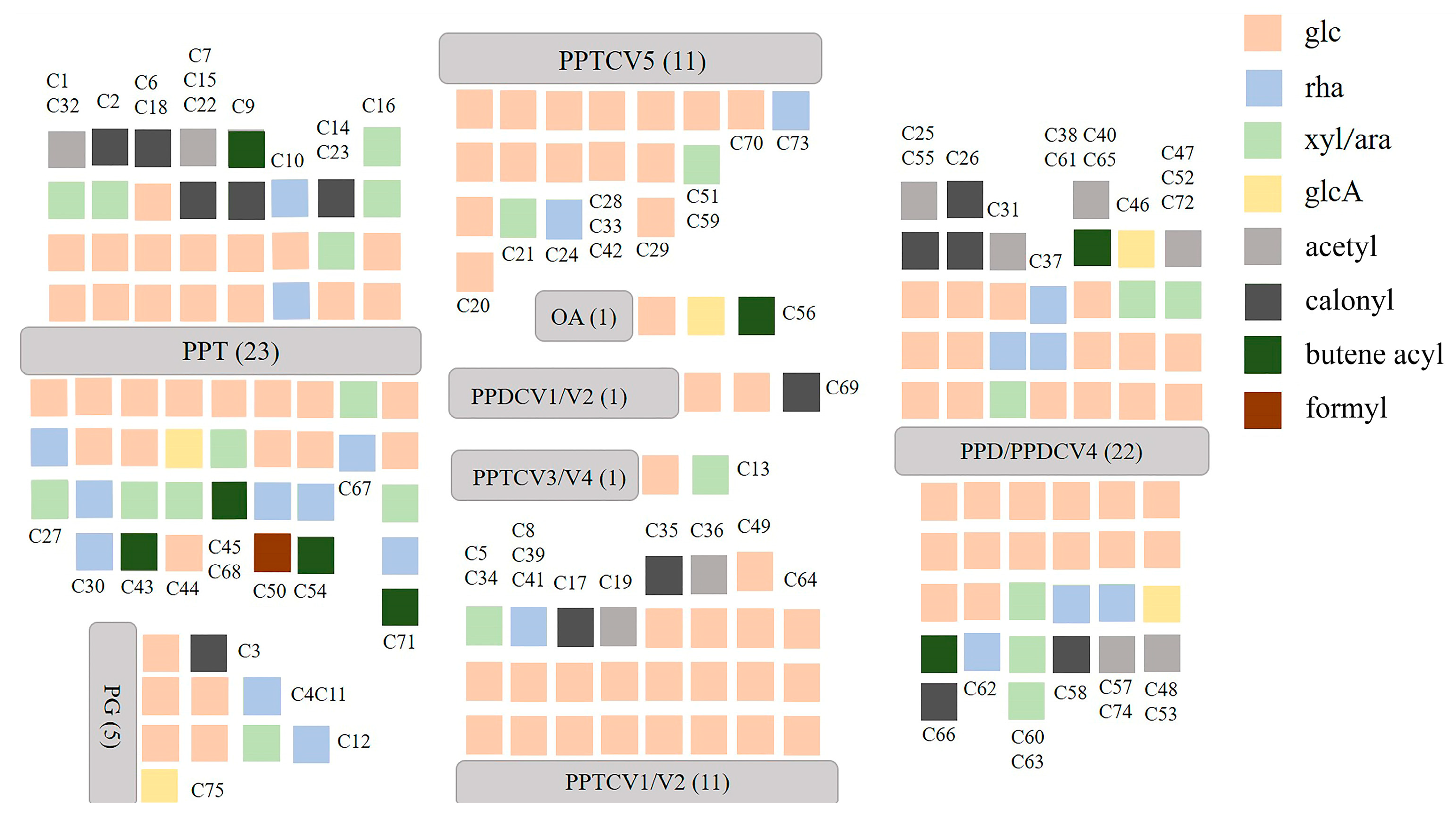
Accurate molecular weight and corresponding chemical formula can be obtained from primary mass spectrometry. MS/MS analysis was performed in a data-dependent acquisition (DDA) mode to produce a pattern of fragment masses unique to the single compound’s fragmentation. Thus, even if two compounds have the same retention time, they can be distinguished by their MS/MS spectra. On the basis of previous studies, compound identification was performed. A total of 312 ginsenosides, including the 63 standard compounds, were finally detected. Among the 312 ginsenosides, 237 are known compounds (Table S1) [20,21,22,23,24,25,26,27,28,29,30,31,32,33,34,35,36,37,38,39,40,41,42,43,44] and 75 are potential new ginsenosides (Table S2). A total of 63 ginsenosides were identified by comparison the references standards, 237 ginsenosides were assigned by comparison of the precise molecular weights, molecular formulae, the types of aglycones, glycosyl groups, chemical structures, and fragmental pathway with those in the literature. The type of aglycone and glycone are shown in Figure 1. There were 75 new potential ginsenosides, identified through the following steps: (a) the molecular formula of the deduced compounds was imported into the website of SCI-Finder to further confirm their structures; (b) compared with the searched compounds, if there is a consistent chemical group (regardless of the connection order of each group), the imported compound is considered to be known, otherwise, the ginsenoside is considered to be a new potential ginsenoside. According to MS and MS/MS data of reference compounds, some fragment ions were used as criteria to identify aglycons of the compounds. The unique fragment ions at m/z 487 [aglycone−H] (C31H51O4), 359 (C24H39O2), 31 (OCH3), 127 (C7H11O2, detached side-chain fragment) were evidence of PPDV3 ginsenosides. In the same way, the ions at m/z 441 [aglycone−H] (C30H49O2), 305 (C20H34O2) were evidence of aglycones PPDV1/V2, which were regarded as a product of H2O-removed PPD at C20–C21 or C20–C22. PPD and PPDV4 ginsenosides shared the same [aglycone−H] ion at m/z 459 (C30H51O3) and fragmental ion at m/z 305 (C20H33O2). The difference between them was the ion at m/z 373 (C25H41O2) and a neutral loss of 88 (C5H12O), which presented only in PPDV4 ginsenosides. However, the ion at m/z 373 was too weak in some PPDV4 ginsenosides to distinguish them from the PPD-type compounds. In addition, aglycones of PPTV1/V2, considered as H2O-removed PPT, would produce ions at m/z 457 [aglycone−H] (C30H49O3) and 321 (C20H34O3). Aglycones of PPTV3 and PPTV4, a pair of configuration isomer and additive products of PPT and H2O, showed unique ions at m/z 493 [aglycone−H] (C30H53O4) and 321 (C20H34O3). Aglycone of PPTV5, an isomer of hydroxylated PPTV1−type aglycone, had fragment ions at m/z 473 [aglycone−H] (C30H49O4) and 321 (C20H34O3). Thus, m/z 475 (C30H51O4) and 391 (C26H39O4) were the feature fragment ions of PPT−type aglycone. Aglycone of PG showed unique [aglycone−H] at m/z 491 (C30H51O5) and another ion at m/z 373 (C25H41O2), while aglycone of OA produced fragment ions at m/z 247 (C15H37O2) and 207 (C14H23O1) due to RDA cleavage. The structural classification of new compounds is shown in Figure 2. There are 46 of PPT-type, 23 of PPD-type, 5 of PG-type, and 1 of OA-type ginsenosides, and most of the saponins contain three or more glycosides. The following is the structural analysis of ginsenosides Rd and Rg1 as examples, and their fragmental pathway is shown in Figure 3.
The molecular formula of Rg1 was deduced as C42H72O14 with [M−H] at 799.4884. It can be inferred that the aglycone is connected to two molecules of glucose through glycosidic bonds according to the fragmental ions of 637.4348 [M−H−Glc], 475.3798 [M−H−2Glc], and 391.2859 [M−H−2Glc−C4H12]. The structure of Rg1 was determined to be PPT-type ginsenoside according to the fragmental ions at 475.3798 [M−H−2Glc] and 391.2859 [M−H−2Glc−C4H12].
The molecular formula of Rd was deduced as C48H82O18 with [M−H] at 945.5496. It can be inferred that the aglycone is connected to three molecules of glucose through glycosidic bonds according to the fragments of 783.4966 [M−H−Glc], 621.4398 [M−H−2Glc], 459.3846 [M−H−3Glc]. The type of Rd was determined to be PPD-type ginsenoside according to the fragmental ions at 375.2973 [M−H−3Glc], 391.2859 [M−H−2Glc−C4H12].

2.2. Multivariate Statistical Analysis of Samples

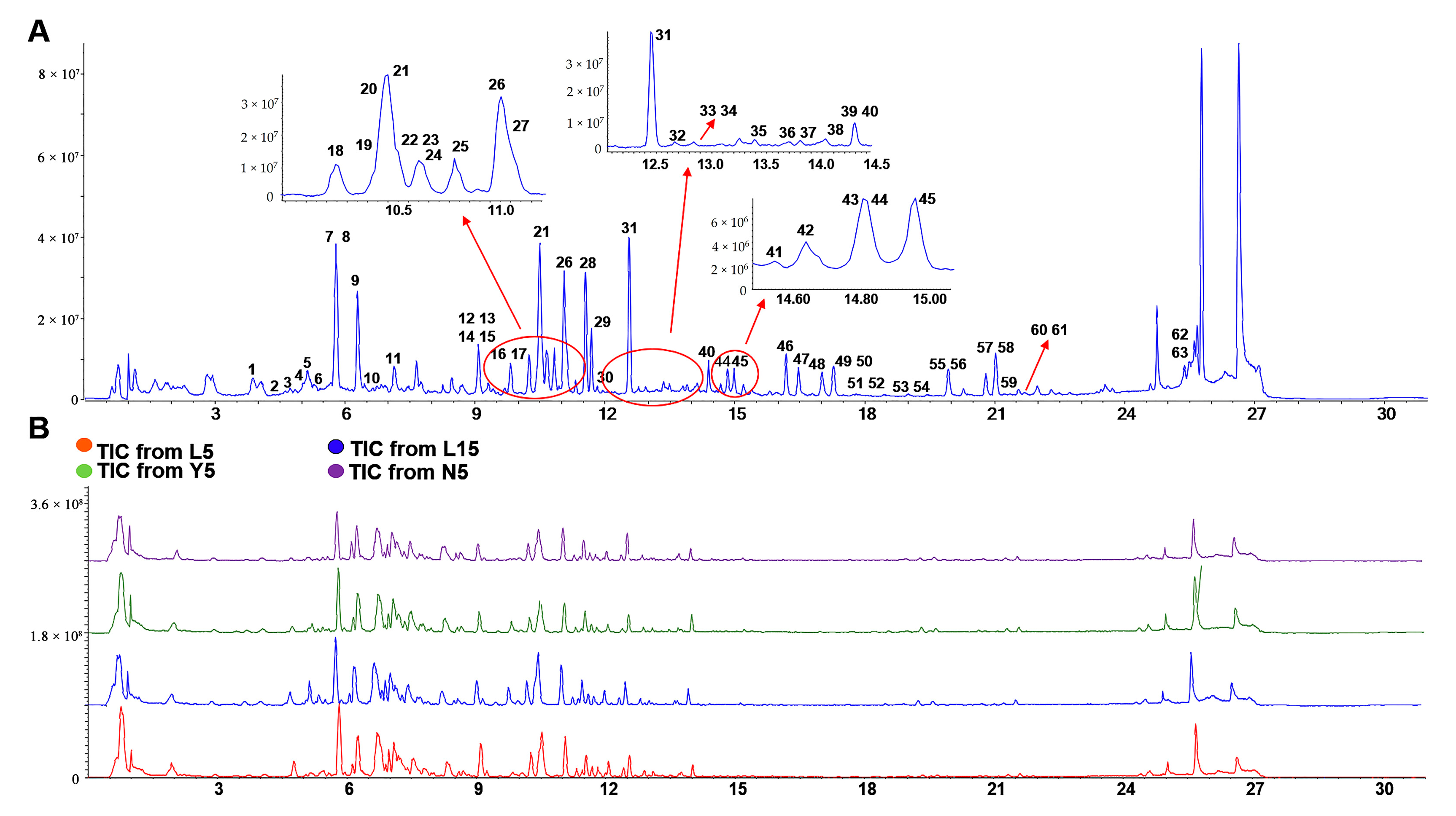
All of the 63 standard references were detected in four kinds of P. ginseng extracts. The TIC (total ion chromatogram) chart of the standard references and four kinds of samples was shown in Figure 4A,B. After a comprehensive analysis of the retention times, molecular weights, and ion fragments, a total of 261 ginsenosides including 54 potential new compounds from L5, 292 ginsenosides including 65 potential new compounds from L15, 273 ginsenosides including 59 potential new compounds from Y5, and 266 ginsenosides including 56 potential new compounds from N5 were identified, respectively.
In order to systematically evaluate the discrepancy of ginsenosides and identify their characteristic components in roots of P. ginsengs growing in four different environments, a workflow was constructed on the application of multivariate statistical techniques for further quantitative analysis.
Metabolomic-based strategy can be analyzed for semi-quantitative results at the same retention time due to its advantage of eliminating instrumental errors and correcting the retention time of the same compound between each sample. We investigated two analytical methods, PCA analysis and OPLS-DA [45]. A number of compounds that could influence the classification results were detected. The PCA and OPLS-DA analysis including four groups L5 (3 samples), L15 (2 samples), N5 (3 samples), and Y5 (3 samples), which was carried out with software SIMCA−14.0 (metrics) based on mass spectrometry semi-quantitative data. The total mass spectrometry data of several groups of samples were used for PCA and OPLS-DA analysis. Due to the existence of instrument errors, intra-group differences are inevitable. The type in the parameter of Scale was set to UV, and the Mode Type of analysis was set to OPLS-DA (supervised cluster analysis). In order to ensure the reliability of data analysis, all blank values will not be discarded. As shown in Figure S1, L5 and L15 performed similarly except for one sample in L5. Due to the irreducible differences between the samples, this PCA analysis convincingly reflects the differences between the samples in each group. With the exception of one sample that showed a clear separation, the sample parallelism of Y5 was recognized and shows a clear separation from L5, L15, and N5. The results for N5 were identical to those for Y5. Four groups of samples showed clear separation under supervision. As shown in Figures S2 and S3, the difference between the L5 and L15 groups, which is insignificant for N5 and Y5, is sufficient to distinguish L5 from L15. For the same species of ginseng, age has an effect on ginseng. L5 and L15 can be considered as two parts or as a whole distinct from N5 and Y5. This implies the influence of environment and age on ginseng. There was a small statistical difference between L5 and L15 due to different growth ages. It could be that L15 has more complex chemistry. However, it is worth noting that the difference between L5 and L15 was smaller than that between the other groups, which reminds us that growing environment has a greater influence on the chemical composition of ginseng than growing age. This can also be reflected in N5 and Y5. Although the two groups of samples are of the same age, in OPLS-DA analysis, they show great differences due to different growing environments. As shown in Figure S3, intra-group sample parallelism was trustworthy, with four groups of samples showing clear separation under supervision. Unsupervised PCA analysis retained the original data of the samples and clearly showed the differences between the samples. In the premise that PCA analysis is accepted, supervised OPLS-DA analysis will show more intuitive separation. In the cluster analysis, there were obvious differences among the four groups, which fully reflected the influence of growing environment and growing age on the chemical composition of ginseng.
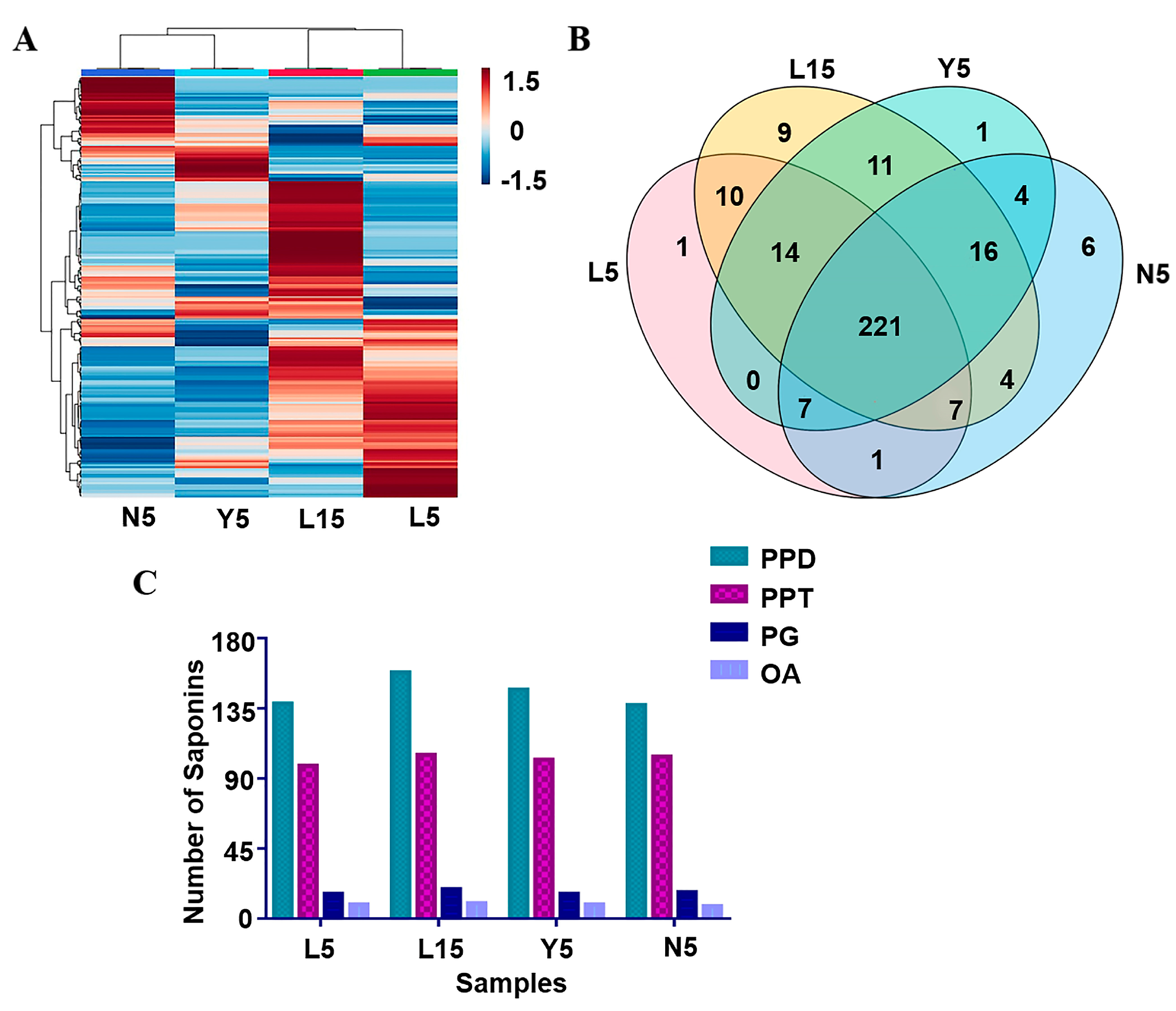
To further verify the accuracy of the result of PCA and OPLS-DA, semi-quantitative results of 312 identified ginsenosides was further submitted for heat map analysis (Figure 5A). The result showed that L5 and L15 could be grouped together due to the similarity of ingredients, while there was still a difference between these two groups on the color depth, which could be speculated that the growth time played an important role in the quantity of P. ginseng [3]. Y5 and N5 can be divided into another group, indicating that N5 and Y5 have a greater similarity compared with the forest-growing ginseng. The results showed that the growth environment plays an important role in the growth process of ginseng.
The Venn diagram of 312 P. ginseng saponins in the four group samples was shown in Figure 5B. Among the detected ginsenosides, 221 kinds of P. ginseng saponins were shared by the four groups of P. ginseng, while 9 ginsenosides were unique in L15 which was significantly higher than others. There were 6 unique ginsenosides in N5, while only 1 unique ginsenoside were detected in L5 and Y5. The results suggested that the variety of chemical composition in P. ginseng is closely related to its growth period.

2.3. Discussion

Semi-quantitative contents of 312 ginsenosides are shown in the Table S3. The S-Plot analysis for the pairwise comparison of the four groups of samples is shown in Figure S4, and there are obvious analyte differences among the groups, which validates the conclusion that the influence of growth environment on chemical constituents in ginseng. In S-plot analysis, the Type in the parameter of Scale was set to Par, and the Mode Type of analysis was set to OPLS-DA (supervised cluster analysis) to ensure the reliability of data analysis. As far as ginsenosides are concerned, the differences from S-plot analysis between the groups were attributed to the unique ginsenosides in the four groups of samples, such as Yesanchinoside E/isomer (L5, rt = 5.19), new ginsenosides 3 (N5, rt = 1.48), new ginsenosides 12 (N5, rt = 2.36), 20(R)-G-Rf2 isomer (N5, rt = 5.28), 20(R)-G-Rf2 isomer (N5, rt = 8.69), G-Rh14 isomer (N5, rt = 10.78), G-Rh4 isomer (N5, rt = 19.07), Diacetyl-G-Rd isomer (Y5, rt = 6.26), new ginsenosides 5 (L15, rt = 1.54), Vinaginsenoside R6 isomer (L15, rt = 3.30), 3β,12β,20,25-tetrahydroxydammarane-6-O-β-d-xylopyranosyl-(1→2)-β-d-glucopyranoside isomer (L15, rt = 4.15), new ginsenosides 25 (L15, rt = 5.62), chikusetsusaponin LM2 isomer (L15, rt = 6.64), new ginsenosides 38 (L15, rt = 7.45), new ginsenosides 40 (L15, 7.84), ZB-RI-6‘ methyl ester (L15, rt = 18.44), Rs4 (L15, rt = 18.44). In addition, ginsenosides with large differences in content also contributed greatly to the difference between groups, such as Pseudoginsenoside Rs1/isomer, G-Re2, G-Re1 isomer, NG-R1, G-Re. In L15 and Y5, the highest content of ginsenoside was G-Rg1, the contents of G-Rg1, G-Ro, G-Rb1, and 20-G-Rf are the top four compounds. While in N5 and L5, the highest content of ginsenoside was G-Re, the top four compounds are G-Re, G-Ro, G−Rb1, and 20-G-Rf. The L15 P. ginseng has the most diversity of ingredients in the four groups in terms of the total number and unique types of ginsenosides. To a certain extent, its chemical composition was most similar to L5 P. ginsengs and had significant differences from the other two groups of P. ginsengs, according to the results of PCA and OPLS-DA analysis. Comparing the difference of chemical components between L5 and L15 P. ginseng, the identified differential compounds were found lower in L5 P. ginseng by semi-quantitative analysis, which is presumably due to the transformation of the related components in L5 to other compounds in L15 after a long growth time. Compared with Y5, 42 different ginsenosides were detected in L15 P. ginseng. Except for the differential growth time, it was speculated that different growth environments took an important role in the formation of compounds especially the change of trace components in P. ginseng, and 58 different ginsenosides were found between N5 and L15 P. ginseng [46].
The N5 P. ginseng grew in farmland, while Y5 P. ginseng grew in a more natural area in mountains. Although having the same growth period, there were 44 different components between P. ginsengs of N5 and Y5. Thus, the growth environment, as moisture, air, and trace elements, might be the main reason for the difference between them. In terms of the growth years, L15 could better reflect the diversity and specificity of ginsenosides than L5. This shows that the growth years and ginsenoside types show a positive correlation. Y5 and N5 have similar exposure times and humidity, the only difference between them is that Y5 is at a higher altitude which results in lower temperature for Y5 than for N5. It can be concluded that the low temperature environment is conducive to the development of ginsenoside diversity. L5 is surrounded by forests and receives less sunlight, resulting in a long-term humid environment for L5; however, these are absent in N5. It can be seen that the moist, cool and low temperature environment provides a positive support for the amount of saponins in ginseng [47].
The proportions of different types of ginsenosides in the four groups of P. ginseng are shown in Figure 5C. The PPD-type and PPT-type ginsenosides account for the largest proportion of ginsenosides in our research, similarly to relevant studies on P. ginseng [48]. It showed that protopanaxadiol and protopanaxatriol saponins were the main constituents in P. ginseng, and the proportions of different ginsenoside types are similar in the four groups. Except for the protopanaxadiol saponins, not only the quantity of total saponins but also each other types saponins of P. ginsengs in L15 were higher than that in other groups. Comparing L15 with L5, it was concluded that the growth time was an important cause of the chemical difference of P. ginseng under the same environmental conditions. As for the difference between L5, Y5, and N5, it can also be concluded that the ecological environments also play a key role in the growth process of ginseng.

3. Experimental

3.1. Materials

Three batches of five-year-old P. ginseng cultivated in the forest (ginseng is grown in forest environments similar to wild ginseng.) (L5), two batches of fifteen-year-old P. ginseng cultivated in the forest same as L5 (L15), three batches of five-year-old P. ginseng cultivated in mountains (ginseng is grown on hill farms that have been cleared of previous plants and made manageable) (Y5), and three batches of five-year-old P. ginseng cultivated in farmland (ordinary farmland in the plains) (N5) (Table S4) were collected for analysis. The differences among the three growth environments were natural factors such as temperature, humidity, and sunlight. Intervention factors such as fertilization and watering remained the same. All samples were from Ji’an City, Jilin Province, China, and identified by Professor Xiu-Wei Yang of the School of Pharmaceutical Sciences of Peking University. The voucher specimens were deposited in the State Key Laboratory of Natural and Biomimetic Drugs of Peking University.

3.2. Standard Samples, Chemicals, and Reagents

Sixty-three kinds of ginsenosides of P. ginseng were used for qualitative analysis: G-Re3 (1), 20-glu-G-Rf (2), G-Re4 (3), G-Re2 (4), NG-R1 (5), G-Re1 (6), G-Rg1 (7), G-Re (8), G-Ro (9), NG-R4 (12), 20(S)-G-Rf-1a (13), 20(S)-G-Rf (14), 20(R)-G-Rf (15), 20(S)-NG-R2 (16), G-Ra2 (18), G-Ra3 (19), 20(R)-NG-R2 (20), G-Rb1 (21), 20(S)-G-Rg2 (22), 20(R)-G-Rg2 (23), G-Ra1 (24), G-Rc (25), 20(S)-G-Rh1 (26), 20(R)-G-Rh1 (27), G-Rb2 (28), G-Rb3 (29), Q-R1 (30), G-Rd (31), G-Rk1 (32), Rs2 (33), Rs1 (34), G-Ro methyl ester (35),G-Rh19 (36), DHDGG (37), G-Rg6 (38), G-F4 (39), DHDXG (40), CS-Iva methyl ester (41), G-Rk3 (42), G-Rh4 (43), G-Rg9 (44), G-F2 (45), 20(S)-G-Rg3 (46), 20(R)-G-Rg3 (47), DEDT (48), 20(S)-PPT (49), 20(R)-PPT (50), 20(S)-G-Rs3 (51), 20(R)-G-Rs3 (52), Rs4 (53), ZB-RI-6′ methyl ester (54), G-Rg5 (55), 20(S)-G-Rh2 (57), 20(R)-G-Rh2 (58), DDT (59), 20(S)-PPD (62), and 20(R)-PPD (63). Among them, CS-IV (10), CS-Iva (11), G-F3 (17), G-C-K (56), G-Rh3 (60), and G-Rk2 (61) were purchased from Chengdu Pusi Biotechnology Co., Ltd., and the other ginsenosides were from the reference’s library of our research group. The purity of each reference compound was more than 98.0% determined by RP-HPLC. The chemical structures of the above 63 compounds were shown in Table S5.
LC-MS grade acetonitrile (ACN), methanol (MeOH), and ammonium formate were purchased from Thermo Fisher Scientific (Fair Lawn, NJ, USA). The deionized water was prepared by a Millipore Alpha-Q water purification system (Bedford, MA, USA).

3.3. Subsubsection Instruments Chromatographic Conditions and Parameters

SCIEX Triple TOF 6600+ was accompanied with the SCIEX Exion LC AD system. The mass spectrometry parameters were as below: auxiliary gas (GS1): 60 psi; atomizing gas (GS2): 60 psi; curtain gas (CUR): 35 psi; ion source temperature (TEM): 600 °C; ion spray voltage (IS): positive ion 5500 V, negative ion −4500 V; declustering voltage (DP): 80/−80 V; collision energy (CE): 40 ± 20 V. The software SCIEX OS combined with Peakview was used to process the data. Composition of the eluent was selected by multi-dimensional investigation of sample resolution, peak shape, and sensitivity. After investigating different elution proportions such as H2O-acetonitrile, 0.1% formic acid–acetonitrile, 0.5 mmol/L ammonium formate–acetonitrile, the eluent made up with 0.5 mmol/L ammonium formate (A)–acetonitrile (B) was finally selected to analyze the samples. A programmed ingredient was carried out as follows: 0–3 min 78% A; 3–5 min 78–70% A; 5–9 min 70–65% A; 9–11 min 65–60% A; 11–13 min 60–52% A; 13–18 min 52–45% A; 18–22 min 45–30% A; 22–25 min 30–10% A; 25–26 min 10% A. An ACQUITY UPLC® CSH (C18 1.7 μm, 2.1 × 00 mm; Waters) coupled with an AC QUITY UPLC® BEH Shield RP 18 1.7 μm pre-column was used for chromatographic separation. The injection volume was 2 μL. The flow rate was 0.35 mL/min. The temperature of the column oven was kept at 40 °C, while the temperature of the autosampler was kept at 25 °C.
MS scan conditions were optimized both in negative and positive ion modes by using reference standards. Due to more peaks and much clearer fragmental information, the negative ion mode was chosen to analyze most of the ginsenosides, while compounds 49 and 60 could be observed only in positive ion mode. For negative ion mode, the [M−H] or [M+FA−H] was shown as molecular ion peaks, and the molecular ion peaks for 49 and 60 were [M+H]+ in positive ion mode. The base peak chromatographic profiles of the standards and samples are shown in Figure 1.

3.4. Preparation of Samples and References Standards

After being washed with water, the roots of P. ginseng were dried at 40 °C for 40 h. Then the materials were crushed to powder and filtered with a 60-mesh sieve. The 0.5 g fine powder of each sample (60 mesh size) was ultrasonic extracted (250 W, 40 kHz) with 20 mL of 70% MeOH (v/v) for 30 min. The samples were then set aside to room temperature and refilled the lost weight with 70% MeOH. After centrifugation at 3000 rpm for 10 min, the supernatant was passed through a 0.2 microns filter membrane to obtain the tested solution and stored at −20 °C for analysis. Each reference was precisely weighed and dissolved with MeOH to form the final concentration at 1 mg/mL. An appropriate volume of each reference solution was mixed to obtain the working solution for qualitative analysis.

3.5. Statistical Analysis

Orthogonal partial least-squares discriminant analysis (OPLS-DA) was used for analysis with SIMCA-P software 14.0 (Umetrics, Umea, Sweden). The semi-quantitative content of 320 ginsenosides in the four groups samples were compared by hierarchical clustering analysis (the website at https://www.metaboanalyst.ca/, accessed on 5 September 2022). The number of saponins in the four groups was used to produce Venn diagram. GraphPad Prism 7 was used to visually display the proportion of each type of ginsenosides in the four groups.

4. Conclusions

This study optimized the method of extracting complicated compounds from P. ginseng, and established a sensitive chromatographic method for components separation. Using positive and negative ion modes, 320 kinds of ginsenosides were identified in P. ginseng by Triple-TOF-MS, in which 75 ginsenosides were identified as potential new compounds. The chemical structures of ginsenosides were initially verified by systematic elucidation of the accurate mass, retention times, and fragment ions. The cluster analysis showed that the growth environment had a great influence on the chemical composition of P. ginseng. The discovery of this study allowed us to further understand the pharmacodynamic substances and growth habits of P. ginseng, and will provide a reference to study on quality evaluation of P. ginseng in the future.

Supplementary Materials

The following supporting information can be downloaded at: https://www.mdpi.com/article/10.3390/molecules28052068/s1, Figure S1: The PCA analysis for the four groups of samples; Figure S2: The OPLS-DA analysis for the four groups of samples; Figure S3: The OPLS-DA analysis for the pairwise comparison of the four groups of samples; Figure S4: The S-Plot analysis for the pairwise comparison of the four groups of sample; Table S1: MS and MS/MS of the 237 known ginsenosides; Table S2: MS and MS/MS of the 75 potential new ginsenosides; Table S3: Semi-quantitative contents of 312 ginsenosides; Table S4: Ginseng samples analyzed in this study; Table S5: Structure of the 63 ginsenoside standards.

Author Contributions

Conceptualization, Y.Z., Q.G. and Y.S.; methodology, Y.S.; software, Y.S.; validation, Y.S., Y.Q. and X.F.; formal analysis, Y.S., X.L. and L.C.; investigation, Y.S., Z.H., J.X. and J.Y.; resources, Y.S. and Y.Z.; data curation, Y.S.; writing—original draft preparation, Y.S.; writing—review and editing, Y.S.; visualization, Y.S., W.M. and J.L.; All authors have read and agreed to the published version of the manuscript.

Funding

This research was funded by the National Natural Science Foundation of China, grant number 81891011 and 81973446.

Institutional Review Board Statement

Not applicable.

Informed Consent Statement

Not applicable.

Data Availability Statement

Not applicable.

Conflicts of Interest

The authors declare no conflict of interest.

Sample Availability

Samples of the compounds are not available from the authors.

Abbreviations

P. ginseng: Panax ginseng C. A. Meyer; UPLC-Triple-TOF-MS/MS: An ultra-performance liquid chromatography–tandem triple quadrupole time-of-flight mass spectrometry; PPD: protopanaxadiol; PPT: protopanaxatriol; OA: oleanolic acid; PG: pseudo; L5: Three batches of five-year-old P. ginseng cultivated in the forest (nature forest in mountain); L15: 2 batches of fifteen-year-old P. ginseng cultivated in the forest; Y5: 3 batches of five-year-old P. ginseng cultivated in mountains (after logging, removing the root system); N5: three batches of five-year-old P. ginseng cultivated in farmland (ordinary farmland in the plains).

References

  1. Qi, L.W.; Wang, C.Z.; Yuan, C.S. Isolation and analysis of ginseng: Advances and challenges. Nat. Prod. Rep. 2011, 28, 467–495. [Google Scholar] [CrossRef] [Green Version]
  2. Mancuso, C.; Santangelo, R. Panax ginseng and Panax quinquefolius: From pharmacology to toxicology. Food Chem. Toxicol. 2017, 107, 362–372. [Google Scholar] [CrossRef] [PubMed]
  3. Chen, W.; Balan, P.; Popovich, D.G. Ginsenosides analysis of New Zealand-grown Forest Panax ginseng by LC-QTOF-MS/MS. J. Ginseng Res. 2020, 44, 552–562. [Google Scholar] [CrossRef] [PubMed]
  4. Zhou, Q.L.; Zhu, D.N.; Yang, X.W.; Xu, W.; Wang, Y.P. Development and validation of a UFLC-MS/MS method for simultaneous quantification of sixty-six saponins and their six aglycones: Application to comparative analysis of red ginseng and white ginseng. J. Pharm. Biomed. Anal. 2018, 159, 153–165. [Google Scholar] [CrossRef] [PubMed]
  5. Kiefer, D.; Pantuso, T. Panax ginseng. Am. Fam. Physician 2003, 68, 1539–1542. [Google Scholar] [PubMed]
  6. Shergis, J.L.; Zhang, A.L.; Zhou, W.; Xue, C.C. Panax ginseng in randomised controlled trials: A systematic review. Phytother. Res. 2013, 27, 949–965. [Google Scholar] [CrossRef] [PubMed]
  7. Wang, L.; Huang, Y.; Yin, G.; Wang, J.; Wang, P.; Chen, Z.Y.; Wang, T.; Ren, G. Antimicrobial activities of Asian ginseng, American ginseng, and notoginseng. Phytother. Res. 2020, 34, 1226–1236. [Google Scholar] [CrossRef] [PubMed]
  8. Arring, N.M.; Millstine, D.; Marks, L.A.; Nail, L.M. Ginseng as a Treatment for Fatigue: A Systematic Review. J. Altern. Complement Med. 2018, 24, 624–633. [Google Scholar] [CrossRef]
  9. Lorz, L.R.; Kim, M.Y.; Cho, J.Y. Medicinal potential of Panax ginseng and its ginsenosides in atopic dermatitis treatment. J. Ginseng Res. 2020, 44, 8–13. [Google Scholar] [CrossRef]
  10. Yun, M.; Yi, Y.S. Regulatory roles of ginseng on inflammatory caspases, executioners of inflammasome activation. J. Ginseng Res. 2020, 44, 373–385. [Google Scholar] [CrossRef] [PubMed]
  11. Chen, J.T. Advances in Ginsenosides. Biomolecules 2020, 10, 681. [Google Scholar] [CrossRef] [PubMed]
  12. Liu, W.L.; He, Y.F.; Li, L.L.; Liu, S.Y. Fast quantitative analysis of ginsenosides in Asian ginseng (Panax ginseng C. A. Mayer) by using solid-phase methylation coupled to direct analysis in real time. Rapid Commun. Mass Spectrom 2016, 1, 111–115. [Google Scholar] [CrossRef] [Green Version]
  13. Wong, Y.L.; Chen, X.; Li, W.; Wang, Z.; Hung, Y.L.; Wu, R.; Chan, T.W. Differentiation of Isomeric Ginsenosides by Using Electron-Induced Dissociation Mass Spectrometry. Anal. Chem. 2016, 88, 5590–5594. [Google Scholar] [CrossRef] [PubMed] [Green Version]
  14. Lee, J.; Han, H.; Yuan, X.; Park, E.; Lee, J.; Kim, J.H. A rapid, simultaneous and quantitative analysis of 26 ginsenosides in white and red Panax ginseng using LC–MS/MS. Appl. Biol. Chem. 2021, 6, 13. [Google Scholar] [CrossRef]
  15. Wang, H.P.; Zhang, Y.B.; Yang, X.W.; Zhao, D.Q.; Wang, Y.P. Rapid characterization of ginsenosides in the roots and rhizomes of Panax ginseng by UPLC-DAD-QTOF-MS/MS and simultaneous determination of 19 ginsenosides by HPLC-ESI-MS. J. Ginseng Res. 2016, 40, 382–394. [Google Scholar] [CrossRef] [PubMed] [Green Version]
  16. Li, H.; Song, Y.; Zhang, H.; Wang, X.; Cong, P.; Xu, J.; Xue, C. Comparative lipid profile of four edible shellfishes by UPLC-Triple TOF-MS/MS. Food Chem. 2020, 310, 125947. [Google Scholar] [CrossRef] [PubMed]
  17. Wang, Y.; Luan, G.; Zhou, W.; Meng, J.; Wang, H.; Hu, N.; Suo, Y. Subcritical water extraction, UPLC-Triple-TOF/MS analysis and antioxidant activity of anthocyanins from Lycium ruthenicum Murr. Food Chem. 2018, 249, 119–126. [Google Scholar] [CrossRef]
  18. Peng, X.; Luo, Y.; Wang, J.; Ji, T.; Yuan, L.; Kai, G. Integrated analysis of the transcriptome, metabolome and analgesic effect provide insight into potential applications of different parts of Lindera aggregata. Food Res. Int. 2020, 138, 109799. [Google Scholar] [CrossRef]
  19. Li, J.; Wang, H.; Shi, X.; Zhao, L.; Lv, T.; Yuan, Q.; Hao, W.; Zhu, J. Anti-proliferative and an-ti-migratory effects of Scutellaria strigillosa Hemsley extracts against vascular smooth muscle cells. J. Ethnopharmacol 2019, 235, 155–163. [Google Scholar] [CrossRef]
  20. Li, K.K.; Yao, C.M.; Yang, X.W. Four new dammarane-type triterpene saponins from the stems and leaves of Panax ginseng and their cytotoxicity on HL-60 cells. Planta Med. 2012, 78, 189–192. [Google Scholar] [CrossRef] [Green Version]
  21. Tava, A.; Mella, M.; Avato, P.; Biazzi, E.; Pecetti, L.; Bialy, Z.; Jurzysta, M. New triterpenic saponins from the aerial parts of Medicago arabica (L.) huds. J. Agric. Food Chem. 2009, 57, 2826–2835. [Google Scholar] [CrossRef] [PubMed]
  22. Chen, G.; Yang, M.; Lu, Z.; Zhang, J.; Huang, H.; Liang, Y.; Guan, S.; Song, Y.; Wu, L.; Guo, D.A. Microbial transformation of 20(S)-protopanaxatriol-type saponins by Absidia coerulea. J. Nat. Prod. 2007, 70, 1203–1206. [Google Scholar] [CrossRef] [PubMed]
  23. Lee, D.G.; Lee, A.Y.; Kim, K.T.; Cho, E.J.; Lee, S. Novel Dammarane-Type Triterpene Saponins from Panax ginseng Root. Chem. Pharm. Bull. 2015, 63, 927–934. [Google Scholar] [CrossRef] [PubMed] [Green Version]
  24. Wu, W.; Qin, Q.; Guo, Y.; Sun, J.; Liu, S. Studies on the chemical transformation of 20(S)-protopanaxatriol (PPT)-type ginsenosides R(e), R(g2), and R(f) using rapid resolution liquid chromatography coupled with quadruple-time-of-flight mass spectrometry (RRLC-Q-TOF-MS). J. Agric. Food Chem. 2012, 60, 10007–10014. [Google Scholar] [CrossRef]
  25. Vinh, L.B.; Lee, Y.; Han, Y.K.; Kang, J.S.; Park, J.U.; Kim, Y.R.; Yang, S.Y.; Kim, Y.H. Two new dammarane-type triterpene saponins from Korean red ginseng and their anti-inflammatory effects. Bioorg. Med. Chem. Lett. 2017, 27, 5149–5153. [Google Scholar] [CrossRef] [PubMed]
  26. Zhang, Y.; Han, L.F.; Sakah, K.J.; Wu, Z.Z.; Liu, L.L.; Agyemang, K.; Gao, X.M.; Wang, T. Bio-active protopanaxatriol type saponins isolated from the roots of Panax notoginseng (Burk.) F. H. Chen. Molecules 2013, 18, 10352–10366. [Google Scholar] [CrossRef] [PubMed]
  27. Zhou, Q.L.; Yang, X.W. Four new ginsenosides from red ginseng with inhibitory activity on melanogenesis in melanoma cells. Bioorg. Med. Chem. Lett. 2015, 25, 3112–3116. [Google Scholar] [CrossRef] [PubMed]
  28. Yang, W.Z.; Ye, M.; Qiao, X.; Liu, C.F.; Miao, W.J.; Bo, T.; Tao, H.Y.; Guo, D.A. A strategy for efficient discovery of new natural compounds by integrating orthogonal column chromatography and liquid chromatography/mass spectrometry analysis: Its application in Panax ginseng, Panax quinquefolium and Panax notoginseng to characterize 437 potential new ginsenosides. Anal. Chim. Acta 2012, 739, 56–66. [Google Scholar] [PubMed]
  29. Yoshizaki, K.; Devkota, H.P.; Yahara, S. Four new triterpenoid saponins from the leaves of Panax japonicus grown in southern Miyazaki Prefecture (4). Chem. Pharm. Bull. 2013, 61, 273–278. [Google Scholar] [CrossRef] [PubMed] [Green Version]
  30. Liang, Z.; Chen, Y.; Xu, L.; Qin, M.; Yi, T.; Chen, H.; Zhao, Z. Localization of ginsenosides in the rhizome and root of Panax ginseng by laser microdissection and liquid chromatography-quadrupole/time of flight-mass spectrometry. J. Pharm. Biomed. Anal. 2015, 105, 121–133. [Google Scholar] [CrossRef]
  31. Shi, J.; Cai, Z.; Chen, S.; Zou, L.; Liu, X.; Tang, R.; Ma, J.; Wang, C.; Chen, J.; Tan, M. Qualitative and quantitative analysis of saponins in the flower bud of Panax ginseng (Ginseng Flos) by UFLC-Triple TOF-MS/MS and UFLC-QTRAP-MS/MS. Phytochem. Anal. 2020, 31, 287–296. [Google Scholar] [CrossRef]
  32. Yoshizaki, K.; Yahara, S. New triterpenoid saponins from fruits specimens of Panax japonicus collected in Kumamoto and Miyazaki prefectures (1). Chem. Pharm. Bull. 2012, 60, 354–362. [Google Scholar] [CrossRef] [PubMed] [Green Version]
  33. Liu, C.Y.; Zhou, R.X.; Sun, C.K.; Jin, Y.H.; Yu, H.S.; Zhang, T.Y.; Xu, L.Q.; Jin, F.X. Preparation of minor ginsenosides C-Mc, C-Y, F2, and C-K from American ginseng PPD-ginsenoside using special ginsenosidase type-I from Aspergillus niger g.848. J. Ginseng Res. 2015, 39, 221–229. [Google Scholar] [CrossRef] [PubMed] [Green Version]
  34. Xu, X.F.; Xu, S.Y.; Zhang, Y.; Zhang, H.; Liu, M.N.; Liu, H.; Gao, Y.; Xue, X.; Xiong, H.; Lin, R.C.; et al. Chemical Comparison of Two Drying Methods of Mountain Cultivated Gin-seng by UPLC-QTOF-MS/MS and Multivariate Statistical Analysis. Molecules 2017, 22, 717. [Google Scholar] [CrossRef] [PubMed] [Green Version]
  35. Vu-Huynh, K.L.; Nguyen, H.T.; Van Le, T.H.; Ma, C.T.; Lee, G.J.; Kwon, S.W.; Park, J.H.; Nguyen, M.D. Accumulation of Saponins in Underground Parts of Panax vietnamensis at Different Ages Analyzed by HPLC-UV/ELSD. Molecules 2020, 25, 3086. [Google Scholar] [CrossRef] [PubMed]
  36. Liu, X.Y.; Wang, S.; Li, C.J.; Ma, J.; Chen, F.Y.; Peng, Y.; Wang, X.L.; Zhang, D.M. Dammarane-type saponins from the leaves of Panax notoginseng and their neuroprotective effects on damaged SH-SY5Y cells. Phytochemistry 2018, 145, 10–17. [Google Scholar] [CrossRef]
  37. Wan, J.B.; Zhang, Q.W.; Hong, S.J.; Li, P.; Li, S.P.; Wang, Y.T. Chemical investigation of saponins in different parts of Panax notoginseng by pressurized liquid extraction and liquid chromatography-electrospray ionization-tandem mass spectrometry. Molecules 2012, 17, 5836–5853. [Google Scholar] [CrossRef] [Green Version]
  38. Zhang, C.X.; Wang, X.Y.; Lin, Z.Z.; Wang, H.D.; Qian, Y.X.; Li, W.W.; Yang, W.Z.; Guo, D.A. Highly selective monitoring of in-source fragmentation sapogenin product ions in positive mode enabling group-target ginsenosides profiling and simultaneous identification of seven Panax herbal medicines. J. Chromatogr. A 2020, 1618, 460850. [Google Scholar] [CrossRef]
  39. Liao, P.Y.; Wang, D.; Zhang, Y.J.; Yang, C.R. Dammarane-type glycosides from steamed notoginseng. J. Agric. Food Chem. 2008, 56, 1751–1756. [Google Scholar] [CrossRef] [PubMed]
  40. Yoshikawa, M.; Murakami, T.; Ueno, T.; Yashiro, K.; Hirokawa, N.; Murakami, N.; Yamahara, J.; Matsuda, H.; Saijoh, R.; Tanaka, O. Bioactive saponins and glycosides. VIII. Noto-ginseng (1): New dammarane-type triterpene oligoglycosides, notoginsenosides-A, -B, -C, and -D, from the dried root of Panax notoginseng (Burk.) F.H. Chen. Chem. Pharm. Bull. 1997, 45, 1039–1045. [Google Scholar] [CrossRef] [Green Version]
  41. Li, M.; Liu, F.; Jin, Y.R.; Wang, X.Z.; Wu, Q.; Liu, Y.; Li, X.W. Five New Triterpenoid Saponins from the Rhizomes of Panacis majoris and Their Antiplatelet Aggregation Activity. Planta Med. 2017, 83, 351–357. [Google Scholar] [CrossRef] [PubMed] [Green Version]
  42. Lin, H.; Zhu, H.; Tan, J.; Wang, H.; Dong, Q.; Wu, F.; Liu, Y.; Li, P.; Liu, J. Non-Targeted Metabolomic Analysis of Methanolic Extracts of Wild-Simulated and Field-Grown American Ginseng. Molecules 2019, 24, 1053. [Google Scholar] [CrossRef] [PubMed] [Green Version]
  43. Liu, W.Y.; Zhang, W.D.; Chen, H.S.; Gu, Z.B.; Li, T.Z.; Chen, W.S. New triterpenoid saponins from bulbs of Bolbostemma paniculatum. Planta Med. 2004, 70, 458–464. [Google Scholar] [PubMed]
  44. Le, T.H.; Lee, G.J.; Vu, H.K.; Kwon, S.W.; Nguyen, N.K.; Park, J.H.; Nguyen, M.D. Ginseng Saponins in Different Parts of Panax vietnamensis. Chem. Pharm. Bull. 2015, 63, 950–954. [Google Scholar] [CrossRef] [Green Version]
  45. Chen, L.H.; Zhang, Y.B.; Yang, X.W.; Xu, W.; Wang, Y.P. Characterization and quantification of ginsenosides from the root of Panax quinquefolius L. by integrating untargeted metabolites and targeted analysis using UPLC-Triple TOF-MS coupled with UFLC-ESI-MS/MS. Food Chem. 2022, 384, 132466. [Google Scholar] [CrossRef] [PubMed]
  46. Kim, Y.J.; Zhang, D.; Yang, D.C. Biosynthesis and biotechnological production of ginsenosides. Biotechnol. Adv. 2015, 33, 717–735. [Google Scholar] [CrossRef]
  47. Jang, W.; Jang, Y.; Kim, N.H.; Waminal, N.E.; Kim, Y.C.; Lee, J.W.; Yang, T.J. Genetic diversity among cultivated and wild Panax ginseng populations revealed by high-resolution microsatellite markers. J. Ginseng Res. 2020, 44, 637–643. [Google Scholar] [CrossRef] [PubMed]
  48. Yu, C.; Wang, C.Z.; Zhou, C.J.; Wang, B.; Zhang, C.F.; Wu, X.H.; Yuan, C.S. Adulteration and cultivation region identification of American ginseng using HPLC coupled with multivariate analysis. J. Pharm. Biomed. Anal. 2014, 99, 8–15. [Google Scholar] [CrossRef] [Green Version]
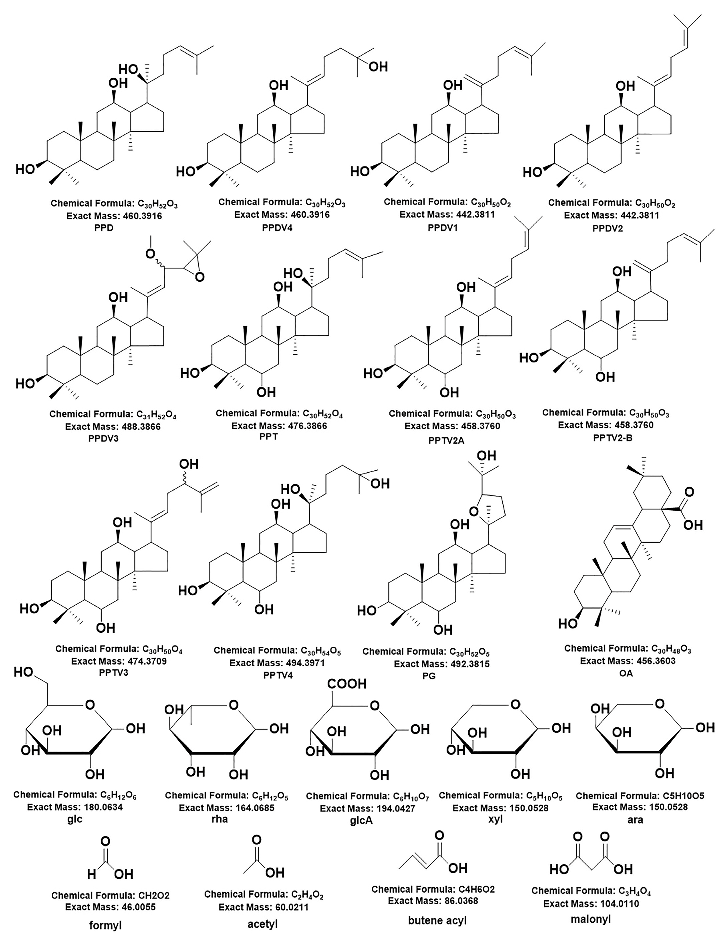
Figure 1. The 75 potential new ginsenosides’ type of aglycone and glycone. PPDCV and PPTCV represent PPD type and PPT type saponins with side chain changes, respectively.
Molecules 28 02068 g001
Figure 2. Structure analysis of 75 potential new ginsenosides.
Molecules 28 02068 g002
Figure 3. The MS/MS spectra and fragmentation process of Rd (A) and Rg1 (B).
Molecules 28 02068 g003
Figure 4. TIC of the ginseng standard references and four ginseng samples. (A) TIC of the ginseng standard references. (B) TIC of the four ginseng samples.
Molecules 28 02068 g004
Figure 5. Multi-statistical analysis of the components in four growth environments ginseng samples. (A) Heat map of four growth environment ginseng samples. (B) The Venn diagram of the 312 P. ginseng saponins. (C) Proportion of different type ginsenosides in the four growth environment ginseng samples.
Molecules 28 02068 g005
Disclaimer/Publisher’s Note: The statements, opinions and data contained in all publications are solely those of the individual author(s) and contributor(s) and not of MDPI and/or the editor(s). MDPI and/or the editor(s) disclaim responsibility for any injury to people or property resulting from any ideas, methods, instructions or products referred to in the content.