Article

Phytochemical Characterizations of Maranthes polyandra (Benth.) Prance

1
H. E. J. Research Institute of Chemistry, International Center for Chemical and Biological Sciences, University of Karachi, Karachi 75270, Pakistan
2
Sino-Pakistan Cooperation Center for Traditional Chinese Medicine, Hunan University of Medicine, Huaihua 418000, China
3
Sino-Pakistan Cooperation Center for Traditional Chinese Medicine, International Center for Chemical and Biological Sciences, University of Karachi, Karachi 75270, Pakistan
4
Third World Center (TWC) for Chemical Sciences, International Center for Chemical & Biological Sciences, University of Karachi, Karachi 75270, Pakistan
5
Department of Pharmacognosy and Drug Development, University of Ilorin, Ilorin 240003, Nigeria
6
Dr. Panjwani Center for Molecular Medicine and Drug Research, International Center for Chemical and Biological Sciences, University of Karachi, Karachi 75270, Pakistan
7
School of Traditional Chinese Pharmacy, China Pharmaceutical University, Nanjing 211198, China
8
Institute of Medicinal Plant Development, Chinese Academy of Medical Sciences, Beijing 100193, China
*
Authors to whom correspondence should be addressed.
Molecules 2022, 27(4), 1316; https://doi.org/10.3390/molecules27041316
Submission received: 31 December 2021 / Revised: 5 February 2022 / Accepted: 12 February 2022 / Published: 15 February 2022
(This article belongs to the Special Issue Theme Issue in Honor of Professor Atta-Ur-Rahman, FRS)

Abstract

:
Two new ursane-type triterpenoids, named Polyanside A (1) and B (2), along with eleven known compounds (313), were isolated and elucidated from Maranthes polyandra (Benth.) Prance. The structures of these compounds were elucidated based on chemical evidence and multiple spectroscopic data. Isolated compounds were evaluated for anti-cancer, anti-inflammatory activities, and cytotoxicity on a normal human cell line (BJ). None of them showed activity and cytotoxicity. The hexane fraction was analyzed by GC-MS, resulting in the identification of forty-one compounds. This is the first comprehensive study on the phytochemistry of M. polyandra.

Graphical Abstract

1. Introduction

The history of medicinal plants is as old as the history of human beings. Natural products have played a vital role in drug discovery. The use of natural components from folk medicines requires a clear understanding of chemistry, efficacy, and safety. Now, there has been a surge in interest in valorizing the biological importance of medicinal plants [1,2]. It is a pressing priority to obtain potent phytoconstituent from different medicinal plants and to explore their promising benefits [3,4].
Maranthes polyandra (Benth.) Prance (Synonym: Parinari polyandra Benth., World Flora Online) belongs to the Chrysobalanaceae family. It is a savannah tree of Africa ranging from Mali to Sudan, some parts of southern states of Nigeria, and largely found in Benin, where it is locally known as Wantuwiwi [5,6]. Different parts of this tree have been used for various ailments, for example, measles [7], diarrhea [8], fertility disorder [9,10], wounds, fracture, fever, and syphilis [11,12]. The extract from the stem bark, fruit, and seed of M. polyandra has shown anti-inflammatory and antinociceptive [9], antihyperlipidemic, hypercalcemic [10], hypertensive, anti-hypercholesterolemia, anti-diabetic [13], and antioxidant effects [14]. However, phytochemistry investigation of this species is extremely limited, except for a few studies [15,16,17,18]. Until now, only three compounds (xanthoxylin, β-eudesmol, luteolin) have been isolated from this plant [15]. The composition of seed oil has been analyzed by GC-MS [17,18]. In addition, GC-MS analysis on the extract and fractions of M. polyandra stem bark just confirmed the presence of some fatty acids [16].
Thus, the current study aimed to explore the phytochemical constituent from M. polyandra through isolation and GC-MS analysis. Finally, two new triterpenoids (1 and 2), and eleven known compounds (313) were isolated. In addition, GC-MS analysis of the hexane fraction also led to the identification of 41 compounds. This is the first comprehensive phytochemistry study of this species. Anti-cancer and anti-inflammatory activities and cytotoxicity of compounds 1, 2, 5, 6, 8, 11, and 13 were evaluated. None of them were active. Based on traditional uses, anti-inflammatory components might exist in this plant. Further study may be required to discover potent anti-inflammatory molecules from this species.

2. Results and Discussion

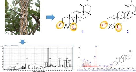
2.1. Structure Elucidation of Isolated Compounds

First, 80% MeOH extract of the stem bark of M. polyandra was fractioned by n-hexane for GC-MS analysis. The remaining residue was then isolated using chromatographic techniques, such as silica gel column chromatography (CC), C18 CC, Sephadex LH 20 CC, and HPLC. Thirteen compounds (113) were obtained, including two new compounds (1 and 2) and eleven known compounds (313) (Figure 1). The structures of 1 and 2 were elucidated mainly through NMR techniques, primarily based on 1D NMR (1H and 13C NMR), 2D NMR (COSY, HSQC, HMBC, and NOESY), and MS techniques including EI-MS and HR-EI-MS.
Polyanside A (1) was obtained as needle-shaped white crystals. The molecular formula was recognized as C30H48O2 based on HR-EI-MS (m/z 440.3648 [M]+, calcd. for 440.3654), representing seven degrees of unsaturation (Figure S2). 1D NMR (Table 1, Figures S6–S13) revealed the presence of thirty carbons, including eight methyl groups at δH 1.50, 1.40, 1.35, 1.15, 1.02, and 0.80 as singlets, along with a broad singlet at δH 0.90 (br s) and a doublet at δH 0.78 (d, J = 5.8 Hz). A group of typical signals consisting of an olefinic proton at δH 5.18 (dd, J = 5.0, 2.5 Hz), two olefinic carbons at δC 124.5, and 139.0, and a carbonyl signal at δC 216.7, were suggestive of a urs-12-en-3-one skeleton. All NMR data showed great similarity with α-amyrone except an extra oxymethine signal at δH 4.49 (br s) that is correlated with δC 69.3 in HSQC [19,20]. The presence of a hydroxyl was confirmed at C-6 through COSY correlations between H-5 (δH 1.22), H-7a (δH 1.81, dd, 14.8, 3.8), H-7b (δH 1.55, overlapped), and δH 4.49, along with HMBC correlations between H-5 (δH 1.22), H-7a (δH 1.81), H-7b (δH 1.55), and δC 69.3. The orientation of the hydroxyl can be confirmed as β, because correlations between H3-25 (δH 1.50), H3-26 (δH 1.35) and H-6 (δH 4.49) were absent; instead, correlation between H-5 (δH 1.22) and H-6 (δH 4.49) was observed. Thus, the structure of compound 1 was elucidated as shown in Figure 1 and named Polyanside A. Key 1H-1H COSY, HMBC, and NOESY correlations are shown in Figure 2 (Figures S14–S21).
Polyanside B (2) was obtained as an amorphous white powder with molecular formula of C30H50O2 deduced by HREIMS (m/z 442.3832 [M]+, calcd. for 442.3811), representing six degrees of unsaturation (Figure S23). 1D NMR data (Table 1, Figures S27–S33) of 2 is in good agreement with 1, except one more oxymethine proton at δH 3.14 (dd, J = 10.0, 5.0 Hz) and the absence of a carbonyl signal. The location of δH 3.14 was confirmed at C-3 through HMBC correlations of δH 3.14 with C-2 (δC 27.4), C-5 (δC 55.5), C-23 (δC 17.0), and C-24 (δC 28.05), along with COSY correlations between δH 3.14 and H-2 (δH 1.61 and 1.63). In addition, H-3 exhibited correlations with H-5 (δH 0.74, d, 2.0 Hz), implying a β-orientation of the hydroxyl at C-3. Therefore, the structure of compound 2 was elucidated as shown in Figure 1 and named Polyanside B. Key COSY, HMBC, and NOESY correlations are shown in Figure 2 (Figures S34–S41).
Compounds 313 were isolated from M. polyandra for the first time and recognized by compared to previously reported data. They were betulonic acid (3) [21], kaur-16-en-19-oic acid (4) [22], n-butyl-β-D-fructopyranoside (5) (Figure 1) [23], β-sitosterol (6) [24,25], stigmasterol (7) [26,27], stigmastane-3,6-dione (8) [28], stigmastane-4-ene-3-one (9), 4,22-stigmastadiene-3-one (10) [29], β-sitosterol β-D-glucoside (11) [30], n-hexadecanol (12), and palmitic acid (13) [31].
Compounds 1, 2, 5, 6, 8, 11, and 13 were performed for anti-cancer activity against MCF-7 cell (breast cancer), NCI-H460 (lung cancer), Hela (cervical cancer), and cytotoxicity against normal human cell line BJ, which were obtained from a cell culture biobank (PCMD, ICCBS) of American Type Culture Collection (ATCC), MTT assay was used for this activity (S3.5) [32]. All of them were observed to be inactive and nontoxic with inhibition < 50% at 50 µM. Compounds 1, 2, 5, 6, 8, 11, and 13 were also screened for nitric oxide (NO) inhibitory activity by a previously described method (S3.6) [33]. Unfortunately, all tested compounds displayed <50% inhibition at 25 µg/mL. The methanol extract and hexane fraction were tested for the same assays. However, they were inactive.
Compound 6 was reported to possess a good antinociceptive effect conferring to hot-plate and tail-flick assays [34]. Compounds 6 and 11 have been claimed to be the responsible components of an active extract to inhibit the growth of A549 cells (lung carcinoma epithelial cells) by analyzing the extract by LC-MS-MS [35]. However, in the current study they were inactive against NCI-H460 (lung cancer). Sari et al. evaluated the antimicrobial potential of 6 and 8. It was observed that 6 inhibited S. aureus with MIC of 9.4 µg/mL. Meanwhile, 8 inhibited S. enterica with MIC of 37.5 µg/mL [36]. To the best of our knowledge, it is the first time to test compounds 1, 2, 5, 6, 8, 11, and 13 for their anti-cancer potential (against MCF-7, HeLa, and H460) as pure compounds.

2.2. Phytochemical Investigation of Hexane Fraction by GC-MS

GC-MS analysis of the hexane fraction revealed the presence of different phytochemicals, which are shown in Figure 3 and listed in Table 2.
The major phytocomponents obtained from the hexane fraction were β-amyrin (8.55%), 4,22-stigmastadiene-3-one (8.33%), β-amyrin methyl ether (7.71%), stigmast-4-en-3-one (7.4%), stigmasterol (6.22%), n-hexadecanoic acid methyl ester (5.17%), (22E)-3 methoxystigmasta-5,22-diene (3.85%), 5α-stigmastane-3,6-dione (3.27%), γ-sitosterol (2.99%), methyl 13-octadecenoate (2.61%), methyl (10E)-10-octadecenoat (2.61%), oleyl alcohol (2.36%), trans-9-octadecen-1-ol (2.36%), hexadecanol (2.15%), 9-hexadecen-1-ol (2.15%), methyl linoleate (2.07%), 1-heptadecanol (1.24%), 3-hydroxy-4-methoxybenzaldehyde, acetate (1.16%), eicosane (0.93%), and friedelan-3-one (0.92%).
By deciphering the results obtained from the GC-MS analysis, it was observed that M. polyandra contained various phytochemicals that are known for their different medicinal and economical importance. These results were acquired firstly through gas chromatogram, in which area of the peaks indicated the relative concentration of the phytoconstituent present in hexane fraction, and their structures were identified through NIST online database for mass spectrometry. The obtained phytochemicals have been reported to possess different biological activities, including antimicrobial, antioxidant, anti-inflammatory, and anti-cancer effects. These results provided new knowledge about the non-polar components from M. polyandra.

3. Materials and Methods

3.1. General Experimental Procedures

Low-resolution mass spectra EI-MS were chronicled on a JEOL MS route JMS 600H instrument, and HR-EI-MS was analyzed on Thermo Finnigan MAT 95XP linked with X-Calibur. The 1H and 13C NMR spectra were recorded on a Bruker Avance NEO-500, 400 NMR spectrometer in CDCl3 at 500, 400, and 125 MHz, respectively. The UV was checked on the EvolutionTM 300 Spectrophotometer, and FT-IR spectra were recorded on a Bruker Vector 22 spectrophotometer. Optical rotations were determined on a JASCO 2000 Polarimeter. The purity of the compounds was verified on TLC (Silica gel, Merck F254, 0.25 mm thickness). Melting points were determined in glass capillary tubes using the Buchi melting point apparatus. For the TLC plate’s visualization, vanillin and ceric sulfate staining reagents were used. All experiments were performed at room temperature using solvents acquired commercially and used without further purification.

3.2. Collection of Plant Material

The stem bark of Maranthes polyandra (Benth.) Prance was collected by Mr. Kayode Muritala Salawu, a Senior Lecturer in the Department of Pharmacognosy and Drug Development, University of Ilorin, Kwara State, Nigeria, in August 2018 in the main campus of the University of Ilorin. The plant was identified and authenticated at the Herbarium Unit of the Department of Plant Biology, University of Ilorin, where the voucher specimen was deposited by the synonym Parinari polyandra Benth and voucher number (UILH/001/582/2021) was issued.

3.3. Extraction and Isolation

The sample was washed properly with distilled water, then air-dried and ground. The powder (1.2 kg) was extracted with 80% of methanol by using a Soxhlet extractor. The extract was concentrated to dryness in vacuum. The residue (104.3 g) was suspended in water, and extracted by hexane. The hexane layer (1.2 g) was used for GC-MS analysis. The remaining residue was extracted by BuOH and the main fraction (38.4 g) was obtained, which was subjected to silica gel (100–200 mesh) column chromatography (CC) and eluted with Hexane/DCM/MeOH (100:0:0–0:0:100). Finally, 20 major fractions (F1–F20) were obtained. F5 (358.7 mg) was separated using silica gel CC and eluted with Hexane/EtOAc (99:1 to 1:1) to afford 10 sub-fractions (F5-1–F5-10). Then F5-5 was subjected to normal phase preparative HPLC (98% hexane/2% EtOAc) and 12 (5.0 mg) was obtained. F7 (450.5 mg) was performed on silica gel CC and eluted with Hexane/EtOAc to afford 14 sub-fractions (F7-1–F7-14). F7-3 (29.2 mg) was subjected to HPLC (Hex/EtOAc 9:1) and gave 9 (3.5 mg). F7-4 (25.5 mg) was chromatographed using HPLC (Hex/EtOAc 9:1) to give 1 (9.4 mg) and 10 (3.5 mg). F7-8 (80.8 mg) and F7-9 (26.4 mg) was subjected to HPLC (Hex/EtOAc 8:2) respectively to yield 6 (10.3 mg), 4 (2.7 mg), 7 (2.8 mg), and 8 (8.3 mg), respectively. While F7-12 (53.0 mg) and F7-13 (25.2 mg) were followed by silica gel CC, then subjected to Sephadex LH-20, and acquired five sub-fractions, respectively. F7-12-2 (29.2 mg) and F7-13-2 (19.5 mg) were purified by HPLC (Hex/EtOAc 7:3) to give 2 (3.0 mg) and 3 (3.5 mg), respectively. F10 (677.8 mg) was chromatographed on silica gel CC and get 10 sub-fractions (F10-1–F10-10). F10-1 (74.8 mg) was purified via HPLC using hexane: ethyl acetate (7:3), compound 13 (7.0 mg) was obtained. 5 (18.2 mg) was crystalized from F15 (650.5 mg), and the remaining residue was purified using silica gel CC (EtOAc/MeOH, 99:1–100:0) and gave 11 (8.2 mg).
Polyanside A (1): needle-like crystals. [α]27D −23 (c 0.001, MeOH); UV (MeOH) λmax 213 nm (logε) (2.52) (Figure S3); m.p. 260–262 °C; IR (KBR) vmax 3734 broad (O-H), 3263 (=C-H), 2919 (C-H), 1691 (C=O), 1453(C=C), 1058 (C-O), and 914 cm−1 (=C-H) (Figure S4); CD nm [mdeg] 370 (2.66), 356 (−0.37), 336 (0.80); 314 (−3.46), 206 (50.14) (Figure S5); 1H NMR (CDCl3 500 MHz) and 13C NMR (CDCl3 150 MHz) data, see Table 1; EI-MS m/z 440.4 [M]+ (Figure S1); HR-EI-MS (m/z 440.3648 [M]+ calcd. for C30H48O2 440.3654) (Figure S2).
Polyanside B (2): an amorphous powder. [α]27D +107 (c 0.001, MeOH); UV (MeOH) λmax 214 nm (logε) (2.98) (Figure S24); m.p. 228–230 °C; IR (KBR) vmax 3729 broad (O-H), 3431(C=C-H), 2930 (C-H), and 1455 (C=C) cm−1 (Figure S25); CD nm [mdeg] 392 (−0.65), 382 (−1.89), 370 (−0.08), 356 (−2.95), 338 (−1.29), 324 (−4.01), 212 (52.15) (Figure S26); 1H NMR (CDCl3 500 MHz) and 13C NMR (CDCl3 150 MHz) data see Table 1; EI-MS m/z 442.4 [M]+ (Figure S22); HR-EI-MS (m/z 442.3832 [M]+ calcd. for C30H48O2 442.3811) (Figure S23).

3.4. Gas Chromatography–Mass Spectrometry (GC-MS) Analysis

The hexane fraction was analyzed through Agilent 7000 GC/MS triple Quad, and Agilent 7890A GC system. The Agilent 7890A GC detector was used to accomplish the analysis. OPTIMA SN 23102-72 OPTIMA-5 was used to give temperature the maximum temperature during the analysis was 325 °C (30 m × 250 µm × 0.25 µm) and the phytocomponent were separated using helium as a carrier gas at a constant flow of 1.129 mL/min. A 2 µL volume of sample was injected, then analyzed by the Agilent 7000 triple quad mass detector. Initially, the temperature was maintained at 50 °C for 3 min, then it increased with 7 °C/min till 200 °C in 20 min, and then 7 °C/min till 300 °C in 25 min. Total runtime was 83.71 min. During this process, the injector temperature was maintained at 250 °C. Agilent 6890 gas chromatograph equipped with ZB-5MS (30 m × 0.32 ID and 0.25 μm film thickness) was combined with a Jeol, JMS-600H mass spectrometer operating in EI mode with ion source at 250 °C, and electron energy at 70 eV. Carrier gas volume was adjusted between 1.0 and 5.0 µL depending upon the detector response. The library used to identify the constituents was NIST Mass Spectral Search Program and Kovat’s retention indices.

4. Conclusions

Two undescribed ursane-type triterpenoids, named Polyanside A (1) and B (2), along with eleven known compounds (3-13), were isolated and elucidated from Maranthes polyandra (Benth.) Prance. The structures of these compounds were elucidated based on chemical evidence and multiple spectroscopic data. The hexane faction was analyzed by GC-MS, resulting the identification of forty-one compounds. The results contributed new knowledge to the phytochemistry of M. polyandra. Unfortunately, the tested compounds 1, 2, 5, 6, 8, 11, and 13 were found to be inactive on the anti-cancer and inflammatory assay. In addition, other compounds were not able to be employed for activity evaluation due to the poor quantity. The limited quantity of initial material presented difficulty in isolating more pure components or a greater quantity of obtained compounds. Further study with a sufficient quantity of initial material is required to discover potent molecules from this plant.

Supplementary Materials

The following supporting information can be downloaded online. S3.5 Anti-cancer and cytotoxicity assay. S3.6 Nitric oxide (NO) inhibition assay. Figure S1. EI-MS spectrum of compound 1. Figure S2. HR-EI-MS spectrum of compound 1. Figure S3. UV spectrum of compound 1. Figure S4. IR spectrum of compound 1. Figure S5. CD spectrum of compound 1. Figure S6. 1H NMR spectrum of compound 1 (500 MHz, CDCl3). Figure S7. 1H NMR assignment-1 of compound 1. Figure S8. 1H NMR assignment-2 of compound 1. Figure S9. 13C NMR spectrum of compound 1 (150 MHz, CDCl3). Figure S10. 13C NMR assignment-1 of compound 1. Figure S11. 13C NMR assignment-2 of compound 1. Figure S12. 13C NMR assignment-3 of compound 1. Figure S13. DEPT spectrum of compound 1. Figure S14. 1H–1H COSY spectrum-1 of compound 1. Figure S15. 1H–1H COSY spectrum-2 of compound 1. Figure S16. HSQC spectrum-1 of compound 1. Figure S17. HSQC spectrum-2 of compound 1. Figure S18. HMBC spectrum-1 of compound 1. Figure S19. HMBC spectrum-2 of compound 1. Figure S20. NOESY spectrum-1 of compound 1. Figure S21. NOESY spectrum-2 of compound 1. Figure S22. EI-MS spectrum of compound 2. Figure S23. HR-EI-MS spectrum of compound 2. Figure S24. UV spectrum of compound 2. Figure S25. IR spectrum of compound 2. Figure S26. CD spectrum of compound 2. Figure S27. 1H NMR spectrum of compound 2 (500 MHz, CDCl3). Figure S28. 1H NMR assignment-1 of compound 2. Figure S29. 1H NMR assignment-2 of compound 2. Figure S30. 13C NMR spectrum of compound 2 (150 MHz, CDCl3). Figure S31. 13C NMR assignment-1 of compound 2. Figure S32. 13C NMR assignment-2 of compound 2. Figure S33. 13C NMR assignment-3 of compound 2. Figure S34. 1H–1H COSY spectrum-1 of compound 2. Figure S35. 1H–1H COSY spectrum-2 of compound 2. Figure S36. HSQC spectrum-1 of compound 2. Figure S37. HSQC spectrum-2 of compound 2. Figure S38. HMBC spectrum-1 of compound 2. Figure S39. HMBC spectrum-2 of compound 2. Figure S40. NOESY spectrum-1 of compound 2. Figure S41. NOESY spectrum-2 of compound 2.

Author Contributions

X.-M.L. and Y.W. designed of the topic; Y.W. supervised the whole procedure; N.A. conducted the phytochemistry investigation and prepared the first draft of the manuscript; F.-A.K. revised the manuscript and improved the language; K.M.S. collected the material and prepared the extract, R.I. conducted anti-cancer and cytotoxicity assay; A.J. conducted anti-inflammatory activity investigation; C.-L.Z. revised the manuscript; M.I.C. reviewed and provided critical comments. Y.W. and X.-M.L. finalized the manuscript. All authors have read and agreed to the published version of the manuscript.

Funding

The authors appreciate the National Administration of Traditional Chinese Medicine, China, and Health & Population Welfare Department, Government of Sindh, Pakistan, for the support to “Sino-Pakistan Cooperation Center on Traditional Chinese Medicine (No. GZYYGJ2020023)” located at the Hunan University of Medicine, Huaihua 418000, China, and the International Center for Chemical and Biological Sciences, University of Karachi, Pakistan. The authors are very grateful for the support from Higher Education Commission, Pakistan for the support to the NRPU 2021 #17603.

Institutional Review Board Statement

Not applicable.

Informed Consent Statement

Not applicable.

Data Availability Statement

Not applicable.

Acknowledgments

The authors acknowledged the support of every aspect from the International Center for Chemical and Biological Sciences (ICCBS), H. E. J. Research Institute of Chemistry, and Dr. Panjwani Center for Molecular Medicine and Drug Research, University of Karachi, Pakistan, for providing the research facilities.

Conflicts of Interest

The authors declare no conflict of interest.

Sample Availability

Samples of the compounds are available from the authors.

References

  1. Atanasov, A.G.; Zotchev, S.B.; Dirsch, V.M.; Orhan, I.E.; Banach, M.; Rollinger, J.M.; Barreca, D.; Weckwerth, W.; Bauer, R.; Bayer, E.A.; et al. Natural products in drug discovery: Advances and opportunities. Nat. Rev. Drug Discov. 2021, 20, 200–216. [Google Scholar] [CrossRef] [PubMed]
  2. Atanasov, A.G.; Waltenberger, B.; Pferschy-Wenzig, E.M.; Linder, T.; Wawrosch, C.; Uhrin, P.; Temml, V.; Wang, L.; Schwaiger, S.; Heiss, E.H.; et al. Discovery and resupply of pharmacologically active plant-derived natural products: A review. Biotechnol. Adv. 2015, 33, 1582–1614. [Google Scholar] [CrossRef] [PubMed] [Green Version]
  3. Chintamunnee, V.; Mahomoodally, M.F. Herbal medicine commonly used against non-communicable diseases in the tropical island of Mauritius. J. Herb. Med. 2012, 2, 113–125. [Google Scholar] [CrossRef]
  4. Gurib-Fakim, A. Medicinal plants: Traditions of yesterday and drugs of tomorrow. Mol. Aspects Med. 2006, 27, 1–93. [Google Scholar] [CrossRef] [PubMed]
  5. Odetoye, T.E.; Ogunniyi, D.S.; Olatunji, G.A. Refining and characterization of under-utilised seed oil of Parinari polyandra Benth for industrial utilization. Niger. J. Pure Appl. Sci. 2014, 27, 2538–2551. [Google Scholar]
  6. Keay, R.W.J.; Onochie, C.F.; Stanfield, D.P. Trees of Nigeria; Clarendon Press: Oxford, UK, 1989. [Google Scholar]
  7. Aniama, S.O.; Usman, S.S.; Ayodele, S.M. Ethnobotanical documentation of some plants among Igala people of Kogi State. Int. J. Eng. Sci. 2016, 5, 33–42. [Google Scholar]
  8. Allabi, A.C.; Busia, K.; Ekanmian, V.; Bakiono, F. The use of medicinal plants in self-care in the Agonlin region of Benin. J. Ethnopharmacol. 2011, 133, 234–243. [Google Scholar] [CrossRef]
  9. Vongtau, H.O.; Abbah, J.; Ngazal, I.E.; Kunle, O.F.; Chindo, B.A.; Otsapa, P.B.; Gamaniel, K.S. Anti-nociceptive and anti-inflammatory activities of the methanolic extract of Parinari polyandra stem bark in rats and mice. J. Ethnopharmacol. 2004, 90, 115–121. [Google Scholar] [CrossRef]
  10. Abolaji, A.O.; Adebayo, A.H.; Odesanmi, O.S. Effects of ethanolic fruit extract of Parinari polyandra (rosaceae) on serum lipid profile and some electrolytes in pregnant rabbits. Res. J. Med. Plant 2007, 1, 121–127. [Google Scholar] [CrossRef]
  11. Burkill, H.M. The Useful Plants of West Tropical Africa, 2nd ed.; Royal Botanic Gardens: Richmond, UK, 1985; ISBN 094764301X. [Google Scholar]
  12. Tor-Anyiin, T.A.; Anyam, J.V.; Anger, G.; Anyam, J.N. Preliminary phytochemical screening and antimicrobial activity of dried seed extracts of Maranthes polyandra. J. Chem. Soc. Niger. 2015, 40, 24–27. [Google Scholar]
  13. Ighodaro, O.; Omole, J.; Adejuwon, A.O.; Odunaiya, A.A. Effects of Parinari polyandra seed extract on blood glucose level and biochemical indices in Wistar Rats. Int. J. Diabetes Res. 2012, 1, 68–72. [Google Scholar] [CrossRef]
  14. Biosci, I.J.; Kagambega, W.; Ouattara, L.; Dibala, C.I.; Semporé, J.N.; Dicko, M.H. Determination of phenolic compounds and evaluation of the antioxidant properties of four plants from Burkina Faso. Int. J. Biosci. 2020, 17, 95–105. [Google Scholar] [CrossRef]
  15. Umaru, A.; Serwa, M.; Ahmed, O.M. Isolation and characterization of chemical constituent of methanol leaves extract of Maranthes polyandra and its biological potentials on some selected facultative food borne pathogens. Int. J. Green Herb. Chem. 2020, 9, 398–416. [Google Scholar] [CrossRef]
  16. Otun, K.O.; Olatunji, G.A.; Ajiboye, A.T.; Badeggi, U.M. Isolation and characterization of the chemical constituents of the stem bark of Parinari polyandra Benth. Int. Res. J. Pure Appl. Chem. 2014, 4, 710–717. [Google Scholar] [CrossRef]
  17. Odetoye, T.E.; Ogunniyi, D.S.; Olatunji, G.A. Studies on the preparation of Parinari polyandra Benth seed oil alkyd resins. J. Appl. Polym. Sci. 2013, 127, 4610–4616. [Google Scholar] [CrossRef]
  18. Motojesi, O.; Ogunlaja, A.S.; Amos, O. Variation in lipid composition of the seed oil Parinari polyandra Benth. Asian J. Appl. Sci. 2011, 4, 195–201. [Google Scholar] [CrossRef] [Green Version]
  19. Lau-Cam, C.A.; Plant, M.; Hong, B.; Herbarium, K.; Previous, N. Triterpenoids of Isertia hypoleuca leaves. Phytochemistry 1973, 12, 475–476. [Google Scholar] [CrossRef]
  20. Dekebo, A.; Ermias, D.; Odd, R.G.; Aasen, A.J. Triterpenes from the resin of boswellia neglecta. Bull. Chem. Soc. Ethiop. 2002, 16, 87–90. [Google Scholar] [CrossRef]
  21. González, A.G.; Amaro, J.; Fraga, B.M.; Luis, J.G. 3-oxo-6β-hydroxyolean-18-en-28-oic acid from orthopterygium huancuy. Phytochemistry 1983, 22, 1828–1830. [Google Scholar] [CrossRef] [Green Version]
  22. Ohno, N.; Mabry, T.J.; Zabelt, V.; Watson, W.H. Tetrachyrin, a new rearranged kaurenoid lactone, and diterpene acids from Tetrachyron orizabaensis and Helianthus debilis. Phytochemistry 1979, 18, 1687–1689. [Google Scholar] [CrossRef]
  23. Mi, K.P.; Yun-Choi, H.S.; Yong, K.K. Isolation of n-Butyl-beta-D-fructopyranoside from Gastrodia elata Blume. Nat. Prod. Sci. 2006, 12, 101–103. [Google Scholar]
  24. Chang, I.M.; YunChoi, H.-S.; Yamasaki, K. Revision of 13C NMR assignments of β-sitosterol and β-sitosteryl-3-O-β-D-glucopyranoside isolated from Plantago asiatica Seed. Korean J. Pharmacogn. 1981, 12, 12–14. [Google Scholar]
  25. Chaturvedula, V.S.P.; Prakash, I. Isolation of Stigmasterol and β-Sitosterol from the dichloromethane extract of Rubus suavissimus.pdf. Int. Curr. Pharm. J. 2012, 9, 239–242. [Google Scholar] [CrossRef] [Green Version]
  26. Shwe, H.H.; Thein, W.W.; Win, S.S.; Pe, N.N.; Win, T. Structural characterization of stigmasterol and β-sitosterol from the roots of Premna herbacea Roxb. Int. Eur. Ext. Enablement Sci. Eng. Manag. 2019, 7, 195–201. [Google Scholar]
  27. Pierre, L.L.; Moses, M.N. Isolation and Characterisation of Stigmasterol and Β -Sitosterol from Odontonema Strictum (Acanthaceae). J. Innov. Pharm. Biol. Sci. 2015, 2, 88–95. [Google Scholar]
  28. Wei, K.; Li, W.; Koike, K.; Pei, Y.; Chen, Y.; Nikaido, T. Complete 1H and 13C NMR assignments of two phytosterols from roots of Piper nigrum. Magn. Reson. Chem. 2004, 42, 355–359. [Google Scholar] [CrossRef]
  29. Kontiza, I.; Abatis, D.; Malakate, K.; Vagias, C.; Roussis, V. 3-Keto steroids from the marine organisms Dendrophyllia cornigera and Cymodocea nodosa. Steroids 2006, 71, 177–181. [Google Scholar] [CrossRef]
  30. Faizi, S.; Ali, M.; Saleem, R.; Irfanullah; Bibi, S. Spectral assignments and reference data complete 1H and 13C NMR assignments of stigma-5-en-3-O-β-glucoside and its acetyl derivative. Magn. Reson. Chem. 2001, 39, 399–405. [Google Scholar] [CrossRef]
  31. Yoo, Y.C.; Shin, B.H.; Hong, J.H.; Lee, J.; Chee, H.Y.; Song, K.S.; Lee, K.B. Isolation of fatty acids with anticancer activity from Protaetia brevitarsis larva. Arch. Pharm. Res. 2007, 30, 361–365. [Google Scholar] [CrossRef]
  32. Choudhary, M.I.; Siddiqui, M.; Atia-tul-Wahab; Yousuf, S.; Fatima, N.; Ahmad, M.S.; Choudhry, H. Bio-catalytic structural transformation of anti-cancer steroid, drostanolone enanthate with Cephalosporium aphidicola and Fusarium lini, and cytotoxic potential evaluation of its metabolites against certain cancer cell lines. Front. Pharmacol. 2017, 8, 900. [Google Scholar] [CrossRef] [Green Version]
  33. Gadallah, A.S.; Yousuf, S.; Jabeen, A.; Swilam, M.M.; Khalifa, S.A.; El-Seedi, H.R.; Choudhary, M.I. Anti-inflammatory principles from Tamarix aphylla L.: A bioassay-guided fractionation study. Molecules 2020, 25, 2994. [Google Scholar] [CrossRef] [PubMed]
  34. Şakul, A.A.; Okur, M.E. Beta-sitosterol and its antinociceptive mechanism action. Ankara Univ. Eczac. Fak. Derg. 2021, 45, 238–252. [Google Scholar] [CrossRef]
  35. Rajavel, T.; Mohankumar, R.; Archunan, G.; Ruckmani, K.; Devi, K.P. Beta sitosterol and Daucosterol (phytosterols identified in Grewia tiliaefolia) perturbs cell cycle and induces apoptotic cell death in A549 cells. Sci. Rep. 2017, 7, 1–15. [Google Scholar] [CrossRef] [PubMed] [Green Version]
  36. Sari, D.A.; Harijono; Chang, C. Evaluation of antibacterial activities from major bioactive constituents of the whole plant of Hedyotis corymbosa. Adv. Food Sci. Sustain. Agric. Agroind. Eng. 2019, 2, 39–42. [Google Scholar] [CrossRef]
  37. Dionisio, K.L.; Phillips, K.; Price, P.S.; Grulke, C.M.; Williams, A.; Biryol, D.; Hong, T.; Isaacs, K.K. The Chemical and Products Database, a resource for exposure-relevant data on chemicals in consumer products. Sci. Data 2018, 5, 180125. [Google Scholar] [CrossRef] [Green Version]
  38. FAO/WHO Joint FAO/WHO Expert Committee on Food Additives (JECFA)—PubChem Data Source. Available online: https://www.who.int/foodsafety/areas_work/chemical-risks/jecfa/en/ (accessed on 5 February 2022).
  39. Roche-Molina, M.; Hardwick, B.; Sanchez-Ramos, C.; Sanz-Rosa, D.; Gewert, D.; Cruz, F.M.; Gonzalez-Guerra, A.; Andres, V.; Palma, J.A.; Ibanez, B.; et al. The pharmaceutical solvent N-methyl-2-pyrollidone (NMP) attenuates inflammation through Krüppel-like factor 2 activation to reduce atherogenesis. Sci. Rep. 2020, 10, 11636. [Google Scholar] [CrossRef]
  40. Chuah, X.; Okechukwu, P.; Amini, F.; Teo, S. Eicosane, pentadecane and palmitic acid: The effects in in vitro wound healing studies. Asian Pac. J. Trop. Biomed. 2018, 8, 490–499. [Google Scholar] [CrossRef]
  41. Zhao, C.-C.; Shao, J.-H.; Li, X.; Xu, J.; Zhang, P. Antimicrobial constituents from fruits of Ailanthus altissima SWINGLE. Arch. Pharm. Res. 2005, 28, 1147–1151. [Google Scholar] [CrossRef]
  42. Kaur, N.; Chaudhary, J.; Jain, A.; Kishore, L. Stigmasterol: A Comprehensive review. Int. J. Pharm. Sci. Res. 2011, 2, 2259–2265. [Google Scholar]
  43. de Almeida, P.D.O.; de A Boleti, A.P.; Rüdiger, A.L.; Lourenço, G.A.; da Veiga Junior, V.F.; Lima, E.S. Anti-inflammatory activity of triterpenes isolated from Protium paniculatum oil-resins. Evid. Based Complement. Alternat. Med. 2015, 2015, 293768. [Google Scholar] [CrossRef] [Green Version]
  44. Pornprasertpol, A.; Sereemaspun, A.; Sooklert, K.; Satirapipatkul, C.; Sukrong, S. Anticancer activity of selected Colocasia gigantia fractions. J. Med. Assoc. Thai. 2015, 98 (Suppl. 1), S98–S106. [Google Scholar] [PubMed]
  45. Harley, B.K.; Amponsah, I.K.; Ben, I.O.; Adongo, D.W.; Mireku-Gyimah, N.A.; Baah, M.K.; Mensah, A.Y.; Fleischer, T.C. Myrianthus libericus: Possible mechanisms of hypoglycaemic action and in silico prediction of pharmacokinetics and toxicity profile of its bioactive metabolite, friedelan-3-one. Biomed. Pharmacother. 2021, 137, 111379. [Google Scholar] [CrossRef] [PubMed]
  46. Jiang, R.W.; Ma, S.C.; He, Z.D.; Huang, X.S.; But, P.P.H.; Wang, H.; Chan, S.P.; Ooi, V.E.C.; Xu, H.X.; Mak, T.C.W. Molecular structures and antiviral activities of naturally occurring and modified cassane furanoditerpenoids and friedelane triterpenoids from Caesalpinia minax. Bioorg. Med. Chem. 2002, 10, 2161–2170. [Google Scholar] [CrossRef]
Figure 1. Structures of compounds 15.
Molecules 27 01316 g001
Figure 2. Key 1H-1H COSY, HMBC, and NOESY correlations of compound 1 and 2.
Molecules 27 01316 g002
Figure 3. GC chromatogram of hexane fraction, (a): GC chromatogram of 5–84 min; (b): GC chromatogram of 11–40 min; (c): GC chromatogram of 41–84 min.
Molecules 27 01316 g003
Table 1. 1H NMR and 13C NMR data of compound 1 and 2.
No.12
δH aδC bδH aδC b
1a1.93 (o c)41.71.63 (o)40.9
1b1.34 (o)0.99 (o)
2a2.74 ddd (15.5, 13.5, 6.5) 34.51.63 (o)27.4
2b2.27 ddd (15.5, 5.0, 3.0)1.61 (o)
3-216.73.14 dd (10.0, 5.0)79.1
4-48.7-39.6
51.22 br s56.40.74 br s55.5
64.49 br s69.34.55 br s68.7
7a1.81 dd (14.5, 4.0)40.81.79 dd (14.5, 4.0)40.9
7b1.55 (o)1.52 (o)
8-39.3-39.1
91.63 dd (11.5, 5.5)47.31.56 dd(11.5, 6.0)48.0
10-36.3-36.3
11a2.11 ddd (18.0, 11.5, 3.0) 23.52.05 ddd (18.0, 12.0, 3.0)23.3
11b1.99 (o)1.95 (o)
125.18 dd (5.0, 3.0) 124.55.16 dd (4.5, 3.0)124.8
13-139.0-138.7
14-42.8-42.7
15a 1.89 (o) 26.61.87 (o) 26.6
15b0.97 ddd (13.0, 4.0, 2.0)0.96 (o)
16a1.98 (o) 28.01.98 (o)28.1
16b0.86 (o)0.87 (o)
17-33.8-33.8
181.33 (o)59.11.32 (o)59.1
191.31 (o)39.71.32 (o)39.7
200.88 (o)39.60.87 (o)39.6
21a1.37 (o)31.21.37 (o)31.3
21b1.24 (o)1.23 (o)
22a1.41 (o)41.51.40 (o)41.5
22b1.28 (o)1.28 (o)
231.15 s 26.01.06 s28.0
241.40 s23.91.16 s17.2
251.50 s16.71.32 s17.0
261.35 s18.91.28 s18.6
271.02 s23.31.02 s23.4
280.80 s28.70.79 s28.7
290.78 d (6.0) 17.40.78 d (6.0)17.4
300.90 br s21.40.90 br s21.4
a measured in CDCl3 at 500 MHz. b measured in CDCl3 at 125 MHz. c o = overlapped.
Table 2. Chemical constituents obtained from GC-MS analysis of Hexane fraction of M. polyandra.
Peak NumberRT (min)Compound NameMolecular FormulaMolecular WeightArea Sum%Compound NatureUsesReferences
1.6.362,4-DimethylhexaneC8H181140.1HydrocarbonFlavor[37]
2.10.472-HeptenalC7H12O1120.13AldehydeFlavor[37]
3.12.282-EthylhexanolC8H18O1300.49AlcoholDispersants, printing, dying, and paints[38]
4.12.57N-Methyl-2-pyrrolidoneC5H9NO990.52LactamRecover certain hydrocarbons generated in processing of petrochemicals[39]
5.13.002-OctenalC8H14O1260.06Aldehyde-
6.14.07n-NonanalC9H18O1420.04Aldehyde-
7.14.49Methyl caprylateC9H18O21580.06Ester-
8.15.57Caprylic acidC8H16O21440.44Fatty acid-
9.17.552-DecenolC10H18O1540.42AldehydeFlavor[37]
10.17.59Nonanoic acidC9H18O21580.02AldehydeFlavor[37]
11.18.242,4-DecadienalC9H18O21580.13Fatty acidFlavor[37]
12.18.712,4-DecanedienalC10H16O1520.17AldehydeFlavor[37]
13.19.54n-Decanoic acidC10H20O21720.13Fatty acid-
14.20.463-Hydroxy-4-methoxybenzaldehyde acetateC10H10O41941.16Aromatic compoundFlavor[37]
15.22.60Vanillic acid methyl esterC9H10O41820.06Aromatic compoundFlavor[37]
16.23.90Methyl 4,7,10,13-hexadecatetraenoateC17H26O22620.35Fatty ester-
17.25.55n-heptadecaneC17H362400.1Alkane-
18.27.73n-octadecaneC18H382540.14HydrocarbonA volatile oil[37]
19.30.081-hexadecanolC16H34O2422.15Alcohol-
20.30.66n-NonadecaneC19H402680.86Hydrocarbon-
21.31.62n-Hexadecanoic acid methyl esterC17H34O22705.17Ester-
22.33.5n-Hexadecanoic acidC16H32O22560.73Hydrocarbon-
23.34.71EicosaneC20H422820.93HydrocarbonUsed for the treatment of eczema[40]
24.37.989-Octadecen-1-olC18H36O2682.36Alcohol-
25.39.391-HeptadecanolC17H36O2561.24Alcohol-
26.40.12Methyl linoleateC19H34O22942.07FattyAnti-inflammatory[37]
27.40.54Methyl (10E)-10-octadecenoatC19H36O22962.61Ester-
28.40.88Oleic acid methyl esterC19H36O22960.39Ester-
29.42.25n-Octadecanoic acid, methyl esterC20H40O23120.74AlcoholEmulsifier[37]
30.47.12EicosanolC20H40O22980.63Arachidyl alcoholEmollient and thickener[37]
31.49.08Kauran-16-olC20H34O2900.7Diterpene-
32.55.31Methyl docosanoateC23H46O23540.62Ester-
33.67.96StigmasterolC29H48O4126.22SterolAnti-inflammatory, antipyretic, antiarthritic, anti-ulcer, insulin-releasing, and estrogenic effects[34,41,42]
34.69.53γ-SitosterolC29H50O4142.99SterolAntidiabetic activity[43]
35.71.44β-amyroneC30H50O4261.42TriterpeneAnti-inflammatory activity[41,44]
36.71.884,22-Stigmastadiene-3-oneC29H46O4108.33SteroidAntimicrobial activity[41]
37.73.91Stigmast-4-en-3-oneC29H48O4127.4SterolHypoglycemic activity[45]
38.77.21Friedelan-3-oneC30H50O4260.92TriterpeneAntimicrobial activity[46]
39.77.813-Methoxystigmasta-5,22-dieneC30H50O4263.85Steroid-
40.79.82β-Amyrin methyl etherC31H52O4407.71Pentacyclic triterpene-
41.80.495α-Stigmastane-3,6-dioneC29H48O24283.27Sterol-
Publisher’s Note: MDPI stays neutral with regard to jurisdictional claims in published maps and institutional affiliations.

