

Marigold Metabolites: Diversity and Separation Methods of Calendula Genus Phytochemicals from 1891 to 2022


Abstract
1. Introduction
2. Review Strategy
3. Chemodiversity of Calendula Genus
3.1. Monoterpenes
3.2. Sesquiterpenes
3.3. Diterpenes
3.4. Triterpenes
3.5. Carotenoids
3.6. Phenols
3.7. Benzoic Acid Derivatives
3.8. Hydroxycinnamates
3.9. Coumarins
3.10. Flavonoids and Anthocyanins
3.11. Other Compounds
3.12. Polysaccharides
4. Separation of Calendula Metabolites by GC and LC
4.1. Sterols
4.2. Triterpenes and Glycosides
4.3. Carotenoids
4.4. Fatty Acids

| Assay a, Ref. | Separation Conditions b | Detection | Compounds |
|---|---|---|---|
| Sterols | |||
| GC-FID [123] | C: Zebron ZB-1 (30 m × 0.25 mm, 0.25 µm; Phenomenex, Torrans, CA, USA) | MS: FID | Oleanolic acid, campesterol, cholesterol, isofucosterol, 24-methylenecycloartanol, sitosterol, sitostanol, stigmasterol, stigmast-7-en-3-ol |
| GC-MS/FID [124] | C: HP-5MS UI (30 m × 0.25 mm, 0.25-μm; Agilent Technologies, Santa-Clara, CA, USA) | MS: FID | Oleanolic acid, campesterol, cholesterol, isofucosterol, sitosterol, sitostanol, stigmasterol, tremulone, 24-methylenecycloartanol |
| GC-MS [3] | C: DB 17 (30 m × 0.3 mm; Agilent Technologies, Santa-Clara, CA, USA) | MS: ESI (70 eV) | Helianol; taraxerol; dammaradienol; α/β-amyrins; cycloartenol; tirucalla-7,24-dienol; lupeol; 24-methylene-cycloartanol; ψ-taraxasterol, taraxasterol |
| GC-MS [56] | C: RTX®-1 MS (30 m × 0.25 mm; Restek, Cartersville, GE, USA) | MS: EI (70 eV) | 3-O-Palmitates and 3-O-myristates of arnidiol, arnitriol A, faradiol, lupane-3β,16β,20-triol, and maniladiol |
| HPLC-UV [125] | C: LiChrosphere RP-8 (250 × 15 mm, 5 μm; Merck, Kenilworth, NJ, USA); I; E: MeOH | UV: λ 210 nm | 3-O-Palmitate and 3-O-myristate of faradiol |
| HPLC-UV [126,127,128] | C: LiChrosphere RP-18e (250 × 4 mm, 5 μm; Merck, Kenilworth, NJ, USA); G; E: TFA (A), MeOH (B); 0–50 min 95–100 %B, 50–95 min 100 %B; T 25 °C; ν 1.5 mL/min | UV: λ 210 nm | 3-O-Palmitate, 3-O-myristate and 3-O-laurate of faradiol |
| HPLC-DAD [60] | C: Hypersil ODS (250 × 4.6 mm, 5 μm; Thermo Fisher Scientific, Waltham, MA, USA); I; E: MeOH; ν 1 mL/min | DAD: λ 210 nm | 3-O-Palmitates, 3-O-myristates and 3-O-laurates of faradiol and maniladiol; taraxasterol, β-amyrin |
| HPLC-UV [58] | C: Nucleosil 100-5 C18 (250 × 4 mm, 5 μm; Macherey-Nagel, Düren, Germany); I; E: MeOH-H2O 97:3; ν 1.5 mL/min | UV: λ 210 nm | 3-O-Palmitates, 3-O-myristates and 3-O-laurates of arnidiol, faradiol and calenduladiol |
| HPLC-UV [3] | C: Superiorex ODS C18 (250 × 10 mm, 5 μm; Osaka Soda, Osaka, Japan); I; E: MeOH; ν 4 mL/min | UV: λ 210 nm | Helianol; taraxerol; dammaradienol; α/β-amyrins; cycloartenol; tirucalla-7,24-dienol; lupeol; 24-methylene-cycloartanol; ψ-taraxasterol, taraxasterol |
| LC-APCI-QTOF-MS [56] | 1. C: Kinetex C18 (100 × 3 mm, 2.6 µm; Phenomenex, Torrans, CA, USA); G; E: MeCN (A), MeOH (B); 0–1 min 0%B, 1–10 min 0–100%B, 10–15 min 100%B; ν 400 µL/min 2. C: Kromasil 100Å (50 × 4 mm, 5 µm; Kromasil, Göteborg, Sweden); G; E: MeOH (A), i-PrOH (B); 0–1 min 30%B, 1–25 min 30–100%B, 25–30 min 100%B; ν 1.2 mL/min | MS: CE | 3-O-Palmitates and 3-O-myristates of arnidiol, arnitriol A, faradiol, lupane-3β,16β,20-triol, and maniladiol |
| Triterpenes and Glycosides | |||
| HPLC-UV [129] | C: KromaPhase C18 (250 mm × 4.6, 5 µm; Kromasil, Göteborg, Sweden); I; E: MeCN-H2O 90:10; ν 1 mL/min | UV: λ 210 nm | Oleanolic acid |
| HPLC-UV-MS [130] | C: Eurospher 100 C18 (250 × 4 mm, 5 µm; Knauer, Berlin, Germany); G; E: 0.5% CH3COOH in MeCN (A), 0.5% CH3COOH in H2O (B); 1–15 min 20% A, 15–45 min 46% A, 45–90 min 55% A, 90–100 min 90% A, 100–110 min 20% A; ν 0.6 mL/min | UV: λ 210 nm; MS: neg. | Glycosides A, B; calendulosides H, F, G, E |
| HPLC-UV-MS [111] | C: Waters Sunfire RP C18 (150 × 2.1 mm, 5 µm; Waters, Milford, MA, USA); G; E: 0.12% CH3COOH in 10% MeCN (A), 0.12% CH3COOH in 100% MeCN (B); 0–3 min 75% A, 3–25 min 75–50% A, 25–28 min 50–25% A, 28–33 min 100% B; ν 0.2 mL/min | UV: λ 205, 215 nm; MS: neg. | Glycosides A, B, C, D, D2 |
| HPLC-UV-MS [131] | C: C18 Luna (150 × 4.6, 5 μm; Phenomenex, Torrans, CA, USA); G; E: H2O (A), MeCN (B), CH3COOH in 10% MeCN (C); 0–47 min 90%A-O%B-10%C→43%A-47%B-10%C, 0–47 min 0%A-90%B-10%C | UV: λ 210 nm; MS: neg. | Glycosides A, B, C, D, F; calenduloside A |
| Carotenoids | |||
| HPLC-DAD [86] | C: Nucleosil ODS C18 (250 × 4.6 mm, 5 µm; Macherey-Nagel, Düren, Germany); G; E: MeCN-H2O 9:1 in 0.25% TEA (A), EtOAc in 0.25% TEA (B); 0–10 min 90–50% A, 10–20 min 50–10% A; ν 1 mL/min | DAD: λ 450 nm | Antheraxanthin, carotene (α-, β-, γ-), flavoxanthin, lactucaxanthin, lutein, lycopene, mutatoxanthin, (9Z)-neoxanthin, rubixanthin, zeaxanthin |
| HPLC-DAD [132] | C: YMC (250 × 4.6 mm, 5 µm; YMC Co., Kyoto, Japan); G; E: MeOH-MTBE-H2O 90:6:4 (A); MeOH-MTBE-H2O 25:71:4 (B); 0–12 min 100% A, 12–96 min 0% A; ν 1 mL/min | DAD: λ 450 nm | γ-Carotene, lycopene, rubixanthin |
| HPLC-DAD [133] | C: Bondclone C18 (300 × 3.9 mm, 10 µm; Phenomenex, Torrans, CA, USA); I; E: MeOH-MeCN-MeCl-cyclohexene 22:55:11.5:11.5; ν 0.8 mL/min | DAD: λ 440 nm | β-Carotene, lutein |
| HPLC-DAD [134] | C: Nucleodur C18 (250 × 4.6 mm, 5 µm; Macherey-Nagel, Düren, Germany); I; E: H2O-Me2CO 13:87; ν 1 mL/min | DAD: λ 445 nm | Lutein, zeaxanthin |
| HPLC-DAD [135] | C: Inertsil ODS-3 C18 (250 × 4.6 mm; GL Sciences, Torrance, CA; USA); I; E: MeOH-THF-H2O 37:60:3; ν 1.4 mL/min | DAD: λ 474 nm | Astaxanthin, canthaxanthin, β-carotene |
| HPLC-DAD [136] | C: C18 (250 × 4.6 mm, 5 µm); I; E: MeCN-MeOH 40:60; ν 1 mL/min | DAD: λ 446 nm | Lutein |
| HPLC-DAD-MS [85] | C: C30 YMC column (250 × 4.6 mm, 5 μm; YMC Co., Kyoto, Japan); G; E: MeOH-MTBE-H2O 81:15:4 (A), MeOH-MTBE-H2O 16:80.4:3.6 (B); 0–39 min 99–44% A, 39–45 min 44–0% A; ν 1.0 mL/min | DAD: 450 nm MS: APCI | 74 Compounds |
| Fatty Acids | |||
| GC-MS [105] | C: BPx-70 (60 m × 0.25 mm, 0.25 µm; Trajan Scientific and Medical, Victoria, Australia) | MS: EI (70 eV) | 11 Acids |
| GC-MS [137] | C: DB-23 (30 m × 0.25 mm, 0.25 μm; Agilent Technologies, Santa-Clara, CA, USA) | MS: EI (70 eV) | 12 Acids |
| GC-MS [138] | C: HP-88 (100 m × 25 mm, 0.2 µm; Agilent Technologies, Santa-Clara, CA, USA) | MS: EI (70 eV) | 7 Acids |
| GC-MS [139] | C: Supelco SP-2560 (100 m × 0.25 mm, 0.2 µm; Sigma-Aldrich, Saint Louis, MI, USA) | MS: CI | 17 Acids |
| Phenolic Compounds | |||
| HPLC-UV [140] | C: SiliaChrom C-18 (150 × 4.6 mm, 5 µm; SiliCycle, Quebec, Canada); G; E: 0.08% H3PO4 (A), MeOH (B); 0–1.5 min 35% B, 1.5–4 min 35–50% B, 4–12 min 55% B, 12–13 min 50–100% B, 13–20 min 100% B, 20–21 min 100–35% B, 21–30 min 35% B; ν 1 mL/min | UV: λ 370 nm | Quercetin |
| HPLC-UV [141] | C: Hypersyl C18 (250 × 4.6 mm, 5 µm; Thermo Fisher Scientific, Waltham, MA, USA); I; E: MeCN-2% CH3COOH in H2O 15:85; ν 1 mL/min | UV: λ 340 nm | Narcissin, rutin |
| HPLC-UV [142] | C: Phenomenex C18 (100 × 4.6 mm, 5 µm; Phenomenex, Torrance, CA, USA); I; E: MeCN-2% HCOOH 15:85; ν: 0.5 mL/min | UV: λ 254 nm | Chlorogenic, caffeic acids, rutin |
| HPLC-UV [143] | C: Zorbax SB-C18 (100 × 3 mm, 3.5 µm; Agilent Technologies, Santa-Clara, CA, USA); G; E: 0.1% HCOOH in H2O (A), MeOH (B); 0–35 min 5–42% B; ν 1 mL/min; T 48 °C | UV: λ 330, 370 nm | Caffeic, chlorogenic, p-coumaric, ferulic acids, isoquercitrin, rutin, quercetin |
| HPLC-UV [96] | K: Schim-pack C-18 (250 × 4.6 mm, 5 µm; Shimadzu, Columbia, MA, USA); G; E: 0.1% HCOOH in H2O (A), 0.1% HCOOH in MeCN (B); 0–1 min 5% B, 1–12 min 5–100% B, 12–16 min 100% B, 16–18 min 100–5% B; ν 200 µL/min | UV: λ 280, 335 nm | Isoquercitrin, isorhamnetin, isorhamnetin-3-O-glucoside, rutin, scopolin |
| HPLC-UV [133] | C: Bondclone C18 (300 × 3.9 mm, 10 µm; Phenomenex, Torrance, CA, USA); G; E: 15% CH3COOH in H2O (A), MeOH (B); 0–15 min 5% B; ν 1.5 mL/min | UV: λ 254 nm | Isoquercitrin, narcissin, quercetin, scopolin |
| HPLC-UV [90,114,144] | C: ProntoSIL-120-5-C18 AQ (75 × 2 mm, 5 µm; Knauer, Berlin, Germany); G; E: 0.2 M LiClO4 in 0.006 M HClO4 (A), MeCN (B); 0–7.5 min 11–18% B, 7.5–13.5 min 18% B, 13.5–15 min 18–20% B, 15–18 min 20–25% B, 18–24 min 25% B, 24–30 min 25–100% B; ν: 150 µL/min; T 35 °C | UV: λ 270 nm | 3-O-Caffeoylquinic, caffeic acids, thyphaneoside, isoquercitrin, rutin, quercetin-3-O-(6″-acetyl)-β-d-glycoside, 3,5-di-O-caffeoylquinic, 1,5-di-O-caffeoylquinic, 4,5-di-O-caffeoylquinic acids, isorhamnetin-3-O-β-d-glucoside, isorhamnetin-3-O-(6″-acetyl)-β-d-glycoside |
| HPLC-PDA [145] | C: X-Bridge C18 (250 × 4.6 mm, 5 µm; Waters, Milford, MA, USA); I; E: MeCN-MeOH-H2O 30:2:68; ν: 0.5 mL/min | PDA: λ 254 nm | Rutin |
| HPLC-DAD [146] | C: Eclipse XDB-C18 (150 × 4.6 mm, 5 µm; Agilent Technologies, Santa-Clara, CA, USA); G; E: 0.1% H3PO4 in MeOH (A), 0.1% H3PO4 in iPrOH (B); 0–10 min 10–15% B, 10–20 min 15–20% B | DAD: λ 280, 330 nm | Caffeic, chlorogenic, vanilic, p-coumaric, t-2-hydroxycinnamic acids |
| HPLC-DAD [147] | C: ODS Hypersil C18 (250 × 4.6 mm, 5 µm; Thermo Fisher Scientific, Waltham, MA, USA); G; E: 0.33 M CH3COOH (A), MeOH (B); 0–80 min 8–70% B; ν 80 µL/min | DAD: λ 327, 356 nm | Quercetin, rutin |
| HPLC-DAD [148] | C: Phenomenex C18 (250 × 4.6 mm, 5 μm; Phenomenex, Torrance, CA); G; E: 0.5% CH3COOH (A), MeOH (B); 0–2 min 1–5% B, 2–10 min 5–20% B, 10–40 min 20–45% B, 40–55 min 70% B, 55–75 min 100% B; ν 0.6 mL/min | DAD: λ 327, 366 nm | Chlorogenic, caffeic, rutin, quercetin, kaempferol |
| HPLC-DAD [149] | C: Spherisorb S3 ODS-2 C18 (150 × 4.6 mm, 3 µm); G; E: 0.1% HCOOH (A), MeCN (B); 0–5 min 15% B, 5–10 min 15–20% B, 10–20 min 20–25% B, 20–30 min 25–35% B, 30–40 min 35–50% B | DAD: λ 280, 370 nm | 5-O-Caffeoylquinic acid, quercetin-3-O-rhamnosylrutinoside, quercetin-3-O-rutinoside, kaempferol-O-rhamnosylrutinoside, isorhamnetin-3-O-rhamnosylrutinoside, isorhamnetin-3-O-neohesperidoside, quercetin-3-O-(6″-acetyl)-glucoside, isorhamnetin-3-O-rutinoside, isorhamnetin-3-O-glucoside, isorhamnetin-3-O-(6″-acetyl)-glucoside |
| HPLC-DAD [150] | C: Phenomenex Kinetex Phenyl-hexyl (150 × 4.6 mm, 2.6 μm; Phenomenex, Torrance, CA); G; E: 0.1% HCOOH (A), 0.1% HCOOH in MeCN (B); 0–5 min 10% B, 5–35 min 15–45 % B, 35–40 min 45–100 % B; ν 500 μL/min | DAD: λ 330 nm | Chlorogenic acid, thyphaneoside, manghaslin, rutin, calendoflavoside, narcissin |
| HPLC- UV-MS [151] | C: RP Zorbax Eclipse Plus C18 (150 × 4.6 mm, 1.8 µm; Agilent Technologies, Santa-Clara, CA, USA); G; E: 0.2% HCOOH in H2O (A), MeCN (B); 0–3 min 5–24% B, 3–6 min 24% B, 6–24 min 24–38% B, 24–30 min 38–99% B, 30–33 min 99% B, 33–34 min 99–5% B; ν 0.8 mL/min | UV: λ 356 nm MS: neg. | 3-O-Caffeoylquinic acid, isorhamnetin-3-O-glucoside, isorhamnetin-3-O-acetylglucoside, manghaslin, narcissin, rutin, thyphaneoside |
| HPLC- UV-MS [152] | C: Aquapore RP-300 (220 × 4.6 mm, 5 µm; PerkinElmer, Waltham, MA, USA); I; E: iPrOH-THF-CH3COONH4 pH 4.5 10:5:85; ν 1.2 mL/min | UV: λ 360 nm MS: neg. | Thyphaneoside |
| HPLC- UV-MS [100,153] | C: LiChrosorb RP18 (10 × 4 mm, 5 µm; Merck, Kenilworth, NJ, USA); G; E: MeCN (A), phosphate buffer pH 3.0 (B); 0–10 min 12% B, 10–15 min 12–18% B, 15–30 min 18–45% B, 30–42 min 45–100% B, 42–50 min 100–12% B; ν 1.3 mL/min; T 26 °C | UV: λ 254, 330, 350 nm MS: neg. | 3-O-Caffeoylquinic acid, isoquercitrin, isorhamnetin-3-O-glucoside, isorhamnetin-3-O-acetylglucoside, manghaslin, narcissin, rutin, thyphaneoside |
| HPLC- UV-MS [131] | C: C18 Luna (150 × 4.6 mm, 5 μm; Phenomenex, Torrans, CA, USA); G; E: H2O (A), MeCN (B), CH3COOH in 10% MeCN (C); 0–47 min 90%A-0%B-10%C→43%A-47%B-10%C, 0–47 min 0%A-90%B-10%C | UV: λ 254 nm; MS: neg. | Narcissin, thyphaneoside |
| HPLC- UV-MS [75] | C: Hypersil gold column (1000 × 20 mm, 1.9 µm; Thermo Fisher Scientific, Waltham, MA, USA); G; MeCN (A), 0.1% HCOOH (B); 0–14 min 5% B, 14–16 min 5–40 % B, 16–23 min 40–100 % B, 23–33 min 100–5 % B; ν 0.2 mL/min; T 30 °C | UV: λ 280 nm; MS: neg. | 40 Compounds |
| UHPLC-DAD [154] | C: Acquity UPLC HSS T3 (150 × 2.1 mm, 1.8 µm; Waters, Milford, MA, USA); G; E: H2O (A), MeCN (B); 0.0–4.0 min 3–13% B, 4.0–5.0 min 13–17.5% B, 5.0–9.0 min 17.5% B, 9.0–12.5 min 17.5–24.5% B, 12.5–17.0 min 24.5–30.0% B, 17.0–25.0 min 30.0% B, 25.0 min 3.0% B, 25.0–30.0 min 3.0% B; ν 275 µL/min | UV: λ 330 nm | Chlorogenic acid, typhaneoside, narcissin |
4.5. Phenolic Compounds
5. Concluding Remarks and Future Perspectives of Calendula Metabolites Research
Supplementary Materials
Author Contributions
Funding
Institutional Review Board Statement
Informed Consent Statement
Data Availability Statement
Conflicts of Interest
References
- Plants of the World Online. Available online: https://powo.science.kew.org/taxon/urn:lsid:ipni.org:names:30120329-2 (accessed on 20 October 2022).
- Pommier, P.; Gomez, F.; Sunyach, M.P.; D’Hombres, A.; Carrie, C.; Montbarbon, X. Phase III randomized trial of Calendula officinalis compared with trolamine for the prevention of acute dermatitis during irradiation for breast cancer. J. Clin. Oncol. 2004, 22, 1447–1453. [Google Scholar] [CrossRef]
- Akihisa, T.; Yasukawa, K.; Oinuma, H.; Kasahara, Y.; Yamanouchi, S.; Takido, M.; Kumaki, K.; Tamura, T. Triterpene alcohols from the flowers of Compositae and their anti-inflammatory effects. Phytochemistry 1996, 43, 1255–1260. [Google Scholar] [CrossRef] [PubMed]
- Chaparzadeh, N.; D’Amico, M.L.; Khavari-Nejad, R.-A.; Izzo, R.; Navari-Izzo, F. Antioxidative responses of Calendula officinalis under salinity conditions. Plant Physiol. Biochem. 2004, 42, 695–701. [Google Scholar] [CrossRef] [PubMed]
- Fronza, M.; Heinzmann, B.; Hamburger, M.; Laufer, S.; Merfort, I. Determination of the wound healing effect of Calendula extracts using the scratch assay with 3T3 fibroblasts. J. Ethnopharmacol. 2009, 126, 463–467. [Google Scholar] [CrossRef] [PubMed]
- Loggia, R.; Tubaro, A.; Sosa, S.; Becker, H.; Saar, S.; Isaac, O. The role of triterpenoids in the topical anti-Inflammatory activity of Calendula officinalis flowers. Planta Med. 1994, 60, 516–520. [Google Scholar] [CrossRef] [PubMed]
- Ukiya, M.; Akihisa, T.; Yasukawa, K.; Tokuda, H.; Suzuki, T.; Kimura, Y. Anti-inflammatory, anti-tumor-promoting, and cytotoxic activities of constituents of marigold (Calendula officinalis) flowers. J. Nat. Prod. 2006, 69, 1692–1696. [Google Scholar] [CrossRef] [PubMed]
- Wagner, H.; Proksch, A.; Riess-Maurer, I.; Vollmar, A.; Odenthal, S.; Stuppner, H.; Jurcic, K.; Le Turdu, M.; Fang, J.N. Immunstimulierend wirkende Polysaccharide (Heteroglykane) aus hoheren Pflanzen. Arzneim. Forsch. 1985, 35, 1069–1075. [Google Scholar]
- Yoshikawa, M.; Murakami, T.; Kishi, A.; Kageura, T.; Matsuda, H. Medicinal flowers. III. Marigold. (1): Hypoglycemic, gastric emptying inhibitory, and gastroprotective principles and new oleanane-type triterpene oligoglycosides, calendasaponins A, B, C, and D, from Egyptian Calendula officinalis. Chem. Pharm. Bull. 2001, 49, 863–870. [Google Scholar] [CrossRef]
- Ćetković, G.S.; Djilas, S.M.; Čanadanović-Brunet, J.M.; Tumbas, V.T. Antioxidant properties of marigold extracts. Food Res. Int. 2004, 37, 643–650. [Google Scholar] [CrossRef]
- Jiménez-Medina, E.; Garcia-Lora, A.; Paco, L.; Algarra, I.; Collado, A.; Garrido, F. A new extract of the plant Calendula officinalis produces a dual in vitro effect: Cytotoxic anti-tumor activity and lymphocyte activation. BMC Cancer 2006, 6, 119. [Google Scholar] [CrossRef]
- Basch, E.; Bent, S.; Foppa, I.; Haskmi, S.; Kroll, D.; Mele, M.; Szapary, P.; Ulbricht, C.; Vora, M.; Yong, S. Marigold (Calendula officinalis L.). J. Herb. Pharmacother. 2006, 6, 135–159. [Google Scholar] [CrossRef] [PubMed]
- Leach, M.J. Calendula officinalis and wound healing: A systematic review. Wounds 2008, 20, 236–243. [Google Scholar] [PubMed]
- Muley, B.; Khadabadi, S.; Banarase, N. Phytochemical constituents and pharmacological activities of Calendula officinalis Linn (Asteraceae): A review. Trop. J. Pharm. Res. 2009, 8, 455–465. [Google Scholar] [CrossRef]
- Mishra, A.K.; Mishra, A.; Chattopadhayay, P. Calendula officinalis: An important herb with valuable therapeutic dimensions—An overview. J. Global Pharma Technol. 2010, 2, 14–23. [Google Scholar]
- Arora, D.; Rani, A.; Sharma, A. A review on phytochemistry and ethnopharmacological aspects of genus Calendula. Pharmacogn. Rev. 2013, 7, 179. [Google Scholar] [CrossRef] [PubMed]
- Kodiyan, J.; Amber, K.T. A review of the use of topical Calendula in the prevention and treatment of radiotherapy-induced skin reactions. Antioxidants 2015, 4, 293–303. [Google Scholar] [CrossRef] [PubMed]
- Ghédira, K.; Goetz, P. Calendula officinalis L. (Asteraceae): Souci. Phytothérapie 2016, 14, 62–67. [Google Scholar] [CrossRef]
- Cruceriu, D.; Balacescu, O.; Rakosy, E. Calendula officinalis: Potential roles in cancer treatment and palliative care. Integr. Cancer Ther. 2018, 17, 1068–1078. [Google Scholar] [CrossRef]
- Chitrakar, B.; Zhang, M.; Bhandari, B. Edible flowers with the common name “marigold”: Their therapeutic values and processing. Trends Food Sci. Technol. 2019, 89, 76–87. [Google Scholar] [CrossRef]
- Givol, O.; Kornhaber, R.; Visentin, D.; Cleary, M.; Haik, J.; Harats, M. A systematic review of Calendula officinalis extract for wound healing. Wound Repair Regener. 2019, 27, 548–561. [Google Scholar] [CrossRef]
- Abdelwahab, S.I.; Taha, M.M.E.; Taha, S.M.E.; Alsayegh, A.A. Fifty-year of global research in Calendula officinalis L. (1971−2021): A bibliometric study. Clin. Complement. Med. Pharmacol. 2022, 2, 100059. [Google Scholar] [CrossRef]
- Egeli, D. Calendula officinalis L. In Novel Drug Targets with Traditional Herbal Medicines; Gürağaç, D.F.T., Ilhan, M., Belwal, T., Eds.; Springer: Cham, Switzerland, 2022. [Google Scholar] [CrossRef]
- Wirth, F.A. Über die Bestandteile der Blüten der Ringelblume; Dissert: Erlangen, Germany, 1891. [Google Scholar]
- Kirchner, A. Beiträge zur Kenntnis der in dem Farbstoff der Blüten der Ringelblume (Calendula officinalis) Vorkommenden Cholesterinester; Dissert: Erlangen, Germany, 1892. [Google Scholar]
- Hilger, A. Zur chemischen Kenntnis der Blumenfarbstoffe. Botan. Centr. 1894, 57, 375. [Google Scholar]
- Zechmeister, L.; von Cholnoky, L.V. Über den Farbstoff der Ringelblume (Calendula officinalis). Ein Beitrag zur Kenntnis des Blüten-Lycopins. Hoppe Seylers Z. Physiol. Chem. 1932, 208, 26–32. [Google Scholar] [CrossRef]
- Gedeon, J. Uber die Inhaltsstoffe der Ringelblume, Calendula officinalis L. Pharmazie 1951, 6, 547–548. [Google Scholar] [PubMed]
- Mishra, A.K.; Mishra, A.; Chattopadhyay, P. Assessment of in vitro sun protection factor of Calendula officinalis L. (Asteraceae) essential oil formulation. J. Young Pharm. 2012, 4, 17–21. [Google Scholar] [CrossRef]
- Okoh, O.O.; Sadimenko, A.A.; Afolayan, A.J. The effect of age on the yield and composition of the essential oils of Calendula officinalis. J. Appl. Sci. 2007, 7, 3806–3810. [Google Scholar] [CrossRef]
- Okoh, O.O.; Sadimenko, A.A.; Asekun, O.T.; Afolayan, A.J. The effect of drying on the chemical components of essential oils of Calendula officinalis. Afr. J. Biotechnol. 2008, 7, 1500–1502. [Google Scholar]
- Gazim, Z.C.; Rezende, C.M.; Fraga, S.R.; Filho, B.P.D.; Nakamura, C.V.; Cortez, D.A.G. Analysis of the essential oils from Calendula officinalis growing in Brazil using three different extraction processes. Rev. Bras. Cienc. Farm. 2008, 44, 391–395. [Google Scholar] [CrossRef]
- Belabbes, R.; Dib, M.E.A.; Djabou, N.; Ilias, F.; Tabti, B.; Costa, J.; Muselli, A. Chemical variability, antioxidant and antifungal activities of essential oils and hydrosol extract of Calendula arvensis L. from Western Algeria. Chem. Biodiv. 2017, 14, e1600482. [Google Scholar] [CrossRef]
- Paolini, J.; Barboni, T.; Desjobert, J.-M.; Djabou, N.; Muselli, A.; Costa, J. Chemical composition, intraspecies variation and seasonal variation in essential oils of Calendula arvensis L. Biochem. Syst. Ecol. 2010, 38, 865–874. [Google Scholar] [CrossRef]
- Khalid, A.K.; Teixeira da Silva, J.A. Biology of Calendula officinalis Linn.: Focus on pharmacology, biological activities and agronomic practices. Med. Arom. Plant Sci. Biotechnol. 2012, 6, 12–27. [Google Scholar]
- Kaškonienė, V.; Kaškonas, P.; Jalinskaitė, M.; Maruška, A. Chemical composition and chemometric analysis of variation in essential oils of Calendula officinalis L. during vegetation stages. Chromatographia 2011, 73, 163–169. [Google Scholar] [CrossRef]
- Khalid, A.K. Effect of potassium uptake on the composition of essential oil content in Calendula officinalis L. flowers. Emirates J. Food Agricult. 2013, 25, 189–195. [Google Scholar] [CrossRef]
- Tosun, G.; Yayli, B.; Arslan, T.; Yasar, A.; Karaoglu, S.A.; Yayli, N. Comparative essential oil analysis of Calendula arvensis L. extracted by hydrodistillation and microwave distillation and antimicrobial activities. Asian J. Chem. 2012, 24, 1955–1958. [Google Scholar]
- Petrovic, L.; Lepojevic, Z.; Sovilj, V.; Adamovic, D.; Tesevic, V. An investigation of CO2 extraction of marigold (Calendula officinalis L.). J. Serb. Chem. Soc. 2007, 72, 407–413. [Google Scholar] [CrossRef]
- Ak, G.; Zengin, G.; Ceylan, R.; Fawzi Mahomoodally, M.; Jugreet, S.; Mollica, A.; Stefanucci, A. Chemical composition and biological activities of essential oils from Calendula officinalis L. flowers and leaves. Flavour Fragr. J. 2021, 36, 554–563. [Google Scholar] [CrossRef]
- Faustino, M.V.; Seca, A.M.L.; Silveira, P.; Silva, A.M.S.; Pinto, D.C.G.A. Gas chromatography–mass spectrometry profile of four Calendula L. taxa: A comparative analysis. Ind. Crops Prod. 2017, 104, 91–98. [Google Scholar] [CrossRef]
- Ohse, S.; Marques, M.B.; Silveira, P.C.; Válega, M.S.G.A.; Granato, D.; Silva, A.M.S.; Pinto, D.C.G.A. Inter-individual versus inter-population variability of Calendula suffruticosa subsp. algarbiensis hexane extracts. Chem. Biodiv. 2021, 18, e2100120. [Google Scholar] [CrossRef]
- Pizza, C.; De Tommasi, N. Plants metabolites. A new sesquiterpene glycoside from Calendula arvensis. J. Nat. Prod. 1987, 50, 784–789. [Google Scholar] [CrossRef]
- Pizza, C.; De Tommasi, N. Sesquiterpene glycosides based on the alloaromadendrane skeleton from Calendula arvensis. Phytochemistry 1988, 27, 2205–2208. [Google Scholar] [CrossRef]
- D’Ambrosio, M.; Ciocarlan, A.; Colombo, E.; Guerriero, A.; Pizza, C.; Sangiovanni, E.; Dell’Agli, M. Structure and cytotoxic activity of sesquiterpene glycoside esters from Calendula officinalis L.: Studies on the conformation of viridiflorol. Phytochemistry 2015, 117, 1–9. [Google Scholar] [CrossRef] [PubMed]
- Jakupovic, J.; Grenz, M.; Bohlmann, F.; Rustaiyan, A.; Koussari, S. Sesquiterpene glycosides from Calendula persica. Planta Med. 1988, 54, 254–256. [Google Scholar] [CrossRef] [PubMed]
- De Tommasi, N.; Pizza, C.; Conti, C.; Orsi, N.; Stein, M.L. Structure and in vitro antiviral activity of sesquiterpene glycosides from Calendula arvensis. J. Nat. Prod. 1990, 53, 830–835. [Google Scholar] [CrossRef] [PubMed]
- Ahmed, A.A.; Jakupovic, J.; Mabry, T.J. Sesquiterpene glycosides from Calendula arvensis. J. Nat. Prod. 1993, 56, 1821–1824. [Google Scholar] [CrossRef]
- Zaki, A.A.; Ashour, A.A.; Qiu, L. New sesquiterpene glycoside ester with antiprotozoal activity from the flowers of Calendula officinalis L. Nat. Prod. Res. 2021, 35, 5250–5254. [Google Scholar] [CrossRef]
- Marukami, T.; Kishi, A.; Yoshikawa, M. Medicinal flowers. IV. Marigold. (2): Structures of new ionone and sesquiterpene glycosides from Egyptian Calendula officinalis. Chem. Pharm. Bull. 2001, 49, 974–978. [Google Scholar] [CrossRef]
- Adler, G.; Kasprzyk, Z. Free sterols, steryl esters, glucosides, acylated glucosides and water-soluble complexes in Calendula officinalis. Phytochemistry 1975, 14, 627–631. [Google Scholar] [CrossRef]
- Pintea, A.; Dulf, F.V.; Bele, C.; Andrei, S. Fatty acid distribution in the lipid fraction of Calendula officinalis L. seed oil. Chem. Listy 2008, 102, 749–750. [Google Scholar]
- Wojciechowski, Z.; Bocheńska-Hryniewicz, M.; Kucharczak, B.; Kasprzyk, Z. Sterol and triterpene alcohol esters from Calendula officinalis. Phytochemistry 1972, 11, 1165–1168. [Google Scholar] [CrossRef]
- Niżyński, B.; Alsoufi, A.S.M.; Pączkowski, C.; Długosz, M.; Szakiel, A. The content of free and esterified triterpenoids of the native marigold (Calendula officinalis) plant and its modifications in in vitro cultures. Phytochem. Lett. 2015, 11, 410–417. [Google Scholar] [CrossRef]
- Mukhtar, H.; Ansari, S.H.; Ali, M.; Naved, T. A new δ-lactone containing triterpene from the flowers of Calendula officinalis. Pharm. Biol. 2004, 42, 305–307. [Google Scholar] [CrossRef]
- Nicolaus, C.; Sievers-Engler, A.; Murillo, R.; D’Ambrosio, M.; Lämmerhofer, M.; Merfort, I. Mastering analytical challenges for the characterization of pentacyclic triterpene mono- and diesters of Calendula officinalis flowers by non-aqueous C30 HPLC and hyphenation with APCI-QTOF-MS. J. Pharm. Biomed. Anal. 2016, 118, 195–205. [Google Scholar] [CrossRef] [PubMed]
- Kasprzyk, Z.; Pyrek, J. Triterpenic alcohols of Calendula officinalis L. flowers. Phytochemistry 1968, 7, 1631–1639. [Google Scholar] [CrossRef]
- Neukirch, H.; D’Ambrosio, M.; Via, J.D.; Guerriero, A. Simultaneous quantitative determination of eight triterpenoid monoesters from flowers of 10 varieties of Calendula officinalis L. and characterisation of a new triterpenoid monoester. Phytochem. Anal. 2004, 15, 30–35. [Google Scholar] [CrossRef]
- Wiłkomirski, B. Pentacyclic triterpene triols from Calendula officinalis flowers. Phytochemistry 1985, 24, 3066–3067. [Google Scholar] [CrossRef]
- Hamburger, M.; Adler, S.; Baumann, D.; Förg, A.; Weinreich, B. Preparative purification of the major anti-inflammatory triterpenoid esters from Marigold (Calendula officinalis). Fitoterapia 2003, 74, 328–338. [Google Scholar] [CrossRef]
- Matysik, G.; Wójciak-Kosior, M.; Paduch, R. The influence of Calendulae officinalis flos extracts on cell cultures, and the chromatographic analysis of extracts. J. Pharm. Biomed. Anal. 2005, 38, 285–292. [Google Scholar] [CrossRef] [PubMed]
- Śliwowski, J.; Dziewanowska, K.; Kasprzyk, Z. Ursadiol: A new triterpene diol from Calendula officinalis flowers. Phytochemistry 1973, 12, 157–160. [Google Scholar] [CrossRef]
- Kasprzyk, Z.; Wiłkomirski, B. Structure of a new triterpene triol from Calendula officinalis flowers. Phytochemistry 1973, 12, 2299–2300. [Google Scholar] [CrossRef]
- Kasprzyk, Z.; Wojciechowski, Z. Incorporation of 14C-acetate into triterpenoids in Calendula officinalis. Phytochemistry 1969, 8, 1921–1926. [Google Scholar] [CrossRef]
- Naved, T.; Ansari, S.H.; Mukhtar, H.M.; Ali, M. New triterpenic esters of oleanene-series from the flowers of Calendula officinalis L. Ind. J. Chem. B 2005, 44, 1088–1091. [Google Scholar] [CrossRef]
- Długosz, M.; Wiktorowska, E.; Wiśniewska, A.; Pączkowski, C. Production of oleanolic acid glucosides by hairy root established cultures of Calendula officinalis L. Acta Biochim. Polon. 2013, 60, 467–473. [Google Scholar] [CrossRef] [PubMed]
- Kasprzyk, Z.; Wojciechowski, Z. The structure of triterpenic glycosides from the flowers of Calendula officinalis L. Phytochemistry 1967, 6, 69–75. [Google Scholar] [CrossRef]
- Szakiel, A.; Ruszkowski, D.; Janiszowska, W. Saponins in Calendula officinalis L.—Structure, biosynthesis, transport and biological activity. Phytochem. Rev. 2005, 4, 151–158. [Google Scholar] [CrossRef]
- Vecherko, L.P.; Zinkevich, É.P.; Kogan, L.M. Oleanolic acid 3-O-β-D-glucuronopyranoside from the roots of Calendula officinalis. Chem. Nat. Compd. 1973, 9, 530–531. [Google Scholar] [CrossRef]
- Vidal-Ollivier, E.; Balansard, G.; Faure, R.; Babadjamian, A. Revised structures of triterpenoid saponins from the flowers of Calendula officinalis. J. Nat. Prod. 1989, 52, 1156–1159. [Google Scholar] [CrossRef]
- Wojciechowski, Z.; Jelonkiewicz-Konador, A.; Tomaszewski, M.; Jankowski, J.; Kasprzyk, Z. The structure of glycosides of oleanolic acid isolated from the roots of Calendula officinalis. Phytochemistry 1971, 10, 1121–1124. [Google Scholar] [CrossRef]
- Lehbili, M.; Alabdul Magid, A.; Kabouche, A.; Voutquenne-Nazabadioko, L.; Abedini, A.; Morjani, H.; Sarazin, T.; Gangloff, S.C.; Kabouche, Z. Oleanane-type triterpene saponins from Calendula stellata. Phytochemistry 2017, 144, 33–42. [Google Scholar] [CrossRef]
- Pizza, C.; Zhong-Liang, Z.; De Tommasi, N. Plant metabolites. Triterpenoid saponins from Calendula arvensis. J. Nat. Prod. 1987, 50, 927–931. [Google Scholar] [CrossRef]
- Vecherko, L.P.; Sviridov, A.F.; Zinkevich, É.P.; Kogan, L.M. Structures of calendulosides G and H from the roots of Calendula officinalis. Chem. Nat. Compd. 1974, 10, 548–549. [Google Scholar] [CrossRef]
- Faustino, M.V.; Pinto, D.C.G.A.; Gonçalves, M.J.; Salgueiro, L.; Silveira, P.; Silva, A.M.S. Calendula L. species polyphenolic profile and in vitro antifungal activity. J. Funct. Foods 2018, 45, 254–267. [Google Scholar] [CrossRef]
- Vecherko, L.P.; Zinkevich, É.P.; Kogan, L.M. The structure of calenduloside F from the roots of Calendula officinalis. Chem. Nat. Compd. 1973, 9, 532–533. [Google Scholar] [CrossRef]
- Kırmızıbekmez, H.; Bassarello, C.; Piacente, S.; Pizza, C.; Çalış, İ. Triterpene saponins from Calendula arvensis. Z. Naturforsch. B 2006, 61, 1170–1173. [Google Scholar] [CrossRef]
- Chemli, R.; Babadjamian, A.; Faure, R.; Boukef, K.; Balansard, G.; Vidal, E. Arvensoside A and B, triterpenoid saponins from Calendula arvensis. Phytochemistry 1987, 26, 1785–1788. [Google Scholar] [CrossRef]
- Vecherko, L.P.; Zinkevich, É.P.; Libizov, N.I.; Ban’kovskii, A.I. Calenduloside A from Calendula officinalis. Chem. Nat. Compd. 1969, 5, 51. [Google Scholar] [CrossRef]
- Vecherko, L.P.; Sviridov, A.F.; Zinkevich, É.P.; Kogan, L.M. The structure of calendulosides C and D from the roots of Calendula officinalis. Chem. Nat. Compd. 1975, 11, 379–384. [Google Scholar] [CrossRef]
- Vecherko, L.P.; Kabanov, V.S.; Zinkevich, É.P. The structure of calenduloside B from the roots of Calendula officinalis. Chem. Nat. Compd. 1971, 7, 516–517. [Google Scholar] [CrossRef]
- De Tommasi, N.; Conti, C.; Stein, M.; Pizza, C. Structure and in vitro antiviral activity of triterpenoid saponins from Calendula arvensis. Planta Med. 1991, 57, 250–253. [Google Scholar] [CrossRef]
- Bakó, E.; Deli, J.; Tóth, G. HPLC study on the carotenoid composition of Calendula products. J. Biochem. Biophys. Methods 2002, 53, 241–250. [Google Scholar] [CrossRef]
- Kishimoto, S.; Sumitomo, K.; Yagi, M.; Nakayama, M.; Ohmiya, A. Three routes to orange petal color via carotenoid components in 9 Compositae species. J. Jap. Soc. Horticult. Sci. 2007, 76, 250–257. [Google Scholar] [CrossRef]
- Rodrigues, D.B.; Mercadante, A.Z.; Mariutti, L.R.B. Marigold carotenoids: Much more than lutein esters. Food Res. Int. 2018, 119, 653–664. [Google Scholar] [CrossRef] [PubMed]
- Pintea, A.; Bele, C.; Andrei, S.; Socaciu, C. HPLC analysis of carotenoids in four varieties of Calendula officinalis L. flowers. Acta Biol. Szeg. 2003, 47, 37–40. [Google Scholar]
- Swiatek, L.; Góra, J. Phenolic acids in the inflorescences of Arnica montana L. and Calendula officinalis L. Herba Polon. 1978, 24, 187–192. [Google Scholar]
- Góra, J.; Kalemba, D.; Kurowska, A.; Swiatek, L. Chemical substances from inflorescences of Arnica montana L. and Calendula officinalis L. soluble in isopropyl myristate and propylene glycol. Acta Hortic. 1980, 96, 165–172. [Google Scholar] [CrossRef]
- Olennikov, D.N.; Kashchenko, N.I. New isorhamnetin glycosides and other phenolic compounds from Calendula officinalis. Chem. Nat. Compd. 2013, 49, 833–840. [Google Scholar] [CrossRef]
- Olennikov, D.N.; Kashchenko, N.I. Componential profile and amylase inhibiting activity of phenolic compounds from Calendula officinalis L. leaves. Sci. World J. 2014, 2014, 654193. [Google Scholar] [CrossRef]
- Olennikov, D.N.; Kashchenko, N.I. 1,5-Di-O-isoferuloylquinic acid and other phenolic compounds from pollen of Calendula officinalis. Chem. Nat. Compd. 2014, 50, 589–593. [Google Scholar] [CrossRef]
- Al-Rifai, A. Identification and evaluation of in-vitro antioxidant phenolic compounds from the Calendula tripterocarpa Rupr. South Afr. J. Bot. 2018, 116, 238–244. [Google Scholar] [CrossRef]
- Fiorentino, M.; Gravina, C.; Piccolella, S.; Pecoraro, M.T.; Formato, M.; Stinca, A.; Pacifico, S.; Esposito, A. Calendula arvensis (Vaill.) L.: A systematic plant analysis of the polar extracts from its organs by UHPLC-HRMS. Foods 2022, 11, 247. [Google Scholar] [CrossRef] [PubMed]
- Derkach, A.I.; Komissarenko, N.F.; Chernobai, V.T. Coumarins of the inflorescences of Calendula officinalis and Helichrysum arenarium. Chem. Nat. Compd. 1986, 22, 722–723. [Google Scholar] [CrossRef]
- Olennikov, D.N.; Kashchenko, N.I.; Vennos, C. A new esculetin glycoside from Calendula officinalis (Asteraceae) and its bioactivity. Farmacia 2017, 65, 698–702. [Google Scholar]
- Rigane, G.; Younes, B.S.; Ghazghazi, H.; Salem, R.B. Investigation into the biological activities and chemical composition of Calendula officinalis L. growing in Tunisia. Int. Food Res. J. 2013, 20, 3001–3007. [Google Scholar]
- Komissarenko, N.F.; Chernobai, V.T.; Derkach, A.I. Flavonoids of inflorescences of Calendula officinalis. Chem. Nat. Compd. 1988, 24, 675–680. [Google Scholar] [CrossRef]
- Vidal-Ollivier, E.; Elias, R.; Faure, F.; Babadjamian, A.; Crespin, F.; Balansard, G.; Boudon, G. Flavonol glycosides from Calendula officinalis flowers. Planta Med. 1989, 55, 73–74. [Google Scholar] [CrossRef] [PubMed]
- Olennikov, D.N.; Kashchenko, N.I. Calendosides I–IV, new quercetin and isorhamnetin rhamnoglucosides from Calendula officinalis. Chem. Nat. Compd. 2014, 50, 633–637. [Google Scholar] [CrossRef]
- Bilia, A.R.; Salvini, D.; Mazzi, G.; Vincieri, F.F. Characterization of calendula flower, milk-thistle fruit, and passion flower tinctures by HPLC-DAD and HPLC-MS. Chromatographia 2000, 53, 210–215. [Google Scholar] [CrossRef]
- Mašterová, I.; Grančaiová, Z.; Uhrínová, S.; Suchý, V.; Ubik, K.; Nagy, M. Flavonoids in flowers of Calendula officinalis L. Chem. Papers 1991, 45, 105–108. [Google Scholar]
- Samra, R.M.; Maatooq, G.T.; Zaki, A.A. A new antiprotozoal compound from Calendula officinalis. Nat. Prod. Res. 2021, 36, 5747–5752. [Google Scholar] [CrossRef]
- Ul’chenko, N.T.; Glushenkova, A.I.; Mukhamedova, K.S. Lipids of Calendula officinalis. Chem. Nat. Compd. 1998, 34, 272–274. [Google Scholar] [CrossRef]
- Khidoyatova, S.K.; Ul’chenko, N.T.; Gusakova, S.D. Hydroxyacids from seeds and lipids of Calendula officinalis flowers. Chem. Nat. Compd. 2016, 52, 692–694. [Google Scholar] [CrossRef]
- Dulf, F.V.; Pamfil, D.; Baciu, A.D.; Pintea, A. Fatty acid composition of lipids in pot marigold (Calendula officinalis L.) seed genotypes. Chem. Centr. J. 2013, 7, 8. [Google Scholar] [CrossRef] [PubMed]
- Janiszowska, W.; Jasiňska, R. Intracellular localization of labelling of tocopherols with [U-14C]-tyrosine in Calendula officinalis leaves. Acta Biochim. Polon. 1982, 29, 37–44. [Google Scholar] [PubMed]
- Janiszowska, W.; Korczak, G. The intracellular distribution of tocopherols in Calendula officinalis leaves. Phytochemistry 1980, 19, 1391–1392. [Google Scholar] [CrossRef]
- Abasova, R.L.; Aslanov, S.M.; Mamedova, M.É. Amino acids of Calendula officinalis. Chem. Nat. Compd. 1994, 30, 641. [Google Scholar] [CrossRef]
- Willuhn, G.; Westhaus, R.-G. Loliolide (calendin) from Calendula officinalis. Planta Med. 1987, 53, 304. [Google Scholar] [CrossRef]
- Toshihiro, A.; Hirotoshi, O.; Ken, Y.; Yoshimasa, K.; Yumiko, K.; Sei-Ichi, T.; Sakae, Y.; Michiob, T.; Kunio, K.; Toshitake, T. Helianol [3,4-seco-19(10→9)abeo-8α,9β,10α-eupha-2.24-dien-3-ol]. a novel triterpene alcohol from the tabular flowers of Helianthus annuns L. Chem. Pharm. Bull. 1996, 44, 1255–1257. [Google Scholar] [CrossRef][Green Version]
- Balsevich, J.J.; Bishop, G.G.; Deibert, L.K. Use of digitoxin and digoxin as internal standards in HPLC analysis of triterpene saponin-containing extracts. Phytochem. Anal. 2009, 20, 38–49. [Google Scholar] [CrossRef]
- Szakiel, A.; Janiszowska, W. Competition between oleanolic acid glucosides in their transport to isolated vacuoles from Calendula officinalis leaf protoplasts. Acta Biochim. Polon. 1992, 39, 107–112. [Google Scholar]
- Fadda, A.; Palma, A.; Azara, E.; D’Aquino, S. Effect of modified atmosphere packaging on overall appearance and nutraceutical quality of pot marigold held at 5 °C. Food Res. Int. 2020, 134, 109248. [Google Scholar] [CrossRef]
- Olennikov, D.N.; Kashchenko, N.I.; Chirikova, N.K.; Akobirshoeva, A.; Zilfikarov, I.N.; Vennos, C. Isorhamnetin and quercetin derivatives as anti-acetylcholinesterase principles of marigold (Calendula officinalis) flowers and preparations. Int. J. Mol. Sci. 2017, 18, 1685. [Google Scholar] [CrossRef]
- Jaiswal, R.; Kiprotich, J.; Kuhnert, N. Determination of the hydroxycinnamate profile of 12 members of the Asteraceae family. Phytochemistry 2011, 72, 781–790. [Google Scholar] [CrossRef] [PubMed]
- Friedrich, H. Über das Vorkommen von Isorhamnetinglykosiden in den Blüten von Calendula officinalis L. Arch. Pharm. 1962, 295, 59–66. [Google Scholar] [CrossRef] [PubMed]
- Friedrich, H. Untersuchungen über die Isorhamnetinglykoside aus den Blüten von Calendula officinalis L. Arch. Pharm. 1962, 295, 464–471. [Google Scholar] [CrossRef] [PubMed]
- Wagner, H.; Proksch, A.; Riess-Maurer, I. Immunstimulierend wirkende Polysaccharide (Heteroglykane) aus hoheren Pflanzen: Vorlaufige Mitteilung. Arzneim. Forsch. 1984, 34, 659–661. [Google Scholar]
- Varljen, J.; Lipták, A.; Wagner, H. Structural analysis of a rhamnoarabinogalactan and arabinogalactans with immuno-stimulating activity from Calendula officinalis. Phytochemistry 1989, 28, 2379–2383. [Google Scholar] [CrossRef]
- Korzh, A.P.; Gur’ev, A.M.; Belousov, M.V.; Yusubov, M.S.; Belyanin, M.L. Composition of water-soluble polysaccharides from Calendula officinalis L. flowers. Pharm. Chem. J. 2012, 46, 219–221. [Google Scholar] [CrossRef]
- Slavov, A.; Panchev, I.; Kovacheva, D.; Vasileva, I. Physico-chemical characterization of water-soluble pectic extracts from Rosa damascena, Calendula officinalis and Matricaria chamomilla wastes. Food Hydrocoll. 2016, 61, 469–476. [Google Scholar] [CrossRef]
- Slavov, A.; Ognyanov, M.; Vasileva, I. Pectic polysaccharides extracted from pot marigold (Calendula officinalis) industrial waste. Food Hydrocoll. 2020, 101, 105545. [Google Scholar] [CrossRef]
- Markowski, M.; Alsoufi, A.S.M.; Szakiel, A.; Długosz, M. Effect of ethylene and abscisic acid on steroid and triterpenoid synthesis in Calendula officinalis hairy roots and saponin release to the culture medium. Plants 2022, 11, 303. [Google Scholar] [CrossRef]
- Rogowska, A.; Pączkowski, C.; Szakiel, A. Modulation of steroid and triterpenoid metabolism in Calendula officinalis plants and hairy root cultures exposed to cadmium stress. Int. J. Mol. Sci. 2022, 23, 5640. [Google Scholar] [CrossRef]
- Zitterl-Eglseer, K.; Sosa, S.; Jurenitsch, J.; Schubert-Zsilavecz, M.; Della Loggia, R.; Tubaro, A.; Bertoldi, M.; Franz, C. Anti-oedematous activities of the main triterpendiol esters of marigold (Calendula officinalis L.). J. Ethnopharmacol. 1997, 57, 139–144. [Google Scholar] [CrossRef]
- Reznicek, G.; Zitterl-Eglseer, K. Quantitative determination of the faradiol esters in marigold flowers and extracts. Sci. Pharm. 2003, 71, 121–128. [Google Scholar] [CrossRef]
- Zitterl-Eglseer, K.; Reznicek, G.; Jurenitsch, J.; Novak, J.; Zitterl, W.; Franz, C. Morphogenetic variability of faradiol monoesters in marigold Calendula officinalis L. Phytochem. Anal. 2001, 12, 199–201. [Google Scholar] [CrossRef] [PubMed]
- Baumann, D.; Adler, S.; Grüner, S.; Otto, F.; Weinreich, B.; Hamburger, M. Supercritical carbon dioxide extraction of marigold at high pressures: Comparison of analytical and pilot-scale extraction. Phytochem. Anal. 2004, 15, 226–230. [Google Scholar] [CrossRef] [PubMed]
- Villanueva-Bermejo, D.; Vázquez, E.; Villalva, M.; Santoyo, S.; Fornari, T.; Reglero, G.; Rodriguez García-Risco, M. Simultaneous supercritical fluid extraction of heather (Calluna vulgaris L.) and Marigold (Calendula officinalis L.) and anti-inflammatory activity of the extracts. Appl. Sci. 2019, 9, 2245. [Google Scholar] [CrossRef]
- Kowalski, R. Studies of selected plant raw materials as alternative sources of triterpenes of oleanolic and ursolic acid types. J. Agricult. Food Chem. 2007, 55, 656–662. [Google Scholar] [CrossRef] [PubMed]
- Budan, A.; Bellenot, D.; Freuze, I.; Gillmann, L.; Chicoteau, P.; Richomme, P.; Guilet, D. Potential of extracts from Saponaria officinalis and Calendula officinalis to modulate in vitro rumen fermentation with respect to their content in saponins. Biosci. Biotechnol. Biochem. 2014, 78, 288–295. [Google Scholar] [CrossRef]
- Kishimoto, S.; Maoka, T.; Sumitomo, K.; Ohmiya, A. Analysis of carotenoid composition in petals of calendula (Calendula officinalis L.). Biosci. Biotechnol. Biochem. 2005, 69, 2122–2128. [Google Scholar] [CrossRef]
- Piccaglia, R.; Marotti, M.; Chiavari, G.; Gandini, N. Effects of harvesting date and climate on the flavonoid and carotenoid contents of marigold (Calendula officinalis L.). Flavour Fragr. J. 1997, 12, 85–90. [Google Scholar] [CrossRef]
- Varzaru, I.; Untea, A.E.; Van, I. Determination of bioactive compounds with benefic potential on health in several medicinal plants. Rom. Biotechnol. Lett. 2015, 20, 10773–10783. [Google Scholar]
- Jorjani, M.; Sharif, R.M.; Mirhashemi, R.A.; Ako, H.; Tan Shau Hwai, A. Pigmentation and growth performance in the blue gourami, Trichogaster trichopterus, fed marigold, Calendula officinalis, powder, a natural carotenoid source. JWAS 2019, 50, 789–799. [Google Scholar] [CrossRef]
- Campos, L.M.A.S.; Michielin, E.M.Z.; Danielski, L.; Ferreira, S.R.S. Experimental data and modeling the supercritical fluid extraction of marigold (Calendula officinalis) oleoresin. J. Supercrit. Fluids 2005, 34, 163–170. [Google Scholar] [CrossRef]
- Qiu, X.; Reed, D.W.; Hong, H.; MacKenzie, S.L.; Covello, P.S. Identification and analysis of a gene from Calendula officinalis encoding a fatty acid conjugase. Plant Physiol. 2001, 125, 847–855. [Google Scholar] [CrossRef] [PubMed]
- Barut, M.; Tansi, L.S.; Bicen, G.; Karaman, S. Deciphering the quality and yield of heteromorphic seeds of marigold (Calendula officinalis L.) under high temperatures in the Eastern Mediterranean region. S. Afr. J. Bot. 2022, 149, 303–314. [Google Scholar] [CrossRef]
- Goldschmidt, R.; Byrdwell, W. GC analysis of seven seed oils containing conjugated fatty acids. Separations 2021, 8, 51. [Google Scholar] [CrossRef]
- Muñoz, M.J.A.; Morgan, M.J.E.; Trujillo, G.M. Validation of an HPLC method for quantification of total quercetin in Calendula officinalis extracts. Rev. Cubana Farm. 2015, 49, 91–102. [Google Scholar]
- Fonseca, Y.M.; Vicentini, F.T.M.C.; Catini, C.D.; Fonseca, M.J.V. Determination of rutin and narcissin in marigold extract and topical formulations by liquid chromatography: Applicability in skin penetration studies. Quím. Nova 2010, 33, 1320–1324. [Google Scholar] [CrossRef]
- Loescher, C.M.; Morton, D.W.; Razic, S.; Agatonovic-Kustrin, S. High performance thin layer chromatography (HPTLC) and high-performance liquid chromatography (HPLC) for the qualitative and quantitative analysis of Calendula officinalis—Advantages and limitations. J. Pharm. Biomed. Anal. 2014, 98, 52–59. [Google Scholar] [CrossRef]
- Oniga, I.; Toiu, A.; Hanganu, D.; Vlase, L.; Duda, M.; Benedec, D. Influence of fertilizer treatment on the chemical composition of some Calendula officinalis varieties cultivated in Romania. Farmacia 2018, 66, 995–998. [Google Scholar] [CrossRef]
- Ochirov, O.S.; Olennikov, D.N.; Turtueva, T.A.; Grigor’eva, M.N.; Stelmakh, S.A.; Mognonov, D.M. Sorption activity of polyhexamethylene guanidine hydrochloride hydrogels towards extracts of medicinal plants. J. Phys. 2020, 1611, 012049. [Google Scholar] [CrossRef]
- Martins, F.S.; da Conceição, E.C.; Bandeira, E.S.; Silva, J.O.C.; Costa, R.M. The effects of extraction method on recovery rutin from Calendula officinalis L. (Asteraceae). Pharmacogn. Mag. 2014, 10, S569–S573. [Google Scholar] [CrossRef] [PubMed]
- Guler, E.; Barlas, F.B.; Yavuz, M.; Demir, B.; Gumus, Z.P.; Baspinar, Y.; Coskunol, H.; Timur, S. Bio-active nanoemulsions enriched with gold nanoparticle, marigold extracts and lipoic acid: In vitro investigations. Colloids Surf. B Biointerfaces 2014, 121, 299–306. [Google Scholar] [CrossRef]
- Deuschle, V.C.K.N.; Deuschle, R.A.N.; Piana, M.; Boligon, A.A.; Bortoluzzi, M.R.B.; Dalprá, V.; Dolwisch, C.B.; Lima, F.O.; Carvalho, L.M.; Athayde, M.L. Phytochemical evaluation and in vitro antioxidant and photo-protective capacity of Calendula officinalis L. leaves. Rev. Bras. Plantas Med. 2015, 17, 693–701. [Google Scholar] [CrossRef]
- Mubashar Sabir, S.; Khan, M.F.; Rocha, J.B.T.; Boligon, A.A.; Athayde, M.L. Phenolic profile, antioxidant activities and genotoxic evaluations of Calendula officinalis. J. Food Biochem. 2015, 39, 316–324. [Google Scholar] [CrossRef]
- Miguel, M.; Barros, L.; Pereira, C.; Calhelha, R.C.; Garcia, P.A.; Castro, A.; Santos-Buelga, C.; Ferreira, I.C.F.R. Chemical characterization and bioactive properties of two aromatic plants: Calendula officinalis L. (flowers) and Mentha cervina L. (leaves). Food Funct. 2016, 7, 2223–2232. [Google Scholar] [CrossRef]
- Engel, R.; Szabó, K.; Abrankó, L.; Rendes, K.; Füzy, A.; Takács, T. Effect of arbuscular mycorrhizal fungi on the growth and polyphenol profile of marjoram, lemon balm, and marigold. J. Agric. Food Chem. 2016, 64, 3733–3742. [Google Scholar] [CrossRef]
- Matić, I.Z.; Juranić, Z.; Šavikin, K.; Zdunić, G.; Nađvinski, N.; Gođevac, D. Chamomile and marigold tea: Chemical characterization and evaluation of anticancer activity. Phytother. Res. 2012, 27, 852–858. [Google Scholar] [CrossRef]
- Pietta, P.; Facino, R.M.; Carini, M.; Mauri, P. Thermospray liquid chromatography-mass spectrometry of flavonol glycosides from medicinal plants. J. Chromatogr. A 1994, 661, 121–126. [Google Scholar] [CrossRef]
- Bilia, A.R.; Bergonzi, M.C.; Gallori, S.; Mazzi, G.; Vincieri, F.F. Stability of the constituents of Calendula, Milk-thistle and Passionflower tinctures by LC-DAD and LC-MS. J. Pharm. Biomed. Anal. 2002, 30, 613–624. [Google Scholar] [CrossRef]
- Chanaj-Kaczmarek, J.; Paczkowska, M.; Osmałek, T.; Kaproń, B.; Plech, T.; Szymanowska, D.; Karaźniewicz-Łada, M.; Kobus-Cisowska, J.; Cielecka-Piontek, J. Hydrogel delivery system containing Calendulae flos lyophilized extract with chitosan as a supporting strategy for wound healing applications. Pharmaceutics 2020, 12, 634. [Google Scholar] [CrossRef]
- Colombo, E.; Sangiovanni, E.; D’Ambrosio, M.; Bosisio, E.; Ciocarlan, A.; Fumagalli, M.; Dell’Agli, M. A bio-guided fractionation to assess the inhibitory activity of Calendula officinalis L. on the NF-κB driven transcription in human gastric epithelial cells. Evid. Based Compl. Altern. Med. 2015, 2015, 727342. [Google Scholar] [CrossRef] [PubMed]
- Yatcyno, A.I.; Belova, L.F.; Lipkina, G.S.; Sokolov, S.Y.; Trutneva, E.A. Pharmacology of calenduloside B, a new triterpenic glycoside from Calendula officinalis roots. Farmakol. Toxicol. 1978, 41, 556–560. [Google Scholar]
- Elias, R.; Méo, M.D.; Vidal-Ollivier, E.; Laget, M.; Balansard, G.; Dumenil, G. Antimutagenic activity of some saponins isolated from Calendula officinalis L., C. arvensis L. and Hedera helix L. Mutagenesis 1990, 5, 327–332. [Google Scholar] [CrossRef] [PubMed]
- Szakiel, A.; Ruszkowski, D.; Grudniak, A.; Kurek, A.; Wolska, K.; Doligalska, M.; Janiszowska, W. Antibacterial and antiparasitic activity of oleanolic acid and its glycosides isolated from marigold (Calendula officinalis). Planta Med. 2008, 74, 1709–1715. [Google Scholar] [CrossRef] [PubMed]


| Year | First Author, Title, Journal, Ref. | Total Count of Metabolites Referred |
|---|---|---|
| 2006 | Basch, E. et al. Marigold (Calendula officinalis L.): An evidence-based systematic review by the natural standard research collaboration. J. Herb. Pharmacother. [12] | 17 |
| 2008 | Leach, M.J. Calendula officinalis and wound healing: A systematic review. Wounds [13] | 10 |
| 2009 | Muley, B. et al. Phytochemical constituents and pharmacological activities of Calendula officinalis Linn (Asteraceae): A review. Trop. J. Pharm. Res. [14] | 155 |
| 2010 | Mishra, A. et al. Calendula officinalis: An important herb with valuable therapeutic dimensions—An overview. J. Global Pharma Technol. [15] | 12 |
| 2013 | Arora, D. et al. A review on phytochemistry and ethnopharmacological aspects of genus Calendula. Pharmacogn. Rev. [16] | 92 |
| 2015 | Kodiyan, A. et al. A review of the use of topical Calendula in the prevention and treatment of radiotherapy-induced skin reactions. Antioxidants [17] | - |
| 2016 | Ghédira, K. Calendula officinalis L. (Asteraceae): Souci. Phytothérapie [18] | 67 |
| 2018 | Cruceriu, D. et al. Calendula officinalis: Potential roles in cancer treatment and palliative care. Integr. Cancer Ther. [19] | 4 |
| 2019 | Chitrakar, B. et al. Edible flowers with the common name “marigold”: Their therapeutic values and processing. Trends Food Sci. Technol. [20] | 17 |
| 2019 | Givol, O. et al. A systematic review of Calendula officinalis extract for wound healing. Wound Repair Regener. [21] | - |
| 2022 | Abdelwahab, S.I. et al. Fifty-year of global research in Calendula officinalis L. (1971–2021): A bibliometric study. Clin. Complement. Med. Pharmacol. [22] | 28 |
| 2022 | Egeli, D. Calendula officinalis L. [23] | 123 |
| No | Compound a | Species (organ) b | Ref. |
|---|---|---|---|
| Monoterpenes | |||
| 1 | Artemisia ketone | C. officinalis (f) | [29] |
| 2 | Bornyl acetate | C. officinalis (ae,l) | [30,31] |
| 3 | Camphene | C. officinalis (f) | [29] |
| 4 | Camphor | C. officinalis (f) | [29] |
| 5 | δ-3-Carene | C. officinalis (ae,l) | [30,31] |
| 6 | Carvenone | C. officinalis (f) | [29] |
| 7 | 1,8-Cyneol | C. officinalis (ae,f,l) | [30,31] |
| 8 | β-Cyclocitral | C. officinalis (f) | [32] |
| 9 | p-Cymene | C. arvensis (ae) | [33] |
| 10 | Dihydrotagenone | C. officinalis (f) | [29] |
| 11 | Dill ether | C. arvensis (ae) | [34] |
| 12 | Geraniol | C. officinalis (ae) | [30] |
| 13 | Geranyl acetate | C. arvensis (ae) | [34] |
| 14 | Geranyl acetone | C. officinalis (f) | [32] |
| 15 | Linalool | C. arvensis (ae) C. stellata (f) | [16,34] |
| 16 | Linalyl acetate | C. stellata (f) | [16] |
| 17 | Limonene | C. arvensis (ae) C. officinalis (ae,f,l) C. stellata (f) | [16,30,31,34] |
| 18 | p-Menth-1-en-9-ol | C. arvensis (ae) | [34] |
| 19 | p-Metha-2,4-diene | C. officinalis (f) | [35] |
| 20 | Menthone | C. officinalis (f) | [35] |
| 21 | β-Myrcene | C. arvensis (ae) C. officinalis (f) | [36] |
| 22 | trans-β-Ocymene | C. officinalis (ae,l) | [30,31] |
| 23 | neo-allo-Ocymene | C. officinalis (f) | [29] |
| 24 | trans-Ocymenone | C. officinalis (f) | [29] |
| 25 | α-Phellandrene | C. officinalis (ae) | [30] |
| 26 | β-Phellandrene | C. arvensis (ae) | [34] |
| 27 | α-Pinene | C. arvensis (ae) C. officinalis (ae,f,l) | [30,31,34] |
| 28 | α-Pinene epoxide | C. officinalis (f) | [29] |
| 29 | β-Pinene | C. arvensis (ae) C. officinalis (ae,f,l) | [30,31,34] |
| 30 | trans-Pinocarveol | C. officinalis (f) | [29] |
| 31 | iso-Piperitenone | C. officinalis (f) | [29] |
| 32 | cis-Piperitol | C. arvensis (ae) | [34] |
| 33 | Sabinene | C. arvensis (ae) C. officinalis (ae,f,l) | [30,31,34] |
| 34 | cis-Sabinene hydrate | C. arvensis (ae) | [34] |
| 35 | Sabinyl acetate | C. officinalis (ae) | [30] |
| 36 | cis-Sesquisabinene hydrate | C. arvensis (ae) | [34] |
| 37 | cis-Tagetone | C. officinalis (f) | [29] |
| 38 | α-Terpinene | C. officinalis (ae) | [30] |
| 39 | α-Terpinene-7-al | C. arvensis (ae) | [34] |
| 40 | γ-Terpinene | C. arvensis (ae) C. officinalis (ae,f,l) | [30,31,33] |
| 41 | α-Terpineol | C. officinalis (ae,l) | [30,31] |
| 42 | Terpinene-4-ol | C. arvensis (ae) C. officinalis (ae,f,l) | [30,31,34] |
| 43 | α-Terpinolene | C. arvensis (ae) C. officinalis (f) | [29,34] |
| 44 | α-Thujene | C. arvensis (ae) C. officinalis (ae,f,l) | [30,31,34] |
| Sesquiterpenes | |||
| 45 | β-Acoradiene | C. arvensis (ae) | [33] |
| 46 | β-Acorenol | C. officinalis (f) | [37] |
| 47 | α-Agarofuran | C. arvensis (ae) | [34] |
| 48 | α-Amorphene | C. officinalis (f,l) | [31] |
| 49 | δ-Amorphene | C. arvensis (ae) | [38] |
| 50 | Aromadendrene | C. arvensis (ae) C. officinalis (ae,f,l) | [30,31,34,37] |
| 51 | allo-Aromadendrene | C. arvensis (ae) C. officinalis (ae,f,l) | [30,31,34] |
| 52 | α-Bergamotene | C. arvensis (ae) | [34] |
| 53 | Bicyclogermacrene | C. arvensis (ae) C. officinalis (ae,f,l) | [34,36] |
| 54 | epi-Bicyclosesquiphellandrene | C. officinalis (ae,l) | [30,31] |
| 55 | α-Bisabolene | C. arvensis (ae) | [33] |
| 56 | β-Bisabolene | C. arvensis (ae) | [34] |
| 57 | α-Bisabolol | C. arvensis (ae) C. officinalis (f) | [34,37] |
| 58 | α-Bourbonene | C. officinalis (ae,l) | [30,31] |
| 59 | β-Bourbonene | C. arvensis (ae) C. officinalis (ae) | [34,36] |
| 60 | Bulnesol | C. officinalis (f) | [37] |
| 61 | cadalene | C. officinalis (f) | [32] |
| 62 | α-Cadinene | C. arvensis (ae) C. officinalis (ae,f,l) | [30,31,34,39] |
| 63 | γ-Cadinene | C. arvensis (ae) C. officinalis (ae,f,l) | [30,31,39] |
| 64 | δ-Cadinene | C. arvensis (ae) C. officinalis (ae,f,l) | [31,34,36] |
| 65 | Cadina-1,4-diene | C. officinalis (f,l) | [31,39] |
| 66 | trans-Cadina-1(6),4-diene | C. officinalis (ae) | [36] |
| 67 | cis-Cadina-1,4-diene | C. arvensis (ae) C. officinalis (ae) | [34,36] |
| 68 | trans-Cadina-1,4-diene | C. arvensis (ae) C. officinalis (ae) | [34,36] |
| 69 | Cadin-4-en-7-ol | C. arvensis (ae) | [34] |
| 70 | α-Cadinol | C. arvensis (ae) C. officinalis (ae) | [30,34] |
| 71 | τ-Cadinol | C. arvensis (ae) C. officinalis (ae,f) | [34,36,39] |
| 72 | α-Calacorene | C. arvensis (ae) C. officinalis (f) | [34,39] |
| 73 | β-Calacorene | C. arvensis (ae) | [34] |
| 74 | γ-Calacorene | C. officinalis (f,l) | [40] |
| 75 | cis-Calamene | C. arvensis (ae) | [34] |
| 76 | trans-Calamene | C. arvensis (ae) | [34] |
| 77 | Calamenene | C. officinalis (f,l) | [40] |
| 78 | Calarene | C. officinalis (ae,l) | [30,31] |
| 79 | Carota-3,8-diene | C. arvensis (ae) | [34] |
| 80 | Carotol | C. officinalis (f) | [35] |
| 81 | Caryophylla-2(12),6(13)-dien-5-one | C. officinalis (f) | [40] |
| 82 | α-Caryophyllene | C. officinalis (ae) | [36] |
| 83 | β-Caryophyllene | C. arvensis (ae) C. officinalis (ae,f,l) | [30,31,34,39] |
| 84 | Caryophyllene oxide | C. arvensis (ae) C. officinalis (ae) | [34,36] |
| 85 | Cedryl methyl ketone | C. arvensis (ae) | [34] |
| 86 | 8,14-Cedranoxide | C. arvensis (ae) C. officinalis (ae) C. suffruticosa (ae) | [41] |
| 87 | α-Copaene | C. arvensis (ae) C. officinalis (ae,f,l) | [30,31,34,39] |
| 88 | β-Copaene | I (AE) | [36] |
| 89 | Copaene-4-ol | C. officinalis (f) | [32] |
| 90 | Cubebane-11-ol | C. arvensis (ae) | [34] |
| 91 | α-Cubebene | C. arvensis (ae) C. officinalis (ae,f,l) | [30,31,34,39] |
| 92 | β-Cubebene | C. arvensis (ae) C. officinalis (ae,f,l) | [30,31,34] |
| 93 | Cubebol | C. arvensis (ae) C. officinalis (f,l) | [34,40] |
| 94 | epi-Cubebol | C. arvensis (ae) C. officinalis (f,l) | [34,40] |
| 95 | Cubenol | C. arvensis (ae) C. officinalis (f) | [34,39] |
| 96 | epi-1-Cubenol | C. arvensis (ae) C. officinalis (ae) | [34,36] |
| 97 | epi-1,10-Dicubenol | C. arvensis (ae) C. officinalis (ae,f,l) | [32,34,36] |
| 98 | α-Curcumene | C. arvensis (ae) | [34] |
| 99 | β-Curcumene | C. arvensis (ae) | [33] |
| 100 | γ-Curcumene | C. arvensis (ae) | [33] |
| 101 | Elemene | C. officinalis (f) | [35] |
| 102 | β-Endobourbonene | C. officinalis (ae,f,l) | [30,31] |
| 103 | Epizonaren | C. suffruticosa (ae) | [42] |
| 104 | Eremoligenol | C. arvensis (ae) | [33] |
| 105 | Eremophylla-1(10),7-diene | C. arvensis (ae) | [34] |
| 106 | 4β-5H-α-Eremophil-1(10)-en | C. suffruticosa (ae) | [42] |
| 107 | 7-epi-α-Eudesmol | C. officinalis (f) | [35] |
| 108 | β-Eudesmol | C. officinalis (f) | [39] |
| 109 | γ-Eudesmol | C. suffruticosa (ae) | [42] |
| 110 | 10-epi-γ-Eudesmol | C. arvensis (ae) | [34] |
| 111 | α-Farnesene | C. arvensis (ae) | [33] |
| 112 | β-Farnesene | C. arvensis (ae) C. officinalis (f) | [33,37] |
| 113 | (E, Z)-Farnesol | C. arvensis (ae) | [34] |
| 114 | (Z, Z)-Farnesol | C. arvensis (ae) | [34] |
| 115 | Germacradiene-11-ol | C. arvensis (ae) | [34] |
| 116 | Germacrene D | C. arvensis (ae) C. officinalis (ae,f,l) | [31,34,36,39] |
| 117 | Germacrene D-4-ol | C. arvensis (ae) | [34] |
| 118 | Gleenol | C. arvensis (ae) | [34] |
| 119 | Globulol | C. arvensis (ae) | [34] |
| 120 | epi-Globulol | C. arvensis (ae) | [33] |
| 121 | Guaiol | C. arvensis (ae) C. officinalis (f) | [34,37] |
| 122 | cis-β-Guaiene | C. officinalis (f) C. suffruticosa (ae) | [35,41] |
| 123 | α- Gurjunene | C. arvensis (ae) C. officinalis (ae,f,l) | [30,31,34,39] |
| 124 | β-Gurjunene | C. officinalis (f) | [39] |
| 125 | γ-Gurjunene | C. officinalis (f) | [37] |
| 126 | α-Himachalene | C. officinalis (f,l) | [40] |
| 127 | γ-Himachalene | C. arvensis (ae) C. officinalis (f,l) | [33,40] |
| 128 | α-Humulene | C. arvensis (ae) C. officinalis (ae,f,l) | [30,31,34,39] |
| 129 | γ-Humulene | C. arvensis (ae) | [34] |
| 130 | Isochiapin D | C. suffruticosa (ae) | [42] |
| 131 | Isocedranol | C. officinalis (f) | [37] |
| 132 | Isoledene | C. arvensis (ae) | [34] |
| 133 | α-Ionone | C. officinalis (f) | [39] |
| 134 | β-Ionone | C. officinalis (f) | [39] |
| 135 | Ledane | C. officinalis (f) | [39] |
| 136 | Ledene | C. arvensis (ae) C. officinalis (f) C. suffruticosa (ae) | [34,39,42] |
| 138 | Ledol | C. arvensis (ae) C. officinalis (f) | [34,39] |
| 139 | Longifolene | C. officinalis (f) | [35] |
| 140 | Longipinene | C. arvensis (ae) | [33] |
| 141 | α-Muurolene | C. arvensis (ae) C. officinalis (ae,f,l) | [30,31,34,39] |
| 142 | γ-Muurolene | C. arvensis (ae) C. officinalis (ae,f) | [34,36,39] |
| 143 | epi-α-Muurolol | C. officinalis (f) | [32] |
| 144 | τ-Muurolol | C. arvensis (ae) C. officinalis (f,l) | [31,34,39] |
| 145 | cis-Muurola-3,5-diene | C. arvensis (ae) C. officinalis (ae) | [34,36] |
| 146 | trans-Muurola-3,5-diene | C. arvensis (ae) | [36] |
| 147 | cis-Muurola-4(14),5-diene | C. arvensis (ae) C. officinalis (ae) | [34,36] |
| 148 | Muurol-5-en-4-B-ol | C. officinalis (f) | [37] |
| 149 | Nerolidol | C. arvensis (ae) C. officinalis (ae,f) | [30,31,34] |
| 150 | β-Oplopenone | C. arvensis (ae) C. officinalis (ae,l) | [30,31,34] |
| 151 | α-Oxobisabolene | C. arvensis (ae) | [33] |
| 152 | Palustrol | C. arvensis (ae) C. officinalis (ae,l) | [30,31,34] |
| 153 | α-Patchoulene | C. officinalis (f) | [37] |
| 154 | α-Patchouli alcohol | C. officinalis (f) | [37] |
| 155 | β-Patchouli alcohol | C. officinalis (f) | [37] |
| 156 | Presilphiperfolane-9α-ol | C. arvensis (ae) | [34] |
| 157 | α-Santalol | C. arvensis (ae) | [38] |
| 158 | α-Selinene | C. arvensis (ae) | [38] |
| 159 | β-Selinene | C. officinalis (ae,l) | [30,31] |
| 160 | γ-Selinene | C. officinalis (ae) C. suffruticosa (ae) | [36,42] |
| 161 | Z-Sesquilavandulol | C. arvensis (ae) | [38] |
| 162 | β-Sesquiphellandrene | C. arvensis (ae) | [33] |
| 163 | 7-β-Silphiperfol-5-ene | C. arvensis (ae) C. suffruticosa (ae) | [38,42] |
| 164 | Spatulenol | C. arvensis (ae) C. officinalis (f) | [29,34] |
| 165 | Valerianol | C. officinalis (ae) | [41] |
| 166 | Valencene | C. arvensis (ae) | [34] |
| 167 | Verbenol | C. officinalis (f) | [29] |
| 168 | Viridiflorene | C. arvensis (ae) | [38] |
| 169 | Viridiflorol | C. arvensis (ae) C. officinalis (f) | [32,33] |
| 170 | α-Ilangene | C. officinalis (ae,l) | [30,31] |
| 171 | Zingiberene | C. arvensis (ae) | [33] |
| 172 | Zingiberenol | C. arvensis (ae) | [34] |
| 173 | Zonarene | C. arvensis (ae) | [34] |
| Sesquiterpene glycosides | |||
| 174 | 4-epi-Cubebol O-βDFucp (arvoside A) | C. arvensis (ae) | [43] |
| 175 | Viridiflorol O-βDFucp (arvoside B) | C. arvensis (ae) | [44] |
| 176 | Viridiflorol O-βDFucp 2′-O-acetate | C. arvensis (ae) | [44] |
| 177 | Viridiflorol O-βDFucp 2′-O-isobutyrate | C. officinalis (f) | [45] |
| 178 | Viridiflorol O-βDFucp 2′-O-isovalerate | C. arvensis (ae) | [44] |
| 179 | Viridiflorol O-βDFucp 2′-O-methylpentenoate | C. arvensis (ae) | [44,46] |
| 180 | Viridiflorol O-βDFucp 2′-O-methylpropanoate | C. arvensis (ae) | [47,48] |
| 181 | Viridiflorol O-βDFucp 2′-O-methylbutenoate | C. arvensis (ae) | [46,47] |
| 182 | Viridiflorol O-βDFucp 2′-O-senecioate | C. officinalis (ae,f) | [45,46] |
| 183 | Viridiflorol O-βDFucp 2′-O-(4-methylsenecioate) | C. arvensis (ae) | [46] |
| 184 | Viridiflorol O-βDFucp 2′-O-angelate | C. officinalis (f) | [45] |
| 185 | Viridiflorol O-βDFucp 2′-O-tiglate | C. officinalis (f) | [45] |
| 186 | Viridiflorol O-βDFucp 2′-O-(3-methyl-2-pentenoate) | C. officinalis (f) | [45] |
| 187 | Viridiflorol O-βDChip | C. arvensis (ae) | [46] |
| 188 | Viridiflorol O-βDChip 2′-O-senecioate | C. arvensis (ae) | [46] |
| 189 | Viridiflorol O-βDChip 2′-O-(3-methyl-2-pentenoate) | C. officinalis (f) | [49] |
| 190 | β-Eudesmol O-βDFucp 2′-O-angelate | C. officinalis (ae,f) | [45,46] |
| 191 | β-Eudesmol O-βDFucp 2′-O-tiglate | C. officinalis (f) | [45] |
| 192 | β-Eudesmol O-βDFucp 2′-O-senecioate | C. officinalis (f) | [45] |
| 193 | β-Eudesmol O-βDFucp 2′-O-isobutyrate | C. officinalis (f) | [45] |
| 194 | β-Eudesmol O-βDFucp 2′-O-(2-methylbutyrate) | C. officinalis (f) | [45] |
| 195 | β-Eudesmol O-βDFucp 2′-O-(3-methyl-2-pentenoate) | C. officinalis (f) | [45] |
| 196 | β-Eudesmol O-βDChip 2′-O-angelate157 | C. arvensis (ae) | [46] |
| 197 | 4α-Hydroxygermacra-1(10)E,5E-diene O-βDFucp 2′-O-angelate | C. arvensis (ae) | [46] |
| 198 | 3,7,11-Trimethy1-1,6-dodecadien-3,10,11-triol 3-O-βDGlcp (icariside C3) | C. officinalis (f) | [9] |
| 199 | (3S,5R,8S,9ζ)-5,8-Epoxy-6-megastigmene-3,9-diol 3-O-βDGlcp (officinoside A) | C. officinalis (f) | [50] |
| 200 | (3S,5R,8R,9R)-5,8-Epoxy-6-megastigmene-3,9-diol 3-O-βDGlcp (officinoside B) | C. officinalis (f) | [50] |
| 201 | Selin-4(15)-ene-3β,11-diol 3-O-βDGlcp-12-O-βDFucp (officinoside C) | C. officinalis (f) | [50] |
| 202 | Flourensadiol 10-O-βDGlcp-12-O-βDFucp (officinoside D) | C. officinalis (f) | [50] |
| 203 | 3α,7β-Dihydroxy-5β,6β-epoxyeudesm-4(15)-ene 11-O-βDFucp 2′,4′-di-O-angelate-3′-O-acetate | C. arvensis (ae) | [48] |
| 204 | 3α,7β-Dihydroxy-5β,6β-epoxyeudesm-4(15)-ene 11-O-βDFucp 2′,4′-di-O-angelate-3′-O-isobutyrate | C. arvensis (ae) | [48] |
| 205 | 3α,7β-Dihydroxy-5β,6β-epoxyeudesm-4(15)-ene 11-O-βDFucp 2′,4′-di-O-angelate-3′-O-methylbutyrate | C. arvensis (ae) | [48] |
| 206 | 3α,7β-Dihydroxy-15-acetoxyeudesm-4(5)-ene 11-O-βDFucp 2′,4′-di-O-angelate-3′-O-acetate | C. arvensis (ae) | [48] |
| 207 | α-Elemol O-βDFucp 2′-O-angelate | C. officinalis (f) | [45] |
| Diterpenes | |||
| 208 | Neophytadiene | C. arvensis (ae) C. officinalis (ae,f,l) C. suffruticosa (ae) | [40,41] |
| 209 | Phytol | C. arvensis (ae) C. officinalis (ae) C. suffruticosa (ae) | [34,41] |
| Triterpenes: aliphatic | |||
| 210 | Squalene | C. suffruticosa (ae) | [42] |
| Triterpenes: stigmastane derivatives | |||
| 211 | Stigmastane-5-ene | C. arvensis (ae) C. officinalis (ae) C. suffruticosa (ae) | [41] |
| 212 | Stigmastane-3β-ol (stigmastanol) | C. officinalis (l) | [51] |
| 213 | Stigmast-5-en-3β-ol (β-sitosterol) | C. officinalis (f,l,r,s) | [51,52,53,54] |
| 214 | Stigmast-7-en-3β-ol (Δ-7-sitosterol) | C. officinalis (l,s) | [51,52] |
| 215 | Stigmasta-5,22-dien-3β-ol (stigmasterol) | C. arvensis (ae) C. officinalis (f,l,r,s) C. suffruticosa (ae) | [41,51,52,53,54] |
| 216 | Stigmasta-5,24(28)-dien-3β-ol (Δ-5-avenasterol, isofucosterol) | C. officinalis (f,l,r,s) | [52,53,54] |
| 217 | Stigmasta-5,25-dien-3β-ol (clerosterol) | C. officinalis (l) | [51] |
| 218 | Stigmasta-7,24(28)-dien-3β-ol (Δ-7-avenasterol) | C. officinalis (s) | [52] |
| 219 | Stigmasta-7,24-dien-3β-ol 4-methyl ester (citrostadienol) | C. officinalis (s) | [52] |
| 220 | Stigmasta-3,6-dione | C. officinalis (l) | [54] |
| Triterpenes: ergostane derivatives | |||
| 221 | Ergostan-3β-ol (campestanol) | C. officinalis (l,s) | [51,52] |
| 222 | Ergost-5-en-3β-ol (campesterol) | C. officinalis (f,l,r,s) | [51,52,53,54] |
| 223 | Ergost-7-en-3β-ol (Δ-7-campesterol) | C. officinalis (s) | [52] |
| 224 | Ergosta-5,22-dien-3β-ol (brassicasterol) | C. officinalis (l) | [51] |
| Triterpenes: cholestane derivatives | |||
| 225 | Cholestan-3β-ol | C. officinalis (l) | [51] |
| 226 | Cholest-5-en-3β-ol (cholesterol) | C. officinalis (l,s) | [51,52] |
| 227 | 24-Methylen-cholesterol | C. officinalis (l) | [51] |
| 228 | Cholest-7-en-3β-ol | C. officinalis (l) | [51] |
| 229 | 4β-Metylcholest-20-en-12-ol-3β-olide (calendulosterolide) | C. officinalis (f) | [55] |
| Triterpenes: lanostane derivatives | |||
| 230 | Lanost-20(22)-en-3β-ol | C. officinalis (f) | [55] |
| 231 | Lanosta-8,24-dien-3β-ol (lanosterol) | C. suffruticosa (ae) | [42] |
| Triterpenes: dammarane derivatives | |||
| 232 | Dammara-20,24-dien-3β-ol (dammaradienol) | C. officinalis (f) | [3] |
| Triterpenes: cycloartane derivatives | |||
| 233 | 9,19-Cyclolanost-24-en-3β-ol (cycloartenol) | C. officinalis (f) | [3] |
| 234 | 24-Methylenecycloartanol | C. officinalis (f,l,r) | [3,54] |
| Triterpenes: friedelane derivatives | |||
| 235 | Friedelane-3β-ol (friedelanol) | C. officinalis (r) | [54] |
| 236 | Friedelane-3-one (friedelin) | C. officinalis (r) | [54] |
| Triterpenes: lupane derivatives | |||
| 237 | Lupane-3β,16β,20-triol | C. officinalis (f) | [56] |
| 238 | Lupane-3β,16β,20-triol 3-O-myristate | C. officinalis (f) | [56] |
| 239 | Lupane-3β,16β,20-triol 3-O-palmitate | C. officinalis (f) | [56] |
| 240 | Lup-20(29)-en-3β-ol (lupeol) | C. officinalis (f,l,r,s) C. suffruticosa (ae) | [41,53,54,57] |
| 241 | Lup-20(29)-ene-3β,16β-diol (calenduladiol) | C. officinalis (f) | [53,57] |
| 242 | Calenduladiol 3-O-laurate | C. officinalis (f) | [58] |
| 243 | Calenduladiol 3-O-myristate | C. officinalis (f) | [58] |
| 244 | Calenduladiol 3-O-palmitate | C. officinalis (f) | [58] |
| 245 | Lup-20(29)-eh-3β,16β,28-triol | C. officinalis (f) | [59] |
| 246 | Lup-20(29)-en-28-al | C. arvensis (ae) C. officinalis (ae) C. suffruticosa (ae) | [41] |
| Triterpenes: ursane derivatives | |||
| 247 | Urs-12-en-3β-ol (α-amyrin) | C. officinalis (f,l,r,s) C. suffruticosa (ae) | [42,53,54,57] |
| 248 | α-Amyrin 3-O-laurate | C. officinalis (f) | [53] |
| 249 | α-Amyrin 3-O-myristate | C. officinalis (f) | [53] |
| 250 | α-Amyrin 3-O-palmitate | C. officinalis (f) | [53] |
| 251 | Urs-20(30)-en-3β-ol (taraxasterol) | C. officinalis (f) | [53,54] |
| 252 | Urs-20-en-3β-ol (ψ-taraxasterol) | C. officinalis (f) | [53,54,57] |
| 253 | Urs-20-ene-3β,12β-diol (faradiol) | C. officinalis (f) | [53,57] |
| 254 | Faradiol 3-O-laurate | C. officinalis (f) | [60] |
| 255 | Faradiol 3-O-myristate | C. officinalis (f) | [60] |
| 256 | Faradiol 3-O-palmitate | C. officinalis (f) | [60] |
| 257 | Urs-20(30)-ene-3β,16β-diol (arnidiol) | C. officinalis (f) | [53,57] |
| 258 | Arnidiol 3-O-laurate | C. officinalis (f) | [58] |
| 259 | Arnidiol 3-O-myristate | C. officinalis (f) | [58] |
| 260 | Arnidiol 3-O-palmitate | C. officinalis (f) | [58] |
| 261 | 3-Hydroxyurs-2,20-dien-28-al | C. officinalis (ae) C. suffruticosa (ae) | [41] |
| 262 | 3-Oxoursan-28-oic acid | C. arvensis (ae) C. officinalis (ae) C. suffruticosa (ae) | [41] |
| 263 | Brein 205 | C. officinalis (f) | [53,57] |
| 264 | Ursa-12-ene-3β,16β,21-triol | C. officinalis (f) | [59] |
| 265 | Urs-12-en-3-on (α-amyrenone) | C. officinalis (f) | [54] |
| 266 | Tarax-20-en-3β,16β,21α-triol (arnitriol A) | C. officinalis (f) | [56] |
| 267 | Arnitriol A 3-O-myristate | C. officinalis (f) | [56] |
| 268 | Arnitriol A 3-O-palmitate | C. officinalis (f) | [56] |
| 269 | Tarax-20-en-3β,16β,22α-triol (heliantriol C) | C. officinalis (f) | [59] |
| 270 | Tarax-20-en-3β,16β,30-triol (heliantriol F) | C. officinalis (f) | [59] |
| Triterpenes: oleanane derivatives | |||
| 271 | Olean-12-en-3β-ol (β-amyrin) | C. officinalis (f,l,r,s) C. suffruticosa (ae) | [42,52,53,54,57] |
| 272 | β-Amyrin 3-O-laurate | C. officinalis (f) | [53] |
| 273 | β-Amyrin 3-O-myristate | C. officinalis (f) | [53] |
| 274 | β-Amyrin 3-O-palmitate | C. officinalis (f) | [53] |
| 275 | β-Amyrin acetate | C. officinalis (f) | [61] |
| 276 | Olean-13(18)-ene-3β,16β-diol (ursadiol) | C. officinalis (f) | [53,62] |
| 277 | Ursadiol 3-O-laurate | C. officinalis (f) | [53] |
| 278 | Ursadiol 3-O-myristate | C. officinalis (f) | [53] |
| 279 | Ursadiol 3-O-palmitate | C. officinalis (f) | [53] |
| 280 | Olean-13(18)-ene-3β,16β-dion (ursadione) | C. officinalis (f) | [63] |
| 281 | Olean-12-ene-3β,16β-diol (maniladiol) | C. officinalis (f) | [60] |
| 282 | Maniladiol 3-O-myristate | C. officinalis (f) | [60] |
| 283 | Maniladiol 3-O-palmitate | C. officinalis (f) | [60] |
| 284 | Olean-12-ene-3,28-diol (erythrodiol) | C. officinalis (f) | [53,64] |
| 285 | Oleane-12-en-3β,16β,28-triol | C. officinalis (f) | [59] |
| 286 | 3β-Hydroxyolean-12-en-28-oic acid (oleanolic acid) | C. officinalis (ae,r) | [57] |
| 287 | Oleanolic acid methyl ester | C. officinalis (f,r) | [54] |
| 288 | Oleanolic acid methyl ester 3-O-acetate | C. officinalis (r) | [54] |
| 289 | Oleanolic acid 3-O-acetate | C. officinalis (ae) | [65] |
| 290 | Oleanolic acid 3-O-acetate methyl ester | C. officinalis (r) | [66] |
| 291 | Oleanolic acid 3-O-βDGlcAp (glucoside F, glucuronide F, calenduloside E, calendulaglycoside F, momordin Ib, polysciasaponin P7, silphioside F) | C. officinalis (ae,r) | [9,67,68,69,70] |
| 292 | Oleanolic acid 3-O-(6′-O-Me)-βDGlcAp (glucoside F methyl ester) | C. officinalis (ae,r) | [71] |
| 293 | Oleanolic acid 3-O-(2′-O-βDGlcp)-βDGlcAp (glucoside E, zingibroside R1, ginsenoside Z-R1, polysciasaponin P5, deglucosylchikusetsusaponin V) | C. officinalis (ae) C. stellata (w) | [67,72] |
| 294 | Oleanolic acid 3-O-(2′-O-βDGalp)-βDGlcAp (udosaponin B) | C. stellata (w) | [72] |
| 295 | Oleanolic acid 3-O-(3′-O-βDGalp)-βDGlcAp (glucoside D, glucuronide D, calenduloside G, calendulaglycoside G) | C. arvensis (ae) C. officinalis (ae,r) C. stellata (w) C. suffruticosa (ae) | [9,67,68,70,72,73,74,75] |
| 296 | Oleanolic acid 3-O-(2′-O-βDGlcp-3′-O-βDGalp)-βDGlcAp (glucoside B, glucuronide B, calendulaglycoside B) | C. officinalis (ae) | [67,68,70] |
| 297 | Oleanolic acid 3-O-(3′-O-βDGalp-6′-O-Me)-βDGlcAp (calenduloside G methyl ester) | C. officinalis (f) | [7] |
| 298 | Oleanolic acid 3-O-(2′-O-βDGlcp-3′-O-βDGalp-6′-O-But)-βDGlcAp (glucoside B butyl ester, calendulaglycoside B butyl ester) | C. officinalis (f) | [7] |
| 299 | Oleanolic acid 3-O-βDGlcAp-28-O-βDGlcp (glucoside D2, glucuronide D2, calenduloside F, momordin IIb, silphioside G, chikusetsusaponin IVa) | C. officinalis (ae,r) C. stellata (w) | [9,68,70,72,76] |
| 300 | Oleanolic acid 3-O-(3′-O-βDGalp)-βDGlcAp-28-O-βDGlcp (glucoside C, glucuronide C, calendulaglycoside C, calenduloside H) | C. arvensis (ae) C. officinalis (ae,r) C. stellata (w) | [67,68,70,72,73,74] |
| 301 | Oleanolic acid 3-O-(3′-O-dCrt)-βDGlcAp-28-O-βDGlcp (arvensoside C) | C. arvensis (ae) | [77] |
| 302 | Oleanolic acid 3-O-(6′-O-Me)-βDGlcAp-28-O-βDGlcp (glucoside D2 butyl ester, calenduloside F butyl ester) | C. officinalis (f) | [7] |
| 303 | Oleanolic acid 3-O-(2′-O-βDGlcp-3′-O-βDGalp)-βDGlcAp-28-O-βDGlcp (glucoside A, glucuronide A, calendulaglycoside A) | C. officinalis (ae) C. stellata (w) | [67,68,70,72] |
| 304 | Oleanolic acid 3-O-(2′-O-βDGlcp-3′-O-βDGalp-6′-O-Me)-βDGlcAp-28-O-βDGlcp (glucoside A methyl ester, calendulaglycoside A methyl ester) | C. officinalis (f) | [7] |
| 305 | Oleanolic acid 3-O-(3′-O-βDGalp-6′-O-Me)-βDGlcAp-28-O-βDGlcp (glucoside C methyl ester, calendulaglycoside C methyl ester) | C. officinalis (f) | [7] |
| 306 | Oleanolic acid 3-O-(3′-O-βDGalp-6′-O-But)-βDGlcAp-28-O-βDGlcp (calendulaglycoside C butyl ester) | C. officinalis (f) | [7] |
| 307 | Oleanolic acid 3-O-(2′-O-βDGlcp-3′-O-βDGalp-6′-O-But)-βDGlcAp-28-O-βDGlcp (calendulaglycoside A butyl ester) | C. officinalis (f) | [7] |
| 308 | Oleanolic acid 3-O-βDGlcp (glucoside I) | C. officinalis (ae,r) | [71] |
| 309 | Oleanolic acid 3-O-(3′-O-βDGalp)-βDGlcp (arvensoside B) | C. arvensis (ae) C. stellata (w) | [72,73,78] |
| 310 | Oleanolic acid 3-O-(4′-O-βDGalp)-βDGlcp (glucoside II, calenduloside A) | C. officinalis (ae,r) | [68,71,79] |
| 311 | Oleanolic acid 3-O-(4′-O-(4″-O-βDGalp)-βDGalp)-βDGlcp (glucoside III) | C. officinalis (ae,r) | [71] |
| 312 | Oleanolic acid 3-O-(2′-O-βDGlcp-3′-O-βDGalp)-βDGlcp (calenduloside C, osteosaponin-I, elateroside B, 2′′,28-dideglucosylosteosaponin II) | C. arvensis (ae) C. officinalis (r) C. stellata (w) | [72,80] |
| 313 | Oleanolic acid 3-O-(3′-O-βDGlcp-4′-O-βDGalp)-βDGlcp (glucoside IV) | C. officinalis (ae,r) | [71] |
| 314 | Oleanolic acid 3-O-(3′-O-(3″-O-βDGlcp)-βDGlcp-4′-O-βDGalp)-βDGlcp (glucoside V) | C. officinalis (ae,r) | [71] |
| 315 | Oleanolic acid 3-O-(3′-O-(3″-O-βDGlcp)-βDGlcp-4′-O-(4‴-O-βDGalp)-βDGalp)-βDGlcp (glucoside VI) | C. officinalis (ae,r) | [71] |
| 316 | Oleanolic acid 3-O-(3′-O-(3″-O-(3″-O-βDGlcp)-βDGlcp)-βDGlcp-4′-O-βDGalp)-βDGlcp (glucoside VII) | C. officinalis (ae,r) | [71] |
| 317 | Oleanolic acid 3,28-O-βDGlcp2 (silphioside B) | C. stellata (w) | [72] |
| 318 | Oleanolic acid 3-O-(3′-O-βDGalp)-βDGlcp-28-O-βDGlcp (arvensoside A) | C. arvensis (ae) C. officinalis (f) | [78] |
| 319 | Oleanolic acid 3-O-(4′-O-βDGalp)-βDGlcp-28-O-βDGlcp (calenduloside B) | C. officinalis (r) C. stellata (w) | [72,81] |
| 320 | Oleanolic acid 3-O-(2′-O-βDGlcp-3′-O-βDGalp)-βDGlcp-28-O-βDGlcp (calenduloside D) | C. arvensis (ae) C. officinalis (f,r) C. stellata (w) | [72,80] |
| 321 | Oleanolic acid 3-O-(3′-O-(3″-O-βDGlcp)-βDGlcp-4′-O-βDGalp)-βDGlcp-28-O-βDGlcp (glucoside VIII) | C. officinalis (ae,r) | [71] |
| 322 | Oleanolic acid 3-O-(3′-O-βDGalp-4′-O-βDGlcp)-βDGlcp-28-O-βDGlcp | C. arvensis (ae) | [82] |
| 323 | 3β-Hydroxyolean-18-en-28-oic acid (morolic acid) | C. stellata (w) | [72] |
| 324 | Morolic acid 3-O-(2′-O-βDGlcp-3′-O-βDGalp)-βDGlcp (calendustellatoside D) | C. stellata (w) | [72] |
| 325 | 3-Oxoolean-18-en-28-oic acid (moronic acid) | C. officinalis (f) | [9] |
| 326 | Moronic acid 3-O-(2′-O-βDGlcp-3′-O-βDGalp)-βDGlcAp-28-O-βDGlcp (calendasaponin A) | C. officinalis (f) | [9] |
| 327 | 3β,16α-Dihydroxyolean-12-en-28-oic acid (echinocystic acid) | C. stellata (w) | [72] |
| 328 | Echinocystic acid 3-O-βDGlcAp-28-O-βDGlcp (acanthopanaxoside E) | C. stellata (w) | [72] |
| 329 | Echinocystic acid 3-O-(3′-O-βDGalp)-βDGlcp-28-O-βDGlcp (calendustellatoside B) | C. stellata (w) | [72] |
| 330 | Echinocystic acid 3-O-(3′-O-(6‴-O-Mal)-βDGalp)-βDGlcp-28-O-βDGlcp (calendustellatoside C) | C. stellata (w) | [72] |
| 331 | Echinocystic acid 3-O-(2′-O-βDGlcp-3′-O-βDGalp)-βDGlcp-28-O-βDGlcp (calendustellatoside A) | C. stellata (w) | [72] |
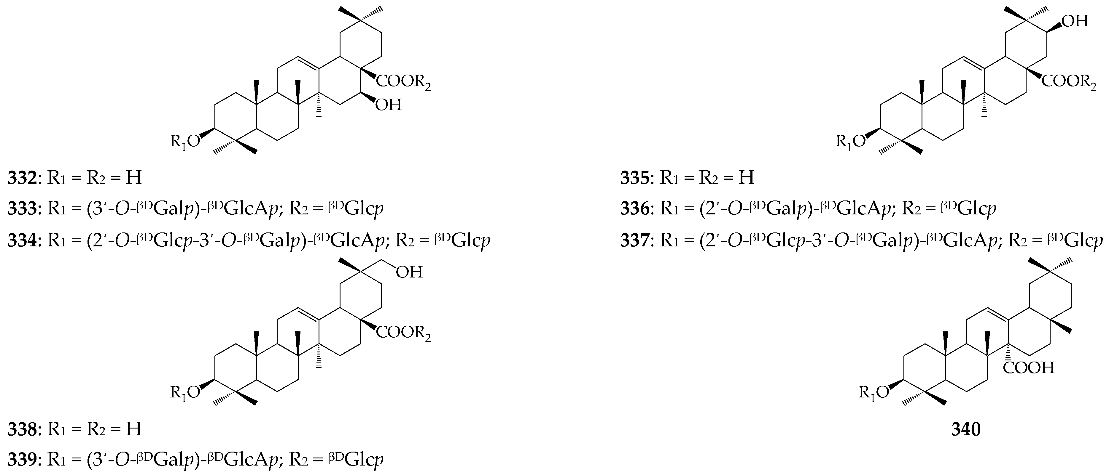
| 332 | 3β,16β-Dihydroxyolean-12-en-28-oic acid (cochalic acid) | C. officinalis (f) | [9] |
| 333 | Cochalic acid 3-O-(3′-O-βDGalp)-βDGlcAp-28-O-βDGlcp (calendasaponin B) | C. officinalis (f) C. suffruticosa (ae) | [9,75] |
| 334 | Cochalic acid 3-O-(2′-O-βDGlcp-3′-O-βDGalp)-βDGlcAp-28-O-βDGlcp (calendasaponin C) | C. officinalis (f) | [9] |
| 335 | 3β,21β-Dihydroxyolean-12-en-28-oic acid (machaerinic acid) | C. officinalis (f) | [9] |
| 336 | Machaerinic acid 3-O-(2′-O-βDGalp)-βDGlcAp-28-O-βDGlcp | C. stellata (w) | [72] |
| 337 | Machaerinic acid 3-O-(2′-O-βDGlcp-3′-O-βDGalp)-βDGlcAp-28-O-βDGlcp (calendasaponin D) | C. officinalis (f) | [9] |
| 338 | 3β,29-Dihydroxyolean-12-en-28-oic acid (mesembryanthemoidigenic acid) | C. stellata (w) | [72] |
| 339 | Mesembryanthemoidigenic acid 3-O-(3′-O-βDGalp)-βDGlcAp-28-O-βDGlcp (calendustellatoside E) | C. stellata (w) | [72] |
| 340 | 3β-Acetoxyoleane-12-en-27-oic acid (cornulacic acid) | C. officinalis (ae) | [65] |
| Triterpenes: tirucallane derivatives | |||
| 341 | Helianol | C. officinalis (f) | [3] |
| 342 | Tirucalla-7,24-dienol | C. officinalis (f) | [3] |
| Carotenoids | |||
| 343 | Antheraxanthin | C. officinalis (f) | [83] |
| 344 | (9Z)-Antheraxanthin | C. officinalis (f) | [84] |
| 345 | (all-E)-Antheraxanthin | C. officinalis (f) | [85] |
| 346 | Auroxanthin | C. officinalis (f) | [83] |
| 347 | Auroxanthin stearate | C. officinalis (f) | [85] |
| 348 | (all-E)-Auroxanthin | C. officinalis (f) | [85] |
| 349 | (all-E)-Auroxanthin palmitate | C. officinalis (f) | [85] |
| 350 | α-Carotene | C. officinalis (f) | [83] |
| 351 | β-Carotene | C. officinalis (f) | [83] |
| 352 | (13Z)-β-Carotene | C. officinalis (f) | [85] |
| 353 | (15Z)-β-Carotene | C. officinalis (f) | [85] |
| 354 | (all-E)-β-Carotene | C. officinalis (f) | [85] |
| 355 | (Z)-β-Carotene | C. officinalis (f) | [84] |
| 356 | γ-Carotene | C. officinalis (f) | [84] |
| 357 | γ-Carotene 1′,2′-epoxide | C. officinalis (f) | [85] |
| 358 | (5′Z)-γ-Carotene | C. officinalis (f) | [84] |
| 359 | δ-Carotene | C. officinalis (f) | [84] |
| 360 | δ-Carotene 1′,2′-epoxide | C. officinalis (f) | [85] |
| 361 | Chrysanthemaxanthin | C. officinalis (f) | [83] |
| 362 | α-Cryptoxanthin | C. officinalis (f) | [83] |
| 363 | β-Cryptoxanthin | C. officinalis (f) | [84] |
| 364 | β-Cryptoxanthin laurate | C. officinalis (f) | [85] |
| 365 | β-Cryptoxanthin palmitate | C. officinalis (f) | [85] |
| 366 | β-Cryptoxanthin stearate | C. officinalis (f) | [85] |
| 367 | (all-E)-β-Cryptoxanthin | C. officinalis (f) | [85] |
| 368 | (all-E)-β-Cryptoxanthin myristate | C. officinalis (f) | [85] |
| 369 | (Z)-Cryptoxanthin | C. officinalis (f) | [84] |
| 370 | Flavoxanthin | C. officinalis (f) | [83] |
| 371 | Lactucaxanthin | C. officinalis (f) | [86] |
| 372 | Lycopene | C. officinalis (f) | [83] |
| 373 | (5Z, 9Z)-Lycopene | C. officinalis (f) | [84] |
| 374 | (5Z, 9Z, 5′Z)-Lycopene | C. officinalis (f) | [84] |
| 375 | (5Z, 9Z, 5′Z, 9′Z)-Lycopene | C. officinalis (f) | [84] |
| 376 | (9/9′)-Lutein | C. officinalis (f) | [83] |
| 377 | (13/13′)-Lutein | C. officinalis (f) | [84] |
| 378 | (9Z)-Lutein | C. officinalis (f) | [84] |
| 379 | (13′Z)-Lutein | C. officinalis (f) | [85] |
| 380 | (all-E)-Lutein | C. officinalis (f) | [85] |
| 381 | (all-E)-Lutein 3-O-myristate | C. officinalis (f) | [85] |
| 382 | (all-E)-Lutein 3′-O-myristate | C. officinalis (f) | [85] |
| 383 | (all-E)-Lutein 3-O-palmitate | C. officinalis (f) | [85] |
| 384 | (all-E)-Lutein 3′-O-palmitate | C. officinalis (f) | [85] |
| 385 | (all-E)-Lutein 3-O-stearate | C. officinalis (f) | [85] |
| 386 | (all-E)-Lutein 3′-O-stearate | C. officinalis (f) | [85] |
| 387 | (all-E)-Lutein dilaurate | C. officinalis (f) | [85] |
| 388 | (all-E)-Lutein dimyristate | C. officinalis (f) | [85] |
| 389 | (all-E)-Lutein distearate | C. officinalis (f) | [85] |
| 390 | (all-E)-Lutein 3-O-laurate-3′-O-caprate | C. officinalis (f) | [85] |
| 391 | (all-E)-Lutein 3-O-laurate-3′-O-myristate | C. officinalis (f) | [85] |
| 392 | (all-E)-Lutein 3-O-myristate-3′-O-laurate | C. officinalis (f) | [85] |
| 393 | (all-E)-Lutein 3-O-myristate-3′-O-palmitate | C. officinalis (f) | [85] |
| 394 | (all-E)-Lutein 3-O-palmitate-3′-O-myristate | C. officinalis (f) | [85] |
| 395 | (all-E)-Lutein 3-O-myristate-3′-O-stearate | C. officinalis (f) | [85] |
| 396 | (all-E)-Lutein 3-O-stearate-3′-O-myristate | C. officinalis (f) | [85] |
| 397 | (all-E)-Lutein 3-O-palmitate-3′-O-stearate | C. officinalis (f) | [85] |
| 398 | (all-E)-Lutein 3-O-stearate-3′-O-palmitate | C. officinalis (f) | [85] |
| 399 | (Z)-Lutein dilaurate | C. officinalis (f) | [85] |
| 400 | (Z)-Lutein dimyristate | C. officinalis (f) | [85] |
| 401 | Lutein dibutyrate | C. officinalis (f) | [85] |
| 402 | Lutein dicaprylate | C. officinalis (f) | [85] |
| 403 | Lutein dicaprate | C. officinalis (f) | [85] |
| 404 | Lutein dipalmitate | C. officinalis (f) | [85] |
| 405 | Lutein-5,6-epoxide | C. officinalis (f) | [83] |
| 406 | (9′Z)-Lutein-5,6-epoxide | C. officinalis (f) | [84] |
| 407 | (Z)-Lutein dilaurate | C. officinalis (f) | [85] |
| 408 | (8′R)-Luteoxanthin | C. officinalis (f) | [83] |
| 409 | Mutatoxanthin | C. officinalis (f) | [83] |
| 410 | (9Z)-Neoxanthin | C. officinalis (f) | [83] |
| 411 | (13Z)-Neoxanthin | C. officinalis (f) | [84] |
| 412 | Neochrome | C. officinalis (f) | [83] |
| 413 | Phytoene | C. officinalis (f) | [85] |
| 414 | (Z)-Phytofluene | C. officinalis (f) | [85] |
| 415 | (all-E)-Phytofluene | C. officinalis (f) | [85] |
| 416 | (5′Z)-Rubixanthin | C. officinalis (f) | [84] |
| 417 | (5′Z, 9′Z)-Rubixanthin | C. officinalis (f) | [84] |
| 418 | Violaxanthin | C. officinalis (f) | [83] |
| 419 | Violaxanthin dipalmitate | C. officinalis (f) | [83] |
| 420 | Violaxanthin palmitate-stearate | C. officinalis (f) | [85] |
| 421 | (9Z)-Violaxanthin | C. officinalis (f) | [85] |
| 422 | (9Z)-Violaxanthin myristate | C. officinalis (f) | [85] |
| 423 | (all-E)-Violaxanthin | C. officinalis (f) | [85] |
| 424 | (all-E)-Violaxanthin myristate | C. officinalis (f) | [85] |
| 425 | (all-E)-Violaxanthin palmitate | C. officinalis (f) | [85] |
| 426 | (all-E)-Violaxanthin laurate | C. officinalis (f) | [85] |
| 427 | (all-E)-Violaxanthin dimyristate | C. officinalis (f) | [85] |
| 428 | (all-E)-Violaxanthin myristate-palmitate | C. officinalis (f) | [85] |
| 429 | Zeaxanthin | C. officinalis (f) | [83] |
| 430 | Zeaxanthin palmitate | C. officinalis (f) | [85] |
| 431 | Zeaxanthin myristate-palmitate | C. officinalis (f) | [85] |
| 432 | (all-E)-Zeaxanthin | C. officinalis (f) | [85] |
| 433 | (all-E)-Zeaxanthin myristate | C. officinalis (f) | [85] |
| 434 | (all-E)-Zeaxanthin dipalmitate | C. officinalis (f) | [85] |
| 435 | (all-E)-Zeinoxanthin | C. officinalis (f) | [85] |
| 436 | (all-E)-Zeinoxanthin myristate | C. officinalis (f) | [85] |
| 437 | Zeinoxanthin laurate | C. officinalis (f) | [85] |
| Phenols | |||
| 438 | p-Cymene | C. arvensis (ae) C. officinalis (ae) | [30,34] |
| 439 | p-Cymenene | C. arvensis (ae) | [34] |
| 440 | Carvacrol | C. officinalis (ae) | [30] |
| 441 | Thymol | C. officinalis (f) | [39] |
| 442 | p-Anethole | C. officinalis (f) | [35] |
| 443 | Estragole | C. officinalis (ae) | [36] |
| Benzoic acids and derivatives | |||
| 444 | 2-Hydroxybenzoic acid (salycilic acid) | C. officinalis (f) | [87,88] |
| 445 | 2-Methoxybenzoic acid (o-anisic acid) | C. officinalis (f) | [35] |
| 446 | 4-Hydroxybenzoic acid (PHBA) | C. officinalis (f) | [87,88] |
| 447 | 3,4-Dihydroxybenzoic acid (protocathechuic acid) | C. officinalis (f) | [87] |
| 448 | 3-Methoxy-4-hydroxybenzoic acid (vanillic acid) | C. officinalis (f) | [87,88,89] |
| 449 | 2,5-Dihydroxybenzoic acid (gentisic acid) | C. officinalis (f) | [87] |
| 450 | 3,5-Dimethoxy-4-hydroxybenzoic acid (syringic acid) | C. officinalis (f) | [87,88,89] |
| 451 | 1-O-p-Hydroxybenzoyl glucose | C. officinalis (l) | [90] |
| 452 | 6-O-p-Hydroxybenzoyl glucose | C. officinalis (l) | [90] |
| 453 | 1-O-Protocatechuoyl glucose | C. officinalis (l) | [90] |
| 454 | 6-O-Protocatechuoyl glucose | C. officinalis (l) | [90] |
| 455 | 1-O-Vanilloyl glucose | C. officinalis (l) | [90] |
| 456 | 6-O-Vanilloyl glucose | C. officinalis (l,p) | [90,91] |
| Hydroxycinnamates | |||
| 457 | Cinnamic acid | C. officinalis (f) | [89] |
| 458 | 2-Hydroxycinnamic acid (o-coumaric acid) | C. officinalis (f) | [89] |
| 459 | 4-Hydroxycinnamic acid (p-coumaric acid) | C. officinalis (f) C. tripterocarpa (ae) | [87,88,89,92] |
| 460 | 3,4-Dihydroxycinnamic acid (caffeic acid) | C. arvensis (ae) C. officinalis (f,l,p,s,r) | [75,87,88,89] |
| 461 | 3-Methoxy-4-hydroxycinnamic acid (ferulic acid) | C. officinalis (f,p) | [87,89,91] |
| 462 | 3-Hydroxy-4-methoxycinnamic acid (isoferulic acid) | C. officinalis (f,p) | [89,91] |
| 463 | 3-O-p-Coumaroylquinic acid | C. officinalis (f) | [89] |
| 464 | 1-O-Caffeoylquinic acid | C. officinalis (f) | [89] |
| 465 | 3-O-Caffeoylquinic acid | C. officinalis (f,l,p,s,r) | [61,89,91] |
| 466 | 4-O-Caffeoylquinic acid | C. arvensis (ae) C. officinalis (f,l,p,r) C. suffruticosa (ae) | [75,89,91] |
| 467 | 5-O-Caffeoylquinic acid | C. arvensis (ae,r) C. officinalis (f,l,s,r) C. suffruticosa (ae) | [75,89,93] |
| 468 | 1,3-Di-O-caffeoylquinic acid | C. officinalis (f,s,r) C. suffruticosa (ae) | [75,89] |
| 469 | 1,5-Di-O-caffeoylquinic acid | C. officinalis (l) C. suffruticosa (ae) | [75,90] |
| 470 | 3,4-Di-O-caffeoylquinic acid | C. arvensis (ae) C. officinalis (f) C. suffruticosa (ae) | [75,89] |
| 471 | 3,5-Di-O-caffeoylquinic acid | C. officinalis (f,l,s,r) | [89] |
| 472 | 4,5-Di-O-caffeoylquinic acid | C. officinalis (f,l,p,s,r) | [89,90,91] |
| 473 | 1,3,5-Tri-O-caffeoylquinic acid | C. officinalis (f) | [89] |
| 474 | 3,4,5-Tri-O-caffeoylquinic acid | C. officinalis (f) | [89] |
| 475 | 5-O-Feruloylquinic acid | C. arvensis (ae,r) C. officinalis (f) C. suffruticosa (ae) | [75,89,93] |
| 476 | 1,5-Di-O-feruloylquinic acid | C. officinalis (p) | [91] |
| 477 | 1,5-Di-O-isoferuloylquinic acid | C. officinalis (p) | [91] |
| 478 | 1-O-Caffeoyl glucose | C. officinalis (f) | [89] |
| Coumarins | |||
| 479 | Umbelliferone | C. officinalis (f,l) | [90,94] |
| 480 | Esculetin | C. officinalis (f,l) | [90,94] |
| 481 | Esculetin 6-O-βDGlcp (esculin) | C. officinalis (l) | [90] |
| 482 | Esculetin 7-O-βDGlcp (cichoriin) | C. officinalis (l) | [90] |
| 483 | Esculetin 7-O-(2″-O-αLRhap)-βDGlcp (neoisobaisseoside) | C. officinalis (f) | [95] |
| 484 | Esculetin 7-O-(6″-O-αLRhap)-βDGlcp (haploperoside A) | C. officinalis (f) | [95] |
| 485 | Scopoletin | C. officinalis (f,l) C. tripterocarpa (ae) | [90,92,94] |
| 486 | Scopoletin 7-O-βDGlcp (scopolin) | C. officinalis (f) C. tripterocarpa (ae) | [92,96] |
| 487 | Scopoletin 7-O-(2″-O-αLRhap)-βDGlcp (haploperoside D) | C. officinalis (f) | [95] |
| 488 | Scopoletin 7-O-(6″-O-αLRhap)-βDGlcp (isobaisseoside) | C. officinalis (f) | [95] |
| Flavonols | |||
| 489 | Kaempferol | C. tripterocarpa (ae) | [92] |
| 490 | Kaempferol 3-O-(6″-O-αLRhap)-βDGlcp (nicotiflorin) | C. arvensis (ae,r) | [93] |
| 491 | Kaempferol 7-O-(6″-O-αLRhap)-βDGlcp | C. arvensis (ae,r) | [93] |
| 492 | Quercetin | C. officinalis (f) C. tripterocarpa (ae) | [88,89,92,97] |
| 493 | Quercetin 3-O-αLRhap (quercitrin) | C. officinalis (f,p) | [89,91] |
| 494 | Quercetin 3-O-βDGlcp (isoquercitrin) | C. arvensis (ae) C. officinalis (f,l,st) | [77,88,89,97,98] |
| 495 | Quercetin 3-O-(2″-O-Ac)-βDGlcp | C. officinalis (f) | [89] |
| 496 | Quercetin 3-O-(6″-O-Ac)-βDGlcp | C. officinalis (f,l,st) | [89] |
| 497 | Quercetin 3-O-(2″,6″-O-Ac2)-βDGlcp | C. officinalis (f) | [89] |
| 498 | Quercetin 3-O-βDGalp (hyperoside) | C. arvensis (ae) C. stellata (w) | [72,77] |
| 499 | Quercetin 3-O-(2″-O-αLRhap)-αLRhap | C. officinalis (f,l) | [89] |
| 500 | Quercetin 3-O-(2″-O-αLRhap)-βDGlcp (calendoflavobioside) | C. arvensis (ae) C. officinalis (f,p,l,st) | [75,89,91,97,98] |
| 501 | Quercetin 3-O-(3″-O-αLRhap)-βDGlcp (calendoside II) | C. officinalis (f) | [99] |
| 502 | Quercetin 3-O-(4″-O-αLRhap)-βDGlcp (calendoside I) | C. officinalis (f) | [99] |
| 503 | Quercetin 3-O-(6″-O-αLRhap)-βDGlcp (rutin) | C. arvensis (ae,r) C. officinalis (f,st) C. suffruticosa (ae) | [75,89,91,93,97] |
| 504 | Quercetin 3-O-(2″,6″-O-αLRhap2)-βDGlcp (manghaslin) | C. officinalis (f,p,st) C. suffruticosa (ae) | [75,89,91,98,100] |
| 505 | Isorhamnetin | C. officinalis (f) | [89,97] |
| 506 | Isorhamnetin 3-O-αLRhap | C. officinalis (f) | [89,101] |
| 507 | Isorhamnetin 3-O-βDGlcp | C. arvensis (ae) C. officinalis (f,l,p,st) | [77,89,91,97,98] |
| 508 | Isorhamnetin 3-O-(2″-O-Ac)-βDGlcp | C. officinalis (f) | [89] |
| 509 | Isorhamnetin 3-O-(6″-O-Ac)-βDGlcp | C. officinalis (f,l,p,st) | [89,91] |
| 510 | Isorhamnetin 3-O-(2″,6″-O-Ac2)-βDGlcp | C. officinalis (f) | [89] |
| 511 | Isorhamnetin 3-O-(2″-O-αLRhap)-βDGlcp (calendoflavoside) | C. officinalis (f,p,st) | [89,91,97,98] |
| 512 | Isorhamnetin 3-O-(3″-O-αLRhap)-βDGlcp (calendoside IV) | C. officinalis (f) | [99] |
| 513 | Isorhamnetin 3-O-(4″-O-αLRhap)-βDGlcp (calendoside III) | C. officinalis (f) | [99] |
| 514 | Isorhamnetin 3-O-(6″-O-αLRhap)-βDGlcp (narcissin) | C. officinalis (f,p,st) C. suffruticosa (ae) C. stellata (w) | [72,75,89,91,97,98,101] |
| 515 | Isorhamnetin 3-O-(2″-O-αLRhap)-αLRhap (calendoflaside) | C. officinalis (f) | [97] |
| 516 | Isorhamnetin 3-O-(2″,6″-O-αLRhap2)-βDGlcp (thyphaneoside) | C. officinalis (f,p,s,st) | [89,91,98,100,101] |
| Anthocyanins | |||
| 517 | Cyanidin 3-O-βDGlcp | C. officinalis (f) | [89] |
| 518 | Cyanidin 3,5-O-βDGlcp2 | C. officinalis (f) | [89] |
| 519 | Cyanidin 3-O-(6″-O-αLRhap)-βDGlcp | C. officinalis (f) | [89] |
| 520 | Delphinidin 3-O-βDGlcp | C. officinalis (f) | [89] |
| 521 | Malvidin 3-O-βDGlcp | C. officinalis (f) | [89] |
| 522 | Paeonidin 3-O-βDGlcp | C. officinalis (f) | [89] |
| 523 | Pelargonidin 3,5-O-βDGlcp2 | C. officinalis (f) | [89] |
| 524 | Petunidin 3-O-βDGlcp | C. officinalis (f) | [89] |
| Alkanes | |||
| 525 | Tridecane | C. officinalis (ae) | [36] |
| 526 | Heptadecane | C. arvensis (ae) C. officinalis (f) | [33,39] |
| 527 | Octadecane | C. officinalis (f) | [39] |
| 528 | Nonadecane | C. arvensis (ae) C. officinalis (f) | [33,39] |
| 529 | Tricosane | C. arvensis (ae) C. officinalis (f,l) | [33,40] |
| 530 | Tetracosane | C. arvensis (ae) C. officinalis (f,l) | [33,40] |
| 531 | Pentacosane | C. arvensis (ae) C. officinalis (f,l) | [33,37,40] |
| 532 | Hexadecene | C. officinalis (ae,f,l) | [36,40] |
| 533 | Heptacosane | C. officinalis (f,l) C. suffruticosa (ae) | [40,41] |
| 534 | Octacosane | C. officinalis (f,l) | [40] |
| 535 | Nonacosane | C. officinalis (f,l) C. suffruticosa (ae) | [40,42] |
| 536 | Eicosane | C. arvensis (ae) C. officinalis (f) | [33,39] |
| 537 | Heneicosane | C. arvensis (ae) | [33] |
| 538 | Triacontane | C. suffruticosa (ae) | [42] |
| 539 | Untriacontane | C. arvensis (ae) C. officinalis (ae) C. suffruticosa (ae) | [41] |
| 540 | Tetratriacontane | C. suffruticosa (ae) | [42] |
| 550 | Cyclohexadecane | C. officinalis (f,l) | [40] |
| Aliphatic alcohols | |||
| 551 | (Z)-Hex-3-en-1-ol | C. arvensis (ae) | [34] |
| 552 | 2-Methyl-6-heptene-3-ol | C. officinalis (f) | [29] |
| 553 | 6-Methyl-5-heptene-2-ol | C. officinalis (f) | [35] |
| 554 | Hexadecan-1-ol | C. suffruticosa (ae) | [41] |
| 555 | 9-Octadecen-1-ol | C. suffruticosa (ae) | [41] |
| 556 | 6-Undecanol | C. suffruticosa (ae) | [41] |
| 557 | 1-Tetracosanol | C. suffruticosa (ae) | [42] |
| 558 | 1-Hexacosanol | C. arvensis (ae) C. suffruticosa (ae) | [41] |
| 559 | 1-Octacosanol | C. suffruticosa (ae) | [41] |
| Aliphatic Aldehydes and Ketones | |||
| 560 | Nonanal | C. arvensis (ae) C. officinalis (ae,l) | [30,31,33] |
| 561 | Decanal | C. arvensis (ae) | [34] |
| 562 | (E, E)-2,4-Decadienal | C. arvensis (ae) | [34] |
| 563 | 6-Methyl-5-heptene-2-one | C. officinalis (f) | [35] |
| 564 | 2-Pentadecanone | C. officinalis (f) | [39] |
| 565 | (6Z,9Z)-Heptadeca-6,9-diene-5,11-dione | C. officinalis (f) | [102] |
| Fatty Acids | |||
| 566 | 2-Methylpropanoic acid | C. officinalis (ae) | [36] |
| 567 | Capric acid | C. officinalis (f,l,s) | [103,104] |
| 568 | Lauric acid | C. officinalis (f,l,s) | [35,103,104,105] |
| 569 | Lauric acid methyl ester | C. officinalis (f) | [35] |
| 570 | Tridecanoic acid | C. officinalis (l) | [103,104] |
| 571 | Myristic acid | C. officinalis (f,l,s) C. suffruticosa (ae) | [41,103,104,105] |
| 572 | Myristic acid methyl ester | C. officinalis (f) | [35] |
| 573 | Myristic acid ethyl ester | C. officinalis (f) | [35] |
| 574 | Pentadecanoic acid | C. officinalis (f,l,s) | [103,104,105] |
| 575 | Palmitic acid | C. arvensis (ae) C. officinalis (f,l,s) C. suffruticosa (ae) | [39,41,103,104] |
| 576 | Palmitic acid methyl ester | C. officinalis (f) | [35] |
| 577 | Palmitic acid ethyl ester | C. officinalis (f) | [35] |
| 578 | cis-7-Hexadecanoic acid | C. officinalis (s) | [105] |
| 579 | Palmitoleic acid | C. officinalis (f,l,s) | [103,104,105] |
| 580 | Margaric acid | C. officinalis (f,l,s) | [103,104,105] |
| 581 | Margaric acid methyl ester | C. officinalis (f) | [35] |
| 582 | Stearic acid | C. arvensis (ae) C. officinalis (f,l,s) C. suffruticosa (ae) | [41,103,104,105] |
| 583 | Stearic acid methyl ester | C. officinalis (f) | [35] |
| 584 | Oleic acid | C. officinalis (f,l,s) | [103,104,105] |
| 585 | Elaidic acid | C. officinalis (s) | [105] |
| 586 | Linoelaidic acid | C. officinalis (f,l,s) | [103,104,105] |
| 587 | Linoleic acid | C. arvensis (ae) C. officinalis (s) C. suffruticosa (ae) | [41,105] |
| 588 | Linoleic acid methyl ester | C. officinalis (f) | [35] |
| 589 | Linolenic acid | C. arvensis (ae) C. officinalis (s) C. suffruticosa (ae) | [41,105] |
| 590 | Linolenic acid methyl ester | C. officinalis (f) | [35] |
| 591 | α-Calendic acid | C. arvensis (s) C. officinalis (f, l, s) C. stellata (f, s) C. suffruticosa (s) C. tripterocarpum (s) C. maroccana (s) | [16,103,104,105] |
| 592 | β-Calendic acid | C. officinalis (s) | [105] |
| 593 | 9-Hydroxy-trans-10-cis-12-octadecadienic acid | C. officinalis (s) | [105] |
| 594 | Gondoic acid | C. officinalis (s) | [105] |
| 595 | Arachic acid | C. officinalis (f,l,s) | [103,104,105] |
| 596 | Heneicosanoic acid | C. officinalis (f,l) | [103,104] |
| 597 | Behenic acid | C. officinalis (f,l,s) | [103,104,105] |
| 598 | Tricosanoic acid | C. officinalis (f,l) | [103,104] |
| 599 | Tetracosanoic acid | C. arvensis (ae) C. officinalis (ae) C. suffruticosa (ae) | [41] |
| 600 | Lignoceric acid | C. officinalis (f,l) | [103,104] |
| 601 | Pentacosanoic acid | C. officinalis (f,l) | [103,104] |
| 602 | Octacosanoic acid | C. suffruticosa (ae) | [41] |
| Chromanols | |||
| 603 | 2-Methyl-2-(4,8,12-trimethyltridecyl)chroman-6-ol (tocol) | C. officinalis (ae) | [106] |
| 604 | Tocol 5-methyl ester | C. officinalis (ae) | [106] |
| 605 | Tocol 7-methyl ester | C. officinalis (ae) | [106] |
| 606 | Tocol 8-methyl ester (δ-tocopherol) | C. officinalis (ae) | [106] |
| 607 | Tocol 5,7-dimethyl ester | C. officinalis (ae) | [106] |
| 608 | Tocol 5,8-dimethyl ester (β-tocopherol) | C. officinalis (ae) | [107] |
| 609 | Tocol 5,7-dimethyl ester (γ-tocopherol) | C. officinalis (ae) | [107] |
| 610 | Tocol 5,7,8-dimethyl ester (α-tocopherol) | C. officinalis (ae) C. suffruticosa (ae) | [42,107] |
| 611 | Plastoquinone | C. officinalis (ae) | [107] |
| 612 | Phylloquinone | C. officinalis (ae) | [107] |
| 613 | Ubiquinone | C. officinalis (ae) | [107] |
| Organic Acids | |||
| 614 | Malic acid | C. arvensis (ae,r) | [93] |
| 615 | Citric acid | C. arvensis (ae) C. officinalis (ae) C. suffruticosa (ae) | [41] |
| 616 | Quinic acid | C. arvensis (ae,r) C. officinalis (ae) C. suffruticosa (ae) | [41,93] |
| Carbohydrates | |||
| 617 | Threonic acid | C. arvensis (ae) | [41] |
| 618 | Ribose | C. suffruticosa (ae) | [41] |
| 619 | Tagatose | C. arvensis (ae) C. officinalis (ae) C. suffruticosa (ae) | [41] |
| 620 | Fructose | C. arvensis (ae) C. officinalis (ae) C. suffruticosa (ae) | [41] |
| 621 | Psicose | C. arvensis (ae) C. officinalis (ae) C. suffruticosa (ae) | [41] |
| 622 | Mannose | C. arvensis (ae) C. officinalis (ae) C. suffruticosa (ae) | [41] |
| 623 | Galactose | C. arvensis (ae) C. officinalis (ae) C. suffruticosa (ae) | [41] |
| 624 | Glucose | C. arvensis (ae) C. officinalis (ae) C. suffruticosa (ae) | [41] |
| 625 | Gluconic acid | C. arvensis (ae) C. officinalis (ae) C. suffruticosa (ae) | [41] |
| 626 | Galactaric acid | C. arvensis (ae) C. officinalis (ae) C. suffruticosa (ae) | [41] |
| 627 | Sucrose | C. arvensis (ae) C. officinalis (ae) C. suffruticosa (ae) | [41] |
| 628 | Cellobiose | C. arvensis (ae) C. officinalis (ae) C. suffruticosa (ae) | [41] |
| 629 | scyllo-Inositol | C. officinalis (ae) C. suffruticosa (ae) | [41] |
| 630 | myo-Inositol | C. arvensis (ae) C. officinalis (ae) C. suffruticosa (ae) | [41] |
| Amino Acids | |||
| 631 | Alanine | C. officinalis (f,l,st) | [108] |
| 632 | γ-Aminobutyric acid (GABA) | C. arvensis (ae) C. officinalis (ae) C. suffruticosa (ae) | [41] |
| 633 | Arginine | C. officinalis (f,l,st) | [108] |
| 634 | Aspartic acid | C. officinalis (f,l,st) | [108] |
| 635 | Asparagine | C. officinalis (f,l,st) | [108] |
| 636 | Histidine | C. officinalis (f,l,st) | [108] |
| 637 | Glutamic acid | C. officinalis (f,l,st) | [108] |
| 638 | Leucine | C. officinalis (f,l,st) | [108] |
| 639 | Lysine | C. officinalis (f,l,st) | [108] |
| 640 | Proline | C. officinalis (f,l,st) | [108] |
| 641 | Serine | C. officinalis (f,l,st) | [108] |
| 642 | Tyrosine | C. officinalis (f,l,st) | [108] |
| 643 | Threonine | C. officinalis (f,l,st) | [108] |
| 644 | Methionine | C. officinalis (f,l,st) | [108] |
| 645 | Phenylalanine | C. officinalis (f,l,st) | [108] |
| 646 | Valine | C. officinalis (f,l,st) | [108] |
| Other Compounds | |||
| 647 | 3-Cyclohexene-1-ol | C. officinalis (ae) | [30] |
| 648 | 3-Cyclohexene-1-ol 4-methyl ester | C. officinalis (l) | [31] |
| 649 | Loliolide | C. officinalis (f) | [109] |
| 650 | 1,2,3,5,8,8α-Hexahydronaphthalene 6,7-dimethyl ester | C. officinalis (ae) | [36] |
| 651 | 4-Methylacethophenone | C. arvensis (ae) | [34] |
| 652 | Tricyclene | C. officinalis (f) | [35] |
| 653 | 1H-Benzocyclohepten-9-ol | C. arvensis (ae) | [41] |
| 654 | 2-Pentyl furane | C. arvensis (ae) | [33] |
| 655 | 1-Methyl ethyl hexadecanoate | C. officinalis (f,l) | [40] |
| 656 | Naphthalene | C. suffruticosa (ae) | [42] |
| Source, Extractant [Ref.] | Name | Ara | Gal | Glc | Man | Rha | Xyl | UA | Yield, % | MW, kDa | Fine Structure |
|---|---|---|---|---|---|---|---|---|---|---|---|
| Flowers, 0.5 M NaOH [119] | PS-I | 34.2 | 41.0 | 24.8 | 0.08 | 15 | rhamnoarabino-3,6-galactan | ||||
| PS-II | 27.6 | 72.4 | 0.04 | 25 | arabino-3,6-galactan | ||||||
| PS-III | 48.7 | 51.4 | 0.05 | 35 | arabino-3,6-galactan | ||||||
| Flowers, water [120] | PSC-1 | 12.5 | 40.8 | 20.1 | 2.3 | 24.1 | - | - | - | ||
| PSC-2 | 11.0 | 35.2 | 14.0 | 0.9 | 1.6 | 37.2 | - | - | - | ||
| PSC-3 | 8.2 | 25.1 | 20.2 | 3.4 | 42.1 | - | - | - | |||
| PSC-4 | 7.5 | 35.7 | 11.5 | 3.1 | 1.0 | 40.5 | - | - | - | ||
| PSC-5 | 4.0 | 14.1 | 18.1 | 5.2 | 57.2 | - | - | - | |||
| Industrial flowers waste, water [121] | - | 7.5 | 6.1 | 1.7 | 1.8 | 0.5 | 1.2 | 58.3 | 8.90 | 40 | - |
| Industrial flowers waste, 0.1 M HCl [122] | F | 4.3 | 2.5 | 0.2 | 4.1 | 1.5 | 64.0 | - | - | - | |
| HD | 7.7 | 3.1 | 1.8 | 5.4 | 2.2 | 62.5 | - | - | - |
| Group of Metabolites | Calendula Species a,b | ||||
|---|---|---|---|---|---|
| CA | CO | CSt | CSu | CT | |
| Monoterpenes, sesquiterpenes, diterpenes as components of essential oils | AE | AE, F, L | F | AE | × |
| Sesquiterpene glycosides | AE | F | × | × | × |
| Triterpenes: sterols | AE | AE, F, L, R, S | × | AE | × |
| Triterpenes: ursanes and oleananes non-glycosidic | × | AE, F, L, R, S | × | AE | × |
| Triterpenes: glycosides | AE | AE, F, R | W | AE | × |
| Carotenoids | × | F | × | × | × |
| Benzoic acid derivatives | × | F, L, P | × | × | × |
| Hydroxycinnamates | AE | AE, F, L, P, R, S | × | AE | AE |
| Coumarins | × | F, L | × | × | AE |
| Flavonoids | AE, R | AE, F, L, P, R, S | W | AE | AE |
| Anthocyanins | × | F | × | × | × |
| Alkanes, aliphatic alcohols, aldehydes, ketones, fatty acids as components of lipophilic extracts | AE | AE, F, L, S | F | AE | × |
| Organic acids, carbohydrates | AE, R | AE | × | AE | × |
| Amino acids | AE | AE, F, L, St | × | AE | × |
| Chromanols | × | AE | × | AE | × |
| Polysaccharides | × | F | × | × | × |
Publisher’s Note: MDPI stays neutral with regard to jurisdictional claims in published maps and institutional affiliations. |
© 2022 by the authors. Licensee MDPI, Basel, Switzerland. This article is an open access article distributed under the terms and conditions of the Creative Commons Attribution (CC BY) license (https://creativecommons.org/licenses/by/4.0/).
Share and Cite
Olennikov, D.N.; Kashchenko, N.I. Marigold Metabolites: Diversity and Separation Methods of Calendula Genus Phytochemicals from 1891 to 2022. Molecules 2022, 27, 8626. https://doi.org/10.3390/molecules27238626
Olennikov DN, Kashchenko NI. Marigold Metabolites: Diversity and Separation Methods of Calendula Genus Phytochemicals from 1891 to 2022. Molecules. 2022; 27(23):8626. https://doi.org/10.3390/molecules27238626
Chicago/Turabian StyleOlennikov, Daniil N., and Nina I. Kashchenko. 2022. "Marigold Metabolites: Diversity and Separation Methods of Calendula Genus Phytochemicals from 1891 to 2022" Molecules 27, no. 23: 8626. https://doi.org/10.3390/molecules27238626
APA StyleOlennikov, D. N., & Kashchenko, N. I. (2022). Marigold Metabolites: Diversity and Separation Methods of Calendula Genus Phytochemicals from 1891 to 2022. Molecules, 27(23), 8626. https://doi.org/10.3390/molecules27238626