
New Organoselenium (NSAIDs-Selenourea and Isoselenocyanate) Derivatives as Potential Antiproliferative Agents: Synthesis, Biological Evaluation and in Silico Calculations

Abstract
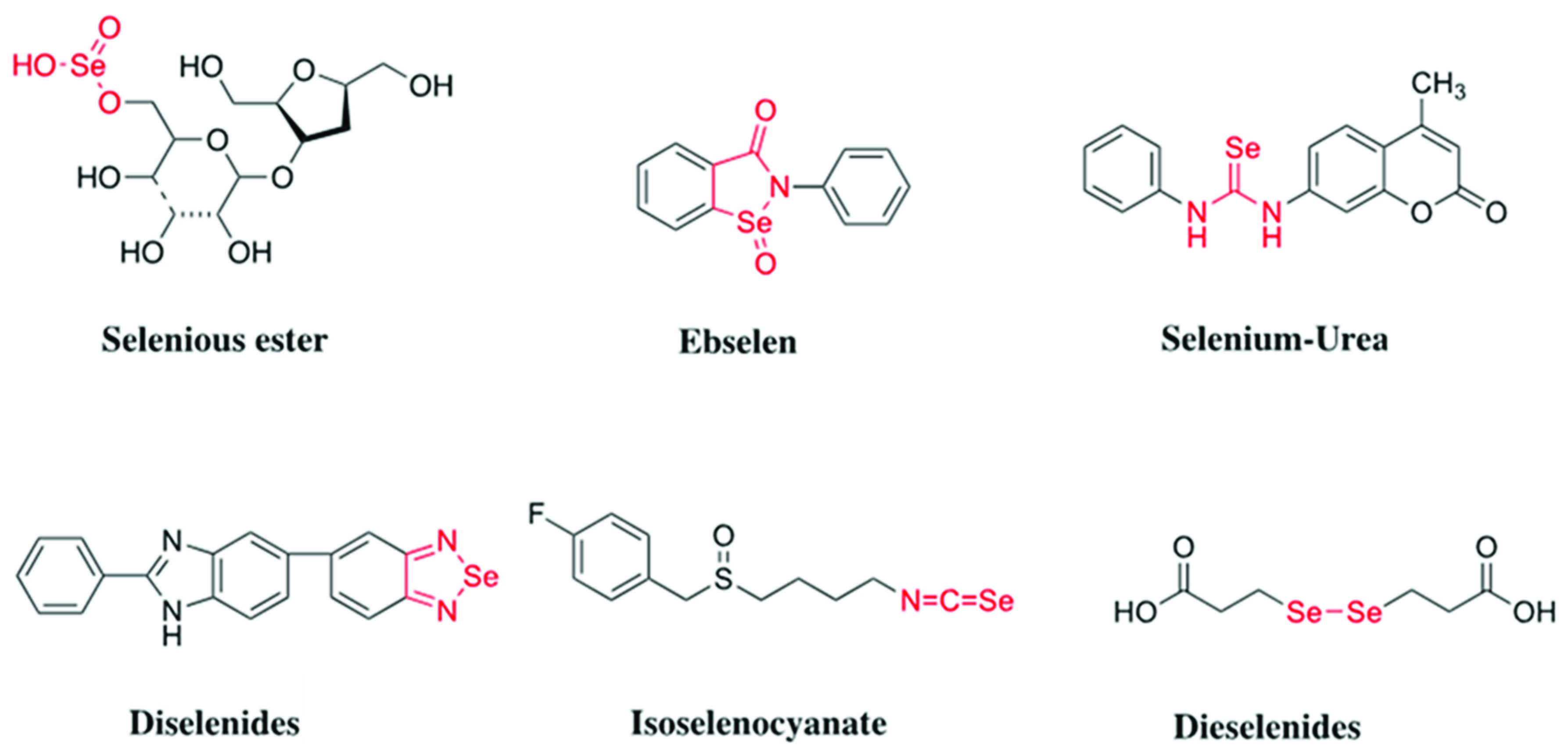
:1. Introduction
2. Results and Discussion
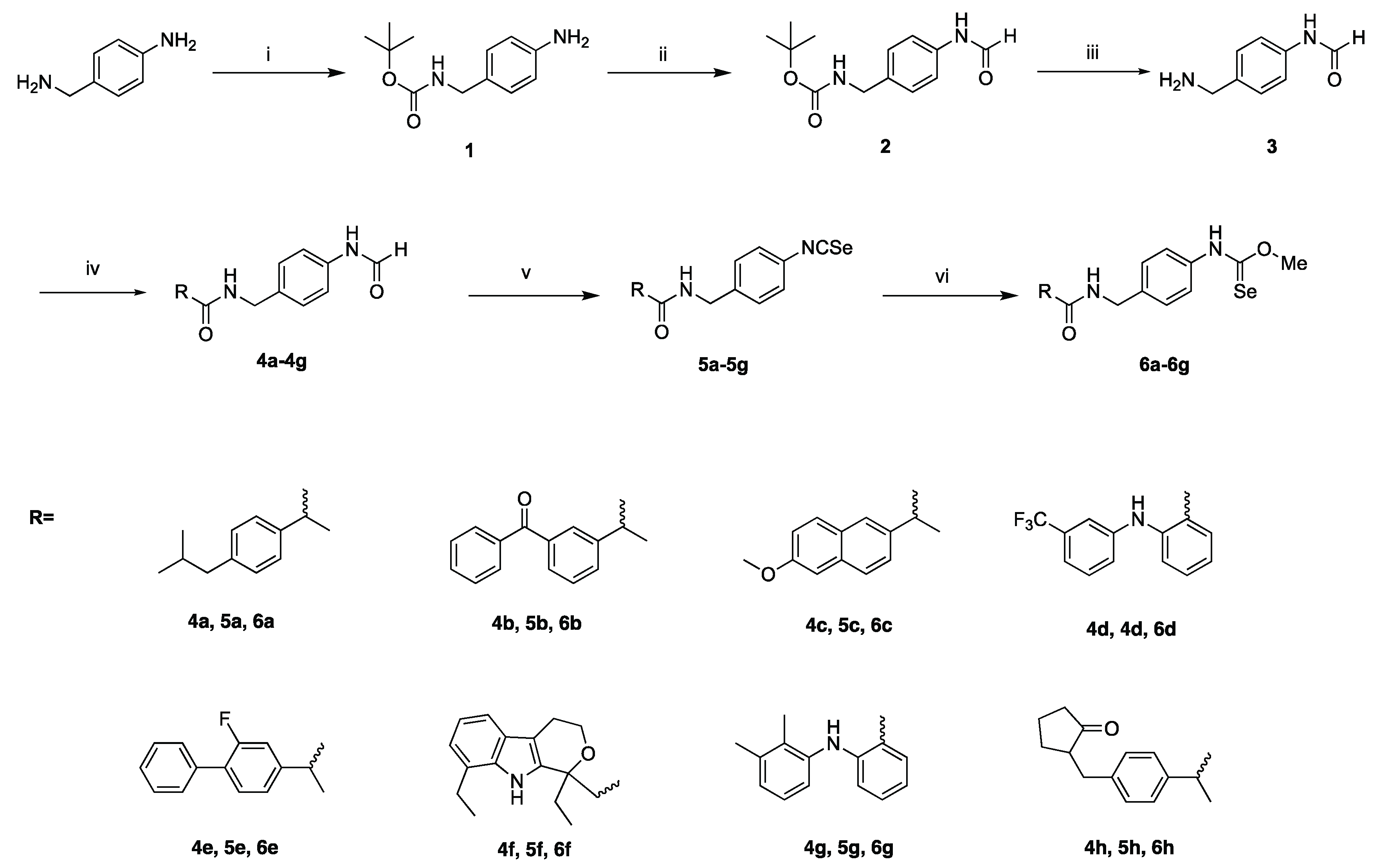
2.1. Chemistry
2.2. Cell Viability Assay
2.3. Evaluation of Bcl-2, IL-2 and Caspase-3 Molecular Biomarkers in MCF-7 Cells
2.4. Antioxidant Assay
2.4.1. Radical Scavenging Capacity (DPPH) Assay
2.4.2. Bleomycin DNA Damage Assay
2.4.3. Glutathione Peroxidase-like Activity Assay
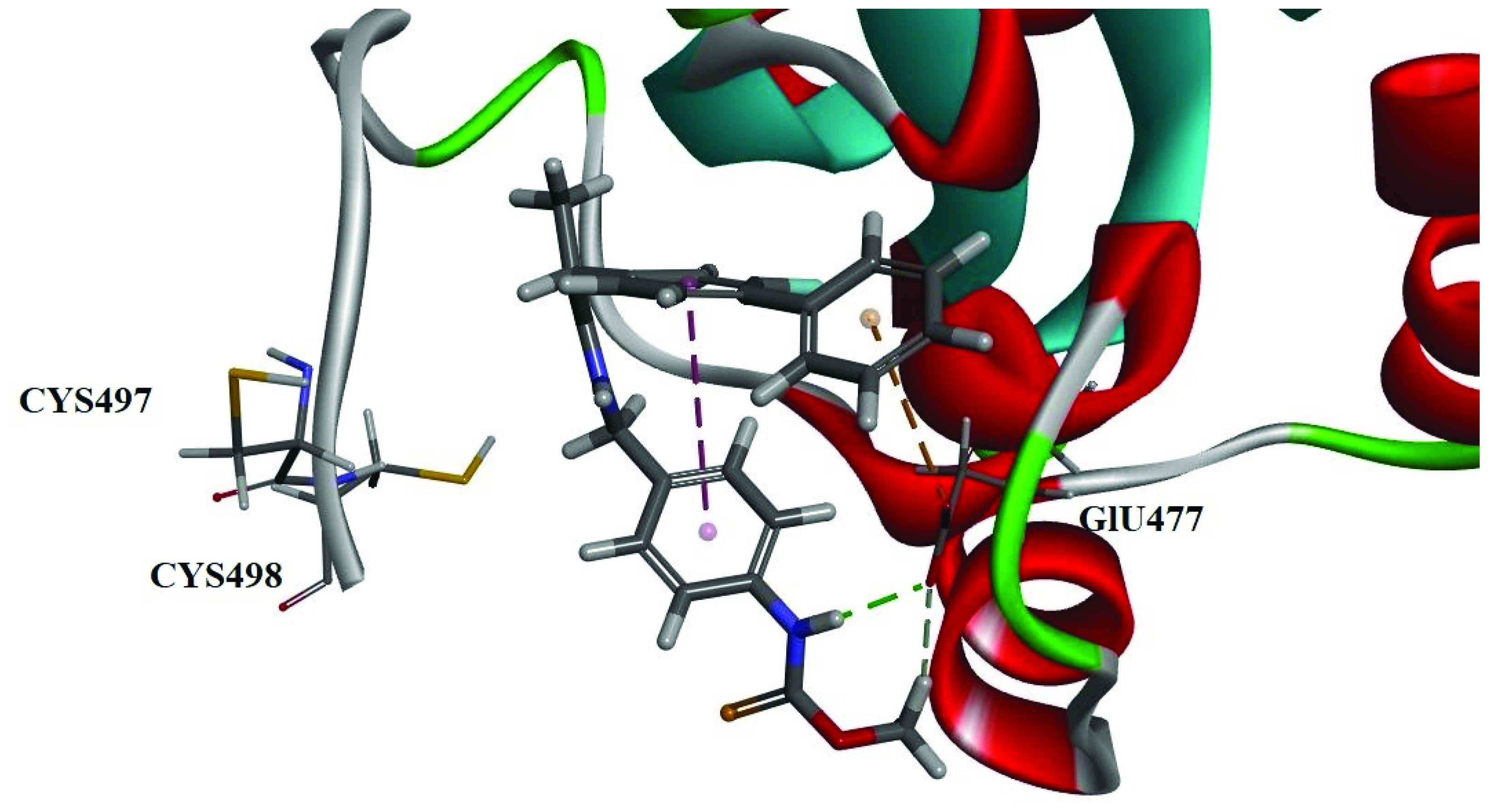
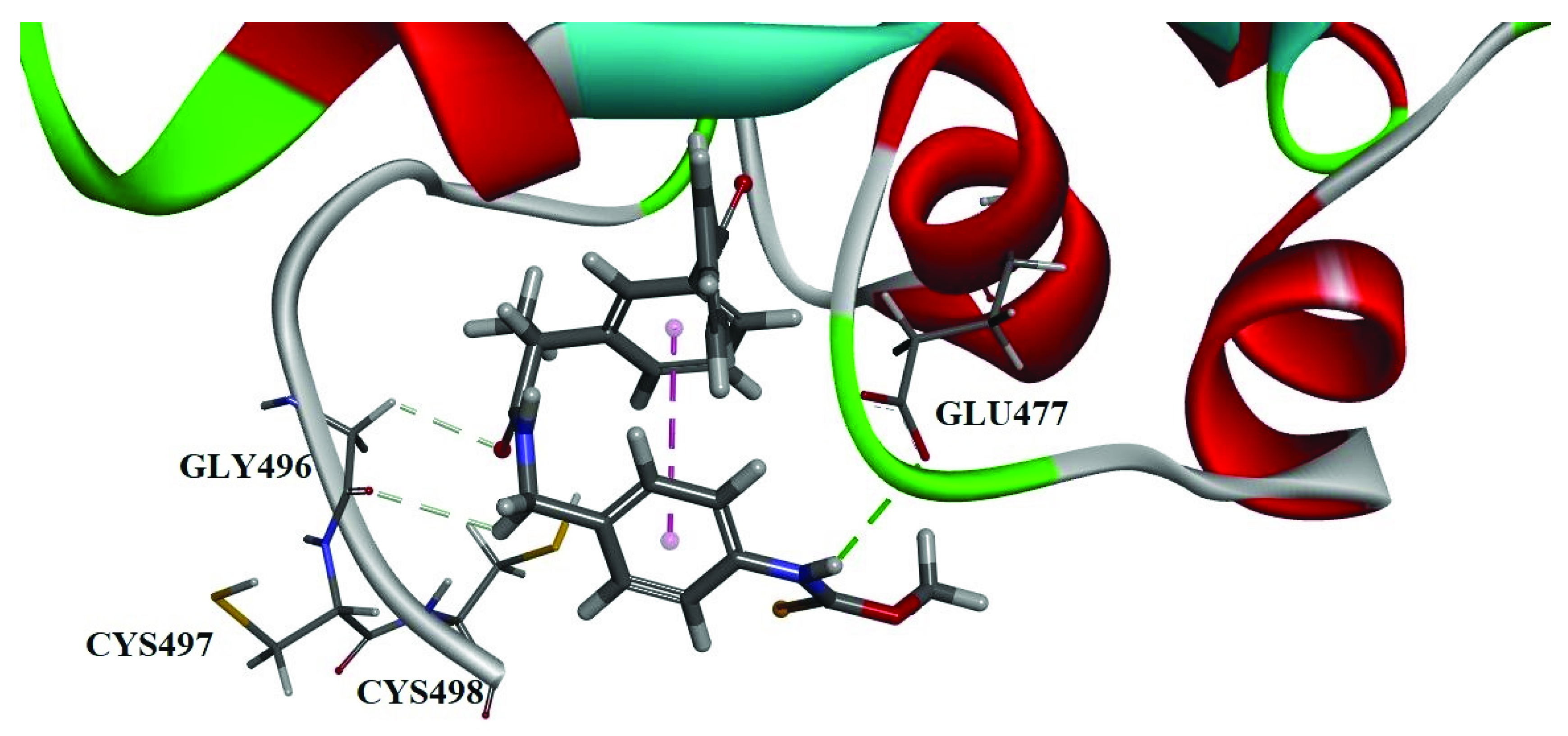
2.5. Docking Studies
3. Conclusions
4. Materials and Methods
4.1. Materials
4.2. Experimental Procedures
4.2.1. N-(4-(aminomethyl)phenyl)formamide 3
4.2.2. General Procedure for the Synthesis of Compounds 4a–4h
N-(4-formamidobenzyl)-2-(4-isobutylphenyl)propenamide (4a)
2-(3-benzoylphenyl)-N-(4-formamidobenzyl)propenamide (4b)
N-(4-formamidobenzyl)-2-(6-methoxynaphthalen-2-yl)propenamide (4c)
N-(4-formamidobenzyl)-2-((3-(trifluoromethyl)phenyl)amino)benzamide (4d)
2-(2-fluoro-[1,1’-biphenyl]-4-yl)-N-(4-formamidobenzyl)propenamide (4e)
2-(1,8-diethyl-1,3,4,9-tetrahydropyrano[3,4-b]indol-1-yl)-N-(4-formamidobenzyl) acetamide (4f)
2-((2,3-dimethylphenyl)amino)-N-(4-formamidobenzyl)benzamide (4g)
N-(4-formamidobenzyl)-2-(4-((2-oxocyclopentyl)methyl)phenyl)propenamide (4h)
4.2.3. General Procedure for the Synthesis of Compounds 5a–5h
2-(4-isobutylphenyl)-N-(4-isoselenocyanatobenzyl)propenamide (5a)
2-(3-benzoylphenyl)-N-(4-isoselenocyanatobenzyl)propenamide (5b)
N-(4-isoselenocyanatobenzyl)-2-(6-methoxynaphthalen-2-yl)propenamide (5c)
N-(4-isoselenocyanatobenzyl)-2-((3-(trifluoromethyl)phenyl)amino)benzamide (5d)
2-(2-fluoro-[1,1’-biphenyl]-4-yl)-N-(4-isoselenocyanatobenzyl)propenamide (5e)
2-(1,8-diethyl-1,3,4,9-tetrahydropyrano[3,4-b]indol-1-yl)-N-(4-isoselenocyanatobenzyl)acetamide (5f)
2-((2,3-dimethylphenyl)amino)-N-(4-isoselenocyanatobenzyl)benzamide (5g)
N-(4-isoselenocyanatobenzyl)-2-(4-((2-oxocyclopentyl)methyl)phenyl)propenamide (5h)
4.2.4. General Procedure for the Synthesis of Compounds 6a–6h
O-methyl (4-((2-(4-isobutylphenyl)propanamido)methyl)phenyl) carbamoselenoate (6a)
O-methyl (4-((2-(3-benzoylphenyl)propanamido)methyl)phenyl)carbamoselenoate (6b)
O-methyl (4-((2-(6-methoxynaphthalen-2-yl)propanamido)methyl)phenyl)carbamoselenoate (6c)
O-methyl (4-((2-((3-(trifluoromethyl)phenyl)amino)benzamido)methyl)phenyl)carbamoselenoate (6d)
O-methyl (4-((2-(2-fluoro-[1,1’-biphenyl]-4-yl)propanamido)methyl)phenyl)carbamoselenoate (6e)
O-methyl (4-((2-(1,8-diethyl-1,3,4,9-tetrahydropyrano[3,4-b]indol-1-yl)acetamido)methyl)phenyl)carbamoselenoate (6f)
O-methyl (4-((2-((2,3-dimethylphenyl)amino)benzamido)methyl)phenyl)carbamoselenoate (6g)
O-methyl (4-((2-(4-((2-oxocyclopentyl)methyl)phenyl)propanamido)methyl)phenyl)carbamoselenoate (6h)
4.3. Cell Lines and Growth Conditions
4.4. Detection of Bcl-2, IL-2 and Caspase-3 Molecular Biomarkers in MCF-7 Cells
4.5. DPPH Free Radical Scavenging Activity
4.6. Bleomycin-Dependent DNA Damage
4.7. Molecular Modeling
4.7.1. Protein and Ligand Preparation
4.7.2. Ligand Docking
4.7.3. Result Analysis
Supplementary Materials
Author Contributions
Funding
Institutional Review Board Statement
Informed Consent Statement
Data Availability Statement
Conflicts of Interest
Statistical Analysis
References
- Maniar, K.H.; Jones, I.A.; Gopalakrishna, R.; Vangsness, C.T. Lowering side effects of NSAID usage in osteoarthritis: Recent attempts at minimizing dosage. Expert Opin. Pharmacother. 2018, 19, 93–102. [Google Scholar] [CrossRef] [PubMed]
- Benbow, T.; Campbell, J. Microemulsions as transdermal drug delivery systems for nonsteroidal anti-inflammatory drugs (NSAIDs): A literature review. Drug Dev. Ind. Pharm. 2019, 45, 1849–1855. [Google Scholar] [CrossRef] [PubMed]
- Waddell, W.R.; Loughry, R.W. Sulindac for polyposis of the colon. J. Surg. Oncol. 1983, 24, 83–87. [Google Scholar] [CrossRef] [PubMed]
- Palayoor, S.T.; Tofilon, P.J.; Coleman, C.N. Ibuprofen-mediated reduction of hy- poxia-inducible factors HIF-1alpha and HIF-2alpha in prostate cancer cells. Clin. Cancer Res. 2003, 9, 3150–3157. [Google Scholar]
- Takiuchi, T.; Blake, E.A.; Matsuo, K.; Sood, A.K.; Brasky, T.M. Aspirin use and endometrial cancer risk and survival. Gynecol. Oncol. 2018, 148, 222–232. [Google Scholar] [CrossRef]
- Wong, R.S.Y. Role of nonsteroidal anti-inflammatory drugs (NSAIDs) in cancer. prevention and cancer promotion. Adv. Pharmacol. Sci. 2019, 2019, 3418975. [Google Scholar] [CrossRef] [Green Version]
- Wrobel, J.K.; Power, R.; Toborek, M. Biological activity of selenium: Revisited. IUBMB Life 2016, 68, 97–105. [Google Scholar] [CrossRef]
- Hosnedlova, B.; Kepinska, M.; Skalickova, S.; Fernandez, C.; Ruttkay-Nedecky, B.; Malevu, T.D.; Sochor, J.; Baron, M.; Melcova, M.; Zidkova, J.; et al. A summary of new findings on the biological effects of selenium in selected animal species-a critical review. Int. J. Mol. Sci. 2017, 18, 2209. [Google Scholar] [CrossRef]
- Vinceti, M.; Filippini, T.; Cilloni, S.; Crespi, C.M. The Epidemiology of Selenium and Human Cancer. Adv. Cancer Res. 2017, 136, 1–48. [Google Scholar]
- Vinceti, M.; Filippini, T.; del Giovane, C.; Dennert, G.; Zwahlen, M.; Brinkman, M.; Zeegers, M.P.; Horneber, M.; D’Amico, R.; Crespi, C.M. Selenium for preventing cancer. Cochrane Database Syst. Rev. 2018, 1, CD005195. [Google Scholar] [CrossRef]
- Murdolo, G.; Bartolini, D.; Tortoioli, C.; Piroddi, M.; Torquato, P.; Galli, F. Selenium and Cancer Stem Cells. Adv. Cancer Res. 2017, 136, 235–257. [Google Scholar] [PubMed]
- El-Bayoumy, K. Overview: The late Larry C. Clark showed the bright side of the moon element (selenium) in a clinical cancer prevention trial. Nutr. Cancer 2001, 40, 4–5. [Google Scholar] [CrossRef] [PubMed]
- Guo, P.; Wang, Q.; Liu, J.; Liu, L.; Zhao, P.; Cao, Y.; Liu, Y.; Qi, C. Preparation of two organoselenium compounds and their induction of apoptosis to SMMC-7221 cells. Biol. Trace Elem. Res. 2013, 154, 304–311. [Google Scholar] [CrossRef]
- Desai, D.; Salli, U.; Vrana, K.E.; Amin, S. SelSA, selenium analogs of SAHA as potent histone deacetylase inhibitors. Bioorg. Med. Chem. Lett. 2010, 20, 2044–2047. [Google Scholar] [CrossRef] [PubMed] [Green Version]
- Pinto, J.T.; Sinha, R.; Papp, K.; Facompre, N.D.; Desai, D.; El-Bayoumy, K. Differential effects of naturally occurring and synthetic organoselenium compounds on biomarkers in androgen responsive and androgen independent human prostate carcinoma cells. Int. J. Cancer 2007, 120, 1410–1417. [Google Scholar] [CrossRef]
- Singh, U.; Null, K.; Sinha, R. In vitro growth inhibition ofmouse mammary epithelial tumor cells by methylseleninic acid: Involvement of protein kinases. Mol. Nutr. Food Res. 2008, 52, 1281–1288. [Google Scholar] [CrossRef]
- Sharma, A.K.; Sharma, A.; Desai, D.; Madhunapantula, S.V.; Huh, S.J.; Robertson, G.P.; Amin, S. Synthesis and antiproliferative activity comparison of phenylalkyl isoselenocyanates with corresponding naturally occurring and synthetic isothiocyanates. J. Med. Chem. 2008, 51, 7820–7826. [Google Scholar] [CrossRef] [Green Version]
- Moreno, E.; Plano, D.; Lamberto, I.; Font, M.; Encio, I.; Palop, J.A.; Sanmartin, C. Sulfur and selenium derivatives of quinazoline and pyrido [2,3-d]pyrimidine: Synthesis and study of their potential cytotoxic activity in vitro. Eur. J. Med. Chem. 2012, 47, 283–298. [Google Scholar] [CrossRef]
- Desai, D.; Sinha, I.; Null, K.; Wolter, W.; Suckow, M.A.; King, T.; Amin, S.; Sinha, R. Synthesis and antitumor properties of selenocoxib-1 against rat prostate adenocarcinoma cells. Int. J. Cancer 2010, 127, 230–238. [Google Scholar] [CrossRef]
- Plano, D.; Karelia, D.N.; Pandey, M.K.; Spallholz, J.E.; Amin, S.; Sharma, A.K. Design, synthesis, and biological evaluation of novel selenium (Se-NSAID) molecules as anticancer agents. J. Med. Chem. 2016, 59, 1946–1959. [Google Scholar] [CrossRef]
- Liu, L.; Li, S.; Li, X.; Zhong, M.; Lu, Y.; Yang, J.; Zhang, Y.; He, X. Synthesis of NSAIDs-Se derivatives as potent anticancer agents. Med. Chem. Res. 2018, 27, 2071–2078. [Google Scholar] [CrossRef]
- Nie, Y.; Zhong, M.; Li, S.; Li, X.; Zhang, Y.; Zhang, Y.; He, X. Synthesis and potential. anticancer activity of some novel selenocyanates and diselenides. Chem. Biodivers. 2020, 17, e1900603. [Google Scholar] [CrossRef]
- He, X.; Zhong, M.; Li, S.; Li, X.; Li, Y.; Li, Z.; Gao, Y.; Ding, F.; Wen, D.; Lei, Y.; et al. Synthesis and biological evaluation of organoselenium (NSAIDs-SeCN and SeCF3) derivatives as potential anticancer agents. Eur. J. Med. Chem. 2020, 208, 112864. [Google Scholar] [CrossRef] [PubMed]
- He, X.; Nie, Y.; Zhong, M.; Li, S.; Li, X.; Guo, Y.; Liu, Z.; Gao, Y.; Ding, F.; Wen, D.; et al. New organoselenides (NSAIDs-Se derivatives) as potential anticancer agents: Synthesis, biological evaluation and in silico calculations. Eur. J. Med. Chem. 2021, 218, 113384. [Google Scholar] [CrossRef]
- Wittmann, C.; Chockley, P.; Singh, S.K.; Pase, L.; Lieschke, G.J.; Grabher, C. Hydrogen peroxide in inflammation: Messenger, guide, and assassin. Adv. Hematol. 2012, 2012, 541471. [Google Scholar] [CrossRef]
- Jamier, V.; Ba, L.A.; Jacob, C. Selenium-and tellurium-containing multifunctional redox agents as biochemical redox modulators with selective cytotoxicity. Chem. A Eur. J. 2010, 16, 10920–10928. [Google Scholar] [CrossRef]
- Pathania, D.; Sechi, M.; Palomba, M.; Sanna, V.; Berrettini, F.; Sias, A.; Taheri, L.; Neamati, N. Design and discovery of novel quinazolinedione-based redox modulators as therapies for pancreatic cancer. Biochim. Biophys. Acta (BBA) Gen. Subj. 2014, 1840, 332–343. [Google Scholar] [CrossRef] [PubMed]
- Plano, D.; Baquedano, Y.; Ibáñez, E.; Jiménez, I.; Palop, J.A.; Spallholz, J.E.; Sanmartín, C. Antioxidant-prooxidant properties of a new organoselenium compound library. Molecules 2010, 15, 7292–7312. [Google Scholar] [CrossRef]
- Lagunes, I.; Begines, P.; Silva, A.; Galán, A.R.; Puerta, A.; Fernandes, M.X.; Maya, I.; Fernández-Bolaños, J.G.; López, Ó.; Padrón, J.M. Selenocoumarins as new multitarget antiproliferative agents: Synthesis, biological evaluation and in silico calculations. Eur. J. Med. Chem. 2019, 179, 493–501. [Google Scholar] [CrossRef]
- Meriane, D.; Genta-Jouve, G.; Kaabeche, M.; Michel, S.; Boutefnouchet, S. Rapid identification of antioxidant compounds of Genista saharae coss. & dur. By combination of DPPH scavenging assay and HPTLC-MS. Molecules 2014, 19, 4369–4379. [Google Scholar]
- Uddin, R.; Saha, M.R.; Subhan, N.; Hossain, H.; Jahan, I.A.; Akter, R.; Alam, A. HPLC-analysis of polyphenolic compounds in gardenia jasminoides and determination of antioxidant activity by using free radical scavenging assays. Adv. Pharm. Bull. 2014, 4, 273–281. [Google Scholar] [PubMed]
- Huang, D.; Qu, B.; Prior, R.L. The chemistry behind antioxidant capacity assays. J. Agric. Food Chem. 2005, 53, 1841–1856. [Google Scholar] [CrossRef] [PubMed]
- Prior, R.L.; Wu, X.; Schaich, K. Standardized methods for the determination of. antioxidant capacity and phenolics in foods and dietary supplements. J. Agric. Food Chem. 2005, 53, 4290–4302. [Google Scholar] [CrossRef] [PubMed]
- Tian, X.; Schaich, K.M. Effects of molecular structure on kinetics and dynamics of the trolox equivalent antioxidant capacity assay with ABTS(t*). J. Agric. Food Chem. 2013, 61, 5511–5519. [Google Scholar] [CrossRef]
- Mira, A.; Gimenez, E.M.; Bolzan, A.D.; Bianchi, M.S.; Lopez-Larraza, D.M. Effect of thiol compounds on bleomycin-induced DNA and chromosome damage in human cells. Arch. Environ. Occup. Health 2013, 68, 107–116. [Google Scholar] [CrossRef]
- Quispe-Tintaya, W.; Lee, M.; Dong, X.; Weiser, D.A.; Vijg, J.; Maslov, A.Y. Bleomycin-induced genome structural variations in normal, non-tumor cells. Sci. Rep. 2018, 8, 16523. [Google Scholar] [CrossRef] [Green Version]
- Laffon, B.; Valdiglesias, V.; Pásaro, E.; Méndez, J. The Organic selenium compound selenomethionine modulates bleomycin-induced DNA damage and repair in human leukocytes. Biol. Trace. Elem. Res. 2010, 133, 12–19. [Google Scholar] [CrossRef]
- Bermingham, E.N.; Hesketh, J.E.; Sinclair, B.R.; Koolaard, J.P.; Roy, N.C. Selenium-enriched foods are more effective at increasing glutathione peroxidase (GPx) activity compared with selenomethionine: A meta-analysis. Nutrients 2014, 6, 4002–4031. [Google Scholar] [CrossRef]
- Nogueira, C.W.; Rocha, J.B.T. Toxicology and pharmacology of selenium: Emphasis on synthetic organoselenium compounds. Arch. Toxicol. 2011, 85, 1313–1359. [Google Scholar] [CrossRef]
- Hodage, A.S.; Phadnis, P.P.; Wadawale, A.; Priyadarsini, K.I.; Jain, V.K. Synthesis, characterization and structures of 2-(3,5-dimethylpyrazol-1-yl)ethylseleno derivatives and their probable glutathione peroxidase (GPx) like activity. Org. Biomol. Chem. 2011, 9, 2992–2998. [Google Scholar] [CrossRef]
- Nascimento, V.; Alberto, E.E.; Tondo, D.W.; Dambrowski, D.; Detty, M.R.; Nome, F.; Braga, A.L. GPx-Like activity of selenides and selenoxides: Experimental evidence for the involvement of hydroxy perhydroxy selenane as the active species. J. Am. Chem. Soc. 2012, 134, 138–141. [Google Scholar] [CrossRef] [PubMed]
- Gromer, S.; Wessjohann, L.A.; Eubel, J.; Brandt, W. Mutational studies confirm the catalytic triad in the human selenoenzyme thioredoxin reductase predicted by molecular modeling. Chem. Biochem. 2006, 7, 1649–1652. [Google Scholar] [CrossRef] [PubMed]
- Brandt, W.; Wessjohann, L.A. The functional role of selenocysteine (Sec) in the catalysis mechanism of large thioredoxin reductases: Proposition of a swapping catalytic triad including a Sec-His-Glu state. Chembiochem 2005, 6, 386–394. [Google Scholar] [CrossRef] [PubMed]
- Sandalova, T.; Zhong, L.; Lindqvist, Y.; Holmgren, A.; Schneider, G. Three dimensional structure of a mammalian thioredoxin reductase: Implications for mechanism and evolution of a selenocysteine-dependent enzyme. Proc. Natl. Acad. Sci. USA 2001, 98, 9533–9538. [Google Scholar] [CrossRef] [Green Version]
- Shaaban, S.; Negm, A.; Ashmawy, A.M.; Ahmed, D.M.; Wessjohann, L.A. Combinatorial synthesis, in silico, molecular and biochemical studies of tetrazole-derived organic selenides with increased selectivity against hepatocellular carcinoma. Eur. J. Med. Chem. 2016, 122, 55–71. [Google Scholar] [CrossRef] [PubMed]


| Compound | IC50 (μM) a | |||
|---|---|---|---|---|
| SW480 | HeLa | A549 | MCF-7 | |
| Aspirin b | >50 | >50 | >50 | >50 |
| Ibuprofen b | >50 | >50 | >50 | >50 |
| Naproxen b | >50 | >50 | >50 | >50 |
| 5a | 24.3 ± 1.8 | 20.4 ± 2.1 | 16.4 ± 1.0 | 27.3 ± 1.7 |
| 5b | 16.5 ± 1.2 | 32.5 ± 2.4 | 21.4 ± 1.4 | 24.5 ± 2.3 |
| 5c | 29.4 ± 2.1 | 32.6 ± 3.1 | 26.3 ± 1.7 | 19.8 ± 1.4 |
| 5d | 18.2 ± 1.3 | 27.1 ± 1.5 | 27.3 ± 2.2 | 30.2 ± 3.1 |
| 5e | 13.3 ± 1.2 | 18.3 ± 1.4 | 24.3 ± 2.2 | 28.4 ± 2.5 |
| 5f | 31.4 ± 2.8 | 19.3 ± 1.7 | 30.8 ± 2.7 | 24.4 ± 1.8 |
| 5g | 13.4 ± 0.8 | 9.7 ± 1.2 | 14.2 ± 1.5 | 7.5 ± 1.2 |
| 5h | 26.6 ± 2.2 | 19.7 ± 1.4 | 35.7 ± 3.0 | 24.3 ± 1.9 |
| 5i | 31.6 ± 2.6 | 12.6 ± 1.6 | 8.8 ± 0.7 | 9.6 ± 0.7 |
| 5j | 17.3 ± 1.3 | 15.4 ± 1.3 | 9.2 ± 0.5 | 12.2 ± 1.3 |
| 5h | 30.4 ± 2.8 | 20.2 ± 1.7 | 10.4 ± 0.8 | 15.4 ± 1.4 |
| 6a | 24.4 ± 1.6 | 32.4 ± 2.8 | 19.7 ± 1.2 | 9.3 ± 0.5 |
| 6b | 8.5 ± 0.6 | 2.3 ± 0.3 | 4.9 ± 0.7 | 2.5 ± 0.5 |
| 6c | 27.5 ± 1.9 | 19.3 ± 1.2 | 21.6 ± 1.8 | 11.8 ± 1.3 |
| 6d | 13.3 ± 1.6 | 19.6 ± 2.1 | 8.5 ± 1.3 | 24.3 ± 2.3 |
| 6e | 11.4 ± 1.2 | 16.5 ± 1.4 | 18.7 ± 0.9 | 21.7 ± 1.8 |
| 6f | 9.3 ± 0.4 | 7.6 ± 0.4 | 6.2 ± 0.4 | 6.4 ± 0.5 |
| 6g | 11.5 ± 1.2 | 8.7 ± 0.3 | 12.4 ± 1.7 | 9.8 ± 0.7 |
| 6h | 16.4 ± 2.2 | 14.5 ± 1.3 | 21.3 ± 2.4 | 14.6 ± 0.9 |
| 6i | 19.9 ± 1.2 | 8.8 ± 0.3 | 8.5 ± 0.6 | 8.4 ± 1.1 |
| 6j | 11.3 ± 1.5 | 13.2 ± 0.7 | 7.7 ± 0.8 | 10.3 ± 0.7 |
| 6h | 21.4 ± 2.0 | 17.2 ± 1.5 | 9.9 ± 0.6 | 12.4 ± 1.3 |
| 5-Fu c | 12.3 ± 1.3 | 9.4 ± 1.3 | 15.4 ± 2.1 | 7.5 ± 0.9 |
| Compound | Protein Expression Levels | ||
|---|---|---|---|
| Bcl-2 | Caspase-3 | IL-2 | |
| Vehicle | 57 | 1.95 | 70 |
| 6b | 44 | 10.88 | 150 |
| 6f | 28 | 8.01 | 112 |
| Compd. No. | DPPH Assay | Bleomycin-Dependent DNA Damage Assay | |
|---|---|---|---|
| Inhibition % | Fold | Absorbance | |
| Vitamin C | 91.5 ± 1.8 | 1 | 287 ± 2.95 |
| 5a | 17.2 ± 1.3 | 0.2 | 78.6 ± 0.63 |
| 5b | 21.3 ± 1.5 | 0.2 | 65.4 ± 0.54 |
| 5c | 22.5 ± 1.4 | 0.2 | 72.8 ± 0.44 |
| 5d | 51.3 ± 2.7 | 0.5 | 99.6 ± 1.84 |
| 5e | 29.4 ± 1.3 | 0.3 | 74.7 ± 0.55 |
| 5f | 17.6 ± 1.1 | 0.2 | 82.4 ± 0.78 |
| 5g | 22.0 ± 1.3 | 0.2 | 94.2 ± 0.73 |
| 5h | 40.3 ± 2.3 | 0.4 | 101.4 ± 1.29 |
| 6a | 30.4 ± 2.0 | 0.3 | 87.5 ± 0.62 |
| 6b | 60.5 ± 3.5 | 0.6 | 97.5 ± 1.23 |
| 6c | 42.6 ± 2.4 | 0.4 | 88.3 ± 1.48 |
| 6d | 28.5 ± 1.6 | 0.3 | 94.3 ± 1.55 |
| 6e | 44.4 ± 2.7 | 0.4 | 80.4 ± 1.40 |
| 6f | 68.2 ± 3.0 | 0.7 | 91.6 ± 1.24 |
| 6g | 28.6 ± 1.7 | 0.3 | 102.7 ± 2.24 |
| 6h | 60.3 ± 2.8 | 0.6 | 87.6 ± 1.23 |
| Compound | GPx-like Activity Assay |
|---|---|
| Rate (μM·min−1) | |
| Ebselen | 0.008 |
| 5a | 0.005 |
| 5b | 0.0062 |
| 5c | 0.0068 |
| 5d | 0.0071 |
| 5e | 0.0068 |
| 5f | 0.0077 |
| 5g | 0.0063 |
| 5h | 0.0051 |
| 6a | 0.011 |
| 6b | 0.013 |
| 6c | 0.012 |
| 6d | 0.0088 |
| 6e | 0.0085 |
| 6f | 0.01 |
| 6g | 0.014 |
| 6h | 0.009 |
| Pose Index | -CDOCKER_ENERGY | -CDOCKER_INTERRATION _ENERGY | Distance Cys497Se | Distance Cys498Se |
|---|---|---|---|---|
| 1 | 26.7498 | 38.7932 | 13.309 | 11.180 |
| 2 | 25.9117 | 37.1133 | 11.189 | 7.489 |
| 3 | 24.7841 | 37.3573 | 12.661 | 9.895 |
| 4 | 24.4515 | 36.8086 | 13.352 | 11.245 |
| 5 | 23.9085 | 36.3628 | 13.360 | 11.270 |
| 6 | 23.2581 | 35.6687 | 13.004 | 10.744 |
| 7 | 23.2402 | 35.5194 | 13.424 | 11.342 |
| 8 | 22.888 | 34.7432 | 10.364 | 11.288 |
| 9 | 22.8783 | 35.474 | 15.857 | 9.295 |
| 10 | 22.7819 | 34.0384 | 11.241 | 9.814 |
| 11 | 21.8846 | 35.9701 | 12.512 | 10.023 |
| 12 | 21.8833 | 34.1865 | 11.286 | 8.155 |
| 13 | 20.9353 | 33.4574 | 12.187 | 10.603 |
| 14 | 20.9345 | 33.2165 | 15.031 | 8.258 |
| 15 | 20.5401 | 32.9407 | 12.311 | 10.654 |
| 16 | 20.0725 | 32.6116 | 15.034 | 6.869 |
| 17 | 20.0267 | 32.588 | 14.556 | 7.882 |
| 18 | 19.9849 | 36.4132 | 15.780 | 14.165 |
| 19 | 19.8432 | 32.9191 | 13.107 | 10.920 |
| 20 | 19.7226 | 30.3231 | 10.969 | 7.250 |
| 21 | 19.5466 | 34.0942 | 13.325 | 11.205 |
| 22 | 19.5319 | 34.3359 | 14.603 | 13.686 |
| 23 | 19.4702 | 30.0589 | 10.964 | 5.421 |
| 24 | 19.166 | 31.5076 | 12.256 | 9.907 |
| 25 | 19.0302 | 34.1211 | 15.695 | 14.076 |
| 26 | 18.9631 | 33.1387 | 13.075 | 10.797 |
| 27 | 18.8205 | 33.4487 | 14.428 | 13.446 |
| 28 | 18.7366 | 32.7512 | 13.253 | 11.014 |
| 29 | 18.4295 | 33.7184 | 13.295 | 11.188 |
| 30 | 18.3109 | 32.5716 | 12.272 | 10.744 |
| Pose Index | -CDOCKER_ENERGY | -CDOCKER_INTERRATION _ENERGY | Distance Cys497Se | Distance Cys498Se |
|---|---|---|---|---|
| 1 | 33.6616 | 41.3973 | 11.712 | 5.167 |
| 2 | 33.6457 | 41.2952 | 12.744 | 6.590 |
| 3 | 33.291 | 40.9393 | 13.088 | 8.328 |
| 4 | 33.2132 | 41.1079 | 12.913 | 6.682 |
| 5 | 33.1387 | 40.3404 | 12.501 | 6.358 |
| 6 | 31.9766 | 41.0476 | 12.968 | 7.063 |
| 7 | 31.6378 | 40.634 | 12.750 | 10.394 |
| 8 | 31.4432 | 38.4133 | 11.476 | 4.274 |
| 9 | 29.5153 | 36.1452 | 13.351 | 11.311 |
| 10 | 29.2182 | 38.8037 | 13.339 | 8.316 |
| 11 | 29.1943 | 36.9631 | 13.361 | 11.327 |
| 12 | 28.8669 | 37.3377 | 11.982 | 5.747 |
| 13 | 28.0394 | 37.8138 | 15.914 | 14.320 |
| 14 | 27.88 | 37.6957 | 15.932 | 14.339 |
| 15 | 27.5992 | 39.9785 | 12.622 | 10.630 |
| 16 | 27.4292 | 34.7973 | 12.462 | 7.700 |
| 17 | 27.2011 | 37.7382 | 13.024 | 10.879 |
| 18 | 27.1549 | 36.954 | 12.233 | 10.787 |
| 19 | 26.3713 | 36.2804 | 12.058 | 5.521 |
| 20 | 26.2274 | 37.3707 | 12.736 | 10.403 |
| 21 | 26.0031 | 36.2979 | 14.981 | 13.686 |
| 22 | 25.8325 | 37.6781 | 14.471 | 13.512 |
| 23 | 25.6065 | 36.9761 | 13.394 | 11.342 |
| 24 | 25.3846 | 35.7122 | 15.463 | 14.029 |
| 25 | 25.1377 | 36.0201 | 15.712 | 14.094 |
| 26 | 24.6081 | 34.284 | 10.748 | 6.542 |
| 27 | 24.4729 | 36.2445 | 15.743 | 14.126 |
| 28 | 23.8165 | 35.693 | 14.575 | 13.631 |
| 29 | 23.4664 | 36.1819 | 15.040 | 13.836 |
| 30 | 23.2005 | 34.206 | 14.575 | 13.673 |
Publisher’s Note: MDPI stays neutral with regard to jurisdictional claims in published maps and institutional affiliations. |
© 2022 by the authors. Licensee MDPI, Basel, Switzerland. This article is an open access article distributed under the terms and conditions of the Creative Commons Attribution (CC BY) license (https://creativecommons.org/licenses/by/4.0/).
Share and Cite
Nie, Y.; Li, S.; Lu, Y.; Zhong, M.; Li, X.; Zhang, Y.; He, X. New Organoselenium (NSAIDs-Selenourea and Isoselenocyanate) Derivatives as Potential Antiproliferative Agents: Synthesis, Biological Evaluation and in Silico Calculations. Molecules 2022, 27, 4328. https://doi.org/10.3390/molecules27144328
Nie Y, Li S, Lu Y, Zhong M, Li X, Zhang Y, He X. New Organoselenium (NSAIDs-Selenourea and Isoselenocyanate) Derivatives as Potential Antiproliferative Agents: Synthesis, Biological Evaluation and in Silico Calculations. Molecules. 2022; 27(14):4328. https://doi.org/10.3390/molecules27144328
Chicago/Turabian StyleNie, Yousong, Shaolei Li, Ying Lu, Min Zhong, Xiaolong Li, Youhong Zhang, and Xianran He. 2022. "New Organoselenium (NSAIDs-Selenourea and Isoselenocyanate) Derivatives as Potential Antiproliferative Agents: Synthesis, Biological Evaluation and in Silico Calculations" Molecules 27, no. 14: 4328. https://doi.org/10.3390/molecules27144328
APA StyleNie, Y., Li, S., Lu, Y., Zhong, M., Li, X., Zhang, Y., & He, X. (2022). New Organoselenium (NSAIDs-Selenourea and Isoselenocyanate) Derivatives as Potential Antiproliferative Agents: Synthesis, Biological Evaluation and in Silico Calculations. Molecules, 27(14), 4328. https://doi.org/10.3390/molecules27144328