ijms-logo

Journal Browser

Journal Browser

Molecular Research of Epidermal Stem Cells

A special issue of International Journal of Molecular Sciences (ISSN 1422-0067). This special issue belongs to the section "Biochemistry".

Deadline for manuscript submissions: closed (30 June 2013) | Viewed by 140402

Special Issue Editor

Department of Dermatology, NYU School of Medicine, New York, NY, USA
Interests: molecular biology and genetics of human keratin genes; transcriptional profiling of skin cells using DNA microarrays; effects of UV light on skin; signal transduction in skin during inflammatory and proliferative processes; epidermal stem cells and differentiation
Special Issues, Collections and Topics in MDPI journals

Special Issue Information

Dear Colleagues,

Arguably among the most exciting research areas, stem cell biology recently burst out with extraordinary thrill and promise. Because of its accessibility, epidermis was among the first organs targeted by stem cell researchers. Several crucial discoveries relating to stem cells biology originated in skin research, the origins of cancers, the influence of the niche, role in wound healing and use in gene replacement therapy, to name a few. The field is fast-moving, but sufficiently mature to warrant a special inclusive and comprehensive overview to define its range, challenges and future directions.

The goal of this special issue is to provide a summary of the field, describe its impact as well as introduce the recent advances in the Molecular Research of Epidermal Stem Cells. Both keratinocyte and melanocyte stem cells will be addressed, mainly those in the hair follicles, but the extrafollicular ones as well. This issue will address the markers of epidermal stem cells, the role of the niche, the regulatory processes governing quiescence and emergence into proliferation, epigenetics, interface of stem cells with cancer and wound healing, and their use in treating dermatologic disorders.

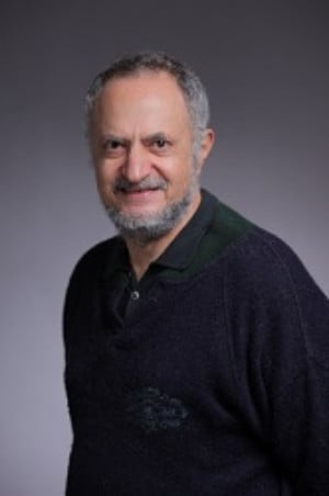
Dr. Miroslav Blumenberg
Guest Editor

Submission

Manuscripts should be submitted online at www.mdpi.com by registering and logging in to this website. Once you are registered, click here to go to the submission form. Manuscripts can be submitted until the deadline. Papers will be published continuously (as soon as accepted) and will be listed together on the special issue website. Research articles, review articles as well as communications are invited. For planned papers, a title and short abstract (about 100 words) can be sent to the Editorial Office for announcement on this website.

Submitted manuscripts should not have been published previously, nor be under consideration for publication elsewhere (except conference proceedings papers). All manuscripts are refereed through a peer-review process. A guide for authors and other relevant information for submission of manuscripts is available on the Instructions for Authors page. International Journal of Molecular Sciences is an international peer-reviewed Open Access semimonthly journal published by MDPI.

Please visit the Instructions for Authors page before submitting a manuscript. The Article Processing Charge (APC) for publication in this open access journal is 1600 CHF.

Keywords

  • bulge region
  • cancer
  • epidermis
  • epigenetics
  • gene replacement therapy
  • hair
  • ichthyosis
  • melanocyte
  • niche
  • sebaceous gland
  • skin
  • wound healing

Related Special Issues

Published Papers (13 papers)

Order results
Result details
Select all
Export citation of selected articles as:

Research

Jump to: Review

2169 KiB  
Article
Hypoxic Conditioned Medium from Human Amniotic Fluid-Derived Mesenchymal Stem Cells Accelerates Skin Wound Healing through TGF-β/SMAD2 and PI3K/Akt Pathways
by Eun Kyoung Jun, Qiankun Zhang, Byung Sun Yoon, Jai-Hee Moon, Gilju Lee, Gyuman Park, Phil Jun Kang, Jung Han Lee, Areee Kim and Seungkwon You
Int. J. Mol. Sci. 2014, 15(1), 605-628; https://doi.org/10.3390/ijms15010605 - 06 Jan 2014
Cited by 140 | Viewed by 11838
Abstract
In a previous study, we isolated human amniotic fluid (AF)-derived mesenchymal stem cells (AF-MSCs) and utilized normoxic conditioned medium (AF-MSC-norCM) which has been shown to accelerate cutaneous wound healing. Because hypoxia enhances the wound healing function of mesenchymal stem cell-conditioned medium (MSC-CM), it [...] Read more.
In a previous study, we isolated human amniotic fluid (AF)-derived mesenchymal stem cells (AF-MSCs) and utilized normoxic conditioned medium (AF-MSC-norCM) which has been shown to accelerate cutaneous wound healing. Because hypoxia enhances the wound healing function of mesenchymal stem cell-conditioned medium (MSC-CM), it is interesting to explore the mechanism responsible for the enhancement of wound healing function. In this work, hypoxia not only increased the proliferation of AF-MSCs but also maintained their constitutive characteristics (surface marker expression and differentiation potentials). Notably, more paracrine factors, VEGF and TGF-β1, were secreted into hypoxic conditioned medium from AF-MSCs (AF-MSC-hypoCM) compared to AF-MSC-norCM. Moreover, AF-MSC-hypoCM enhanced the proliferation and migration of human dermal fibroblasts in vitro, and wound closure in a skin injury model, as compared to AF-MSC-norCM. However, the enhancement of migration of fibroblasts accelerated by AF-MSC-hypoCM was inhibited by SB505124 and LY294002, inhibitors of TGF-β/SMAD2 and PI3K/AKT, suggesting that AF-MSC-hypoCM-enhanced wound healing is mediated by the activation of TGF-β/SMAD2 and PI3K/AKT. Therefore, AF-MSC-hypoCM enhances wound healing through the increase of hypoxia-induced paracrine factors via activation of TGF-β/SMAD2 and PI3K/AKT pathways. Full article
(This article belongs to the Special Issue Molecular Research of Epidermal Stem Cells)
Show Figures

5943 KiB  
Article
Cultivation of Keratinocytes and Fibroblasts in a Three-Dimensional Bovine Collagen-Elastin Matrix (Matriderm®) and Application for Full Thickness Wound Coverage in Vivo
by Jasper Killat, Kerstin Reimers, Claudia Y. Choi, Sabrina Jahn, Peter M. Vogt and Christine Radtke
Int. J. Mol. Sci. 2013, 14(7), 14460-14474; https://doi.org/10.3390/ijms140714460 - 11 Jul 2013
Cited by 39 | Viewed by 9550
Abstract
New skin substitutes for burn medicine or reconstructive surgery pose an important issue in plastic surgery. Matriderm® is a clinically approved three-dimensional bovine collagen-elastin matrix which is already used as a dermal substitute of full thickness burn wounds. The drawback of an [...] Read more.
New skin substitutes for burn medicine or reconstructive surgery pose an important issue in plastic surgery. Matriderm® is a clinically approved three-dimensional bovine collagen-elastin matrix which is already used as a dermal substitute of full thickness burn wounds. The drawback of an avital matrix is the limited integration in full thickness skin defects, depending on the defect size. To further optimize this process, Matriderm® has also been studied as a matrix for tissue engineering of skin albeit long-term cultivation of the matrix with cells has been difficult. Cells have generally been seeded onto the matrix with high cell loss and minimal time-consuming migration. Here we developed a cell seeded skin equivalent after microtransfer of cells directly into the matrix. First, cells were cultured, and microinjected into Matriderm®. Then, cell viability in the matrix was determined by histology in vitro. As a next step, the skin substitute was applied in vivo into a full thickness rodent wound model. The wound coverage and healing was observed over a period of two weeks followed by histological examination assessing cell viability, proliferation and integration into the host. Viable and proliferating cells could be found throughout the entire matrix. The presented skin substitute resembles healthy skin in morphology and integrity. Based on this study, future investigations are planned to examine behaviour of epidermal stem cells injected into a collagen-elastin matrix under the aspects of establishment of stem cell niches and differentiation. Full article
(This article belongs to the Special Issue Molecular Research of Epidermal Stem Cells)
Show Figures

2693 KiB  
Article
Isolation of Multipotent Nestin-Expressing Stem Cells Derived from the Epidermis of Elderly Humans and TAT-VHL Peptide-Mediated Neuronal Differentiation of These Cells
by Hiroshi Kanno, Atsuhiko Kubo, Tetsuya Yoshizumi, Taro Mikami and Jiro Maegawa
Int. J. Mol. Sci. 2013, 14(5), 9604-9617; https://doi.org/10.3390/ijms14059604 - 03 May 2013
Cited by 10 | Viewed by 8009
Abstract
A specialized population of cells residing in the hair follicle is quiescent but shows pluripotency for differentiating into epithelial-mesenchymal lineage cells. Therefore, such cells are hoped to be useful as implantable donor cells for regenerative therapy. Recently, it was reported that intracellular delivery [...] Read more.
A specialized population of cells residing in the hair follicle is quiescent but shows pluripotency for differentiating into epithelial-mesenchymal lineage cells. Therefore, such cells are hoped to be useful as implantable donor cells for regenerative therapy. Recently, it was reported that intracellular delivery of TAT-VHL peptide induces neuronal differentiation of skin-derived precursors. In the present study, we successfully isolated multipotent stem cells derived from the epidermis of elderly humans, characterized these cells as being capable of sphere formation and strong expression of nestin, fibronectin, and CD34 but not of keratin 15, and identified the niche of these cells as being the outer root sheath of the hair follicles. In addition, we showed that TAT-VHL peptide induced their neuronal differentiation in vitro, and confirmed by fluorescence immunohistochemistry the neuronal differentiation of such peptide-treated cells implanted into rodent brains. These multipotent nestin-expressing stem cells derived from human epidermis are easily accessible and should be useful as donor cells for neuronal regenerative cell therapy. Full article
(This article belongs to the Special Issue Molecular Research of Epidermal Stem Cells)
Show Figures

688 KiB  
Article
Irradiated Human Dermal Fibroblasts Are as Efficient as Mouse Fibroblasts as a Feeder Layer to Improve Human Epidermal Cell Culture Lifespan
by Francis Bisson, Éloise Rochefort, Amélie Lavoie, Danielle Larouche, Karine Zaniolo, Carolyne Simard-Bisson, Odile Damour, François A. Auger, Sylvain L. Guérin and Lucie Germain
Int. J. Mol. Sci. 2013, 14(3), 4684-4704; https://doi.org/10.3390/ijms14034684 - 26 Feb 2013
Cited by 62 | Viewed by 9600
Abstract
A fibroblast feeder layer is currently the best option for large scale expansion of autologous skin keratinocytes that are to be used for the treatment of severely burned patients. In a clinical context, using a human rather than a mouse feeder layer is [...] Read more.
A fibroblast feeder layer is currently the best option for large scale expansion of autologous skin keratinocytes that are to be used for the treatment of severely burned patients. In a clinical context, using a human rather than a mouse feeder layer is desirable to reduce the risk of introducing animal antigens and unknown viruses. This study was designed to evaluate if irradiated human fibroblasts can be used in keratinocyte cultures without affecting their morphological and physiological properties. Keratinocytes were grown either with or without a feeder layer in serum-containing medium. Our results showed that keratinocytes grown either on an irradiated human feeder layer or irradiated 3T3 cells (i3T3) can be cultured for a comparable number of passages. The average epithelial cell size and morphology were also similar. On the other hand, keratinocytes grown without a feeder layer showed heavily bloated cells at early passages and stop proliferating after only a few passages. On the molecular aspect, the expression level of the transcription factor Sp1, a useful marker of keratinocytes lifespan, was maintained and stabilized for a high number of passages in keratinocytes grown with feeder layers whereas Sp1 expression dropped quickly without a feeder layer. Furthermore, gene profiling on microarrays identified potential target genes whose expression is differentially regulated in the absence or presence of an i3T3 feeder layer and which may contribute at preserving the growth characteristics of these cells. Irradiated human dermal fibroblasts therefore provide a good human feeder layer for an effective expansion of keratinocytes in vitro that are to be used for clinical purposes. Full article
(This article belongs to the Special Issue Molecular Research of Epidermal Stem Cells)
Show Figures

464 KiB  
Article
Transcriptional Analysis of Hair Follicle-Derived Keratinocytes from Donors with Atopic Dermatitis Reveals Enhanced Induction of IL32 Gene by IFN-γ
by Yoshie Yoshikawa, Yusuke Sasahara, Katsuyuki Takeuchi, Yoshimasa Tsujimoto, Takashi Hashida-Okado, Yukio Kitano and Tomoko Hashimoto-Tamaoki
Int. J. Mol. Sci. 2013, 14(2), 3215-3227; https://doi.org/10.3390/ijms14023215 - 05 Feb 2013
Cited by 8 | Viewed by 6703
Abstract
We cultured human hair follicle-derived keratinocytes (FDKs) from plucked hairs. To gain insight into gene expression signatures that can distinguish atopic dermatitis from non-atopic controls without skin biopsies, we undertook a comparative study of gene expression in FDKs from adult donors with atopic [...] Read more.
We cultured human hair follicle-derived keratinocytes (FDKs) from plucked hairs. To gain insight into gene expression signatures that can distinguish atopic dermatitis from non-atopic controls without skin biopsies, we undertook a comparative study of gene expression in FDKs from adult donors with atopic dermatitis and non-atopic donors. FDK primary cultures (atopic dermatitis, n = 11; non-atopic controls, n = 7) before and after interferon gamma (IFN-γ) treatment were used for microarray analysis and quantitative RT-PCR. Comparison of FDKs from atopic and non-atopic donors indicated that the former showed activated pathways with innate immunity and decreased pathways of cell growth, as indicated by increased NLRP2 expression and decreased DKK1 expression, respectively. Treatment with IFN-γ induced the enhanced expression of IL32, IL1B, IL8, and CXCL1 in the cells from atopic donors compared to that in cells from non-atopic donors at 24 h after treatment. IL1B expression in FDKs after IFN-γ treatment correlated with IL32 expression. We hypothesized that overexpression of IL32 in hair follicle keratinocytes of patients with atopic dermatitis would lead to the excessive production of pro-IL1β and that the activation of IL1β from pro-IL1β by inflammasome complex, in which NLRP2 protein might be involved, would be augmented. This is the first report to show enhanced induction of cytokine/chemokine genes by IFN-γ in atopic dermatitis using cultured FDKs. Full article
(This article belongs to the Special Issue Molecular Research of Epidermal Stem Cells)
Show Figures

Review

Jump to: Research

415 KiB  
Review
Signaling Involved in Hair Follicle Morphogenesis and Development
by Pisal Rishikaysh, Kapil Dev, Daniel Diaz, Wasay Mohiuddin Shaikh Qureshi, Stanislav Filip and Jaroslav Mokry
Int. J. Mol. Sci. 2014, 15(1), 1647-1670; https://doi.org/10.3390/ijms15011647 - 22 Jan 2014
Cited by 241 | Viewed by 21777
Abstract
Hair follicle morphogenesis depends on Wnt, Shh, Notch, BMP and other signaling pathways interplay between epithelial and mesenchymal cells. The Wnt pathway plays an essential role during hair follicle induction, Shh is involved in morphogenesis and late stage differentiation, Notch signaling determines stem [...] Read more.
Hair follicle morphogenesis depends on Wnt, Shh, Notch, BMP and other signaling pathways interplay between epithelial and mesenchymal cells. The Wnt pathway plays an essential role during hair follicle induction, Shh is involved in morphogenesis and late stage differentiation, Notch signaling determines stem cell fate while BMP is involved in cellular differentiation. The Wnt pathway is considered to be the master regulator during hair follicle morphogenesis. Wnt signaling proceeds through EDA/EDAR/NF-κB signaling. NF-κB regulates the Wnt pathway and acts as a signal mediator by upregulating the expression of Shh ligand. Signal crosstalk between epithelial and mesenchymal cells takes place mainly through primary cilia. Primary cilia formation is initiated with epithelial laminin-511 interaction with dermal β-1 integrin, which also upregulates expression of downstream effectors of Shh pathway in dermal lineage. PDGF signal transduction essential for crosstalk is mediated through epithelial PDGF-A and PDGFRα expressed on the primary cilia. Dermal Shh and PDGF signaling up-regulates dermal noggin expression; noggin is a potent inhibitor of BMP signaling which helps in counteracting BMP mediated β-catenin inhibition. This interplay of signaling between the epithelial and dermal lineage helps in epithelial Shh signal amplification. The dermal Wnt pathway helps in upregulation of epithelial Notch expression. Dysregulation of these pathways leads to certain abnormalities and in some cases even tumor outgrowth. Full article
(This article belongs to the Special Issue Molecular Research of Epidermal Stem Cells)
Show Figures

513 KiB  
Review
Genetic Correction of Stem Cells in the Treatment of Inherited Diseases and Focus on Xeroderma Pigmentosum
by Sophie Rouanet, Emilie Warrick, Yannick Gache, Sabine Scarzello, Marie-Françoise Avril, Françoise Bernerd and Thierry Magnaldo
Int. J. Mol. Sci. 2013, 14(10), 20019-20036; https://doi.org/10.3390/ijms141020019 - 09 Oct 2013
Cited by 5 | Viewed by 8079
Abstract
Somatic stem cells ensure tissue renewal along life and healing of injuries. Their safe isolation, genetic manipulation ex vivo and reinfusion in patients suffering from life threatening immune deficiencies (for example, severe combined immunodeficiency (SCID)) have demonstrated the efficacy of ex vivo gene [...] Read more.
Somatic stem cells ensure tissue renewal along life and healing of injuries. Their safe isolation, genetic manipulation ex vivo and reinfusion in patients suffering from life threatening immune deficiencies (for example, severe combined immunodeficiency (SCID)) have demonstrated the efficacy of ex vivo gene therapy. Similarly, adult epidermal stem cells have the capacity to renew epidermis, the fully differentiated, protective envelope of our body. Stable skin replacement of severely burned patients have proven life saving. Xeroderma pigmentosum (XP) is a devastating disease due to severe defects in the repair of mutagenic DNA lesions introduced upon exposure to solar radiations. Most patients die from the consequences of budding hundreds of skin cancers in the absence of photoprotection. We have developed a safe procedure of genetic correction of epidermal stem cells isolated from XP patients. Preclinical and safety assessments indicate successful correction of XP epidermal stem cells in the long term and their capacity to regenerate a normal skin with full capacities of DNA repair. Full article
(This article belongs to the Special Issue Molecular Research of Epidermal Stem Cells)
Show Figures

445 KiB  
Review
Keratin K15 as a Biomarker of Epidermal Stem Cells
by Amrita Bose, Muy-Teck Teh, Ian C. Mackenzie and Ahmad Waseem
Int. J. Mol. Sci. 2013, 14(10), 19385-19398; https://doi.org/10.3390/ijms141019385 - 25 Sep 2013
Cited by 82 | Viewed by 8777
Abstract
Keratin 15 (K15) is type I keratin protein co-expressed with the K5/K14 pair present in the basal keratinocytes of all stratified epithelia. Although it is a minor component of the cytoskeleton with a variable expression pattern, nonetheless its expression has been reported as [...] Read more.
Keratin 15 (K15) is type I keratin protein co-expressed with the K5/K14 pair present in the basal keratinocytes of all stratified epithelia. Although it is a minor component of the cytoskeleton with a variable expression pattern, nonetheless its expression has been reported as a stem cell marker in the bulge of hair follicles. Conversely, suprabasal expression of K15 has also been reported in both normal and diseased tissues, which is inconsistent with its role as a stem cell marker. Our recently published work has given evidence of the molecular pathways that seem to control the expression of K15 in undifferentiated and differentiated cells. In this article, we have critically reviewed the published work to establish the reliability of K15 as an epidermal stem cell marker. Full article
(This article belongs to the Special Issue Molecular Research of Epidermal Stem Cells)
Show Figures

532 KiB  
Review
EGFR-Ras-Raf Signaling in Epidermal Stem Cells: Roles in Hair Follicle Development, Regeneration, Tissue Remodeling and Epidermal Cancers
by Eszter Doma, Christian Rupp and Manuela Baccarini
Int. J. Mol. Sci. 2013, 14(10), 19361-19384; https://doi.org/10.3390/ijms141019361 - 25 Sep 2013
Cited by 36 | Viewed by 12566
Abstract
The mammalian skin is the largest organ of the body and its outermost layer, the epidermis, undergoes dynamic lifetime renewal through the activity of somatic stem cell populations. The EGFR-Ras-Raf pathway has a well-described role in skin development and tumor formation. While research [...] Read more.
The mammalian skin is the largest organ of the body and its outermost layer, the epidermis, undergoes dynamic lifetime renewal through the activity of somatic stem cell populations. The EGFR-Ras-Raf pathway has a well-described role in skin development and tumor formation. While research mainly focuses on its role in cutaneous tumor initiation and maintenance, much less is known about Ras signaling in the epidermal stem cells, which are the main targets of skin carcinogenesis. In this review, we briefly discuss the properties of the epidermal stem cells and review the role of EGFR-Ras-Raf signaling in keratinocyte stem cells during homeostatic and pathological conditions. Full article
(This article belongs to the Special Issue Molecular Research of Epidermal Stem Cells)
Show Figures

307 KiB  
Review
Epidermal Stem Cells and Their Epigenetic Regulation
by Qi Shen, Hongchuan Jin and Xian Wang
Int. J. Mol. Sci. 2013, 14(9), 17861-17880; https://doi.org/10.3390/ijms140917861 - 30 Aug 2013
Cited by 14 | Viewed by 7423
Abstract
Stem cells play an essential role in embryonic development, cell differentiation and tissue regeneration. Tissue homeostasis in adults is maintained by adult stem cells resident in the niches of different tissues. As one kind of adult stem cell, epidermal stem cells have the [...] Read more.
Stem cells play an essential role in embryonic development, cell differentiation and tissue regeneration. Tissue homeostasis in adults is maintained by adult stem cells resident in the niches of different tissues. As one kind of adult stem cell, epidermal stem cells have the potential to generate diversified types of progeny cells in the skin. Although its biology is still largely unclarified, epidermal stem cells are widely used in stem cell research and regenerative medicine given its easy accessibility and pluripotency. Despite the same genome, cells within an organism have different fates due to the epigenetic regulation of gene expression. In this review, we will briefly discuss the current understanding of epigenetic modulation in epidermal stem cells. Full article
(This article belongs to the Special Issue Molecular Research of Epidermal Stem Cells)
Show Figures

717 KiB  
Review
Stem Cells behind the Barrier
by Michael Cangkrama, Stephen B. Ting and Charbel Darido
Int. J. Mol. Sci. 2013, 14(7), 13670-13686; https://doi.org/10.3390/ijms140713670 - 28 Jun 2013
Cited by 19 | Viewed by 8551
Abstract
Epidermal stem cells sustain the adult skin for a lifetime through self-renewal and the production of committed progenitors. These stem cells generate progeny that will undergo terminal differentiation leading to the development of a protective epidermal barrier. Whereas the molecular mechanisms that govern [...] Read more.
Epidermal stem cells sustain the adult skin for a lifetime through self-renewal and the production of committed progenitors. These stem cells generate progeny that will undergo terminal differentiation leading to the development of a protective epidermal barrier. Whereas the molecular mechanisms that govern epidermal barrier repair and renewal have been extensively studied, pathways controlling stem cell differentiation remain poorly understood. Asymmetric cell divisions, small non-coding RNAs (microRNAs), chromatin remodeling complexes, and multiple differentiation factors tightly control the balance of stem and progenitor cell proliferation and differentiation, and disruption of this balance leads to skin diseases. In this review, we summarize and discuss current advances in our understanding of the mechanisms regulating epidermal stem and progenitor cell differentiation, and explore new relationships for maintenance of skin barrier function. Full article
(This article belongs to the Special Issue Molecular Research of Epidermal Stem Cells)
Show Figures

Graphical abstract

318 KiB  
Review
Epidermal Stem Cells in Orthopaedic Regenerative Medicine
by Jin Li, Gehua Zhen, Shin-Yi Tsai and Xiaofeng Jia
Int. J. Mol. Sci. 2013, 14(6), 11626-11642; https://doi.org/10.3390/ijms140611626 - 31 May 2013
Cited by 4 | Viewed by 8204
Abstract
In the last decade, great advances have been made in epidermal stem cell studies at the cellular and molecular level. These studies reported various subpopulations and differentiations existing in the epidermal stem cell. Although controversies and unknown issues remain, epidermal stem cells possess [...] Read more.
In the last decade, great advances have been made in epidermal stem cell studies at the cellular and molecular level. These studies reported various subpopulations and differentiations existing in the epidermal stem cell. Although controversies and unknown issues remain, epidermal stem cells possess an immune-privileged property in transplantation together with easy accessibility, which is favorable for future clinical application. In this review, we will summarize the biological characteristics of epidermal stem cells, and their potential in orthopedic regenerative medicine. Epidermal stem cells play a critical role via cell replacement, and demonstrate significant translational potential in the treatment of orthopedic injuries and diseases, including treatment for wound healing, peripheral nerve and spinal cord injury, and even muscle and bone remodeling. Full article
(This article belongs to the Special Issue Molecular Research of Epidermal Stem Cells)
Show Figures

Graphical abstract

877 KiB  
Review
Epidermal Development in Mammals: Key Regulators, Signals from Beneath, and Stem Cells
by Shuang Liu, Huishan Zhang and Enkui Duan
Int. J. Mol. Sci. 2013, 14(6), 10869-10895; https://doi.org/10.3390/ijms140610869 - 24 May 2013
Cited by 81 | Viewed by 18555
Abstract
Epidermis is one of the best-studied tissues in mammals that contain types of stem cells. Outstanding works in recent years have shed great light on behaviors of different epidermal stem cell populations in the homeostasis and regeneration of the epidermis as well as [...] Read more.
Epidermis is one of the best-studied tissues in mammals that contain types of stem cells. Outstanding works in recent years have shed great light on behaviors of different epidermal stem cell populations in the homeostasis and regeneration of the epidermis as well as hair follicles. Also, the molecular mechanisms governing these stem cells are being elucidated, from genetic to epigenetic levels. Compared with the explicit knowledge about adult skin, embryonic development of the epidermis, especially the early period, still needs exploration. Furthermore, stem cells in the embryonic epidermis are largely unstudied or ambiguously depicted. In this review, we will summarize and discuss the process of embryonic epidermal development, with focuses on some key molecular regulators and the role of the sub-epidermal mesenchyme. We will also try to trace adult epidermal stem cell populations back to embryonic development. In addition, we will comment on in vitro derivation of epidermal lineages from ES cells and iPS cells. Full article
(This article belongs to the Special Issue Molecular Research of Epidermal Stem Cells)
Show Figures

Back to TopTop