Computer-Aided Maxillofacial Surgery

A special issue of Applied Sciences (ISSN 2076-3417). This special issue belongs to the section "Applied Biosciences and Bioengineering".

Deadline for manuscript submissions: closed (20 March 2023) | Viewed by 4733

Special Issue Editors


E-Mail Website
Guest Editor
1. Department of Surgery, University of Freiburg, 79098 Freiburg, Germany
2. Department of Anesthesiology, Perioperative and Pain Medicine, Brigham and Women's Hospital, Harvard Medical School, Boston, MA 02115, USA
Interests: imaging; CAD/CAM; biostatistics; prediction modelling; artificial intelligence; deep learning; machine learning
Special Issues, Collections and Topics in MDPI journals

E-Mail Website
Guest Editor
Oral and Maxillofacial Surgery, University Clinic Ruppin-Brandenburg, Fehrbelliner Str. 38, 16816 Neuruppin, Germany
Interests: imaging; deep learning; surgical techniques; confocal microscopy; prediction modeling; oral and maxillofacial surgery

E-Mail Website
Guest Editor
Department of Oral and Maxillofacial Surgery, Ruppiner Kliniken, Neuruppin, Germany
Interests: oral and maxillofacial surgery; implantology; plastic and aesthetic operations
Special Issues, Collections and Topics in MDPI journals

Special Issue Information

Dear Colleagues,

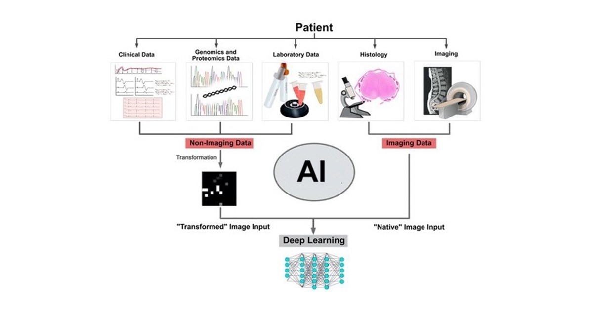
In what direction are technical developments in oral and maxillofacial surgery heading? We are in the midst of a digital transformation, but what trends will shape the future? As we examine innovations and future technical standards, we will explore topics, such as artificial intelligence in diagnostics and digital manufacturing, as well as the fundamental changes that will occur to the "surgical practice enterprise". The use of artificial intelligence (AI) will be an integral part of the clinical team in the future. It is used in a variety of applications, such as diagnosis and evaluation of digital images, planning surgical interventions (e.g., implant position), and billing and accounting.

Optimum cognitive capabilities are one of the advantages of AI. On the basis of learning algorithms, cognitive systems can derive conclusions and make decisions based on digital information. As a result, algorithms are able to process and recognize significantly more information and patterns than the human brain. A machine learning algorithm, for example, is able to analyze various symptoms and risk factors in relation to the patient's medical history, and it is able to make recommendations for future actions or diagnostics (probability calculations).

The extent to which machines should and may determine medical therapy is an ethical issue that needs to be addressed elsewhere. There are numerous possibilities available, and, in most cases, they can be used in the patient's best interest. The use of artificial intelligence will become a key component of surgeons' decision-making in the near future. Clinical practice has become increasingly reliant on digital manufacturing. CAD/CAM-manufactured components are now widely used in virtually every practice or laboratory. Dental laboratories are leading the digital transformation with the use of milling machines and 3D printers as common manufacturing technologies.

It is our pleasure to invite you to contribute your work regarding the implementation of artificial intelligence in oral and maxillofacial surgery and the use of digital workflows to advance this field. Research areas may include (but not limited to) the following:

  • Diagnostics and prognostics utilizing AI-based algorithms (machine learning and deep learning techniques);
  • Digital workflows in oral and maxillofacial surgery (CAD/CAM);
  • Telemedicine and digitalization of the patient–surgeon interaction;
  • Virtual Reality (VR) and Augmented Reality (AR);
  • Digital imaging and novel techniques in image processing;
  • Digitalization of surgical techniques and robotics.

The purpose of this Special Issue is to gather evidence regarding computer-aided oral and maxillofacial surgery from around the world. All article types (e.g., reviews, original articles) are welcome.

We look forward to receiving your contributions.

Dr. Babak Saravi
Dr. Veronika Shavlokhova
Prof. Dr. Christian Stoll
Guest Editors

Manuscript Submission Information

Manuscripts should be submitted online at www.mdpi.com by registering and logging in to this website. Once you are registered, click here to go to the submission form. Manuscripts can be submitted until the deadline. All submissions that pass pre-check are peer-reviewed. Accepted papers will be published continuously in the journal (as soon as accepted) and will be listed together on the special issue website. Research articles, review articles as well as short communications are invited. For planned papers, a title and short abstract (about 100 words) can be sent to the Editorial Office for announcement on this website.

Submitted manuscripts should not have been published previously, nor be under consideration for publication elsewhere (except conference proceedings papers). All manuscripts are thoroughly refereed through a single-blind peer-review process. A guide for authors and other relevant information for submission of manuscripts is available on the Instructions for Authors page. Applied Sciences is an international peer-reviewed open access semimonthly journal published by MDPI.

Please visit the Instructions for Authors page before submitting a manuscript. The Article Processing Charge (APC) for publication in this open access journal is 2400 CHF (Swiss Francs). Submitted papers should be well formatted and use good English. Authors may use MDPI's English editing service prior to publication or during author revisions.

Keywords

  • digital workflow
  • CAD/CAM
  • oral and maxillofacial surgery
  • implants
  • machine learning
  • deep learning
  • artificial intelligence
  • manufacturing
  • digitalization
  • telemedicine

Published Papers (3 papers)

Order results
Result details
Select all
Export citation of selected articles as:

Research

15 pages, 2400 KiB  
Article
Design of Unilateral Complete Presurgical Nasoalveolar Molding (PNAM) Corrector Based on Feature Points Extraction of Complex 3D Surface
by Li Li, Tao Liu and Dongshen Fang
Appl. Sci. 2023, 13(9), 5251; https://doi.org/10.3390/app13095251 - 22 Apr 2023
Viewed by 1439
Abstract
Cleft lip and palate is a congenital maxillofacial deformity. Unilateral complete cleft lip and palate is one of the most common clinical types. Nasal alveolar molding (PNAM) is a recognized strategy for the treatment of cleft lip and palate. However, the current design [...] Read more.
Cleft lip and palate is a congenital maxillofacial deformity. Unilateral complete cleft lip and palate is one of the most common clinical types. Nasal alveolar molding (PNAM) is a recognized strategy for the treatment of cleft lip and palate. However, the current design of PNAM devices mainly relies on the subjective experience of doctors. The purpose of this paper is to describe the design and manufacture of a new computer-aided design appliance, which can be applied to the presurgical nasoalveolar molding of unilateral complete cleft lip and palate, eliminate individual differences, and improve production efficiency. In this paper, seven feature points on the healthy side and the affected side are extracted by the method of Gaussian curvature and ridge line extraction, and the healthy side rotation and built-in model are designed by using these seven feature points, which can quickly generate eight treatment stages of PNAM. The correction effects of the PNAM appliance designed in this paper were compared with the original maxillary model and the clinical PNAM appliance (hand-made by subjective experience) from the aspects of alveolar fissure width and symmetry. The PNAM appliance designed in this paper can effectively improve the symmetry of patients with unilateral complete cleft lip and palate (morphological similarity: t = 3.250, p ≤ 0.01; length similarity: t = 1.559, p = 0.150) and reduce the width of alveolar cleft (t = 8.330, p < 0.01). This can fully achieve the therapeutic effect of PNAM appliances prepared by experienced doctors and is more efficient. The method based on complex 3D surface feature point extraction can provide the basis for the design and evaluation of a unilateral complete PNAM correction model, improve the design and production efficiency of unilateral complete cleft lip and palate appliance, eliminate the design problems caused by individual differences, and reduce the burden of doctors. Full article
(This article belongs to the Special Issue Computer-Aided Maxillofacial Surgery)
Show Figures

Figure 1

13 pages, 1723 KiB  
Article
Automated Assessment of Radiographic Bone Loss in the Posterior Maxilla Utilizing a Multi-Object Detection Artificial Intelligence Algorithm
by Andreas Vollmer, Michael Vollmer, Gernot Lang, Anton Straub, Alexander Kübler, Sebastian Gubik, Roman C. Brands, Stefan Hartmann and Babak Saravi
Appl. Sci. 2023, 13(3), 1858; https://doi.org/10.3390/app13031858 - 31 Jan 2023
Viewed by 1622
Abstract
Periodontitis is one of the most prevalent diseases worldwide. The degree of radiographic bone loss can be used to assess the course of therapy or the severity of the disease. Since automated bone loss detection has many benefits, our goal was to develop [...] Read more.
Periodontitis is one of the most prevalent diseases worldwide. The degree of radiographic bone loss can be used to assess the course of therapy or the severity of the disease. Since automated bone loss detection has many benefits, our goal was to develop a multi-object detection algorithm based on artificial intelligence that would be able to detect and quantify radiographic bone loss using standard two-dimensional radiographic images in the maxillary posterior region. This study was conducted by combining three recent online databases and validating the results using an external validation dataset from our organization. There were 1414 images for training and testing and 341 for external validation in the final dataset. We applied a Keypoint RCNN with a ResNet-50-FPN backbone network for both boundary box and keypoint detection. The intersection over union (IoU) and the object keypoint similarity (OKS) were used for model evaluation. The evaluation of the boundary box metrics showed a moderate overlapping with the ground truth, revealing an average precision of up to 0.758. The average precision and recall over all five folds were 0.694 and 0.611, respectively. Mean average precision and recall for the keypoint detection were 0.632 and 0.579, respectively. Despite only using a small and heterogeneous set of images for training, our results indicate that the algorithm is able to learn the objects of interest, although without sufficient accuracy due to the limited number of images and a large amount of information available in panoramic radiographs. Considering the widespread availability of panoramic radiographs as well as the increasing use of online databases, the presented model can be further improved in the future to facilitate its implementation in clinics. Full article
(This article belongs to the Special Issue Computer-Aided Maxillofacial Surgery)
Show Figures

Figure 1

12 pages, 3202 KiB  
Article
Anteroposterior Ethmoidectomy in the Endoscopic Reduction of Medial Orbital Wall Fractures: Does It Really Reduce Stability?
by Antonio Romano, Stefania Troise, Francesco Maffia, Umberto Committeri, Lorenzo Sani, Marco Sarcinella, Antonio Arena, Giorgio Iaconetta, Luigi Califano and Giovanni Dell’Aversana Orabona
Appl. Sci. 2023, 13(1), 98; https://doi.org/10.3390/app13010098 - 21 Dec 2022
Viewed by 1130
Abstract
The surgical treatment of isolated medial orbital wall fractures is still a much-debated topic in the literature due to the choice of many surgical accesses. The main options are represented by transcutaneous versus endonasal endoscopic approaches. Our study aims to clarify the role [...] Read more.
The surgical treatment of isolated medial orbital wall fractures is still a much-debated topic in the literature due to the choice of many surgical accesses. The main options are represented by transcutaneous versus endonasal endoscopic approaches. Our study aims to clarify the role of ethmoidectomy in the pure endoscopic endonasal reduction of medial orbital wall fractures, evaluating the immediate postoperative outcome and its long-term stability. A total of 31 patients affected by isolated medial orbital wall fracture, treated only by endoscopic approach, were included in the study and divided in two groups: (A) 14 patients treated by endoscopic reduction and anterior ethmoidectomy; (B) 17 patients treated by endoscopic reduction and anteroposterior ethmoidectomy. Perioperative and 6-month postoperative follow-up CT scans were performed. With the use of 3D medical software, we evaluated the comparison between the treated orbit and the mirrored contralateral orbit in the two groups, in order to observe the reduction of the fracture. Furthermore, to check the stability of reduction and to evaluate any medial orbital wall changes, we provided a comparison between the 3D CT scan orbital images of immediate postoperative CT and 6-month follow-up. Data obtained showed that the intraoperative surgical reduction was successful in all 31 cases, but it was better in Group B. Stability of the reduction at 6 months was observed in both groups without significant discrepancies. In our opinion, the endonasal endoscopic approach with ethmoidectomy represents a valid and useful technique by which to treat medial orbital wall fractures. The anatomical detail of the buttressing structures of the medial orbital wall, as the second portion of the middle turbinate, grants long-term stability of the surgical outcome. Full article
(This article belongs to the Special Issue Computer-Aided Maxillofacial Surgery)
Show Figures

Figure 1

Back to TopTop