Special Issue "Advances in Orthodontics and Dental Medicine"

A special issue of Applied Sciences (ISSN 2076-3417). This special issue belongs to the section "Applied Dentistry and Oral Sciences".

Deadline for manuscript submissions: 20 January 2024 | Viewed by 8154

Special Issue Editors

Discipline of Orthodontics, 2nd Department, OrthoCenter Research Center, Faculty of Dental Medicine, “Victor Babes” University of Medicine and Pharmacy Timisoara, Eftimie Murgu Square No. 2, 300041 Timisoara, Romania
Interests: digital orthodontics; biomechanics; CBCT; cephalometric analysis; orthodontic implants; functional occlusion; aligners; adult orthodontics; interdisciplinary treatment; three-dimensional (3D) printing; numerical simulations; finite element analysis
Department of Orthodontics and Dento-Facial Orthopedics, 1st Surgical Department, Dental Medicine Faculty, University of Medicine and Pharmacy, Grigore T. Popa” Iași, University Street No. 16, 700115 Iasi, Romania
Interests: adult orthodontics; orthodontic implants; interdisciplinary treatment; dental materials; biomechanics; digital planning; growth and development; dental medicine

Special Issue Information

Dear Colleagues,

Recent advances in orthodontics and dental medicine is this new Special Issue will cover the new up-to-date research results from all areas in dentistry. 

Academics, researchers, and professionals from all fields of dentistry are invited to publish review and original articles that will emphasise research work from orthodontics and dental medicine.

The topics that will be covered include: digital orthodontics, diagnosis, virtual treatment planning, digital dentistry, CBCT, orthodontic implants, adult orthodontics, preventive and interceptive treatment, orthognathic surgery, biomechanics, interdisciplinary treatment, aligners, esthetics in orthodontics, cephalometric analysis, biocompatibility, the cytotoxicity of dental and orthodontic materials, and finite elements analysis.

Prof. Dr. Camelia Szuhanek
Prof. Dr. Irina Nicoleta Zetu
Guest Editors

Manuscript Submission Information

Manuscripts should be submitted online at www.mdpi.com by registering and logging in to this website. Once you are registered, click here to go to the submission form. Manuscripts can be submitted until the deadline. All submissions that pass pre-check are peer-reviewed. Accepted papers will be published continuously in the journal (as soon as accepted) and will be listed together on the special issue website. Research articles, review articles as well as short communications are invited. For planned papers, a title and short abstract (about 100 words) can be sent to the Editorial Office for announcement on this website.

Submitted manuscripts should not have been published previously, nor be under consideration for publication elsewhere (except conference proceedings papers). All manuscripts are thoroughly refereed through a single-blind peer-review process. A guide for authors and other relevant information for submission of manuscripts is available on the Instructions for Authors page. Applied Sciences is an international peer-reviewed open access semimonthly journal published by MDPI.

Please visit the Instructions for Authors page before submitting a manuscript. The Article Processing Charge (APC) for publication in this open access journal is 2300 CHF (Swiss Francs). Submitted papers should be well formatted and use good English. Authors may use MDPI's English editing service prior to publication or during author revisions.

Keywords

  • orthodontics
  • dentistry
  • digital
  • virtual treatment planning
  • intraoral scan
  • orthognathic surgery
  • biomechanics
  • aligners
  • cephalometric analysis
  • interceptive orthodontics
  • dental medicine
  • interdisciplinarity

Published Papers (10 papers)

Order results
Result details
Select all
Export citation of selected articles as:

Research

Jump to: Review, Other

Article
The Surgical Guides for TADs: The Rational and Laboratory Procedures
Appl. Sci. 2023, 13(18), 10332; https://doi.org/10.3390/app131810332 - 15 Sep 2023
Viewed by 307
Abstract
The widespread and popular use of TADs for orthodontic anchoring has become a daily routine in clinical orthodontic treatment. However, as there have been many accidents over the past decade, safety action is needed to help reduce these accidents. We have advocated using [...] Read more.
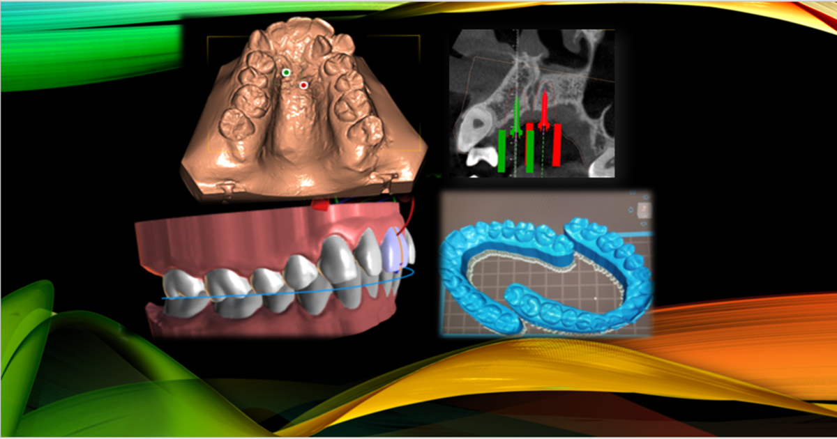
The widespread and popular use of TADs for orthodontic anchoring has become a daily routine in clinical orthodontic treatment. However, as there have been many accidents over the past decade, safety action is needed to help reduce these accidents. We have advocated using the surgical guide and developed a procedure that can benefit patients and orthodontists. The first part of this paper is about the rationale for making the surgical guide for various implant placements that were observed. Due to their anatomical structure, some placements may require particular attention, which is focused on and discussed in length. The second part deals with fabricating the surgical guide in the laboratory procedure. The data from the intraoral SLT acquisition was extracted, and with the DICOM data from CBCT and in a 3-Shapes software, the guidance was designed. The detailed and step-by-step laboratory procedure, CAD/CAM, and 3D printers to make the surgical guide for TADs are explained. The procedure is performed in an easy-to-understand manner to make using the surgical guide possible for the daily practice of orthodontics (Pubmed). Full article
(This article belongs to the Special Issue Advances in Orthodontics and Dental Medicine)
Show Figures

Figure 1

Article
Maxillary Response Induced by Rapid Palatal Expansion vs. Clear Aligners: A Short-Term Retrospective Evaluation of the Dento-Alveolar Effects in Mixed Dentition
Appl. Sci. 2023, 13(15), 8675; https://doi.org/10.3390/app13158675 - 27 Jul 2023
Viewed by 324
Abstract
Background: to analyze the dento-alveolar effects induced by two treatment appliances (i.e., RME and Clear Aligners) in growing subjects presenting with early mixed dentition and mild maxillary deficiency. Methods: digital casts of 32 children treated with RME (RG: 17 subjects) or with Clear [...] Read more.
Background: to analyze the dento-alveolar effects induced by two treatment appliances (i.e., RME and Clear Aligners) in growing subjects presenting with early mixed dentition and mild maxillary deficiency. Methods: digital casts of 32 children treated with RME (RG: 17 subjects) or with Clear aligners (CAG: 15 subjects) were collected. Linear and angular values were measured in both groups on the upper arch for both pre- (T1) and post-treatment (T2) models. An unpaired t-test was used to test significant differences between groups (p < 0.05). Results: the comparison of T2-T1 changes for linear measurements between groups showed a greater increase in the inter-canine width (+1.5 mm ± 0.5 mm) and in the first inter-deciduous molar width (+1.4 ± 0.4 mm), also at the trans-palatal level in the CAG group. Conversely, in the posterior region of the upper arch, a greater increase in the first inter-molar distal width was found in the RG group (+1.2 ± 0.4 mm) when compared with the CAG group. A significant increase in the crown angulation in the CAG group was found for all the teeth except for the first molars. Conclusions: the rapid palatal expander widened the palate, tipping the first upper molars buccally to a greater extent, whereas the Clear aligners caused a greater increase in the canine width. Full article
(This article belongs to the Special Issue Advances in Orthodontics and Dental Medicine)
Show Figures

Figure 1

Article
Interaction Phenomena between Dental Implants and Bone Tissue in Case of Misfit: A Pilot Study
Appl. Sci. 2023, 13(10), 6004; https://doi.org/10.3390/app13106004 - 13 May 2023
Cited by 2 | Viewed by 614
Abstract
The biomechanical response of cortical and trabecular bone tissues represents a fundamental aspect for the interpretation of the functional response of dental implants. In the case of misfit, the interaction phenomena occurring within the surgical and the subsequent healing phases must be interpreted [...] Read more.
The biomechanical response of cortical and trabecular bone tissues represents a fundamental aspect for the interpretation of the functional response of dental implants. In the case of misfit, the interaction phenomena occurring within the surgical and the subsequent healing phases must be interpreted primarily in the light of the response of bone tissue. This is influenced by the specific loadings induced, characterized by intensity and variable trends. The pilot study reported, which intentionally refers to a simple case of a two-implants frame, is addressed to define the method to approach the biomechanical investigation of the problem and to attest the necessity to integrate clinical competences with biomechanical analysis for interpreting different aspects of osseointegration. The action induced in cortical and trabecular bone regions depending on the implant frame conformation, the surgical procedure adopted, the varying condition at the bone–implant interface and the evolutionary trend of healing are the principal aspects to be considered to evaluate the osseointegration process. The biomechanical reliability of the specific implant frame is investigated in terms of bone–implant interaction by means of numerical models. This approach can offer valid information and support clinical practice under the fundamental condition that bone biomechanical behavior is properly characterized and represented in the model, in spite of the complex formulation to be adopted. Full article
(This article belongs to the Special Issue Advances in Orthodontics and Dental Medicine)
Show Figures

Figure 1

Article
Accuracy of Soft Tissue Prediction in Skeletal Type III Relationship Using a Computer-Aided Three-Dimensional Surgical Simulation Planning Program
Appl. Sci. 2023, 13(9), 5623; https://doi.org/10.3390/app13095623 - 03 May 2023
Viewed by 752
Abstract
Using a computer-aided, three-dimensional surgical simulation planning program, soft-tissue planning can help achieve adequate facial esthetics and patient satisfaction after orthognathic surgery. This study aimed to assess the Simplant O&O software’s soft tissue prediction accuracy. Fourteen skeletal type III patients who underwent orthognathic [...] Read more.
Using a computer-aided, three-dimensional surgical simulation planning program, soft-tissue planning can help achieve adequate facial esthetics and patient satisfaction after orthognathic surgery. This study aimed to assess the Simplant O&O software’s soft tissue prediction accuracy. Fourteen skeletal type III patients who underwent orthognathic surgery by the same surgeons were included in this prospective study, and they were separated into two groups: the one-jaw (n = 5) and two-jaw (n = 9) groups. The software was used to analyze the preoperative (T0) and 4-month postoperative computed tomography data (T1), as well as intraoral scans. Data from cone-beam computed tomography and stereolithography from a scanned dental cast were used to reconstruct a composite skull model. Based on the presurgical CT data, the program generated a predicted soft tissue image (TP), which was then superimposed on the T1. The distances between seven T1 and TP landmarks were measured and evaluated using a one-sample t-test. In the one-jaw group, the mean error for all linear measurements was 1.73 ± 1.14 mm, whereas the mean error of the two-jaw group was 1.03 ± 0.83 mm, and both measurements were within clinically acceptable limits. Pronasele had the best correlation (mean error of 0.63 ± 0.45 mm) while soft tissue pogonion and soft tissue point B had the worst correlations (mean error of 2.87 ± 2.22 mm and 1.31 ± 0.98 mm, respectively). Even though there were some limitations, it was possible to conclude that the ability to accurately predict soft tissue changes using Simplant O&O for skeletal type III patients makes it adequate for use in clinical practice. Full article
(This article belongs to the Special Issue Advances in Orthodontics and Dental Medicine)
Show Figures

Figure 1

Article
Comparison of Clear Aligner Treatment in First-Treatment and Re-Treatment Patients: A Retrospective Cohort Study
Appl. Sci. 2023, 13(7), 4303; https://doi.org/10.3390/app13074303 - 28 Mar 2023
Viewed by 833
Abstract
This retrospective real-world study aimed to evaluate the outcomes of clear aligner treatment (CAT) and patient demographics of patients seeking orthodontic re-treatment who had previously received orthodontic treatment in childhood or adolescence. The study was conducted using anonymized data from DrSmile (Berlin, Germany), [...] Read more.
This retrospective real-world study aimed to evaluate the outcomes of clear aligner treatment (CAT) and patient demographics of patients seeking orthodontic re-treatment who had previously received orthodontic treatment in childhood or adolescence. The study was conducted using anonymized data from DrSmile (Berlin, Germany), a CAT provider in Europe. Data were collected from a total of 2080 patients in Germany (71.5% female, 28.5% male, ranging from 18–70 years, median 29.0 years), who completed their CAT between 1 June and 30 June 2022. Patients were divided into two groups: adult first-treatment patients (n = 1587) and adult re-treatment patients (n = 493). Results showed that re-treatment patients had a shorter treatment duration (p < 0.05), required fewer interproximal reductions (IPR) (p < 0.001), and had fewer clinical visits (mean 2.96 versus 4.14 visits) than first-treatment patients (p < 0.001). Data collected from adult orthodontic re-treatment and first-treatment patients indicated that the demographic characteristics of these two groups were very similar. No significant differences were found between re-treatment patients and first-treatment patients in terms of motives for orthodontic treatment (p = 0.068), types of insurance (p = 0.615), and treatment satisfaction (p = 0.673). The study highlights the need for orthodontists and dentists to provide adequate information to patients about long-term retention and decision-making for orthodontic treatment, especially for re-treatment. Full article
(This article belongs to the Special Issue Advances in Orthodontics and Dental Medicine)
Article
Accuracy of Three-Dimensional Printed Dental Models Based on Ethylene Di-Methacrylate-Stereolithography (SLA) vs. Digital Light Processing (DLP)
Appl. Sci. 2023, 13(4), 2664; https://doi.org/10.3390/app13042664 - 19 Feb 2023
Cited by 1 | Viewed by 782
Abstract
Additive manufacturing is a technology that has many uses across a variety of fields. Its usage spans many fields, including the fields of art, design, architecture, engineering and medicine, including dentistry. The study aims to evaluate and compare the accuracy of three-dimensional printed [...] Read more.
Additive manufacturing is a technology that has many uses across a variety of fields. Its usage spans many fields, including the fields of art, design, architecture, engineering and medicine, including dentistry. The study aims to evaluate and compare the accuracy of three-dimensional printed dental models based on ethylene di-methacrylate using the SLA and DLP techniques. For evaluation, a reference model containing 16 maxillary permanent molars was chosen. An ATOS Capsule 3D scanner was used to scan the reference model. Using a photo-cured liquid resin, eight three-dimensional printed models were obtained using the reference model as benchmark. Four of the models (A1–A4) were obtained using SLA printing technology and four models (B1–B4) were manufactured using DLP printing technology. A standard best fit method was used to pre-align the reference and the printed model surfaces. The height of the teeth, and the mesial–distal and buccal–lingual distances were analyzed. The assessment of the two manufacturing methods was achieved by using non-parametric tests to compare the mean ranks for the assessed features. The results show that models obtained through DLP had a higher precision but also a higher bias. Both methods still are within the required accuracy range for dental models. Full article
(This article belongs to the Special Issue Advances in Orthodontics and Dental Medicine)
Show Figures

Figure 1

Review

Jump to: Research, Other

Review
Elastodontic Therapy with Oral Bioactivator Devices: A Review
Appl. Sci. 2023, 13(15), 8868; https://doi.org/10.3390/app13158868 - 01 Aug 2023
Viewed by 369
Abstract
Scientific studies have shown that dysfunctional problems associated with dysmorphisms are the base of malocclusion. The position of the mouth and the head is a component of the general postural arrangement, so any head misalignment creates disharmonies and tensions among the neck muscles. [...] Read more.
Scientific studies have shown that dysfunctional problems associated with dysmorphisms are the base of malocclusion. The position of the mouth and the head is a component of the general postural arrangement, so any head misalignment creates disharmonies and tensions among the neck muscles. Elastodontics is a treatment based on the use of removable elastomeric devices. These functional appliances are made of elastomeric material, acting tridimensionally on a neuromuscular system with orthopaedic and dental effects. Bioactivators have a vestibular and lingual flange with a free central area that permits them to position teeth without constraints. The flanges are linked by an occlusal plane that maintains the two skeletal bases and teeth in first-class normocclusion with propulsive and retropulsive effects on the maxilla and mandible. Elastomeric devices are generally well tolerated by patients requiring simple collaboration and management. This review aim is to describe the indications and limits of orthodontic therapy with elastomeric devices to provide an overview of elastodontics. Full article
(This article belongs to the Special Issue Advances in Orthodontics and Dental Medicine)
Show Figures

Figure 1

Review
Passive Self-Ligating Bracket Systems: A Scoping Review of Their Claims Regarding Efficiency and Effectiveness in Orthodontic Treatment
Appl. Sci. 2023, 13(10), 6322; https://doi.org/10.3390/app13106322 - 22 May 2023
Viewed by 996
Abstract
Passive self-ligating bracket systems (PSLBs), such as the Damon® system (Ormco, US) and SmartClip (3M Unitek®, US) claim advantages over other types of brackets regarding efficiency and effectiveness. However, some studies have questioned their qualities, particularly during the alignment stage, [...] Read more.
Passive self-ligating bracket systems (PSLBs), such as the Damon® system (Ormco, US) and SmartClip (3M Unitek®, US) claim advantages over other types of brackets regarding efficiency and effectiveness. However, some studies have questioned their qualities, particularly during the alignment stage, in arch development, and regarding stability of results. This article aims to conduct a scoping review from the past decade’s literature (2013–2023) regarding these assertions about PSLBs. A comprehensive search was performed on several databases. With at least one comparison between PSLBs and the other two bracket types, the inclusion criteria were restricted to English-written randomized controlled trials and split-mouth design studies involving patients receiving orthodontic treatment using PSLB, active self-ligating bracket systems (ASLBs), or conventional/traditional bracket systems (CBs). After screening and manual analysis of the selected studies, only 39 were chosen for the review. Most of the efficiency and effectiveness results from research conducted in the past ten years did not indicate any appreciable differences between the three different types of brackets; therefore, the evidence on whether PSLB is better than CBs and ASLBs is equivocal. Further research is necessary to understand the distinctions between passive self-ligating and other bracket systems and establish the optimal utilization of each system in clinical settings. Full article
(This article belongs to the Special Issue Advances in Orthodontics and Dental Medicine)
Show Figures

Figure 1

Other

Jump to: Research, Review

Systematic Review
Non-Compliance Distalization Appliances Supported by Mini-Implants: A Systematic Review
Appl. Sci. 2023, 13(8), 5176; https://doi.org/10.3390/app13085176 - 21 Apr 2023
Viewed by 1029
Abstract
Background: A common strategy for the correction of Class II malocclusion is to initially distalize the maxillary molars to create a Class I relationship. Material and Methods: PubMed, Embase, Cochrane Library, Google Scholar, and Clinicaltrials.gov databases were searched to identify and retrieve orthodontic [...] Read more.
Background: A common strategy for the correction of Class II malocclusion is to initially distalize the maxillary molars to create a Class I relationship. Material and Methods: PubMed, Embase, Cochrane Library, Google Scholar, and Clinicaltrials.gov databases were searched to identify and retrieve orthodontic articles that evaluated non-compliance distalization appliances supported by mini-implants up to 11 November 2022. Results: A total of 505 articles were initially identified, and after applying the inclusion criteria, 28 studies were enlisted for evaluation. For the prospective studies, the Risk of bias in non-randomized studies of interventions assessment tool was used, and for the retrospective studies, the Newcastle-Ottawa quality assessment scale. Regarding the palatal devices with mini-implants, the maxillary molars were distalized with a mean value ranging from 2.4 to 5.9 mm, along with a distal tipping ranging between 0.01° and 11°, while when Pendulums were used with mini-implants, the maxillary molars were distalized with a mean value from 1.8 mm to 7.9 mm, and the distal tipping ranged from 7.34° to 22.8°. Further, in the second subgroup, including the appliances placed buccally, the maxillary molars were distalized with a mean value ranging from 1.83 mm to 4.2 mm and a distal tipping ranging between 0.6° and 4.8°. Conclusions: Non-compliance appliances supported by mini-implants are effective in maxillary molar distalization, presenting no anchorage loss of the anterior dental unit in most of the appliances, while distal tipping was found to be more pronounced when the mini-implants were used with Pendulums. Full article
(This article belongs to the Special Issue Advances in Orthodontics and Dental Medicine)
Show Figures

Figure 1

Systematic Review
Complications Arising Due to Orthodontic Treatment—A Systematic Review and Meta-Analysis
Appl. Sci. 2023, 13(6), 4035; https://doi.org/10.3390/app13064035 - 22 Mar 2023
Viewed by 1427
Abstract
A variety of metals and alloys are employed in the field of orthodontics, of which the construction of wires happens to be predominant. Through this systematic review, our primary goal was to review and assess studies focusing on complications that emerged during or [...] Read more.
A variety of metals and alloys are employed in the field of orthodontics, of which the construction of wires happens to be predominant. Through this systematic review, our primary goal was to review and assess studies focusing on complications that emerged during or directly after the completion of an orthodontic treatment plan. We then used a meta-analysis to determine how these complications affected the patients who were receiving orthodontic treatment. A total of 634 documents were discovered after a thorough search of online journals, and 416 of the papers were initially selected. In the end, 14 papers, including in vitro experiments, literature reviews, comparative analyses, observational studies, and retrospective studies, were chosen that met the requisite inclusion and exclusion criteria. There were difficulties related to orthodontic treatment in all of the studies listed in our systematic review, but their severity varied greatly. The effects of gingival mucosa and root resorption were two of the most often mentioned periodontal problems in this review. More long-term studies are required to confirm the involvement of an orthodontic component in these issues, but the majority of the complications were assessed to initially arise after the treatment plan had begun and to resolve with time. Full article
(This article belongs to the Special Issue Advances in Orthodontics and Dental Medicine)
Show Figures

Figure 1

Back to TopTop