
Acta Microbiol. Hell., Volume 70, Issue 3 (September 2025) – 12 articles
Cover Story (view full-size image):
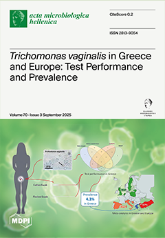
Trichomonas vaginalis infection (TVI) is the most common curable sexually transmitted infection (STI). For the diagnosis of TVI, vaginal microscopy is still in use in many parts of the world, while culture is considered the gold standard. In recent years, the rapid nucleic acid amplification test (NAAT) has been recommended as an alternative gold standard. In this study, we aimed to assess the performances of different tests for TVI diagnosis in symptomatic Greek women, evaluating the TVI prevalence rate (PR) in Greece and comparing the latter with TVI-PR estimates from Europe. Our findings demonstrated that in countries with a relatively high TVI-PR among symptomatic women, automated point-of-care NAAT would facilitate rapid, accurate TVI diagnosis and treatment of this target population. View this paper
- Issues are regarded as officially published after their release is announced to the table of contents alert mailing list.
- You may sign up for e-mail alerts to receive table of contents of newly released issues.
- PDF is the official format for papers published in both, html and pdf forms. To view the papers in pdf format, click on the "PDF Full-text" link, and use the free Adobe Reader to open them.
Previous Issue
Next Issue