Article

Assessment of Bacterial Contamination and Biofilm Formation in Popular Street Foods of Biskra, Algeria

1
Laboratory of Promotion of Agricultural Innovation in Arid Regions (PIARA), Department of Biology, Faculty of Exact Sciences and Natural Life Sciences, Mohamed Khider University, Biskra 07000, Algeria
2
Laboratory of Health, Animal Production and Environment (ESPA), University of Batna, Batna 05000, Algeria
3
Department of Biology, Faculty of Exact Sciences and Natural Life Sciences, Mohamed Khider University, Biskra 07000, Algeria
*
Author to whom correspondence should be addressed.
Acta Microbiol. Hell. 2025, 70(3), 32; https://doi.org/10.3390/amh70030032
Submission received: 25 April 2025 / Revised: 23 June 2025 / Accepted: 14 July 2025 / Published: 28 July 2025

Abstract

This study assessed microbiological contamination in street-sold meat products, focusing on Enterobacterales and coagulase-negative staphylococci (CoNS) species and their antibiotic resistance. Chicken and mutton street foods like shawarma and brochettes were tested for bacterial load, species distribution. and resistance profiles. The results showed significant contamination, with Enterobacter cloacae (5.38 Log 10 CFU/g). Staphylococcus lentus and Staphylococcus xylosus were also common, reaching 6.23 Log 10 CFU/g in some samples. Contamination levels varied significantly by food type, with chicken shawarma showing the highest risk. Antimicrobial susceptibility testing revealed high multidrug resistance, particularly among E. cloacae and Staphylococcus species. Biofilm formation an indicator of resistance was observed mainly in staphylococci and enhanced under fed-batch culture. These findings highlight public health concerns tied to poor hygiene and undercooking in street food environments. The study emphasizes the need for improved hygiene practices, standardized cooking methods, and systematic food safety monitoring to reduce contamination and antibiotic resistance risks.

1. Introduction

Street foods are becoming increasingly popular due to rising industrialization, prompting many urban residents to consume their primary meals outside of the home. The sale of these foods is a prevalent characteristic of urban areas in developing nations. They are valued not only for their convenience and affordability but also for their flavor, which is a product of the vendors’ culinary expertise [1]. The report on collective food poisoning (CFP) published by the Ministry of Trade and Export Promotion of Algeria shows that the number of CFP cases and the number of people poisoned rose by 105% and 109%, respectively, in the first half of 2021 compared with the same period of the previous year [2]. Meat-based foods were selected for this study due to several important factors. First, animal-derived foods, particularly meat products, offer a nutrient-rich environment that favors the growth of microorganisms, including antibiotic-resistant pathogens. This characteristic makes meat a high-risk category for microbial contamination. Secondly, meat products are widely sold as street foods, given their popularity and convenience. Combined with often suboptimal hygiene conditions in street vending, this increases the potential for contamination. Moreover, contaminated meat poses significant public health risks, acting as a vector for pathogen transmission to consumers. Several studies have reported outbreaks of foodborne illness directly linked to shawarma consumption. For example, cases have been reported in India and Lebanon, where poor hygiene practices during shawarma preparation contributed to outbreaks of Salmonella and Campylobacter infections [3,4]. These events highlight the potential public health risks associated with meat-based street foods, especially shawarma, and justify targeted microbial assessments. Finally, meat substrates are particularly conducive to biofilm formation, which enhances bacterial protection and resistance to sanitation efforts. Investigating the biofilm-forming and antibiotic-resistant capacities of Enterobacterales and staphylococci in meat-based street foods allows for a targeted assessment of microbial contamination risks in the food supply chain.
Certain pathogenic species within the Enterobacterales family and the Staphylococcus genus are among the microorganisms most commonly associated with foodborne illnesses. These bacteria can cause a wide range of symptoms, from mild gastrointestinal disorders to severe infections, especially in vulnerable populations like children, senior citizens, and the immunocompromised [5]. Treating these bacterial infections is severely hampered by the emergence of multiple antibiotic resistance (MAR) and multidrug resistance (MDR) [6]. Potential foodborne illnesses may arise from food contamination, primarily caused by bacteria that form biofilms [7]. A community of microorganisms known as a biofilm is formed on biotic or abiotic surfaces by an extracellular polymeric matrix. The food matrix may aid in the horizontal transfer of resistance genes, and reports have already been made regarding the transmission of antibiotic resistance genes to pathogens through the food chain [8].
Understanding the dual role of foodborne bacteria in antibiotic resistance and biofilm formation is therefore crucial for evaluating their persistence and potential virulence in food-related environments. This study aims to evaluate bacterial contamination in various meat-based street foods in Algeria, with a specific focus on Enterobacterales and Staphylococcus coagulase-negative species. We investigated the resistance profiles of isolated strains and assessed their biofilm-forming capacity using two different cultivation methods in order to understand their impact on biofilm development. The findings provide essential data for understanding the health risks associated with street food consumption and for recommending appropriate preventive measures.

2. Materials and Methods

2.1. Sampling Strategy and Food Selection for Microbial Analysis in Street Food

A total of 60 street food samples were obtained from various urban vendors. The samples were categorized into four primary groups, representing the food types most frequently consumed in the study area. The research examined different kinds of popular dishes to evaluate preparation and cooking methods, along with their effects on microbial load. A total of 15 samples of chicken shawarma, which is a marinated, grilled meat preparation, were examined. The samples of chicken shawarma collected alongside by fourteen samples of mutton brochette to assess how avenue vendors practiced the express cooking of meat. We also included nineteen samples of chicken brochettes to assess the microbial risks associated with skewered poultry preparation. And finally, twelve samples of mutton shawarma were analyzed. Though less prevalent than chicken shawarma, these samples were incorporated to evaluate potential discrepancies in contamination levels between the two meat varieties. This variety of samples offered a thorough examination of the practices and risks linked to these popular dishes. Samples were collected under aseptic conditions to prevent external contamination. Then, samples were analyzed within 24 h of collection to guarantee precise results according to ISO 4833-1:2013 [9].
The microbiological quality of the meat-based street food samples was assessed based on the Enterobacteriacae. Samples were classified into the categories of satisfactory, acceptable, unsatisfactory, or unacceptable/potentially hazardous [10], defind as follows:
Satisfactory: Test results indicating good microbiological quality (CFU < 102 CFU/g)
Acceptable: Results reflecting borderline microbial quality (CFU between 102–104 CFU/g).
Unsatisfactory: Indicating potential hygiene issues that may necessitate further inspection (CFU > 104 CFU/g).
Unacceptable/potentially hazardous: N/A: Not applicable—this category is not defined for the corresponding microorganism.

2.2. Microbiological Analysis

2.2.1. Identification of Pathogenic Bacteria

Samples were subjected to standardized microbiological protocols to detect and identify Enterobacterales and staphylococci. For Enterobacterales, homogenized samples were enriched in buffered peptone broth and incubated at 37 °C for 24 h before being plated on selective media, such as MacConkey and EMB agar, to isolate suspect colonies, which were then confirmed biochemically [11]. For staphylococci, enrichment was performed in tryptic soy broth containing 6.5% NaCl to promote growth, followed by incubation and isolation on Mannitol Salt Agar, a selective medium recommended for staphylococcal detection [11,12]. These procedures ensured reliable identification of potential foodborne pathogens, particularly in manually handled food items.

2.2.2. Species Characterization and Antibiotic Resistance

The identification of bacterial isolates comprised two fundamental steps. Initially, Staphylococcus species were identified through the API Staph gallery. Simultaneously, Enterobacterales were identified utilizing the API 20E gallery through specific assays. The outcomes were subsequently validated by the automated VITEK 2 COMPACT system. This integrated method guaranteed swift, dependable, and precise identification of bacterial species.
Bacterial identification was performed using API strips and the VITEK-2 Compact system (bioMérieux). Although widely accepted, these methods may have had limited accuracy for food-derived isolates. VITEK-2 showed 95.8% concordance with API for Gram-positive cocci [13], but without molecular confirmation (e.g., 16S rRNA). Misidentification remained a study limitation.

2.2.3. Qualitative and Quantitative Detection of Biofilm

The capacity of slime (a viscous extracellular material associated with biofilm formation) production was established with Congo Red Agar (CRA) medium. Colony phenotypes were used to interpret the isolates. Red colonies were thought to be non-biofilm producers, but black colonies were a sign of biofilm development [14].
For the quantitative determination of production of biofilm, the microtiter plate method (MTP) was used, with some modifications [12,13]. The microplates were placed in an incubator at a temperature of 37 °C for durations of 1 and 3 days. During the 3-day duration, two different cultivation methods were employed: batch mode and fed batch mode, and each assay was performed in triplicate (n = 3). After this period, the wells were washed three times with PBS (phosphate-buffered saline). Cells adherent to the wells were fixed with 200 μL of 100% ethanol for 15 min; after drying, crystal violet (Merck KGaA, 64271 Darmstadt, Germany) was used to stain the wells, and glacial acetic acid (VWR 20104.334) at a concentration of 33% (v/v) was used for reading the absorbances at OD595nm (Epoch 2, BioTek Instruments, Winooski, VT, USA). To evaluate biofilm capacity, the measurement was made using the ODc of the negative control [15].
The batch mode provides a closed system where nutrients are supplied initially and are gradually depleted as microbial growth progresses, leading to accumulation of metabolic byproducts, which can impact biofilm development [14,15]. Conversely, the fed-batch mode introduces nutrients incrementally, allowing controlled substrate addition to sustain microbial activity while avoiding excessive inhibition by byproducts, which is particularly advantageous for studying biofilm persistence and robustness [16,17]. These differences in nutrient availability and waste accumulation are known to influence bacterial growth dynamics and biofilm architecture, making them suitable for understanding variations in biofilm formation under realistic environmental conditions [18,19]. By utilizing both modes, our study aims to explore the potential effects of nutrient dynamics on the isolates biofilm formation and antibiotic resistance, as nutrient-limited environments and controlled nutrient feeding have been shown to shape biofilm structure and resilience in microbial communities [16].

2.3. Statistical Analysis

Bacterial contamination levels (Log 10 CFU/g) were measured in multiple food types (chicken and mutton shawarma and chicken and mutton brochettes) with descriptive statistics to assess the distribution of microbiological risk. Chi-square tests were used to investigate relationships between food categories in terms of the presence of bacteria.
Chi-square analysis was also applied to examine associations between antibiotic resistance profiles (non-MDR, MDR, and XDR) and biofilm formation (presence/absence). When appropriate, Fisher’s exact test was used for small sample sizes. To assess quantitative differences in biofilm production (optical density values), one-way ANOVA or Kruskal–Wallis tests were used depending on data normality (verified using the Shapiro–Wilk test). Pairwise comparisons were followed by post hoc tests (Tukey or Dunn’s).
Significance levels (p-values) were reported for all key analyses, and results were considered statistically significant when p < 0.05. All analyses were conducted using SPSS software (version 26. IBM Corp).

3. Results

3.1. Distribution of Contamination Levels and Bacteria Isolates from Different Meat-Based Street Food

Microbiological contamination levels for both Enterobacterales and staphylococci differ according to the meats analyzed. Based on the results of the analyses, loading rates and species distribution vary both quantitatively and qualitatively depending on the product (Figure 1).
Enterobacterales contamination levels in chicken shawarma ranged from 1.3 to 5.4 Log10 CFU/g, with a high average. E. cloacae is the most prevalent species, accounting for 64.3% of isolates, followed by Serratia (14.3%), Escherichia coli (7.1%) and Citrobacter (7.1%). For Staphylococcus species, contamination levels ranged from 0 to 6.2 Log10 CFU/g. S. lentus is the dominant species (46%), followed by S. xylosus (34%) and S. lugdunensis (16%). Qualitative classification reveals that 66.7% of samples are unsatisfactory, 26.7% acceptable, and only 6.7% satisfactory.
Enterobacterales contamination levels in mutton brochettes ranged from 0 to 3.1 Log10 CFU/g, with E. cloacae dominating at 66.7%. Serratia and E. coli are less frequent (11.1% each). For Staphylococcus species, levels range from 0 to 3.9 Log10 CFU/g, with S. lentus (50%) and S. xylosus (28.6%) as the most prevalent species, while S. lugdunensis was present at 14.3%. In terms of quality, the results obtained are as follows: 64.3% satisfactory, 35.7% acceptable, and none unsatisfactory.
Enterobacterales counts in chicken brochettes varied within a range of 0 to 3.4 Log10 CFU/g, E. cloacae accounted for 50% of isolates, with Klebsiella and Salmonella making a notable appearance (12.5% each), thereby classifying this item as unsafe to eat. For staphylococci species, levels range from 0 to 4.5 Log10 CFU/g, with S. lentus dominating at 52.6%, followed by S. xylosus (31.6%) and S. lugdunensis (10.5%). Microbiological evaluation revealed 73.7% of samples to be satisfactory, 26.3% acceptable, and none unsatisfactory.
The levels of Enterobacterales contamination in mutton shawarma ranged from 0 to 3.1 Log10 CFU/g, and E. cloacae was the only species identified (100%). For Staphylococcus species, levels ranged from 0 to 22.958 CFU/g, with S. lentus dominating at 64.7%, followed by S. xylosus (23%) and S. lugdunensis (8.3%). Sample distribution shows a 58.3% rate for satisfactory samples, 41.7% for acceptable, and no unsatisfactory quantities.
Regarding the three staphylococcal species, S. capitis, S. hominis, and S. haemolyticus, their occurrence was consistently low and relatively uniform across all food samples. S. capitis was detected at a steady rate of approximately 1.5 to 1.6%, regardless of the type of meat product; S. hominis showed slight variation, ranging between 2.4% and 2.6%, with identical rates found in both chicken and lamb shawarma. Similarly, S. haemolyticus appeared at 1.5%–1.6% in all analyzed samples (Table 1).
A chi-square test of independence was conducted to evaluate the relationship between food types and the bacterial species isolated from them. The results demonstrated a highly significant association (χ2 = 168.2, df = 36, p < 0.000001), indicating that bacterial presence is strongly influenced by the type of food analyzed.
E. cloacae were consistently present at high frequencies across all food types, particularly in shawarma (85%) and chicken-based items. In contrast, certain species, such as Salmonella and C. freundii, were detected only in specific foods, such as chicken brochettes and shawarma mutton, respectively. S. liquefaciens was found exclusively in mutton brochettes and chawarma chicken. These findings suggest that different food matrices provide distinct ecological niches, favoring the growth of specific bacterial communities. This has important implications for food safety monitoring and the development of targeted decontamination strategies depending on food type.

3.2. Antibiotic Resistance Profile of Isolates

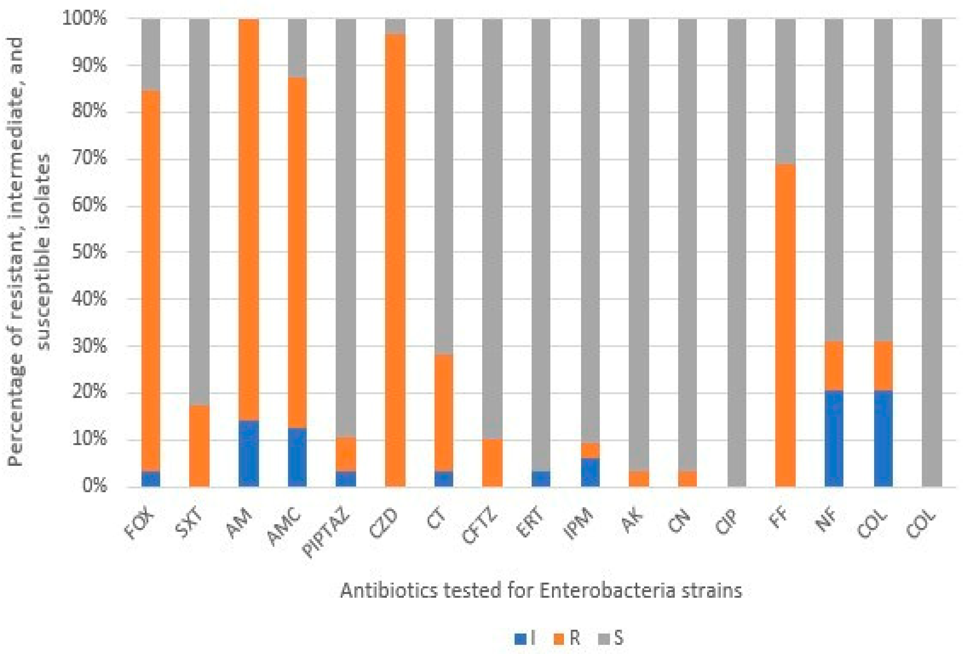
Antibiotic susceptibility testing of Enterobacterales isolates showed considerable variability in profiles of resistance. Susceptibility profiles varied because of both genetic variability and dissimilar resistance mechanisms possessed by this group of bacteria. Ciprofloxacin was the most effective antibiotic tested (100% susceptible), followed by ertapenem (96.5% susceptible) and imipenem (90.6% susceptible). The aminoglycosides amikacin and gentamicin (both at 96.9% susceptible, 3.1% resistant), with ceftazidime also having a fair performance at 90% susceptible. Nitrofurantoin was moderately effective at 68.9% susceptible, 20% intermediate, and 10.3% resistant, and fosfomycin had the worst performance, with 68.9% of the isolates resistant (Figure 2).
The percentage frequency of resistance profiles among Enterobacterales isolates reveals clear variation between species. C. freundii, K. pneumoniae, Salmonella spp., and E. sakazakii demonstrated the highest levels of multidrug resistance (MDR), with 100% of isolates classified as MDR, highlighting their critical role in antimicrobial resistance within this sample. E. cloacae also showed a strikingly high MDR profile (~95%), placing it among the most resistant isolates. Interestingly, S. liquefaciens had a notable presence in both MDR (~65%) and non-MDR/XDR (~30%) categories, reflecting its variable resistance capacity. While E. coli is commonly studied for resistance, it showed a more moderate profile, with about 45% of isolates were MDR, and the remainder were non-MDR/XDR, which suggests a mixed resistance trend. This distribution underlines the heterogeneity of resistance among Enterobacterales and supports the importance of species-level tracking in resistance surveillance (Figure 3).
To further assess the correlation between Enterobacterales species and their antimicrobial resistance classification (MDR vs. non-MDR/XDR), a Pearson chi-square test was conducted. The test revealed a statistically significant association (χ2 = 489.8, df = 48, p < 0.001), confirming that resistance patterns are not randomly distributed across species. Additionally, the likelihood ratio test supported this result (G2 = 412.3, p < 0.001), strengthening the evidence that specific species are more prone to MDR classifications than others.
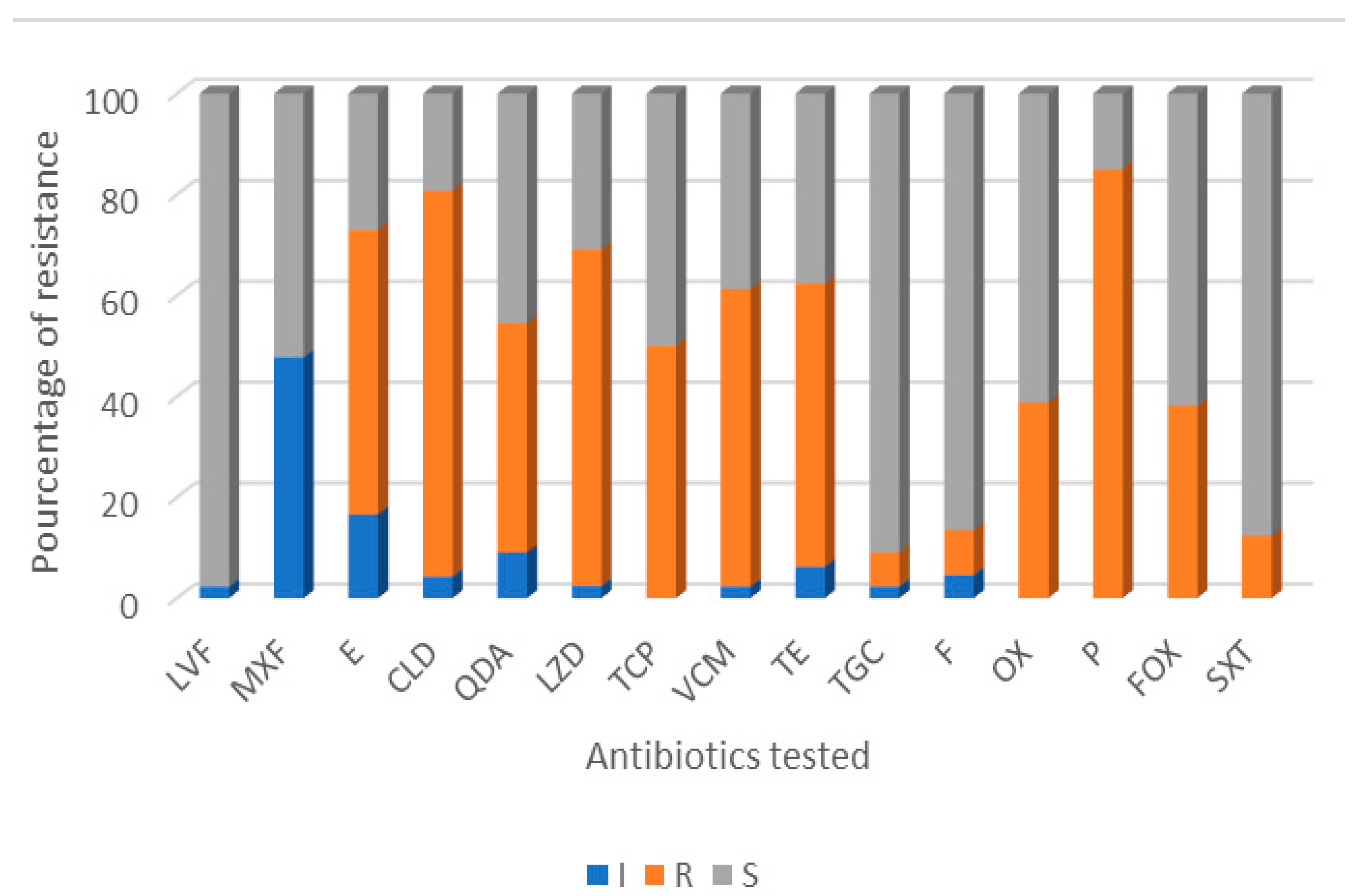
Given that Staphylococcus isolates are frequently implicated in clinical infections, their antibiotic susceptibility profiles were carefully assessed. Using the VITEK 2 system, we found a high level of resistance in all staphylococci isolates tested, with substantial variation between species. S. lentus and S. xylosus had the highest resistance arc, whereas S. haemolyticus was mostly susceptible, with the exception of penicillin, cefoxitin, and trimethoprim-sulfamethoxazole. Penicillin had the highest rate of overall resistance, with 85.1% of the isolates being resistant, which was likely due to its long history and heavy use. Clindamycin also had quite a high resistance rate of 76.59%, which is concerning given its frequent use in treating MRSA infections. Even more alarmingly, resistance was detected against last resort antibiotics, with 66.66% of isolates resistant to linezolid and 59.09% to vancomycin. Erythromycin and tetracycline had the same resistance rates, 56.25%, which reflects their extensive use in both human and veterinary medicine. Overall, these findings highlight the growing issue of antimicrobial resistance amongst staphylococci, and the need for better care with antibiotic prescriptions and targeted therapy (Figure 4).
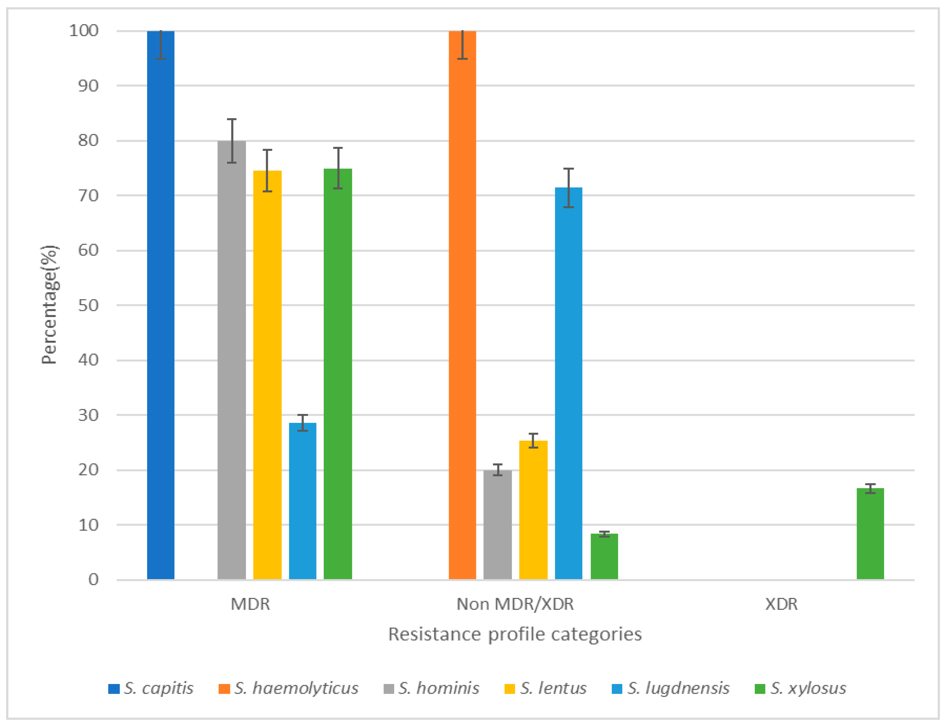
The percentage frequency of resistance profiles amongst staphylococci isolates shows notable differences between species. S. capitis was the most resistant species in this study, with all isolates categorized as multidrug resistant (MDR). S. hominis follows, with approximately 80% of its isolates classified within the MDR group, making it the second most MDR-associated species. S. lentus and S. xylosus had similar percentages of MDR resistance, ~75%, but they still showed notable amounts of involvement with resistance. Interestingly, for S. haemolyticus, the clear majority of isolates (100%) were categorized in the non-MDR/XDR category. The vast majority of S. lugdunensis isolates were similarly non-MDR/XDR (~70%), but it was one of two species (along with S. xylosus) that showed a significant number of both non-MDR/XDR and MDR profiles. S. xylosus was also notably the only species to represent all three categories, including the extensively drug resistant (XDR) group (~15%), which highlights its clinical significance and adaptability in comparison to the other species. Overall, this distribution highlights the intrinsic differences between resistance profiles across all staphylococci isolates and ultimately highlights the need for species level surveillance to monitor antimicrobial resistance (Figure 5).
To examine the relationship between Staphylococcus species and their patterns of antimicrobial resistance (MDR, non-MDR/XDR, and XDR), a Pearson chi-square test was performed. The result was highly statistically significant (χ2 = 569.2, df = 60, p < 0.001), indicating that resistance patterns were not evenly distributed across species. The likelihood ratio test also confirmed this association (G2 = 453.5, p < 0.001). These findings highlight a strong association between species and resistance profiles, emphasizing the importance of species-level antimicrobial resistance surveillance.

3.3. Slime and Biofilm Production Among Strains

Biofilm production among the tested Gram-negative isolates revealed notable interspecies variability. Only 8% of the strains were identified as strong biofilm producers based on the MTP assay, while 48.9% were categorized as non-producers. Post-hoc pairwise comparison following a Kruskal–Wallis test highlighted statistically significant differences between certain species, confirming the non-uniformity of biofilm-forming capabilities. S. liquefaciens exhibited the highest biofilm production rank, significantly outperforming K. pneumoniae and C. freundii, which positions it as a particularly robust biofilm-forming species with potential implications for persistence in both clinical and environmental contexts.
These results were further supported by quantitative CRAV measurements. S. liquefaciens and E. coli reached peak CRAV levels of 100%, while E. cloacae also showed high production (up to 90%), confirming biofilm potential despite rank variability. Other species, such as Salmonella and K. pneumoniae, exhibited minimal or no CRAV production, reinforcing the observed heterogeneity. This distribution illustrates the complex and species-dependent dynamics of biofilm formation, which are critical for understanding bacterial survival, virulence, and resistance potential in foodborne and environmental microbiology.
Among the 50 Staphylococcus isolates tested, 70% exhibited a slime-positive phenotype, visualized as black colonies on Congo red agar. S. lentus showed the highest frequency of slime production, followed by S. capitis, S. haemolyticus, and others. This phenotype was often associated with increased surface adherence and potential virulence.
Regarding biofilm formation (MTP assay), 36% of isolates were classified as strong biofilm producers, while 16.7% were non-producers. A Kruskal–Wallis test followed by post-hoc pairwise comparisons revealed significant differences between species. Notably, S. capitis demonstrated significantly lower biofilm formation than S. hominis and S. lentus. while S. haemolyticus also differed from both S. lentus and S. capitis. Other species such as S. xylosus and S. lugdunensis showed no significant differences from the rest, indicating similar biofilm-forming abilities under the conditions tested. These findings emphasize the species-specific variability in both slime and biofilm production, which are critical traits influencing the persistence and pathogenic potential of Staphylococcus in food and clinical environments (Table 2).

3.4. Correlation Between Biofilm Production and Antibiotic Resistance

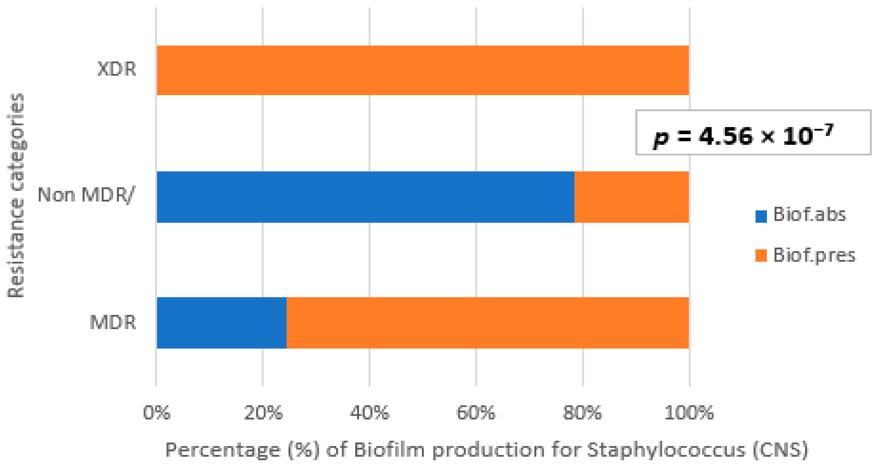
The Figure 6 illustrates the distribution of biofilm production capacity among Staphylococcus strains, categorized by their resistance profiles: MDR, non-MDR, and XDR. The ability to form biofilm was classified as either present (Biof.pres) or absent (Biof.abs). Among MDR strains, the majority demonstrated the ability to form biofilm, with a significantly larger proportion of Biof.pres (74.4%) compared to Biof.abs. Conversely, non-MDR strains showed a higher proportion of biofilm-negative isolates (76%), although a substantial number still retained biofilm-forming potential. Notably, XDR strains represented a minor fraction overall (4.8%), with only a small number displaying biofilm formation capacity.
A chi-square test of independence was performed to examine the association between antibiotic resistance profiles (MDR, non-MDR, and XDR) and the capacity for biofilm production among Staphylococcus strains. The results revealed a statistically significant relationship between these variables (χ2 = 30.4, df = 2, p < 0.000001). This extremely low p-value indicates that the observed distribution is highly unlikely to have occurred by chance. Specifically, MDR strains showed a markedly higher prevalence of biofilm production compared to non-MDR and XDR strains. While XDR strains were fewer in number, their biofilm production rate remained notable, although the absence of biofilm-negative isolates in this group suggests limited diversity. These findings suggest a strong correlation between multidrug resistance and biofilm-forming capacity in Staphylococcus species, potentially implicating biofilm production as a contributing factor to increased antimicrobial tolerance.
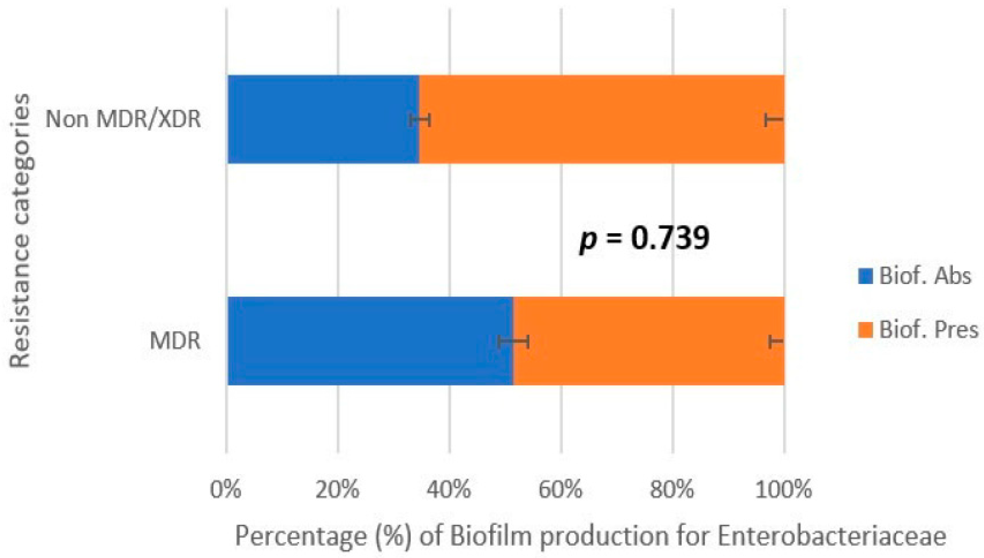
Figure 7 illustrates the distribution of biofilm production capacity among Enterobacterales strains categorized by their resistance profiles: MDR and non-MDR/XDR. The ability to form biofilm is classified as either present (Biof.pres) or absent (Biof.abs). Among MDR strains, an equal proportion of isolates exhibited biofilm-forming and non-forming capacities. In contrast, among non-MDR/XDR strains, both biofilm-producing and non-producing phenotypes were found at very low levels, suggesting a limited biofilm-forming potential within this group. A chi-square test of independence was conducted to evaluate the association between antibiotic resistance profiles and biofilm production among Enterobacterales. The result was not statistically significant (χ2 = 0.11, df = 1, p = 0.7), indicating that the observed distribution could be due to chance. Unlike observations made in the Staphylococcus species, no clear correlation was found between multidrug resistance and the ability to form biofilm in this Enterobacteria dataset. These findings suggest that, in this study, biofilm formation was not significantly associated with the resistance profile among members of the family Enterobacterales, and other factors may play a more influential role in determining their biofilm phenotype.
Our study revealed a statistically significant association between biofilm production capacity and antibiotic resistance profiles among Staphylococcus strains, particularly those classified as multidrug-resistant (MDR). MDR strains exhibited a high rate of biofilm formation (74.4%) compared to non-MDR strains, the majority of which were biofilm-negative (76%). Although extensively drug-resistant (XDR) strains accounted for a small fraction of isolates, all biofilm-producing strains in this group were positive, suggesting a potential link between persistence in clinical settings and biofilm production.
Overall, our results support the hypothesis that biofilm formation may contribute to increased antimicrobial tolerance in Staphylococcus spp., and it may represent a crucial factor in persistent and difficult-to-treat infections. These findings underscore the need to incorporate routine biofilm screening in clinical diagnostics, especially for MDR isolates, to inform appropriate therapeutic strategies and prevent chronic infection development.
In contrast to the strong association observed between biofilm production and multidrug resistance (MDR) in Staphylococcus species, our analysis of Enterobacterales isolates revealed no statistically significant correlation between resistance profiles and biofilm formation (χ2 = 0.11, df = 1, p = 0.7). Among MDR strains, biofilm-producing and non-producing isolates were equally distributed. Interestingly, non-MDR/XDR isolates exhibited very limited biofilm-forming potential, with both phenotypes being present at low frequencies.

3.5. Fed-Batch Mode Enhances Biofilm Formation Compared to Batch Culture

3.5.1. Enterobacterales

A comparison of biofilm production under fed-batch and batch conditions showed that fed-batch mode led to significantly higher biofilm levels (mean OD = 2.925; CI [2.39. 2.70]; SD = 0.971) than batch culture (mean OD = 1.429; 95% CI [1.35. 1.70]; SD = 1.239). Although both modes reached the same maximum OD (4.000), batch cultures showed greater variability and lower consistency, reflected in the lower minimum OD (0.061 vs. 0.609). ANOVA confirmed this difference was statistically significant (p < 0.001), though the explanatory power was limited (R2 = 12%). Importantly, the effect size was moderate to large (Cohen’s d = 0.78), indicating a meaningful difference in biofilm behavior. Regression analysis supported a positive relationship between cultivation mode and biofilm production (β = 0.352, p < 0.001). These results indicate that fed-batch conditions favor more stable and enhanced biofilm formation in Enterobacterales compared to batch mode.

3.5.2. Staphylococcus

Biofilm production by Staphylococcus isolates was assessed under fed-batch and batch conditions to examine the effect of nutrient supply on biofilm development. On average, biofilm formation was higher in fed-batch cultures (mean OD = 2.305; SD = 1.335) than in batch mode (mean OD = 1.6; SD = 1.5), indicating that nutrient-controlled environments favor biofilm growth. ANOVA results confirmed a significant difference between the two modes (F = 47.3, p < 0.0001), with 24% of the variation in OD values (R2 = 24%) in fed-batch OD explained by the cultivation method, and the effect size was moderate (Cohen’s d = 0.55), supporting a substantial biological difference. Regression analysis also showed a positive association between batch and fed-batch OD values (β = 0.4, p < 0.0001), suggesting that strains performing well in batch mode tend to produce even more biofilm in fed-batch conditions. These results highlight the influence of culture conditions on staphylococcal biofilm development and support the use of fed-batch systems for studies on persistent and resilient biofilms.
Our findings reveal a high degree of interspecies and intraspecies variability in biofilm production among both Gram-negative Enterobacterales and Staphylococcus species, highlighting the complexity of biofilm-related phenotypes in different ecological and clinical contexts (Figure 8).

4. Discussion

The study reveals significant microbiological contamination in popular street foods, such as chicken shawarma, mutton shawarma, and chicken and mutton brochettes. The most concerning findings involved the presence of Enterobacterales species indicators of fecal contamination and Staphylococcus species, which indicated poor hygiene, with chicken shawarma exhibiting the highest levels of contamination [20]. The detection of Staphylococcus spp. suggests inadequate hygiene practices during food handling [21]. In mutton shawarma, lower levels of Enterobacterales (0–3.15 Log10 CFU/g) were observed, and in Staphylococcus spp., levels reached 4.36 Log10 CFU/g, dominated by S. lentus (66.7%). Several international studies report similar contamination levels in shawarma across Saudi Arabia [22], Jordan [23], and Egypt [24], with Enterobacterales as primary contaminants. In Iraq, 82% of chicken and 94% of red meat shawarma samples were contaminated, with bacterial loads reaching 14 × 104 CFU/g [25]. Mutton brochettes presented lower contamination by Enterobacterales, led by E. cloacae (66.7%) and E. coli (11.1%), along with Staphylococcus spp., especially S. lentus (50%) and S. xylosus (28.6%). Though high-temperature cooking may reduce bacterial loads, post-cooking contamination remains an issue [26]. Chicken skewers showed slightly higher contamination, confirming Radwana’s findings [27].
In Algeria, contamination varies by region and product type, with pathogen presence ranging from 25% to 97.5%. For example, CoNS was isolated from raw milk and meat in Médéa and Ain Defla [28], and in Constantine, 97.5% of seafood was unfit for consumption due to coliforms and mesophilic bacteria [29]. In M’Sila [30] and Tipaza [31], high levels of Salmonella and thermotolerant coliforms in merguez and fish products highlighted the role of hygiene and storage in contamination. Algerian and international studies suggest that staphylococci especially (CoNS) should be considered emerging foodborne pathogens that require stringent monitoring and control.
While the contamination levels highlight the widespread presence of pathogenic bacteria in street foods, the concurrent detection of multidrug-resistant strains raises additional concerns regarding the therapeutic challenges they may pose. Antibiotic susceptibility testing of Enterobacterales isolates revealed considerable heterogeneity in resistance, largely attributable to species-specific genetic mechanisms. Most isolates remained susceptible to ceftazidime (90%) and ciprofloxacin (100%) [32], and while carbapenem resistance was low—3.1% for imipenem and 3.5% for ertapenem—these findings remain concerning due to carbapenems’ role as last-resort antibiotics [33]. Aminoglycosides such as amikacin and gentamicin retained high efficacy (96.9%), while fosfomycin (68.9%) and nitrofurantoin (10.3%) showed moderate resistance rates [34]. Resistance levels varied significantly between species, with C. freundii exhibited notable resistance to third-generation cephalosporins [35] while K. pneumoniae isolates—both foodborne and clinical—were often MDR, including carbapenemase producers [36,37,38]. E. coli showed moderate resistance to β-lactams and chloramphenicol (~50%) while remaining largely susceptible to carbapenems and fluoroquinolones [39,40,41,42]. Post-COVID increases in fosfomycin resistance are also emerging. E. cloacae demonstrated broad resistance to β-lactams, linked to AmpC β-lactamase, ESBLs, and colistin resistance mechanisms [43,44,45]. Salmonella spp. showed low resistance to amoxicillin and amoxicillin-clavulanate, and were generally susceptible to other antibiotics [46,47,48,49].
While these resistance patterns suggest the presence of underlying genetic mechanisms—such as ampC β-lactamase production, extended-spectrum β-lactamases (ESBLs), or efflux systems like qac—we recognize that our study did not include molecular screening or genotyping to confirm these hypotheses. Therefore, references to these resistance genes are intended only to provide contextual background, drawn from prior literature, and not as direct conclusions from our dataset.
Future studies incorporating PCR assays, whole-genome sequencing, or plasmid profiling would be necessary to confirm the presence and role of such genes in the isolates analyzed here. Until such analyses are performed, any interpretation of resistance mechanisms remains hypothetical, and the phenotypic variability observed should be considered a foundation for guiding molecular research.
The resistance profiles of the Staphylococcus species—particularly S. lentus, S. xylosus, and S. capitis—highlight their growing role as reservoirs of antimicrobial resistance genes. Resistance to penicillin reached 85.1%, echoing findings from Achek et al. (2018) [28] and Saidi et al. (2015) [50], who reported 94% penicillin resistance in bovine milk isolates linked to blaZ and mecA genes. Clindamycin resistance (76.59%) is also critical given its status as a β-lactam alternative, with similar patterns found in meat isolates [28]. Resistance to erythromycin and tetracycline (both 56.25%) aligns with Mikulášová et al. (2014) [8], who reported 45% MDR in S. xylosus from cheese, including resistance to oxacillin and tetracycline. Alarmingly, resistance to last-line antibiotics was also recorded: linezolid (66.66%) and vancomycin (59.09%), with linezolid resistance linked to 23S rRNA mutations, as seen in S. capitis nosocomial infections [51]. Similarly, S. capitis-induced endocarditis, once treatable with vancomycin. now raises concerns over resistance [52]. Overall, 66% of staphylococcal isolates were classified as MDR, echoing Achek et al. [28], who found resistance to ≥3 antibiotic classes in SCN from meat and dairy involving mecA, erm(C), tet(M), and blaZ. Species-level differences were notable: S. capitis showed 100% MDR, supporting its rising clinical relevance [53]; S. hominis followed at ~80% [54]; S. lentus and S. xylosus reached ~75%, with S. xylosus also including XDR strains, reflecting its ecological plasticity [55]. S. haemolyticus was only found in the non-MDR/XDR group, although it is clinically known for high resistance [54,56]. S. lugdunensis had ~70% non-MDR/XDR isolates but was also represented in the MDR group, consistent with emerging resistance trends in hospital settings [57,58].
In light of these findings. it becomes evident that traditional antibiotics alone may not suffice to control biofilm-associated and MDR pathogens. One promising approach is the use of Trojan Horse antibiotics. which exploit bacterial nutrient uptake systems—such as siderophore transporters—to smuggle antibiotics into resistant cells, bypassing classical resistance mechanisms [59]. In line with this strategy is the emerging research suggesting the antimicrobial potential of metallophores, which are metal-bound chelating compounds produced by bacteria to assist in the process of essential metal ion uptake. As bacteria require metal ions for growth and virulence, targeting metallophore-mediated pathways represents an alternative therapeutic option to prevent infection [60]. In chronic infections like those seen in cystic fibrosis, anti-virulence strategies targeting biofilm formation, rather than bacterial viability, have also shown promise [61]. These innovative approaches represent a critical step forward in the global fight against antimicrobial resistance and merit consideration in future research on foodborne pathogens.
While antimicrobial resistance poses a major threat to treatment efficacy, its potential association with biofilm production a key virulence factor warrants further exploration. These findings align with several previous studies. Shrestha et al. (2018) reported that 71.8% of (CoNS) isolated from clinical samples were biofilm producers, and that these strains exhibited significantly higher antimicrobial resistance than non-biofilm formers [62]. Similarly, Farajzadeh Sheikh et al. (2019) observed that 65% of Staphylococcus epidermidis strains isolated from neonatal septicemia cases formed biofilms, with a predominance of polysaccharide matrices, linked to the presence of the icaA and icaD genes [63]. Conversely, other studies—such as that by Donadu et al. (2022)—found no direct correlation between biofilm formation and antibiotic resistance in environmental Staphylococcus spp., highlighting the variability that may arise due to strain origin, genetic background, or methodological differences [64]. Additionally, Medis et al. (2023) demonstrated that biofilm formation plays a key role in the colonization of central venous catheters by S. haemolyticus, underscoring the clinical importance of biofilm in catheter-related bloodstream infections [65].
For the Enterobacterales. our results are consistent with the study by Cepas et al. (2019) [66], who concluded that global multidrug resistance in Gram-negative bacteria, including E. coli and K. pneumoniae, does not necessarily correlate with enhanced biofilm formation. Instead, specific resistance traits, such as resistance to gentamicin or ceftazidime, were associated with biofilm production in certain species, suggesting that the relationship is species- and drug-dependent rather than generalized.
These findings highlight the high interspecies and intraspecies variability in biofilm production among Staphylococcus and Enterobacterales isolates. Although this study focused on phenotypic evaluation of biofilm formation, substantial evidence from the literature supports the pivotal roles of ica genes and global regulators such as agr and sarA in Staphylococcus biofilm development.
The icaADBC operon is critical for the synthesis of polysaccharide intercellular adhesin (PIA), a major component of the biofilm matrix. The transcription of ica genes is modulated by the sarA locus, which acts as a positive regulator by directly binding to promoter regions and enhancing expression, thereby promoting biofilm accumulation [67]. In contrast, the agr quorum-sensing system has been shown to negatively regulate biofilm formation, likely through repression of surface adhesin expression. The balance between agr and sarA activities has been described as a “regulatory see-saw,” influencing whether cells adopt a biofilm or planktonic phenotype [68]. Furthermore, some studies suggest that sarA contributes to both ica-dependent and ica-independent pathways. broadening its role in biofilm regulation beyond a single operon [69].
Even though we did not assess these genes molecularly, our phenotypic findings—particularly the strain-specific variability in biofilm production—may reflect differences in the regulation of these loci. Future investigations incorporating PCR, sequencing, or transcriptomic profiling of ica, sarA, and agr would be valuable to validate these mechanistic insights.
Several studies have examined the relationship between multidrug resistance (MDR) and biofilm formation in foodborne bacteria, with mixed conclusions. Kim et al. (2018) observed that while many MDR E. coli isolates from meat and vegetables could form biofilms, no consistent association was found between MDR status and biofilm strength, suggesting a complex interplay of resistance and persistence traits [70]. Similarly, Castaño-Arriba et al. (2020) reported that although all Enterococcus isolates from meat were biofilm formers and 87.5% exhibited MDR, biofilm intensity did not correlate linearly with resistance levels, implying strain-specific or ecological influences [71]. In Nigeria, Beshiru et al. (2023) found that 87.2% of ESBL- and AmpC-producing Enterobacterales from vegetables formed strong biofilms; however, MDR phenotypes were not limited to biofilm-producing strains, reinforcing that biofilm capacity alone is not a reliable predictor of antibiotic resistance category [72].
In conclusion, the present study underscores the complexity of the biofilm-antibiotic resistance relationship in Enterobacterales. Unlike in Staphylococcus, where a clear trend was observed, biofilm formation among Enterobacteria appears to be influenced by more nuanced and possibly strain-specific factors. These findings stress the importance of assessing both phenotypic and genotypic traits when evaluating the pathogenic potential and persistence of resistant Enterobacteria in clinical and food-related settings.
Among the Gram-negative isolates, only 8% were classified as strong biofilm producers based on the MTP assay, with nearly half (48.9%) being non-producers. Species such as S. liquefaciens exhibited significantly higher biofilm formation compared to K. pneumoniae and C. freundii, suggesting intrinsic differences in their genetic or regulatory pathways. These results are consistent with those reported by Beshiru et al. (2023), who observed strong biofilm-forming capabilities in certain β-lactamase-producing E. coli and Serratia strains isolated from vegetables, emphasizing their potential for environmental persistence and food safety risks [72].
In contrast, E. sakazakii and E. cloacae demonstrated weaker biofilm profiles, which aligns with studies by Cepas et al. (2019) [66], who documented low to moderate biofilm activity in similar species despite their clinical relevance. Interestingly, while E. coli and Salmonella spp. showed intermediate biofilm levels, post-hoc tests revealed no significant differences from either high or low producers, reinforcing the notion that biofilm capacity among Enterobacterales is both strain- and context-dependent [66].
The variability in biofilm production was further confirmed through CRAV (crystal violet absorbance value) quantification, which revealed strong biofilm production by S. liquefaciens and E. coli, with values reaching 100%, and E. cloacae at 90%, despite its low TCP rank. This highlights that methods like CV-staining or CRAV can detect biofilm biomass missed by qualitative ranking alone, as noted by Wilson et al. (2018), who advocated for multimodal quantification strategies to capture biofilm heterogeneity [73].
Staphylococcal isolates also demonstrated species-specific differences in both slime and biofilm production. In total, 70% were Congo red-positive, particularly S. lentus and S. capitis, which is consistent with findings by Shrestha et al. (2018), who linked the slime phenotype to increased surface adherence and antibiotic resistance in (CoNS) [62]. The MTP assay confirmed these patterns, with S. lentus and S. hominis producing significantly more biofilm than S. capitis, reinforcing earlier reports on intra- and inter-species variability among CoNS biofilms by Oliveira et al. (2015) [74].
Environmental conditions further influenced biofilm formation. Under fed-batch conditions, both Gram-negative and Staphylococcus strains produced significantly more biofilm biomass compared to batch cultures. For Enterobacterales, the fed-batch mode led to a higher mean OD (2.925) with reduced variability, a finding mirrored by Hunt et al. (2004), who demonstrated that nutrient availability modulates biofilm structure and detachment dynamics through starvation or metabolic feedback mechanisms [75]. Similarly, Tan et al. (2017) showed that longer incubation times and nutrient replenishment promoted denser and metabolically active biofilms in mixed bacterial-fungal cultures, underscoring the importance of culture conditions [76].
In Staphylococcus, fed-batch cultures yielded higher average OD values (2.305 vs. 1.585), with statistically significant differences confirmed by ANOVA and regression analysis. These observations align with Cerca et al. (2004), who reported that nutrient-rich environments support matrix synthesis and multilayered structure development in S. epidermidis biofilms [77].
In summary, biofilm formation among Enterobacterales and Staphylococcus is a multifactorial trait influenced by species identity, environmental conditions, and quantification methodology. These findings stress the necessity of comprehensive biofilm assessments incorporating both phenotypic assays and environmental variables to accurately evaluate bacterial persistence and pathogenicity in clinical and food safety frameworks.

Limitation of Strain-Level Analysis

One limitation of the present study is the absence of molecular typing, which restricts strain-level resolution and genotypic differentiation. Although phenotypic identification using API strips and the VITEK-2 system enabled classification at the species level, their accuracy—particularly for (CoNS) from food or environmental sources—remains limited. Giger et al. (1984) reported 79.2% accuracy of API Staph-Ident for clinical CoNS isolates [78], while Sampimon et al. (2009) found only 41% correct identification for isolates from bovine milk [79]. Burriel (1997) also showed that ID32 STAPH assigned species to just 54% of isolates compared to 73.8% with API Staph and failed to differentiate slow-growing staphylococci from micrococci [80]. Though the VITEK-2 system demonstrated 95.8% concordance with API for Gram-positive cocci [81], potential misidentifications still exist, especially in the presence of atypical biochemical profiles or when older culture media are used. We acknowledge that the combined use of API and VITEK enhances the reliability of phenotypic identification, yet this approach cannot replace the precision of molecular tools. Furthermore, although we attempted to review recent AOAC or EU validations of these methods, no up-to-date assessments were found. Future studies should incorporate molecular confirmation techniques such as 16S rRNA gene sequencing or whole-genome sequencing to improve taxonomic accuracy, validate phenotypic identifications, and enable robust phylogenetic and epidemiological analyses.

5. Conclusions

This study confirms that meat-based street foods in Biskra, Algeria, are significantly contaminated with antibiotic-resistant and biofilm-producing bacteria, notably E. cloacae and Staphylococcus species such as S. lentus and S. xylosus. These microorganisms present a substantial public health threat, exacerbated by inadequate hygiene and improper food handling. The demonstrated link between multidrug resistance and biofilm production in staphylococci—absent in Enterobacterales—highlights species-specific pathogenic mechanisms that complicate decontamination. Moreover, the enhanced biofilm formation observed under fed-batch conditions mimicking realistic environmental stress suggests that current cleaning protocols may be insufficient.
Taken together, these findings emphasize the urgent need for robust street food regulations, vendor education, and routine microbiological surveillance to limit foodborne illness and the propagation of resistant bacteria. Proactive interventions at the vendor and policy levels could significantly improve consumer safety and public health outcomes in urban food environments.

Author Contributions

Conceptualization, S.B. and A.A.; methodology, S.B.; software, S.B.; validation, S.B., A.A. and W.B.; formal analysis, S.B.; investigation, S.B. and W.B.; resources, S.B. and W.B.; data curation, S.B.; writing—original draft preparation, S.B.; writing—review and editing, S.B. and W.B.; visualization, S.B.; supervision, A.A.; project administration, A.A. All authors have read and agreed to the published version of the manuscript.

Funding

This research received no external funding.

Institutional Review Board Statement

Not applicable.

Informed Consent Statement

Not applicable.

Data Availability Statement

The data presented in this study are available on request from the corresponding author.

Conflicts of Interest

The authors declare no conflicts of interest.

Abbreviations

The following abbreviations are used in this manuscript:
CoNSCoagulase-negative staphylococci
CFUColony forming unit
CRACongo red agar
MDRMultidrug resistance
XDRExtensively drug resistant
MTPMicrotiter plate method

References

  1. Alimi, B.A. Risk factors in street food practices in developing countries: A review. Food Sci. Hum. Wellness 2016, 5, 141–148. [Google Scholar] [CrossRef]
  2. Algeria Press Service Intoxication Alimentaire: Augmentation de 105 Cas Durant le 1er Semestre de 2021. Available online: https://www.aps.dz/sante-science-technologie/125713-intoxication-alimentaire-augmentation-de-105-cas-durant-le-1er-semestre-de-2021 (accessed on 23 August 2024).
  3. Deepanjali, S.; Jharna, M.; Chanaveerappa, B.; Sarumathi, D.; Gopichand, P.; Anupriya, K. An outbreak of Salmonella enteritidis food poisoning following consumption of chicken shawarma: A brief epidemiological investigation. F1000Research 2022, 10, 851. [Google Scholar] [CrossRef]
  4. Banna, H.; Nawas, T. Salmonella: A Common Contaminant of Chicken Shawarma in RasBeirut Restaurants. IOSR J. Environ. Sci. Toxicol. Food Technol. 2016, 10, 19–22. [Google Scholar] [CrossRef]
  5. Fusaro, C.; Miranda-Madera, V.; Serrano-Silva, N.; Bernal, J.E.; Ríos-Montes, K.; González-Jiménez, F.E.; Ojeda-Juárez, D.; Sarria-Guzmán, Y. Antibiotic-Resistant Bacteria Isolated from Street Foods: A Systematic Review. Antibiotics 2024, 13, 481. [Google Scholar] [CrossRef]
  6. Urmi, M.; Ansari, W.; Islam, M.; Sobur, M.; Rahman, M.; Rahman, M. Antibiotic resistance patterns of Staphylococcus spp. isolated from fast foods sold in different restaurants of Mymensingh, Bangladesh. J. Adv. Vet. Anim. Res. 2021, 8, 274–281. [Google Scholar] [CrossRef] [PubMed]
  7. Xu, H.; Lee, H.; Ahn, J. Characteristics of Biofilm Formation by Selected Foodborne Pathogens. J. Food Saf. 2011, 31, 91–97. [Google Scholar] [CrossRef]
  8. Mikulášová, M.; Valáriková, J.; Dušinský, R.; Chovanová, R.; Belicová, A. Multiresistance of Staphylococcus xylosus and Staphylococcus equorum from Slovak Bryndza cheese. Folia Microbiol. 2014, 59, 223–227. [Google Scholar] [CrossRef]
  9. ISO 4833-1:2013; Microbiology of the Food Chain—Horizontal Method for the Enumeration of Microorganisms—Part 1: Colony Count at 30 °C by the Pour Plate Technique. ISO: Geneva, Switzerland. Available online: https://www.iso.org/standard/53728.html (accessed on 24 March 2025).
  10. World Health Organization; Food and Agriculture Organization of the United Nations. Food Safety Risk Analysis: A Guide for National Food Safety Authorities; World Health Organization; Food and Agriculture Organization of the United Nations: Rome, Italy, 2006. [Google Scholar]
  11. Gajewska, J.; Chajęcka-Wierzchowska, W. Biofilm Formation Ability and Presence of Adhesion Genes among Coagulase-Negative and Coagulase-Positive Staphylococci Isolates from Raw Cow’s Milk. Pathogens 2020, 9, 654. [Google Scholar] [CrossRef]
  12. Stepanović, S.; Vuković, D.; Hola, V.; Bonaventura, G.D.; Djukić, S.; Ćirković, I.; Ruzicka, F. Quantification of biofilm in microtiter plates: Overview of testing conditions and practical recommendations for assessment of biofilm production by Staphylococci. APMIS 2007, 115, 891–899. [Google Scholar] [CrossRef]
  13. Sudagidan, M.; Ozalp, V.C.; Öztürk, O.; Yurt, M.N.Z.; Yavuz, O.; Tasbasi, B.B.; Ucak, S.; Mavili, Z.S.; Coban, A.; Aydin, A. Bacterial surface, biofilm and virulence properties of Listeria monocytogenes strains isolated from smoked salmon and fish food contact surfaces. Food Biosci. 2021, 41, 101021. [Google Scholar] [CrossRef]
  14. Shuler, M.L.; Kargi, F. Bioprocess Engineering: Basic Concepts, 2nd ed.; Prentice Hall: Upper Saddle River, NJ, USA, 2002. [Google Scholar]
  15. Chisti, Y. Fermentation Technology. In Industrial Biotechnology: Sustainable Growth and Economic Success; Wiley: Hoboken, NJ, USA, 2010. [Google Scholar]
  16. Petrova, O.E.; Sauer, K. Escaping the biofilm in more than one way: Desorption, detachment or dispersion. Curr. Opin. Microbiol. 2016, 30, 67–78. [Google Scholar] [CrossRef]
  17. Hjersted, J.L.; Henson, M.A.; Mahadevan, R. Genome-scale analysis of Saccharomyces cerevisiae metabolism and ethanol production in fed-batch culture. Biotechnol. Bioeng. 2007, 97, 1190–1204. [Google Scholar] [CrossRef]
  18. Tsao, G.T. Principles of microbe and cell cultivation, S. John Pirt, Halsted Press, Division of John Wiley and Sons, New York, 274 pages, $34.00. AIChE J. 1976, 22, 621. [Google Scholar] [CrossRef]
  19. Yamanè, T.; Shimizu, S. Fed-Batch Techniques in Microbial Processes. In Bioprocess Parameter Control; Springer: Berlin/Heidelberg, Germany, 1984; pp. 147–194. [Google Scholar]
  20. Koumassa, O.A.B.; Ouétchéhou, R.; Hounsou, M.; Zannou, O.; Dabadé, D.S. Factors influencing street-vended foods quality and safety in developing countries: A review. Discov. Food 2025, 5, 18. [Google Scholar] [CrossRef]
  21. Perreten, V.; Giampà, N.; Schuler-Schmid, U.; Teuber, M. Antibiotic Resistance Genes in Coagulase-negative Staphylococci Isolated from Food. Syst. Appl. Microbiol. 1998, 21, 113–120. [Google Scholar] [CrossRef] [PubMed]
  22. Ayaz, M.; Othman, F.A.; Bahareth, T.O.; Al-Sogair, A.M.; Sawaya, W.N. Microbiological Quality of Shawarma in Saudi Arabia. J. Food Prot. 1985, 48, 811–814. [Google Scholar] [CrossRef]
  23. Ahmad, F.; Abbas, S.; Butt, Z.A.; Ali, A.S.; Mahmood, R.; Ahmad, M.; Farhan, M.; Wahid, A.; Nawaz, M.; Iftikhar, A. Bacterial Contamination in Processed Chicken Shawarma (Meat) Sold in Various Parts of Lahore, Pakistan. Pak. J. Nutr. 2013, 12, 130–134. [Google Scholar] [CrossRef]
  24. Salem, N.; Gamel, A.; Khalifa, A.; AbdElHady, H.; Zeid, M. Microbial Status of Shawerma Sandwiches in Kafr El-Sheikh Governorate. Alex. J. Vet. Sci. 2016, 51, 303. [Google Scholar] [CrossRef]
  25. Soran University; Saeed, W.Q.; Mohammad, I.M. University of Duhok Comparison between Chicken and Red Meat Shawarma Food for Bacterial Contamination. J. Duhok Univ. 2021, 24, 39–48. [Google Scholar] [CrossRef]
  26. European Food Safety Authority; European Centre for Disease Prevention and Control. The European Union One Health 2020 Zoonoses Report. Sci. Rep. 2021, 19, e06971. [Google Scholar] [CrossRef]
  27. Radwan, A.G.; El Naghy, W.S.; Shahbab, A.A.; Hassan, A.M. Evaluation of antagonistic activity of an Egyptian probiotic Lactobacillus plantarum against bacteria isolated from ready-to-eat meat products. Tanta Med. J. 2023, 51, 81–87. [Google Scholar] [CrossRef]
  28. Achek, R.; Hotzel, H.; Cantekin, Z.; Nabi, I.; Hamdi, T.M.; Neubauer, H.; El-Adawy, H. Emerging of antimicrobial resistance in staphylococci isolated from clinical and food samples in Algeria. BMC Res. Notes 2018, 11, 663. [Google Scholar] [CrossRef]
  29. Dib, A.L.; Lakhdara, N.; Rodriguez, E.E.; Kabouia, R.; Moreno, E.; García, M.E.; Koutchoukali, H.; Guerraichi, L.; Bouaziz, O. Prevalence of microbial contamination of fresh seafood product sold in Constantine, Algeria. Environ. Skept. Crit. 2014, 3, 83. [Google Scholar]
  30. Mourad, H.; Khelaf, S.; Reguia, N.; Halima, S.B.; Abdelhamid, F.; Ali, B. Microbiological quality of Merguez in some retailing meat shops in the region of MSila (Algeria). Afr. J. Microbiol. Res. 2017, 11, 211–217. [Google Scholar] [CrossRef]
  31. Samira, Z.; Rachida, H.; Norhene, F. Control of bacterial contamination in three types of fish selected from the coastal and inland regions of Tipaza (Algeria). Braz. J. Anim. Environ. Res. 2024, 7, e74375. [Google Scholar] [CrossRef]
  32. Wu, C.; Zhang, X.; Liang, J.; Li, Q.; Lin, H.; Lin, C.; Liu, H.; Zhou, D.; Lu, W.; Sun, Z.; et al. Characterization of florfenicol resistance genes in the coagulase-negative Staphylococcus (CoNS) isolates and genomic features of a multidrug-resistant Staphylococcus lentus strain H29. Antimicrob. Resist. Infect. Control 2021, 10, 9. [Google Scholar] [CrossRef]
  33. Nordmann, P.; Naas, T.; Poirel, L. Global Spread of Carbapenemase-producing Enterobacterales. Emerg. Infect. Dis. 2011, 17, 1791–1798. [Google Scholar] [CrossRef]
  34. Tajik, S.; Shokri, F.; Rostamnezhad, M.; Khoshnood, S.; Mortazavi, S.M.; Sholeh, M.; Kouhsari, E. Fosfomycin: A look at its various aspects. Gene Rep. 2020, 19, 100640. [Google Scholar] [CrossRef]
  35. Pepperell, C.; Kus, J.V.; Gardam, M.A.; Humar, A.; Burrows, L.L. Low-virulence Citrobacter species encode resistance to multiple antimicrobials. Antimicrob. Agents Chemother. 2002, 46, 3555–3560. [Google Scholar] [CrossRef]
  36. Aggoune, N.; Tali-Maamar, H.; Assaous, F.; Benamrouche, N.; Naim, M.; Rahal, K. Emergence of plasmid mediated carbapenemase OXA-48 in a Klebsiella pneumoniae strain in Algeria. J. Glob. Antimicrob. Resist. 2014, 2, 327–329. [Google Scholar] [CrossRef]
  37. Chaalal, N.; Touati, A.; Bakour, S.; Aissa, M.A.; Sotto, A.; Lavigne, J.-P.; Pantel, A. Spread of OXA-48 and NDM-1-Producing Klebsiella pneumoniae ST48 and ST101 in Chicken Meat in Western Algeria. Microb. Drug Resist. 2021, 27, 492–500. [Google Scholar] [CrossRef]
  38. Zekar, F.M.; Granier, S.A.; Touati, A.; Millemann, Y. Occurrence of Third-Generation Cephalosporins-Resistant Klebsiella pneumoniae in Fresh Fruits and Vegetables Purchased at Markets in Algeria. Microb. Drug Resist. 2020, 26, 353–359. [Google Scholar] [CrossRef] [PubMed]
  39. Boudjerda, D.; Lahouel, M. Virulence and antimicrobial resistance of Escherichia coli isolated from chicken meat, beef, and raw milk. Austral J. Vet. Sci. 2022, 54, 115–125. [Google Scholar] [CrossRef]
  40. Tamendjari, S.; Saidani, K.; Chaib, L.; Aggad, H.; Bouzebda, Z.; Bouzebda, F.A. Hygienic quality of food from animal origin and antibiotic resistance of Escherichia coli in a border region of Algeria. Rev. Cient. Cienc. Veter. 2024, 34, 1–8. [Google Scholar] [CrossRef]
  41. Guergueb, N. Fosfomycin Escherichia coli Resistance in Poultry Meat Associated with the Excessive use of Biocides During COVID-19. Folia Vet. 2024, 68, 22–30. [Google Scholar] [CrossRef]
  42. Elkady, S.; Darwish, W.; Tharwat, A.; Said, M.; ElAtriby, D.; Seliem, M.; Alfifi, A.; Ghareeb, W.; Gad, T. Prevalence and antibiogram of shigatoxin-producing E. coli in camel meat and offal. Open Vet. J. 2024, 14, 571–576. [Google Scholar] [CrossRef]
  43. Zaatout, N.; Al-Mustapha, A.I.; Bouaziz, A.; Ouchene, R.; Heikinheimo, A. Prevalence of AmpC, ESBL, and colistin resistance genes in Enterobacterales isolated from ready-to-eat food in Algeria. Braz. J. Microbiol. 2023, 54, 2205–2218. [Google Scholar] [CrossRef] [PubMed]
  44. Khennouchi, N.C.e.H.; Loucif, L.; Boutefnouchet, N.; Allag, H.; Rolain, J.-M. MALDI-TOF MS as a Tool To Detect a Nosocomial Outbreak of Extended-Spectrum-β-Lactamase- and ArmA Methyltransferase-Producing Enterobacter cloacae Clinical Isolates in Algeria. Antimicrob. Agents Chemother. 2015, 59, 6477–6483. [Google Scholar] [CrossRef] [PubMed]
  45. Nouria, L.; Hafida, H.; Fréderic, R.; Richard, B.; Djamel-Eddine, A. Prevalence and molecular typing of extended-spectrum -lactamases in Escherichia coli, Enterobacter cloacae and Citrobacter freundii isolates from Laghouat Hospital, Algeria. Afr. J. Microbiol. Res. 2016, 10, 1430–1438. [Google Scholar] [CrossRef]
  46. Djeffal, S.; Mamache, B.; Elgroud, R.; Hireche, S.; Bouaziz, O. Prevalence and risk factors for Salmonella spp. contamination in broiler chicken farms and slaughterhouses in the northeast of Algeria. Vet. World 2018, 11, 1102–1108. [Google Scholar] [CrossRef]
  47. Merati, R.; Boudra, A. Detection and Antimicrobial Susceptibility of Salmonella spp. Isolated from Commercial Eggs in Tiaret Province, Algeria. World’s Vet. J. 2023, 13, 200–204. [Google Scholar] [CrossRef]
  48. Karim, S.J.I.; Islam, K.B.M.S.; Adnan, M.R.; Sadi, A.H.; Islam, M.; Drlica, K. Antibiotic-Resistant Salmonella enteritidis in Raw Chicken Meat of Dhaka City, Bangladesh. Int. J. Microbiol. 2025, 2025, 5654730. [Google Scholar] [CrossRef]
  49. Lajmorak, F.; Ghasemian, S.O.; Gholami-Ahangaran, M. Isolation, genotyping and antibiotic resistance analysis in Salmonella species isolated from turkey meat in Isfahan, Iran. Heliyon 2025, 11, e42220. [Google Scholar] [CrossRef]
  50. Saidi, R.; Cantekïn, Z.; Khelef, D.; Ergün, Y.; Solmaz, H.; Kaidi, R. Cezayir’de İnek Mastitislerinden İzole Edilen Stafilokokların Antibiyotik Direncinin Fenotipik ve Moleküler Yöntemlerle Belirlenmesi. Kafkas Univ. Vet. Fak. Derg. 2015, 21, 513–520. [Google Scholar] [CrossRef]
  51. Ibrahim, R.; Waked, R.; Choucair, J.; Aubry, A.; Laurent, F.; Simoes, P.M.; Dupieux-Chabert, C.; Haddad, E. Linezolid-resistant Staphylococcus capitis isolate. Infect. Dis. Now 2022, 52, 176–177. [Google Scholar] [CrossRef]
  52. Sandoe, J.A.T.; Kerr, K.G.; Reynolds, G.W.; Jain, S. Staphylococcus capitis endocarditis: Two cases and review of the literature. Heart 1999, 82, e1. [Google Scholar] [CrossRef] [PubMed]
  53. Crepin, D.M.; Chavignon, M.; Verhoeven, P.O.; Laurent, F.; Josse, J.; Butin, M.; Forrest, G.N. Staphylococcus capitis: Insights into epidemiology, virulence, and antimicrobial resistance of a clinically relevant bacterial species. Clin. Microbiol. Rev. 2024, 37, e0011823. [Google Scholar] [CrossRef] [PubMed]
  54. Berends, M.S.; Luz, C.F.; Ott, A.; Andriesse, G.I.; Becker, K.; Glasner, C.; Friedrich, A.W. Trends in Occurrence and Phenotypic Resistance of Coagulase-Negative Staphylococci (CoNS) Found in Human Blood in the Northern Netherlands between 2013 and 2019. Microorganisms 2022, 10, 1801. [Google Scholar] [CrossRef]
  55. Becker, K.; Heilmann, C.; Peters, G. Coagulase-Negative Staphylococci. Clin. Microbiol. Rev. 2014, 27, 870–926. [Google Scholar] [CrossRef]
  56. Fijałkowski, K.; Peitler, D.; Karakulska, J. Staphylococci isolated from ready-to-eat meat—Identification, antibiotic resistance and toxin gene profile. Int. J. Food Microbiol. 2016, 238, 113–120. [Google Scholar] [CrossRef]
  57. Argemi, X.; Hansmann, Y.; Riegel, P.; Prévost, G. Is Staphylococcus lugdunensis Significant in Clinical Samples? J. Clin. Microbiol. 2017, 55, 3167–3174. [Google Scholar] [CrossRef] [PubMed]
  58. Taha, L.; Stegger, M.; Söderquist, B. Staphylococcus lugdunensis: Antimicrobial susceptibility and optimal treatment options. Eur. J. Clin. Microbiol. Infect. Dis. 2019, 38, 1449–1455. [Google Scholar] [CrossRef] [PubMed]
  59. Tillotson, G.S. Trojan Horse Antibiotics–A Novel Way to Circumvent Gram-Negative Bacterial Resistance? Infect. Dis. Res. Treat. 2016, 9, 45–52. [Google Scholar] [CrossRef]
  60. Ezzeddine, Z.; Ghssein, G. Towards new antibiotics classes targeting bacterial metallophores. Microb. Pathog. 2023, 182, 106221. [Google Scholar] [CrossRef]
  61. Scoffone, V.C.; Barbieri, G.; Irudal, S.; Trespidi, G.; Buroni, S. New Antimicrobial Strategies to Treat Multi-Drug Resistant Infections Caused by Gram-Negatives in Cystic Fibrosis. Antibiotics 2024, 13, 71. [Google Scholar] [CrossRef]
  62. Shresthar, L.B.; Bhattarai, N.R.; Khanal, B. Comparative evaluation of methods for the detection of biofilm formation in coagulase-negative staphylococci and correlation with antibiogram. Infect. Drug Resist. 2018, 11, 607–613. [Google Scholar] [CrossRef]
  63. Farajzadeh Sheikh, A.; Dezfuli, A.A.Z.; Navidifar, T.; Fard, S.S.; Dehdashtian, M. Association between biofilm formation, structure and antibiotic resistance in Staphylococcus epidermidis isolated from neonatal septicemia in southwest Iran. Infect. Drug Resist. 2019, 12, 1771–1782. [Google Scholar] [CrossRef]
  64. Donadu, M.G.; Ferrari, M.; Mazzarello, V.; Zanetti, S.; Kushkevych, I.; Rittmann, S.K.-M.R.; Stájer, A.; Baráth, Z.; Szabó, D.; Urbán, E.; et al. No Correlation between Biofilm-Forming Capacity and Antibiotic Resistance in Environmental Staphylococcus spp.: In Vitro Results. Pathogens 2022, 11, 471. [Google Scholar] [CrossRef]
  65. Medis, S.; Dissanayake, T.; Kottahachchi, J.; Namali, D.; Gunasekara, S.; Wijesinghe, G.; Dilrukshi, N.; Weerasekera, M. Biofilm formation and antibiotic resistance among Coagulase Negative Staphylococcus species isolated from central venous catheters of intensive care unit patients. Indian J. Med. Microbiol. 2023, 42, 71–76. [Google Scholar] [CrossRef]
  66. Cepas, V.; López, Y.; Muñoz, E.; Rolo, D.; Ardanuy, C.; Martí, S.; Xercavins, M.; Horcajada, J.P.; Bosch, J.; Soto, S.M. Relationship Between Biofilm Formation and Antimicrobial Resistance in Gram-Negative Bacteria. Microb. Drug Resist. 2019, 25, 72–79. [Google Scholar] [CrossRef] [PubMed]
  67. Peng, Q.; Tang, X.; Dong, W.; Sun, N.; Yuan, W. A Review of Biofilm Formation of Staphylococcus aureus and Its Regulation Mechanism. Antibiotics 2022, 12, 12. [Google Scholar] [CrossRef]
  68. Patel, H.; Rawat, S. A genetic regulatory see-saw of biofilm and virulence in MRSA pathogenesis. Front. Microbiol. 2023, 14, 1204428. [Google Scholar] [CrossRef]
  69. Figueiredo, A.M.S.; Ferreira, F.A.; Beltrame, C.O.; Côrtes, M.F. The role of biofilms in persistent infections and factors involved inica-independent biofilm development and gene regulation in Staphylococcus aureus. Crit. Rev. Microbiol. 2017, 43, 602–620. [Google Scholar] [CrossRef]
  70. Kim, H.J.; Oh, T.; Baek, S.-Y. Multidrug Resistance, Biofilm Formation, and Virulence of Escherichia coli Isolates from Commercial Meat and Vegetable Products. Foodborne Pathog. Dis. 2018, 15, 782–789. [Google Scholar] [CrossRef] [PubMed]
  71. Castaño-Arriba, A.; González-Machado, C.; Igrejas, G.; Poeta, P.; Alonso-Calleja, C.; Capita, R. Antibiotic Resistance and Biofilm-Forming Ability in Enterococcal Isolates from Red Meat and Poultry Preparations. Pathogens 2020, 9, 1021. [Google Scholar] [CrossRef] [PubMed]
  72. Beshiru, A.; Igbinosa, I.H.; Enabulele, T.I.; Ogofure, A.G.; Kayode, A.J.; Okoh, A.I.; Igbinosa, E.O. Biofilm and antimicrobial resistance profile of extended-spectrum β-lactamase (ESBL) and AmpC β-lactamase producing Enterobacterales in vegetables and salads. LWT 2023, 182, 114913. [Google Scholar] [CrossRef]
  73. Wilson, C.; Lukowicz, R.; Merchant, S.; Valquier-Flynn, H.; Caballero, J.; Sandoval, J.; Okuom, M.; Huber, C.; Brooks, T.D.; Wilson, E.; et al. Quantitative and Qualitative Assessment Methods for Biofilm Growth: A Mini-review. Res. Rev. J. Eng. Technol. 2018, 6, 1–42. [Google Scholar]
  74. Oliveira, F.; Lima, C.A.; Brás, S.; França, Â.; Cerca, N.; Rumbaugh, K. Evidence for inter- and intraspecies biofilm formation variability among a small group of coagulase-negative staphylococci. FEMS Microbiol. Lett. 2015, 362, fnv175. [Google Scholar] [CrossRef]
  75. Hunt, S.M.; Werner, E.M.; Huang, B.; Hamilton, M.A.; Stewart, P.S. Hypothesis for the Role of Nutrient Starvation in Biofilm Detachment. Appl. Environ. Microbiol. 2004, 70, 7418–7425. [Google Scholar] [CrossRef] [PubMed]
  76. Tan, Y.; Leonhard, M.; Schneider-Stickler, B. Evaluation of culture conditions for mixed biofilm formation with clinically isolated non- albicans Candida species and Staphylococcus epidermidis on silicone. Microb. Pathog. 2017, 112, 215–220. [Google Scholar] [CrossRef]
  77. Cerca, N.; Pier, G.; Vilanova, M.; Oliveira, R.; Azeredo, J. Influence of batch or fed-batch growth on Staphylococcus epidermidis biofilm formation. Lett. Appl. Microbiol. 2004, 39, 420–424. [Google Scholar] [CrossRef]
  78. Giger, O.; Charilaou, C.C.; Cundy, K.R. Comparison of the API Staph-Ident and DMS Staph-Trac systems with conventional methods used for the identification of coagulase-negative staphylococci. J. Clin. Microbiol. 1984, 19, 68–72. [Google Scholar] [CrossRef] [PubMed]
  79. Sampimon, O.; Zadoks, R.N.; De Vliegher, S.; Supré, K.; Haesebrouck, F.; Barkema, H.; Sol, J.; Lam, T. Performance of API Staph ID 32 and Staph-Zym for identification of coagulase-negative staphylococci isolated from bovine milk samples. Vet. Microbiol. 2009, 136, 300–305. [Google Scholar] [CrossRef] [PubMed]
  80. Burriel, A.R. A comparison of the micromethods API Staph and ID32 STAPH in the identification of coagulase-negative staphylococci isolated from sheep. J. Microbiol. Methods 1997, 28, 109–112. [Google Scholar] [CrossRef]
  81. Nakasone, I.; Kinjo, T.; Yamane, N.; Kisanuki, K.; Shiohira, C.M. Laboratory-based evaluation of the colorimetric VITEK-2 Compact system for species identification and of the Advanced Expert System for detection of antimicrobial resistances: VITEK-2 Compact system identification and antimicrobial susceptibility testing. Diagn. Microbiol. Infect. Dis. 2007, 58, 191–198. [Google Scholar] [CrossRef]
Figure 1. Microbiological quality assessment of different meat-based street foods.
Amh 70 00032 g001
Figure 2. Antibiotic susceptibility profiles of Enterobacterales isolates from street food samples.
Amh 70 00032 g002
Figure 3. Distribution of multidrug-resistant (MDR) and non-MDR/XDR Enterobacterales by species.
Amh 70 00032 g003
Figure 4. Antibiotic susceptibility profiles of staphylococci isolate from street food samples.
Amh 70 00032 g004
Figure 5. Distribution of multidrug-resistant (MDR) and non-MDR/XDR staphylococci by species.
Amh 70 00032 g005
Figure 6. Biofilm formation capacity across resistance categories in Staphylococcus isolates (p = 4.56 × 10⁻⁷).
Amh 70 00032 g006
Figure 7. Biofilm formation capacity across resistance categories in Enterobacteria isolates (p = 0.7).
Amh 70 00032 g007
Figure 8. Comparison between fed-batch and batch conditions was statistically significant (t-test. *** p < 0.001).
Amh 70 00032 g008
Table 1. Distribution (%) of bacterial species isolated from different meat-based street foods.
Type of Food
Bacterial diversity Mutton Brochettes Chicken Brochettes Shawarma Sheep Shawarma Chicken
Percentage (%)
Citrobacter freundii0050
Escherichia coli11.1007.1
Enterobacter cloacae66.7508564.3
Klebsiella pneumonia012.5100
Salmonella012.500
Enterobacter sakazakii11.12507.2
Seratia liquefaciens11.10014.3
Staphylococcus lentus5052.664.746
Staphylococcus xylosus28.631.62334
Staphylococcus lugdunensis14.310.58.316
Staphylococcus capitis1.51.51.51.6
Staphylococcus hominis2.42.42.62.6
Staphylococcus haemolyticus1.51.51.51.6
Table 2. Slime production and biofilm formation by Staphylococcus and Enterobacterales isolates.
Bacteria GroupBacteria StrainCRABiofilm After 24 h
AbsenceProductionAbsenceProduction
STAPHYLOCOCCUS 30%70%16.66%83.33%
S. capitis66.66%33.33%22.22%77.77%
S. haemolyticus33.33%66.66%66.66%33.33%
S. hominis60%40%20%80%
S. lentus5%95%5%95%
S. lugdnensis42.85%57.14%52.38%47.61%
S. xylosus41.66%58.33%0%100%
ENTEROBACTERALES 12.5%87.5%48.95%51.04%
C. freunidi0%100%0%100%
E. coli0%100%50%50%
E. cloacae10%90%61.66%38.33%
E. sakazakii25%75%50%50%
K. pneumonia0%100%33.33%66.66%
Salmonella100%0%0%100%
S. liquefaciens0%100%0%100%
Disclaimer/Publisher’s Note: The statements, opinions and data contained in all publications are solely those of the individual author(s) and contributor(s) and not of MDPI and/or the editor(s). MDPI and/or the editor(s) disclaim responsibility for any injury to people or property resulting from any ideas, methods, instructions or products referred to in the content.