Article

Genetic Diversity and Phylogenetic Analysis Among Multidrug-Resistant Pseudomonas spp. Isolated from Solid Waste Dump Sites and Dairy Farms

1
Department of Biosciences, JIS University, 81 Nilgunj Road, Agarpara, Kolkata 700109, India
2
Department of Microbiology, Ballygunge Science College, University of Calcutta, 35 Ballygunge Circular Road, Kolkata 700019, India
3
Cytoskeleton and Cancer Metastasis Team, The Breast Cancer Now Toby Robins Research Centre, Division of Breast Cancer Research, The Institute of Cancer Research, Chester Beatty Laboratories, 237 Fulham Road, London SW3 6JB, UK
*
Authors to whom correspondence should be addressed.
Acta Microbiol. Hell. 2025, 70(3), 30; https://doi.org/10.3390/amh70030030
Submission received: 29 April 2025 / Revised: 2 July 2025 / Accepted: 4 July 2025 / Published: 16 July 2025

Abstract

The excessive use of antimicrobials drives the emergence of multidrug resistance (MDR) in bacterial strains, which harbor resistance genes to survive under diverse drug pressures. Such resistance can result in life-threatening infections. The predominance of MDR Pseudomonas spp. poses significant challenges to public health and environmental sustainability, particularly in ecosystems affected by human activities. Characterizing MDR Pseudomonas spp. is crucial for developing effective diagnostic tools and biosecurity protocols, with broader implications for managing other pathogenic bacteria. Strains were diagnosed through 16S rRNA PCR and sequencing, complemented by phylogenetic analysis to evaluate local and global evolutionary connections. Antibiotic susceptibility tests revealed extensive resistance across multiple classes, with MIC values surpassing clinical breakpoints. This study examined the genetic diversity, resistance potential, and phylogenetic relationships among Pseudomonas aeruginosa strain DG2 and Pseudomonas fluorescens strain FM3, which were isolated from solid waste dump sites (n = 30) and dairy farms (n = 22) in West Bengal, India. Phylogenetic analysis reveals distinct clusters that highlight significant geographic linkages and genetic variability among the strains. Significant biofilm production under antibiotic exposure markedly increased resistance levels. RAPD-PCR profiling revealed substantial genetic diversity among the isolates, indicating variations in their genetic makeup. In contrast, SDS-PAGE analysis provided insights into the protein expression patterns that are activated by stress, which are closely linked to MDR. This dual approach offers a clearer perspective on their adaptive responses to environmental stressors. This study underscores the need for vigilant monitoring of MDR Pseudomonas spp. in anthropogenically impacted environments to mitigate risks to human and animal health. Surveillance strategies combining phenotypic and molecular approaches are essential to assess the risks posed by resilient pathogens. Solid waste and dairy farm ecosystems emerge as critical reservoirs for the evolution and dissemination of MDR Pseudomonas spp.

1. Introduction

Antimicrobial resistance (AMR) refers to the ability of microbes to resist the effects of one or more antimicrobial agents. The concurrent use of multiple antimicrobials accelerates the development of multidrug resistance (MDR), as bacterial subtypes may carry multiple resistance genes conferring protection against various drugs and can transform minor infections into potentially life-threatening conditions [1,2]. In India, AMR affects nearly 700,000 people annually, with projections indicating up to 10 million cases by 2050, and is projected to reduce global GDP by 2.0–3.5%, with livestock losses of 3.0–8.0%, potentially resulting in an economic cost of approximately USD 100 trillion [3]. Global health organizations, including the World Health Organization (WHO), Centers for Disease Control and Prevention (CDC), and the Indian Council of Medical Research (ICMR), have identified AMR as a critical area of concern [4,5,6,7]. The National Action Plan (NAP) has been adopted by the European Union (EU), the United Nations (UN), the United States of America (USA), and India for AMR. The One Health Approach highlights the interconnection between environmental, animal, and human health in minimizing AMR globally, including in India. Limited research poses a significant challenge in accurately estimating the rise and extent of AMR [8]. The Organization for Economic Cooperation and Development (OECD) declared that MDR illnesses increase mortality rates, prolong hospital stays, and necessitate costly treatments. Data from the European Centre for Disease Prevention and Control (ECDC) revealed that, out of many bacterial species, Pseudomonas aeruginosa and Escherichia coli persist with antibiotic resistance. The treatment of infections has become increasingly challenging because of the rise of antibiotic-resistant bacteria (ABR). These bacteria are resistant to essential broad-spectrum antimicrobials and can lead to life-threatening infections, including systemic infections and pneumonia, as reported by the WHO [9,10,11]. Environmental bacteria act as reservoirs for resistance genes and are becoming a challenge for dealing with infections in animal and human populations. Antibiotic resistance factors in these bacteria are believed to have arisen through evolution, coexistence, and horizontal gene transfer (HGT). Some of these bacteria are resistant to various antibiotics, including vancomycin-resistant Enterococcus (VRE), Klebsiella pneumoniae, E. coli, P. aeruginosa, etc. [12,13].
P. aeruginosa adapts to thermophilic (42 °C) and psychotolerant/psychotropic (15 °C) conditions and is found in various environments, including soil, water, plant surfaces, animals, and humans [14,15,16]. Additionally, they produce enzymes that degrade proteins and lipids, impacting the quality and shelf-life of foods, especially dairy products [17,18]. P. fluorescens is a versatile bacterium with both beneficial and potentially harmful properties and is common in garden soil and food contamination, dominating raw or pasteurized milk. The impact of P. aeruginosa on human health cannot be understood without considering the vast number of virulence factors it possesses. It acquires a wide range of virulence factors, including quorum-sensing, biofilm, and flagella that provide bacterial communication and drug resistance, as well as Pilli, lipopolysaccharides (LPS), and flagella that aid in bacterial adhesion and colonization to the host and secretion systems that transport effectors and toxins into the host [19]. P. aeruginosa forms biofilms that enhance its persistence and virulence and contribute to bloodstream infections, urinary tract infections (UTIs), bronchial pneumonia, ventilator-associated pneumonia (VAP), and other hospital-acquired infections (HAIs) [20]. P. aeruginosa produces exopolysaccharides (known as alginate), which provide a protective safeguard against harsh environmental conditions and can cleave alginate into shorter oligosaccharide units. Type IV pili provide adhesion and accelerate twitching motility, which initiates infection through attachment to gangliosides on the epithelial surfaces of hosts [21,22]. T3SS produces various diverse toxins, such as ExoS, ExoT, ExoU, and ExoY. They are capable of ADP-ribosyl transferase activity (ADPRT), adenylate cyclase activity, GTPase-activating proteinase (GAP) activity, and membrane phospholipid cleavage. ExoA is an ADPRT that inhibits host elongation factor 2 (EF2), which disrupts the synthesis of proteins by cells. P. aeruginosa also employs metallophores and metal-chelating molecules to acquire essential metals in nutrient-limited environments, supporting both survival and virulence. This produces three key metallophores like pyoverdine, pyochelin, and pseudopaline [23]. Pyoverdine, the primary siderophore with high affinity for iron, is synthesized under iron-limited conditions and also acts as a signaling molecule by scavenging iron. Pyoverdine induces virulence factors such as exotoxin A and PrpL protease. Its biosynthesis involves NRPS-encoded genes in the “pvd” locus and begins with the cytoplasmic precursor ferribactin. P. aeruginosa can also utilize pyoverdines from other species, reducing its energy expenditure [24]. On the other hand, Pyochelin, produced in smaller amounts, has a lower iron-binding affinity and a simpler structure. This synthesized earlier than pyoverdine under moderate iron limitation, with gene expression governed by two operons (pchDCBA and pchEFGHI). Though less efficient, pyochelin may contribute to inflammation in chronic infections such as cystic fibrosis [25]. The third metallophore, Pseudopaline, is an opine-type and involved in the zinc, nickel, and cobalt uptake. It is structurally related to nicotianamine and staphylopine and synthesized by the enzymes called CntL and CntM. Its expression is controlled by the cntOLMI operon and is repressed by the zinc-sensitive Zur regulator. Zinc is crucial for P. aeruginosa virulence, antibiotic resistance, and enzymatic activity [26]. Together, these metallophores enable P. aeruginosa to maintain metal homeostasis, adapt to host environments, and enhance pathogenicity, making them attractive targets for antimicrobial strategies.
This study characterizes isolated Pseudomonas spp. using phenotyping and genotyping techniques to develop rapid diagnostics for antibiotic-resistant P. aeruginosa and P. fluorescens. Biochemical tests, antibiotic susceptibility tests (ASTs), minimum inhibitory concentrations (MICs), and biofilm formation assays were performed to obtain information on available strains from various sources, such as soil, water, and milk. PCR-based molecular characterization was used to develop rapid diagnostics. Gene sequencing and bioinformatics analysis were performed for genotypic surveillance and genetic variability monitoring through phylogenetic positions. The findings related to antibiotic resistance in Pseudomonas spp. have elucidated the clustering dynamics among various strains. This study not only proposes a biosecurity protocol for managing Pseudomonas spp. but also sets a precedent for applying similar methodologies to other pathogenic bacteria in future research. This study presents a biosecurity protocol for Pseudomonas spp. and will be applied to other pathogenic bacteria in the future.

2. Methods

2.1. Site Selection, Sample Collection, and Processing

Kolkata’s primary solid waste dumping ground, Dhapa, has been operational since 1987 on the city’s eastern outskirts. The Dhapa landfill has significantly exceeded its intended capacity, leading to serious environmental concerns. Efforts are currently underway to address the issue of legacy waste at Dhapa through biomining, intending to clear the accumulated waste by June 2024, following directives from the National Green Tribunal (NGT).
The township of Agarpara is in the North 24 Parganas district of West Bengal, India, and is a part of the Kolkata Metropolitan Development Authority (KMDA). In the dumping area at Agarpara, various types of waste are regularly dumped, including municipal solid waste (MSW), organic waste (food scraps and garden waste), recyclable materials (paper, plastics, metals, and glass), hazardous waste (batteries and chemicals), and nonrecyclable items. Industrial waste includes manufacturing byproducts and scrap materials, while construction and demolition waste comprises debris like concrete and wood. Agricultural waste arises from farming activities, including crop residues and animal manure, and biomedical waste originates from healthcare facilities and contains potentially infectious materials. Additionally, e-waste includes discarded electronics such as computers and appliances, textile waste consists of unusable fabrics, and special waste refers to materials requiring unique management, such as radioactive waste or contaminated soil. Understanding these categories is essential for effective waste management and recycling strategies. This site was selected for its diverse waste categories and environmental significance for studying microbial diversity.
A total of 30 waste samples and 22 raw milk samples were collected using a convenience sampling method. The sample size was chosen based on availability and logistical feasibility to obtain initial insights into the presence of Pseudomonas spp. and its antimicrobial resistance traits in these sources [27]. Samples were taken from the top layer of the dumping area at depths varying from 10 to 30 cm, stored immediately at 4 °C, and transported to the Molecular Diagnostics and Epidemiology Laboratory of the Department of Biosciences at JIS University, Kolkata, India. Pseudomonas spp. was isolated using standard procedures as described by Baron et al. (1994) [28], where samples were initially enriched in nutrient broth, followed by plating on Pseudomonas species-specific Cetrimide agar (HiMedia, Mumbai, India). Colonies exhibiting typical characteristics (e.g., green pigmentation, grape-like odor) were subjected to oxidase testing and further confirmed biochemically, and 1 mL of each culture was stored in 50% glycerol at −20 °C for further use [28,29].

2.2. Phenotypic Characterization

Morphology, Gram Staining, and Biochemical Tests

Each isolate underwent cultural characterization, and various colony characteristics, including shape, size, elevation, surface texture, margin appearance, color, and pigmentation, were assessed following the guidelines outlined in “Bergey’s Manual of Determinative Bacteriology”, ensuring consistency and accuracy in the identification process [30]. These observations provided valuable insights into the phenotypic diversity of the isolates, aiding in their classification and further analysis. Gram staining was performed to classify the isolates based on their gram reaction [31]. Standard biochemical assays were performed for the identification of all isolates. These assays included the indole test, citrate test, Methy Red–Voges Prauskauer (MR-VP) test, catalase test, starch hydrolysis test, and lipid hydrolysis test. Each assay was carried out three times according to a modified protocol of Bergey’s Manual [30].

2.3. Bacterial Motility Test

Soft agar (0.4%) was prepared by adding NaCl (0.5%), yeast extract (0.3%), and gelatin (1%). The culture mixture was stabbed within 1 cm of the bottom of the tube and incubated at 3 °C for 24–72 h. The tubes were examined three times daily, and the results were recorded after 72 h.

2.4. Antimicrobial Susceptibility Test

Commercially available antimicrobial agents, including penicillin–streptomycin, gentamicin, tetracycline, and chloramphenicol (Thermo Fisher Scientific, Waltham, MA, USA), were used at working concentrations of 10 mg/mL, 40 mg/mL, 20 mg/mL, and 1 mg/mL, respectively. All the antibiotics were diluted to concentrations ranging from 160 µg/mL to 1.25 µg/mL. Muller–Hinton agar plates (HiMedia, Mumbai, India) were inoculated with bacterial cultures (50 µL), and wells were formed using a puncher into which the respective antibiotics (50 µL) were added. After 24 h of incubation at 37 °C, zones of inhibition were observed and measured.
The selection of penicillin–streptomycin (pen–strep), gentamicin (gen), tetracycline (tet), and chloramphenicol (chl) for the antibiogram was based on their broad-spectrum activity and frequent clinical or experimental use in bacterial infection models. These antibiotics represent distinct classes with different mechanisms of action as β-lactams (penicillin), aminoglycosides (streptomycin, gentamicin), tetracyclines, and phenicols (chloramphenicol), allowing for a preliminary assessment of resistance across multiple drug categories. Additionally, these agents are commonly used to screen Pseudomonas spp. for baseline resistance patterns, especially in environmental or non-clinical isolates where multidrug resistance may emerge. This focused panel served as a representative set for initial resistance profiling, with the potential for expansion based on observed resistance phenotypes.

2.5. Biofilm Assay

Antimicrobial susceptibility results were interpreted based on the Clinical and Laboratory Standards Institute (CLSI) guidelines (CLSI M100, 2023) [32]. For antibiotics not covered under CLSI, interpretation was completed using EUCAST breakpoint tables (version 13.0, 2023). Individual wells of Tryptic Soy Broth (TSB-HiMedia, Mumbai, India) colorimetric microtiter plates were used, and 200 μL aliquots of each isolated suspension were transferred into individual wells of a flat-bottom 24-well plate and incubated at 37 °C for 24–72 h. After incubation, the broth was aspirated from each well, and the adherent cells were fixed with 200 µL of 99.9% methanol. Following fixation, each well was rinsed with 200 μL of phosphate-buffered saline (PBS) and air-dried, facilitating biofilm fixation. Crystal violet (0.1%) was added to each well, and the samples were incubated for 15–20 min. After subsequent washing with PBS and drying, the dye bound to the adherent cells was dissolved in 200 μL of 33% acetic acid, and the optical density (OD570) was subsequently measured. Data were finally analyzed using IBM-SPSS v25.0, with p-values < 0.05 considered statistically significant [33,34].

2.6. Genomic DNA Extraction and 16S Ribosomal RNA (16S rRNA)-PCR Amplification

To extract genomic DNA from overnight-grown bacterial cultures, a HiPurATM bacterial genomic DNA purification kit (HiMedia, Mumbai, India) was used. Following the manufacturer’s protocol, extracted DNA was mixed with 6X gel loading buffer (HiMedia-ML015, Mumbai, India) and run on a 0.8% agarose gel stained with EtBr (Sigma, St. Louis, MI, USA), then visualized using a Bio-Rad gel documentation system. The extracted DNA was quantified at OD260/280 for downstream molecular analyses.
Molecular confirmation of all the isolates was conducted via 16S rRNA—PCR analysis with PCR master mix (Takara, Kyoto, Japan) and specific universal primer sets 27F and 1492R (IDT, Coralville, IA, USA). The conditions for amplification were 95 °C for 5 min; 30 cycles of 95 °C for 30 s, 56 °C for 30 s, and 72 °C for 30 s; and a final extension at 72 °C for 10 min. The amplified products were estimated via visual comparison with the 100 bp standard DNA marker (Promega, Madison, WI, USA) and visualized via 1.5% agarose gel electrophoresis [35].

2.7. Amplicon Purification, Sequencing, and Bioinformatics Analysis

The PCR amplicons were purified via Exo-Sap treatment. The concentration of purified DNA was determined, and the DNA was subjected to automated DNA sequencing on an ABI 3730xl Genetic Analyzer (Thermo Fisher Scientific, Waltham, MA, USA). The raw sequences of the forward and reverse strands of the 16S rRNA genes were further edited and analyzed via DNA Baser v5.20 (www.dnabaser.com/download/DNA-Baser-sequence-assembler/, accessed on 1 February 2025), and consensus sequences were formed for further bioinformatics analysis [36].

2.8. BLAST (Basic Local Alignment Search Tool) Analysis

BLASTn (www.ncbi.nlm.nih.gov/geo/query/blast.html, accessed on 10 February 2025) was used for nucleotide analysis, assessing sequence quality, e-values, maximum scores, and taxonomic identification [37]. The edited and assembled sequences were submitted to NCBI-GenBank (www.ncbi.nlm.nih.gov/) through the “BankIt” submission tool (https://www.ncbi.nlm.nih.gov/WebSub/, accessed on 10 February 2025) [38].

2.9. Multiple Sequence Alignment (MSA) and Phylogenetic Analysis

Multiple sequence alignment is a comparative analysis that involves aligning both local and global sequences. In our study, a total of 90 sequences of P. fluorescens and P. aeruginosa were obtained from the NCBI database (www.ncbi.nlm.nih.gov/), along with our sequences, and MEGA v11.0 (Molecular Evolutionary Genetics Analysis, Tokyo Metropolitan University, Tokyo, Japan) was used to perform MSA [39]. Phylogenetic analysis and evolutionary position among antibiotic-resistant Pseudomonas spp. were examined via Bayesian interference analysis [40]. The evolutionary history was inferred via the maximum parsimony method with 500 bootstrap replicates, and clustering of the associated taxa via the bootstrap test was performed in terms of the percentage next to the branches. A one-way distance matrix was used to calculate pairwise evolutionary divergence between sequences [41].

2.10. RAPD-PCR Analysis

The RAPD-PCR technique is an old and conventional technique for identifying genetic polymorphisms in organisms. Genetic changes were identified by analyzing DNA samples treated with various antibiotics. For RAPD-PCR, each tube was prepared for a 25 µL reaction and then subjected to a series of modifications. Each tube consisted of 2.5 µL of 10X reaction buffer (Takara, Japan), 1.25 µL of MgCl2 (Promega, USA), 1.75 µL of dNTPs (Promega, USA), 0.2 µL of Taq polymerase (Promega, USA), 10 µL of each primer (10 picomole) (IDT, USA), 9.2 µL of molecular grade water, and 50 ng of DNA template. Following the addition of each component, the mixture was centrifuged briefly, and PCR was performed in a thermal cycler (Bio-Rad). The PCR method consisted of initial denaturation at 94 °C for 4 min, followed by 30 cycles of denaturation at 94 °C for 1 min, annealing at 36 °C for 1 min, extension at 72 °C for 2 min, and a final extension at 72 °C for 4 min. RAPD fragments were checked and resolved by 2% (w/v) agarose gel electrophoresis with a 100 bp DNA ladder (Promega, USA) to serve as a molecular size standard.

2.11. Protein Extraction and Estimation

The samples were prepared via the addition of different antibiotics and transferred to nutrient media at 37 °C. Untreated (without antibiotics) samples were used as controls. The overnight culture mixture was centrifuged at 5000× g for 15 min, and the supernatant was discarded. The pellet was dissolved with lysozyme and Tris-HCl and incubated for 2 h at room temperature. After 2 h of incubation, the samples were immediately incubated at 4 °C for an hour. Ultrasonication was performed until the solution became transparent, and the solution was subsequently centrifuged at 15,000× g for 10 min at 4 °C. Finally, the supernatant was collected at another MCT and stored at −20 °C for further use. The protein concentration was estimated via the Bradford assay, and a standard curve was generated to calculate the protein concentration [42].

2.12. SDS—PAGE Analysis

Protein samples were routinely prepared from the cells and diluted with commercial 4X gel loading Laemmli buffer (Thermo Scientific, Waltham, MA, USA) to the final concentration. After boiling for 5 min, the samples were rapidly cooled on ice, 50 µg of protein was loaded into each well of a 12% SDS-containing polyacrylamide gel (Bio-Rad vertical slab gel system), and separation of the proteins was performed at 100 V. After separation, the gel was stained and separated until the band was visible. Protein banding patterns were visualized and recorded using a Bio-Rad Chemi Documentation system.

3. Results and Discussion

3.1. Morphological and Phenotypic Characterization

In this study, samples collected from waste soils and raw milk were screened for Pseudomonas spp. using specific media for Pseudomonas culture. The initial screening revealed suspected isolates with cloudy, round, transparent colonies (Supplementary Figure S1), and the results observed from Pseudomonas-specific cetrimide agar and Pseudomonas agar are summarized in Table 1. All the isolates developed into small-to-medium, smooth, and glistening grey colonies. Gram staining revealed that the isolated colonies were gram-negative, and microscopic analysis revealed that the colonies were rod-shaped without sporulation. However, the FM3 isolates presented blue fluorescence in Pseudomonas agar, and DG2 appeared green in cetrimide agar medium (Supplementary Figure S2); revealing how these differences correlate with ABR phenotypes might strengthen this section [43,44]. The morphological characteristics of our isolates were different and are presented in Table 2. Bacterial culture and cell morphology studies of P. fluorescens FM3 and P. aeruginosa DG2 were performed for different biochemical assays following the standard protocol of Bergey’s Manual. Generally, a few species of different strains of Pseudomonas spp. are motile [45]. In the present study, we tested our samples to determine whether they were motile; sample FM3 was found to be less motile, whereas sample DG2 was found to be motile in soft agar media (0.4%).

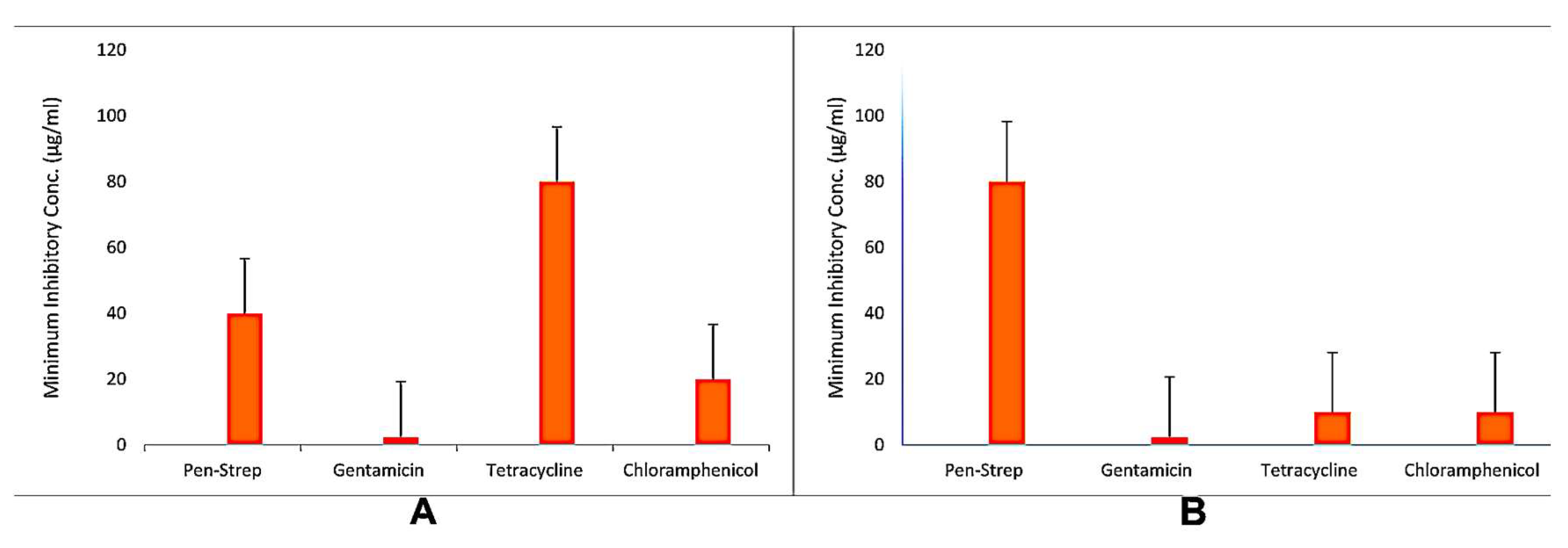
3.2. Antibiotic Resistance Profiling

Antibiotic susceptibility tests detect the vulnerability of bacteria to antibiotics and antimicrobial drugs by subjecting standardized concentrations of bacteria to specific concentrations [46]. This is critical because antibiotic-resistant bacteria can become resistant to drugs, making them incapable of being killed. As a result, we can determine the most effective agent against the bacteria causing the infection. The MIC was measured to visualize bacterial resistance to different concentrations of various antibiotics by following the guidelines of the European Committee on Antimicrobial Susceptibility Testing (EUCAST, 2023) and the European Society of Clinical Microbiology and Infectious Diseases (ESCMID, 2023) [47]. In our study, the results revealed that different antibiotics at different concentrations were remarkably resistant to Pseudomonas spp. Figure 1 shows the percentage of resistance of the isolated species. An MDR assay was performed for P. fluorescens FM3 from ground soil and P. aeruginosa DG2 from raw milk. The results indicated that both strains were multiresistant to four antimicrobial agents among three or more classes. However, phenotypic and genotypic resistance profiles may depend on the species and strains tested. Overall, a high prevalence of phenotypic resistance to ß-lactams (penicillin–streptomycin) and tetracyclines was found in P. fluorescens and P. aeruginosa, probably due to their use as veterinary therapeutic agents and promotion by specific resistance genes (intrinsic or acquired ß-lactamases for resistance to ß-lactams and “tet” genes for resistance to tetracycline under selective stress) or nonspecific multidrug efflux pumps. Concerning the other antibiotics, phenotypic resistance was detected against chloramphenicol and gentamicin for both isolates. Our findings indicate that resistance traits are not invariably phenotypic or genotypic. The existence of these genes may lead to multiple possible explanations for this discrepancy, such as the existence of “silent genes”, which are expressed only under specific circumstances [48]. The MIC values revealed significant resistance in Pseudomonas spp. against multiple antibiotics. Notably, the high levels of resistance observed in P. aeruginosa provide crucial insights into its rapid adaptation to widely used antibiotics (Figure 2). This is quite concerning in clinical settings, especially regarding infections such as skin, urinary tract, and pneumonia, while Pseudomonas spp. is frequently found as pathogens. According to earlier research, P. aeruginosa possesses innate resistance mechanisms, such as poor membrane permeability, biofilm formation, and efflux pumps, which provide it with advantages in harsh conditions where antibiotic pressure is present [49,50]. The genus showed significant resistance to several antibiotic classes, including β-lactams, aminoglycosides, and fluoroquinolones, which is consistent with the results of the MIC testing. β-lactam antibiotics, such as ceftazidime and piperacillin, which are generally effective against gram-negative bacteria, demonstrated decreased activity against the isolated strains. These findings suggest that bacteria produce β-lactamase enzymes, a well-known mechanism of resistance [22,51,52]. Pseudomonas spp. is responsible for enzymatic changes that result in resistance to aminoglycosides such as gentamicin. Similarly, changes in DNA gyrase and topoisomerase-IV can be linked to fluoroquinolone resistance. The rate of resistance (%) is shown in Figure 2, which emphasizes how urgent it is to implement strict infection control procedures and create cutting-edge treatment strategies [53].

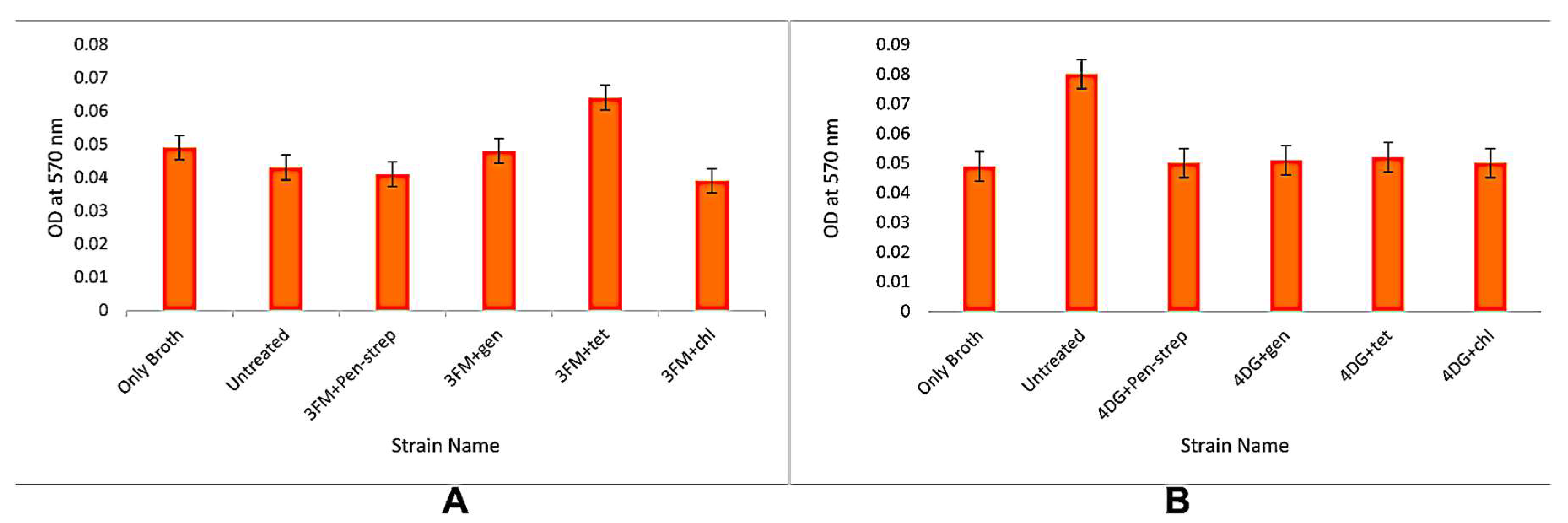
3.3. Biofilm Formation Assays

Biofilm formation in P. aeruginosa significantly contributes to its pathogenesis by reducing antibiotic penetration and enhancing bacterial persistence on surfaces. Bukholm et al. (2002) supported this statement and reported that P. aeruginosa is an opportunistic pathogen that is one of the major concerns for infections in various types of wounds, immunocompromised patients, and intensive care unit (ICU) patients [53]. The ability of P. fluorescens FM3 and P. aeruginosa DG2 to produce biofilms was examined via the use of four different antibiotics (gentamicin, tetracycline, chloramphenicol, and penicillin–streptomycin combination). Spectroscopic analysis at OD570 revealed that the untreated P. fluorescens FM3 strain was not a biofilm producer, whereas the penicillin–streptomycin combination, gentamicin, and chloramphenicol strains of all the selected P. fluorescens strains produced fewer biofilms. Among these strains, only the tetracycline-treated strains of P. fluorescens FM3 were weak biofilm producers. This may be due to nonuniform biofilm conditions, which prevent the eradication of microorganisms. A similar hypothesis was suggested by Luo et al. (2021) [54] and other researchers, who explained that the nutrient level and oxygen content simultaneously decrease at the bottom from the top; therefore, the metabolic activity and growth rate are affected. Sometimes, biofilm components and structures inhibit antibiotic penetration and may lead to the development of enzyme-mediated resistance [53,54]. Moreover, the P. aeruginosa DG2 strain was examined and found to be a weak biofilm producer against all the selected antibiotics (Figure 2). Infectivity studies have demonstrated that P. aeruginosa requires functional flagella to establish infection and colonize a host initially. The formation of biofilms is a complex process and includes various stages, such as adsorption, adhesion, microcolony formation, maturation, and dispersal [55]. In addition, AMR, ABR, and MDR can induce the expression of efflux pumps in different parts of the biofilm, increasing the frequency of mutation. During horizontal gene transfer (HGT), the close contact between extracellular DNA and cells is affected through biofilm formation, and antibiotic resistance occurs, which supports the development of new strategies against infections [56,57,58].

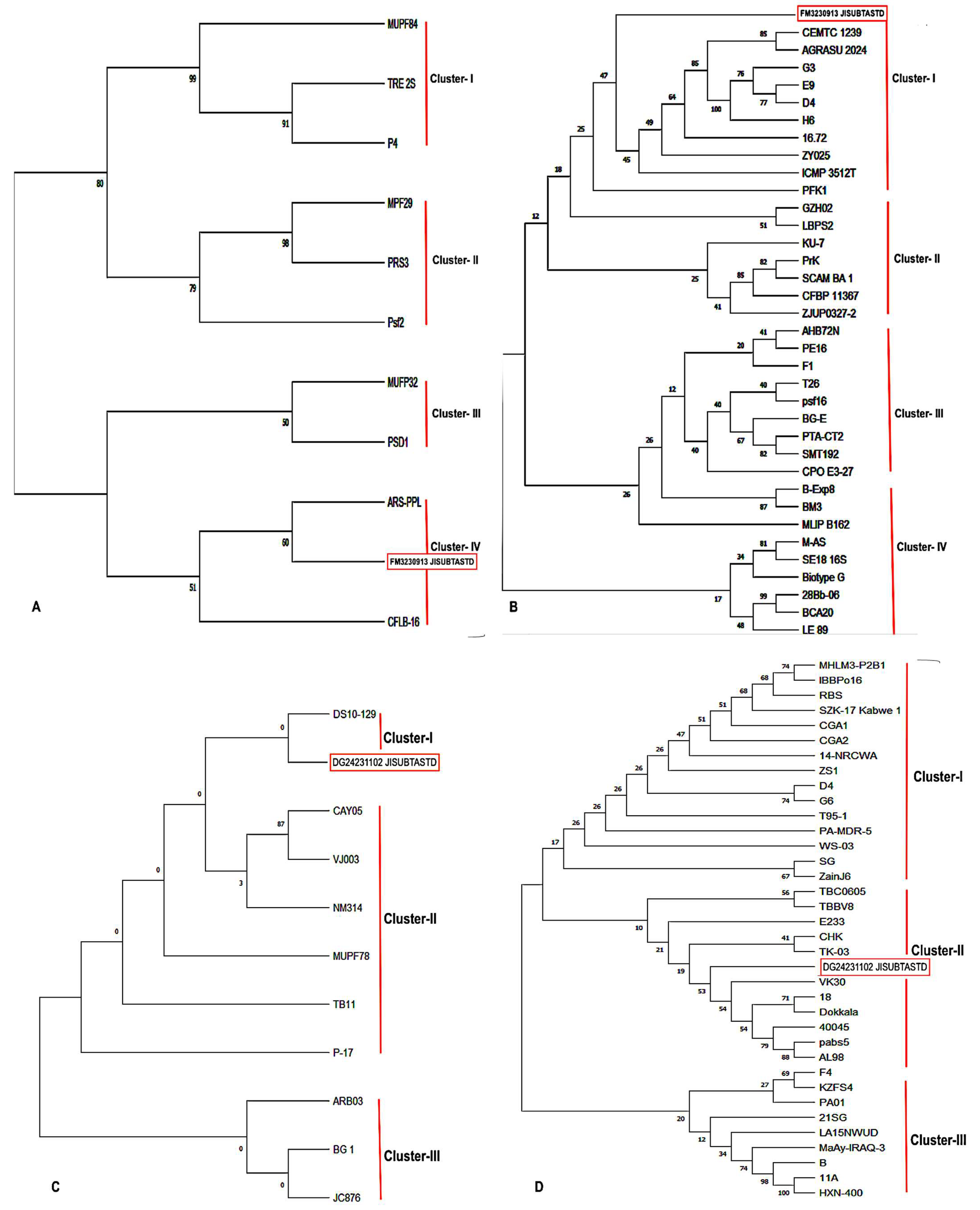
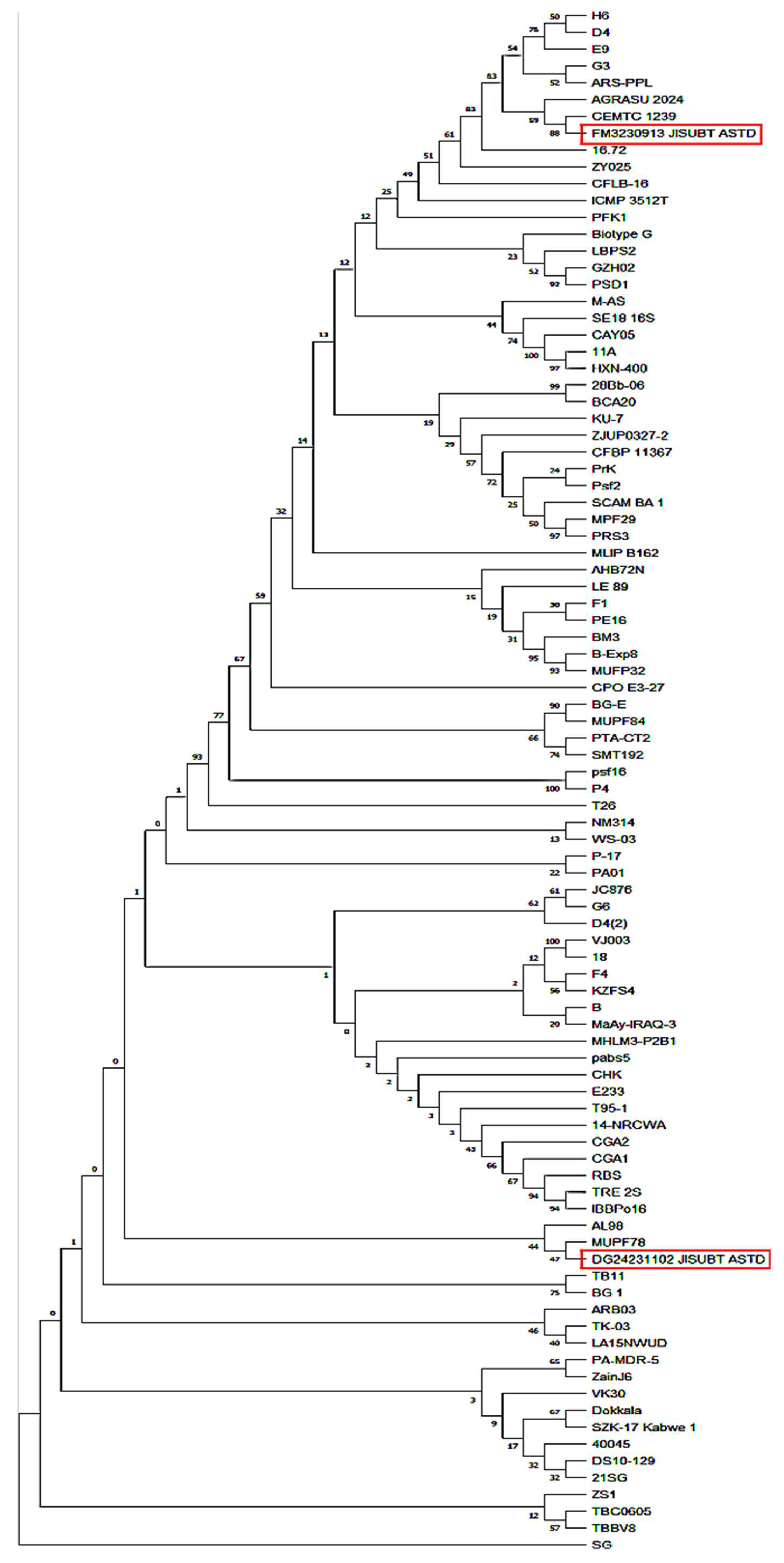
3.4. Molecular Characterization and Phylogenetic Analysis

Molecular characterization is the gold standard method for the identification of microbes over phenotypic methods [59,60]. In this study, molecular identification was performed via 16S rRNA—PCR analysis. The extracted genomic DNA from the P. fluorescens FM3 strain and P. aeruginosa DG2 strain was subjected to PCR amplification, and approximately 1.5 kb amplicons initially confirmed the presence of the 16S rRNA gene in the samples (Supplementary Figure S3). The purified amplicons were sequenced and curated for further bioinformatics analysis. A similarity search was conducted via the NCBI-BLASTn tool, and the BLAST results revealed that the FM3 strain belongs to P. fluorescens with 100% similarity, while the DG2 strain was most closely related (100%) to P. aeruginosa. After the raw sequences were curated, the length of the complementary sequences was confirmed to be 1518 bp for the P. fluorescens FM3 strain and 1528 bp for the P. aeruginosa DG2 strain. All the sequences were submitted to NCBI GenBank with accession numbers (PP406927 for P. fluorescens FM3 and PP406862 for P. aeruginosa DG2).
To assess evolutionary relationships, 90 sequences from the NCBI database were analyzed, comparing our isolated strains with other Pseudomonas spp. from both Indian and global origins (Supplementary Tables S1 and S2). Evolutionary distance was calculated through distance matrix analysis of each strain via MEGA v11.0. The phylogenetic positions of our isolates were compared via Bayesian inference analysis, and phylogenetic trees were constructed. The local position was calculated via comparisons with all sequences from Indian strains. In contrast, the global position was analyzed via comparisons with sequences retrieved from different geographical regions across the world (Supplementary Table S3). The phylogenetic analysis based on 16S rRNA gene sequences is depicted in the maximum likelihood tree presented in Figure 3. Panel (A) illustrates the local phylogenetic analysis of P. fluorescens strains of Indian origin, while panel (B) displays the global phylogenetic relationships among selected P. fluorescens strains from various regions worldwide. Similarly, panel (C) represents the phylogenetic distribution of P. aeruginosa strains of Indian origin, whereas panel (D) depicts the global phylogenetic analysis of selected P. aeruginosa strains obtained from the NCBI database. Figure 3 shows that four unique clusters generated by P. fluorescens from India and strain FM3 (PP406927) closely clustered with the phyllosphere of cauliflower, whereas ARS-PPL (OR002035) and CFLB-16 (ON764429) were isolated from unidentified sources. Further speculation on why such geographic linkages exist, possibly owing to horizontal gene transfer or environmental pressures, could benefit the discussion.
Similarly, a global comparison revealed that a total of four clusters of P. fluorescens and the FM3 strain (PP406927) presented the greatest distance from LE-89 (IN908452) from the skin of healthy brown trout in Spain and the closest relationship with CEMTC (OP602236) from a gasothermal field in Russia. (Figure 3B). As a result, P. fluorescens strains from Spain, Russia, and India are somehow linked to each other, possibly because of genetic variability after evolution [61]. Compared with local and global sequences, P. aeruginosa formed separate clusters in the phylogenetic tree, which indicates that geographical region is also an important factor for genetic variability. A total of three clusters were formed by Indian P. aeruginosa strains (Figure 4), and our strain DG2 (PP406862) was positioned in cluster I with the diesel-contaminated soil strain DS10-129 (AM419153). In Figure 4, a global comparison revealed a total of three clusters again, and our strain DG2 (PP406862) was clustered closely in clade II with the soil strain VK30 (OR647320) in Vietnam. Furthermore, the rhizosphere TK30 (OP365094) and the unidentified source HXN-400 (AJ784812) presented the strongest links within the phylogenetic tree. Additionally, MHLM3-P2B1 (LC789109) from an Indonesian mangrove forest clustered remotely. In this study, a worldwide phylogenetic study of 16S rRNA gene sequences revealed a strong Asian linkage between P. aeruginosa strains. Three distinct clusters were produced upon combining all the strains of P. fluorescens and P. aeruginosa, while SG (KJ995745) from oilfield-generated water in China presented the greatest evolutionary distance from all the other strains. For several decades, 16S rRNA sequence analysis has been used for determining bacterial species, and phylogenetic analysis for identifying taxonomic positions is considered the “gold standard” method [62]. Pseudomonas spp. has been identified and distinguished by denaturing gradient gel electrophoresis as well as RAPD analysis after selective amplification [63]. These findings are in line with recent findings that have highlighted the value of 16S rRNA sequencing in distinguishing closely related Pseudomonas spp., especially in environmental and clinical isolates. For example, P. aeruginosa is a well-known opportunistic pathogen that presents serious healthcare issues because of its resistance to antibiotics, while P. fluorescens is frequently found in soil and water. These species may be reliably identified molecularly via 16S rRNA sequences, which also allows monitoring of species occurrence in a variety of settings and the comprehension of the ecological roles and evolutionary relationships between species [61]. A phylogenetic study of the 16S rRNA genes of P. fluorescens and P. aeruginosa provides insight into the evolutionary relationships and geographic diversity of strains from various regions, with a focus on clusters from India, Spain, Russia, and Vietnam found in this study, which highlights the genetic diversity among species and suggests possible ecological and evolutionary variables affecting these associations [64,65]. In this study, we took advantage of a recent reassessment of the phylogenetic affiliation of the pseudomonads to reexamine the rapidly expanding 16S rRNA sequence data available to the public. However, our PCR and sequence analyses revealed that molecular diagnostic methods are more reliable than traditional phenotypic testing. Thus, when this set of isolates was assessed via 16S rRNA gene sequencing, the sensitivity and specificity of both PCR assays were 100%. It has also been reported that selective amplification of Pseudomonas-16S rRNA is used to detect and differentiate Pseudomonas spp. [66,67]. The results from the phylogenetic tree and evolutionary history were analyzed, and relationships were inferred via the maximum parsimony method. The bootstrap consensus tree inferred from 500 replicates was used to represent the evolutionary history of the taxa analyzed. Branches corresponding to partitions reproduced in less than 50% of the bootstrap replicates are collapsed.

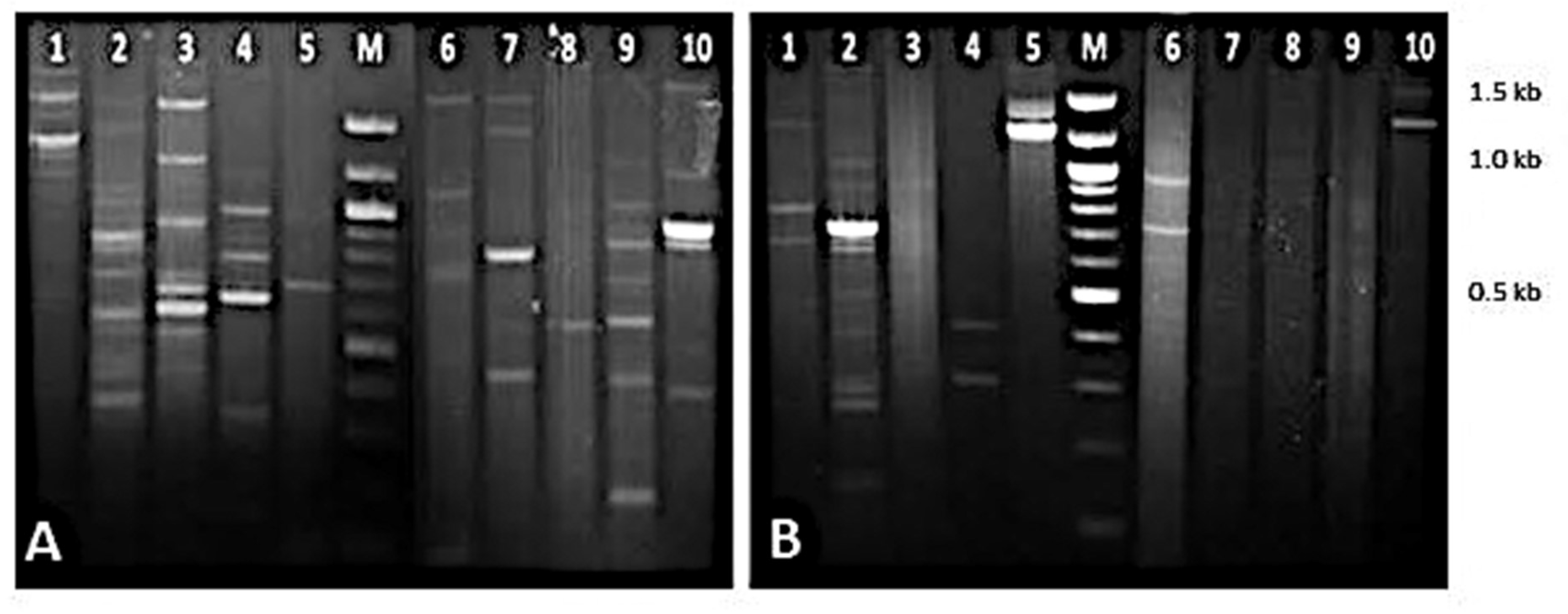
3.5. Genetic Diversity and RAPD-PCR Profiling

The genetic diversity of the antibiotic-resistant P. fluorescens FM3 and P. aeruginosa DG2 strains was also compared to determine their epidemiological and genetic distributions. Genetic polymorphisms were studied and detected via the RAPD-PCR-based conventional genotyping technique. Out of a total of 20 sets of primers (OPA1-OPA10; OPB1-OPB10), both of our strains (FM3 and DG2) produced multiple reproducible bands significantly. The genomic fingerprints of OPA-1 to OPA-10 presented few common bands in every lane (Figure 5), which may be due to the intragenic relationship and can be considered the genus and species confidence. Genomic polymorphisms, reflected in fingerprinting patterns, provide insights into evolutionary changes and mutations within the organism’s DNA [68,69]. Molecular genotyping of Pseudomonas spp. via PCR-based techniques is of foremost importance in the interpretation of routes of transmission compared with phenotyping methods and is less affected by environmental factors. Minor genetic variability can be detected by molecular methods, as compared with phenotypic assays. Hence, RAPD-PCR has proven to be one of the oldest conventional techniques for the detection of genetic polymorphisms [55,70,71].

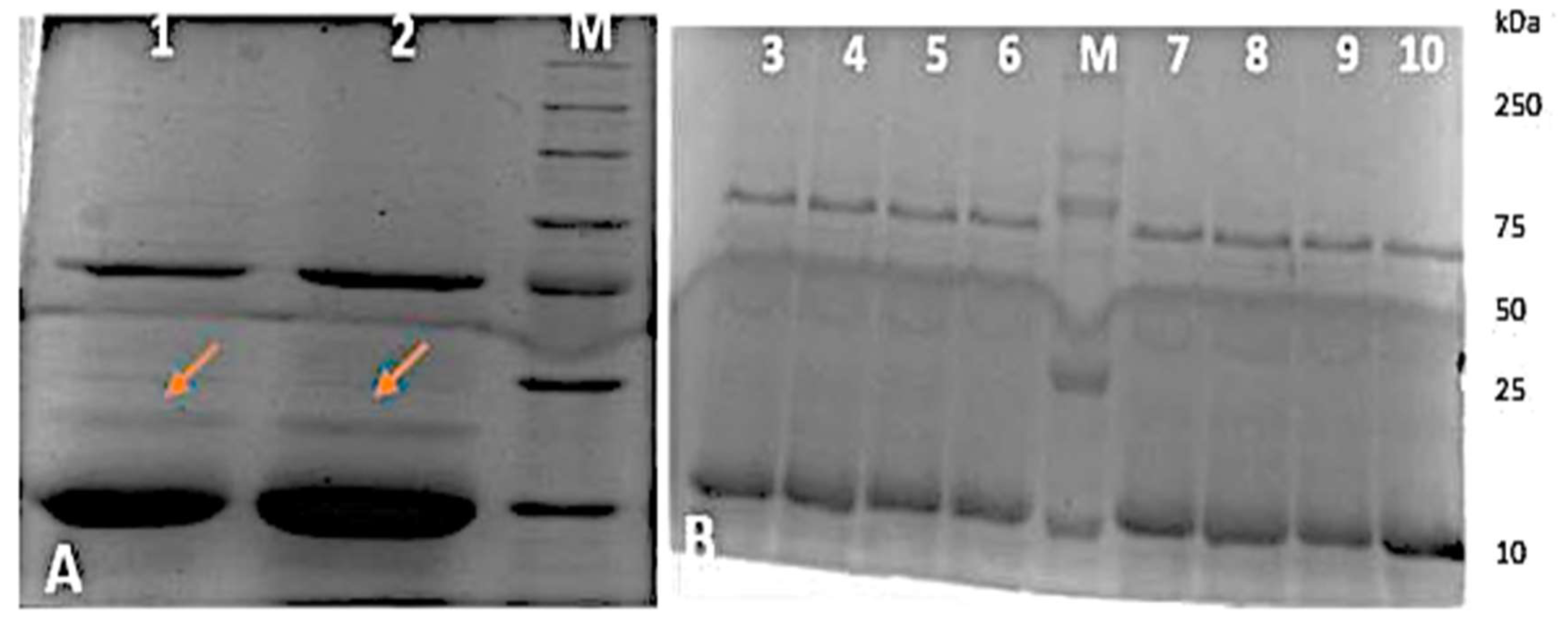
3.6. Antibiotic Resistance Protein Profiling

The present study also focused on antibiotic resistance protein profiling of Pseudomonas strains FM3 and DG2 to compare changes in typical proteins treated with antibiotics at different concentrations with those of untreated strains and bovine serum albumin (BSA) as a control. Whole-cell proteins were extracted, and 12% SDS—PAGE analysis was used to analyze both strains of Pseudomonas spp. (FM3 and DG2). The isolated samples were treated with a penicillin–streptomycin combination, gentamicin, tetracycline, or chloramphenicol. We observed that the untreated protein bands were highly intense, especially those of low-molecular-weight proteins expressed at high density. In contrast, strains treated with different concentrations of antibiotics presented almost the same intensity, but numerous low-molecular-weight protein bands disappeared in each lane, and few new banding patterns were found in the treated strains.
Additionally, the disappearance of several protein bands in treated strains suggests potential impacts of antibiotic exposure or underlying genetic mutations (Figure 6). Other studies also supported our findings, where P. aeruginosa PAR50 strains were isolated from foot ulcer patients and were resistant to a wide range of antibiotics, such as ticarcillin-clavulanic acid, ceftriaxone, piperacillin, amikacin, gentamicin, and tobramycin. Multiple banding patterns of antibiotic-resistant proteins were produced at different concentrations [72].
Bacterial metallophore-mediated metal uptake is a promising target for developing new antimicrobial therapies, as metal ions are essential for bacterial growth, metabolism, and virulence. Metallophores, low-molecular-weight chelators synthesized by bacteria, facilitate the acquisition of essential metals like iron and zinc, playing a critical role in pathogenicity. This section highlights the therapeutic potential of metallophores through various antimicrobial strategies [73].
A notable application is the Trojan horse strategy, which uses bacterial iron transport systems to deliver antibiotics into cells [74]. Sideromycins, such as albomycin and salmycin, are natural siderophore–antibiotic conjugates that enter bacteria via siderophore receptors. Once internalized, the antibiotic is released, bypassing membrane permeability barriers and directly targeting bacterial processes [75]. These conjugates generally consist of a siderophore for iron binding, a linker, and an antibiotic moiety. They enable selective uptake and enhance efficacy, especially against gram-negative bacteria. Synthetic conjugates, e.g., siderophore–ciprofloxacin or siderophore–ampicillin, have shown promising results. Challenges include potential off-target effects due to nonspecific uptake. To address this, metal analogues like gallium (Ga3+) have been used to disrupt iron metabolism and biofilm formation [76]. This strategy is particularly relevant for P. aeruginosa, which exhibits high antibiotic resistance through efflux pumps (e.g., MexAB–OprM). Under iron-limited conditions, these systems are upregulated. Siderophore–drug conjugates, especially when combined with efflux pump inhibitors, have demonstrated enhanced activity against P. aeruginosa [77,78]. Additionally, a reverse application of this concept involves using siderophores to chelate and remove excess iron from the body, such as iron released from degraded iron oxide nanoparticles, thus preventing toxicity. In summary, metallophores offer versatile potential in antimicrobial therapy, serving both as targeted delivery systems and as tools for managing metal-related toxicity.

4. Conclusions

This study provided phenotypic and genotypic insights into the identification and antibiotic resistance mechanisms of Pseudomonas spp., emphasizing their multidrug resistance (MDR) potential. The high prevalence of MDR underscores the critical need for informed antibiotic selection and robust antimicrobial stewardship programs to combat resistance and improve clinical outcomes. Longitudinal studies are essential to further understand resistance mechanisms and their impact on treatment efficacy, particularly in the developing world. Additionally, expanding sample sizes, incorporating diverse antibiotic groups, and developing cost-effective diagnostic techniques will enhance the understanding of Pseudomonas spp. in food and environmental contexts, aiding in the formulation of targeted interventions to manage infections and mitigate resistance.

Supplementary Materials

The following supporting information can be downloaded at: https://www.mdpi.com/article/10.3390/amh70030030/s1.

Author Contributions

Conceptualization, A.S., D.D., M.S. and T.D.; execution of methodology, T.D., N.D., R.M. and A.D.; investigation, T.D., N.D., R.M., A.D. and A.S.; writing—original draft preparation, T.D. and A.D.; writing—review and editing, A.S., M.S. and D.D. All authors have read and agreed to the published version of the manuscript.

Funding

This research received no external funding.

Institutional Review Board Statement

Not applicable.

Informed Consent Statement

Not applicable.

Data Availability Statement

The datasets generated and/or analyzed during the current study are available in the NCBI Sequence Read Archive (SRA) repository under the accession numbers PP406927 for P. fluorescens FM3 and PP406862 for P. aeruginosa DG2 in the submission system.

Conflicts of Interest

The authors declare that they have no competing interests.

References

  1. Magiorakos, A.P.; Srinivasan, A.; Carey, R.B.; Carmeli, Y.; Falagas, M.E.; Giske, C.G.; Harbarth, S.; Hindler, J.F.; Kahlmeter, G.; Olsson-Liljequist, B.; et al. Multidrug-resistant, extensively drug-resistant and pan drug-resistant bacteria: An international expert proposal for interim standard definitions for acquired resistance. Clin. Microbiol. Infect. 2012, 18, 268–281. [Google Scholar] [CrossRef]
  2. Urban-Chmiel, R.; Marek, A.; Stępień-Pyśniak, D.; Wieczorek, K.; Dec, M.; Nowaczek, A.; Osek, J. Antibiotic Resistance in Bacteria-A Review. Antibiotics 2022, 11, 1079. [Google Scholar] [CrossRef]
  3. Taneja, N.; Sharma, M. Antimicrobial resistance in the environment: The Indian scenario, Indian. J. Med. Res. 2019, 149, 119. [Google Scholar] [CrossRef] [PubMed]
  4. World Health Organization. Regional Office for South—East Asia. Jaipur Declaration on Antimicrobial Resistance. 2011. Available online: www.who.int/iris/handle/10665/205397 (accessed on 15 April 2017).
  5. Nelson, R.E.; Hatfield, K.M.; Wolford, H.; Samore, M.H.; Scott, R.D.; Reddy, S.C.; Olubajo, B.; Paul, P.; Jernigan, J.A.; Baggs, J. National estimates of healthcare costs associated with multidrug-resistant bacterial infections among hospitalized patients in the united states. Clin. Infect. Dis. 2021, 72, S17–S26. [Google Scholar] [CrossRef]
  6. In Proceedings of the 61st Conference on Decision and Control (CDC), Cancun, Mexico, 6–9 December 2022; IEEE: Cancun, Mexico. Available online: https://ieeexplore.ieee.org/xpl/conhome/9992315/proceeding (accessed on 20 March 2025).
  7. Division of Descriptive Research; Indian Council of Medical Research. Annual Report on Antimicrobial Resistance Research and Surveillance Network January 2023 to December 2023. AMR Surveillance Network, Indian Council of Medical Research: New Delhi, India. Available online: https://www.icmr.gov.in/icmrobject/uploads/Documents/1725536060_annual_report_2023.pdf (accessed on 20 March 2025).
  8. Gandra, S.; Tseng, K.K.; Arora, A.; Bhowmik, B.; Robinson, M.L.; Panigrahi, B.; Laxminarayan, R.; Klein, E.Y. The Mortality Burden of Multidrug-resistant Pathogens in India: A Retrospective, Observational Study. Clin. Infect. Dis. 2019, 69, 563–570. [Google Scholar] [CrossRef] [PubMed]
  9. Palleroni, N.J. Pseudomonas. In Bergey’s Manual of Systematics of Archaea and Bacteria, 1st ed.; Whitman, W.B., Ed.; Wiley: Hoboken, NJ, USA, 2015; p. 1. [Google Scholar] [CrossRef]
  10. Saha, M.; Sarkar, A. Review on multiple facets of drug resistance: A rising challenge in the 21st century. J. Xenobio. 2021, 11, 197–214. [Google Scholar] [CrossRef] [PubMed]
  11. Mancuso, G.; Midiri, A.; Gerace, E.; Biondo, C. Bacterial Antibiotic Resistance: The Most Critical Pathogens. Pathogens 2012, 10, 1310. [Google Scholar] [CrossRef]
  12. Samreen Ahmad, I.; Malak, H.A.; Abulreesh, H.H. Environmental antimicrobial resistance and its drivers: A potential threat to public health. J. Global Antimicrobial. Resistance 2021, 27, 101–111. [Google Scholar] [CrossRef]
  13. Larsson, D.G.J.; Flach, C.F. Antibiotic resistance in the environment. Nat. Rev. Microbiol. 2022, 20, 257–269. [Google Scholar] [CrossRef]
  14. Mulet, M.; Lalucat, J.; García-Valdés, E. DNA sequence-based analysis of the Pseudomonas species. Environ. Microbiol. 2010, 12, 1513–1530. [Google Scholar] [CrossRef]
  15. Scales, B.S.; Dickso, R.P.; LiPuma, J.J.; Huffnagle, G.B. Microbiology, genomics, and clinical significance of the Pseudomonas fluorescens species complex, an unappreciated colonizer of humans. Clin. Microbiol. Rev. 2014, 27, 927–948. [Google Scholar] [CrossRef] [PubMed]
  16. Raposo, A.; Ferez, E.; Faria, C.T.D.; Ferrus, M.A.; Carrascosa, C. Food spoilage by Pseudomonas spp.: An overview. In Foodborne Pathogens and Antibiotic Resistance, 1st ed.; Singh, O.V., Ed.; Wiley: Hoboken, NJ, USA, 2016; pp. 41–71. [Google Scholar] [CrossRef]
  17. Diggle, S.P.; Whiteley, M. Microbe Profile: Pseudomonas aeruginosa: Opportunistic pathogen and lab rat. Microbe Profiles collection. Microbiology 2020, 166, 30–33. [Google Scholar] [CrossRef] [PubMed]
  18. Del Olmo, A.; Calzada, J.; Nuñez, M. The blue discoloration of fresh cheeses: A worldwide defect associated with specific contamination by Pseudomonas fluorescens. Food Cont. 2018, 86, 359–366. [Google Scholar] [CrossRef]
  19. Bédard, E.; Prévost, M.; Déziel, E. Pseudomonas aeruginosa in premise plumbing of large buildings. Microbiol. Open. 2016, 5, 937–956. [Google Scholar] [CrossRef] [PubMed]
  20. Maurice, N.M.; Bedi, B.; Sadikot, R.T. Pseudomonas aeruginosa biofilms: Host response and clinical implications in lung infections. Am. J. Respp. Cell Mol. Biol. 2018, 58, 428–439. [Google Scholar] [CrossRef]
  21. Boyd, A.; Chakrabarty, A.M. Pseudomonas aeruginosa biofilms: Role of the alginate exopolysaccharide. J. Ind. Microbiol. 1995, 15, 162–168. [Google Scholar] [CrossRef]
  22. Elfadadny, A.; Ragab, R.F.; AlHarbi, M.; Badshah, F.; Ibáñez-Arancibia, E.; Farag, A.; Hendawy, A.O.; De Los Ríos-Escalante, P.R.; Aboubakr, M.; Zakai, S.A.; et al. Antimicrobial resistance of Pseudomonas aeruginosa: Navigating clinical impacts, current resistance trends, and innovations in breaking therapies. Front. Microbiol. 2024, 15, 1374466. [Google Scholar] [CrossRef]
  23. Ghssein, G.; Ezzeddine, Z. A Review of Pseudomonas aeruginosa Metallophores: Pyoverdine, Pyochelin and Pseudopaline. Biology 2022, 11, 1711. [Google Scholar] [CrossRef]
  24. Ackerley, D.F.; Caradoc-Davies, T.T.; Lamont, I.L. Substrate Specificity of the Nonribosomal Peptide Synthetase PvdD from Pseudomonas aeruginosa. J. Bacteriol. 2003, 185, 2848–2855. [Google Scholar] [CrossRef]
  25. Reimmann, C.; Serino, L.; Beyeler, M.; Haa, D. Dihydroaeruginoic acid synthetase and pyochelin synthetase, products of the pchEF, are induced by extracellular pyochelin in Pseudomonas aeruginosa. Microbiology 1998, 144, 3135–3148. [Google Scholar] [CrossRef]
  26. Laffont, C.; Brutesco, C.; Hajjar, C.; Cullia, G.; Fanelli, R.; Ouerdane, L.; Cavelier, F.; Arnoux, P. Simple rules govern the diversity of bacterial nicotianamine-like metallophores. Biochem. J. 2019, 476, 2221–2233. [Google Scholar] [CrossRef] [PubMed]
  27. Soil Science Division Staff, USDA. Soil Survey Manual. 2017. Available online: www.nrcs.usda.gov/sites/default/files/2022-09/The-Soil-Survey-Manual.pdf (accessed on 20 March 2025).
  28. Baron, E.J.; Peterson, L.R.; Finegold, S.M. Bailey and Scott’s Diagnostic Microbiology, 9th ed.; Mosby-Year Book Inc.: St. Louis, MI, USA, 1994. [Google Scholar]
  29. Arai, T.; Otake, M.; Enomoto, S.; Goto, S.; Kuwahara, S. Determination of Pseudomonas aeruginosa by Biochemical Test Methods. II. Acylamidase Test, a Modified Biochemical Test for the Identification of Pseudomonas aeruginosa. Jpn. J. Microbiol. 1970, 14, 279–284. [Google Scholar] [CrossRef]
  30. Bergey, D.H.; Holt, J.G. Bergey’s Manual of Determinative Bacteriology, 9th ed.; Williams and Wilkins: Baltimore, Maryland, 1994; Available online: https://www.biodiversitylibrary.org/page/11178148 (accessed on 12 February 2025).
  31. Hucker, G.J.; Conn, H.J. Methods of Gram Staining. J. Bact. 1923, 8, 343–348. [Google Scholar]
  32. CLSI supplement M100. Performance Standards for Antimicrobial Susceptibility Testing, 33rd ed.; Clinical and Laboratory Standards Institute (CLSI): Malvern, PA, USA, 2023. [Google Scholar]
  33. IBM SPSS Statistics 25 Step by Step: A Simple Guide and Reference, 15th ed.; George, D., Mallery, P., Eds.; Routledge: New York, NY, USA, 2018; p. 404. [Google Scholar] [CrossRef]
  34. Lefterova, M.I.; Budvytiene, I.; Sandlund, J.; Färnert, A.; Banaei, N. Simple real-time PCR and amplicon sequencing method for identification of plasmodium species in human whole blood. J. Clin. Microbiol. 2015, 53, 2251–2257. [Google Scholar] [CrossRef] [PubMed]
  35. Adikesavalu, H.; Patra, A.; Banerjee, S.; Sarkar, A.; Abraham, T.J. Phenotypic and molecular characterization and pathology of Flectobacillus roseus causing flectobacillosis in captive held carp Labeo rohita (Ham.) fingerlings. Aquaculture 2015, 439, 60–65. [Google Scholar] [CrossRef]
  36. Burks, C. DNA sequence assembly. IEEE Eng. Med. Biol. Mag. 1994, 13, 771–773. [Google Scholar] [CrossRef]
  37. McGinnis, S.; Madden, T.L. BLAST: At the core of a powerful and diverse set of sequence analysis tools. Nucleic Acids Res. 2004, 32, W20–W25. [Google Scholar] [CrossRef]
  38. Benson, D.A.; Karsch-Mizrachi, I.; Lipman, D.J.; Ostell, J.; Wheeler, D.L. GenBank. Nucleic Acids Res. 2005, 33, D34–D38. [Google Scholar] [CrossRef]
  39. Tamura, K.; Stecher, G.; Kumar, S. MEGA11: Molecular Evolutionary Genetics Analysis Version 11. Mol. Biol. Evol. 2021, 38, 3022–3027. [Google Scholar] [CrossRef]
  40. Barido-Sottani, J.; Schwery, O.; Warnock, R.C.M.; Zhang, C.; Wright, A.M. Practical guidelines for Bayesian phylogenetic inference using Markov chain Monte Carlo (Mcmc). Open Res. Europe. 2024, 3, 204. [Google Scholar] [CrossRef]
  41. Zhang, R.; Drummond, A. Improving the performance of Bayesian phylogenetic inference under relaxed clock models. BMC Evol. Biol. 2020, 20, 54. [Google Scholar] [CrossRef]
  42. Bradford, M.M. A rapid and sensitive method for the quantitation of microgram quantities of protein utilizing the principle of protein-dye binding. Analyt. Biochem. 1976, 72, 248–254. [Google Scholar] [CrossRef] [PubMed]
  43. Abdelaziz, A.A.; Kamer, A.M.A.; Al-Monofoy, K.B.; Al-Madboly, L.A. Pseudomonas aeruginosa’s greenish-blue pigment pyocyanin: Its production and biological activities. Microb. Cell Fact. 2023, 22, 110. [Google Scholar] [CrossRef]
  44. Murray, T.S.; Kazmierczak, B.I. Pseudomonas aeruginosa exhibits sliding motility in the absence of type iv pili and flagella. J. Bacteriol. 2008, 190, 2700–2708. [Google Scholar] [CrossRef]
  45. Zago, A.; Chugani, S. Pseudomonas. In Encyclopedia of Microbiology, 3rd ed.; Schaechter, M., Ed.; Academic Press: Cambridge, MA, USA, 2009; pp. 245–260. [Google Scholar] [CrossRef]
  46. Kowalska-Krochmal, B.; Dudek-Wicher, R. The minimum inhibitory concentration of antibiotics: Methods, interpretation, clinical relevance. Pathogens 2021, 10, 165. [Google Scholar] [CrossRef] [PubMed]
  47. European Committee for Antimicrobial Susceptibility Testing (EUCAST) of the European Society of Clinical Microbiology and Infectious Diseases (ESCMID). The Minimum Inhibitory Concentrations (MICs) of the Antibacterial Agents Were Determined via Broth Dilution. Eucast Discuss. Doc. E. Dis. 5.1. 2023, 9, 1–7. Available online: www.clinicalmicrobiologyandinfection.com/article/S1198-743X(14)64063-5/fulltext (accessed on 12 February 2025).
  48. Livermore, D.M. Multiple mechanisms of antimicrobial resistance in Pseudomonas aeruginosa: Our worst nightmare? Clin. Infect. Dis. 2002, 34, 634–640. [Google Scholar] [CrossRef]
  49. Strateva, T.; Yordanov, D. Pseudomonas aeruginosa- a phenomenon of bacterial resistance. J. Med. Microbiol. 2009, 58, 1133–1148. [Google Scholar] [CrossRef]
  50. Ramirez, M.S.; Tolmasky, M.E. Aminoglycoside modifying enzymes. Drug Resist. Updates 2010, 13, 151–171. [Google Scholar] [CrossRef]
  51. Duineveld, B.M.; Kowalchuk, G.A.; Keijzer, A.; van Elsas, J.D.; van Veen, J.A. Analysis of bacterial communities in the rhizosphere of chrysanthemum by denaturing gradient gel electrophoresis of PCR-amplified 16S rRNA as well as DNA fragments coding for 16S rRNA. Applied Environ. Microbiology 2001, 67, 172–178. [Google Scholar] [CrossRef]
  52. Kiyaga, S.; Kyany’a, C.; Muraya, A.W.; Smith, H.J.; Mills, E.G.; Kibet, C.; Mboowa, G.; Musila, L. Genetic Diversity, Distribution, and Genomic Characterization of Antibiotic Resistance and Virulence of Clinical Pseudomonas aeruginosa Strains in Kenya. Front. Microbiol. 2022, 14, 835403. [Google Scholar] [CrossRef] [PubMed]
  53. Bukholm, G.; Tannaes, T.; Kjelsberg, A.B.B.; Smith-Erichsen, N. An outbreak of multidrug-resistant Pseudomonas aeruginosa associated with increased risk of patient death in an intensive care unit. Infect. Cont. Hospp. Epidemiol. 2002, 23, 441–446. [Google Scholar] [CrossRef]
  54. Luo, J.; Dong, B.; Wang, K.; Cai, S.; Liu, T.; Cheng, X.; Lei, D.; Chen, Y.; Li, Y.; Cong, J.; et al. Baicalin inhibits biofilm formation, attenuates the quorum sensing-controlled virulence and enhances Pseudomonas aeruginosa clearance in a mouse peritoneal implant infection model. Plos One 2017, 12, e0176883. [Google Scholar] [CrossRef]
  55. Topa, S.H.; Subramoni, S.; Palombo, E.A.; Kingshott, P.; Rice, E.A.; Blackall, L.L. Cinnamaldehyde disrupts biofilm formation and swarming motility of Pseudomonas aeruginosa. Microbiology 2018, 164, 1087–1097. [Google Scholar] [CrossRef] [PubMed]
  56. Cendra, M.D.M.; Torrents, E. Pseudomonas aeruginosa biofilms and their partners in crime. Biotechnol. Adv. 2021, 49, 107734. [Google Scholar] [CrossRef]
  57. Franco-Duarte, R.; Černáková, L.; Kadam, S.S.; Kaushik, K.; Salehi, B.; Bevilacqua, A.; Corbo, M.R.; Antolak, H.; Dybka-Stępień, K.; Leszczewicz, M.; et al. Advances in Chemical and Biological Methods to Identify Microorganisms-From Past to Present. Microorganisms 2019, 7, 130. [Google Scholar] [CrossRef] [PubMed]
  58. Rather, M.A.; Gupta, K.; Mandal, M. Microbial biofilm: Formation, architecture, antibiotic resistance, and control strategies. Braz. J. Microbiol. 2021, 52, 1701–1718. [Google Scholar] [CrossRef]
  59. Sharma, S.; Mohler, J.; Mahajan, S.D.; Schwartz, S.A.; Bruggemann, L.; Aalinkeel, R. Microbial Biofilm: A Review on Formation, Infection, Antibiotic Resistance, Control Measures, and Innovative Treatment. Microorganisms 2023, 11, 1614. [Google Scholar] [CrossRef]
  60. Grooters, K.E.; Ku, J.C.; Richter, D.M.; Krinock, M.J.; Minor, A.; Li, P.; Kim, A.; Sawyer, R.; Li, Y. Strategies for combating antibiotic resistance in bacterial biofilms. Front. Cell. Infect. Microbiol. 2024, 14, 1352273. [Google Scholar] [CrossRef]
  61. Kamel, N.A.; Tohamy, S.T.; Yahia, I.S.; Aboshanab, K.M. Insights on the performance of phenotypic tests versus genotypic tests for the detection of carbapenemase-producing gram-negative bacilli in resource-limited settings. BMC Microbiol. 2022, 22, 248. [Google Scholar] [CrossRef]
  62. Clarridge, J.E. Impact of 16S rRNA gene sequence analysis for identification of bacteria on clinical microbiology and infectious diseases. Clin. Microbiol. Rev. 2004, 17, 840–862. [Google Scholar] [CrossRef]
  63. Woese, C.R. Bacterial evolution. Microbiol. Rev. 1987, 51, 221–271. [Google Scholar] [CrossRef]
  64. Quiroz-Morales, S.E.; García-Reyes, S.; Ponce-Soto, G.Y.; Servín-González, L.; Soberón-Chávez, G. Tracking the Origins of Pseudomonas aeruginosa Phylogroups by Diversity and Evolutionary Analysis of Important Pathogenic Marker Genes. Diversity 2022, 14, 345. [Google Scholar] [CrossRef]
  65. Falagas, M.E.; Rafailidis, P.I.; Kofteridis, D.; Virtzili, S.; Chelvatzoglou, F.C.; Papaioannou, V.; Maraki, S.; Samonis, G.; Michalopoulos, A. Risk factors for carbapenem-resistant Klebsiella pneumoniae infections: A matched case control study. J. Antimicrob. Chemother. 2007, 60, 1124–1130. [Google Scholar] [CrossRef] [PubMed]
  66. Didelot, X.; Bowden, R.; Wilson, D.J.; Peto, T.E.A.; Crook, D.W. Transforming clinical microbiology with bacterial genome sequencing. Nat. Rev. Genet. 2012, 13, 601–612. [Google Scholar] [CrossRef]
  67. Singh, A.; Goering, R.V.; Simjee, S.; Foley, S.L.; Zervos, M.J. Application of molecular techniques to the study of hospital infection. Clin. Microbiol. Rev. 2006, 19, 512–530. [Google Scholar] [CrossRef] [PubMed]
  68. Nanvazadeh, F.; Khosravi, A.D.; Zolfaghari, M.R. Genotyping of Pseudomonas aeruginosa strains isolated from burn patients by RAPD-PCR. Burns 2013, 39, 1409–1413. [Google Scholar] [CrossRef]
  69. Katsanis, S.H.; Katsanis, N. Molecular genetic testing and the future of clinical genomics. Nat. Rev. Genet. 2013, 14, 415–426. [Google Scholar] [CrossRef]
  70. Chang, Y.; Li, J.; Zhang, L. Genetic diversity and molecular diagnosis of Giardia. Infect. Genet. Evol. 2023, 113, 105482. [Google Scholar] [CrossRef]
  71. Moore, E.R.B.; Tindall, B.J.; Martins Dos Santos, V.A.P.; Pieper, D.H.; Ramos, J.L.; Palleroni, N.J. Nonmedical: Pseudomonas. In The Prokaryotes; Dworkin, M., Falkow, S., Rosenberg, E., Schleifer, K.H., Stackebrandt, E., Eds.; Springer: New York, NY, USA, 2006. [Google Scholar] [CrossRef]
  72. Al-Wrafy, F.; Brzozowska, E.; Górska, S.; Drab, M.; Strus, M.; Gamian, A. Identification and characterization of phage protein and its activity against two strains of multidrug-resistant Pseudomonas aeruginosa. Sci. Rep. 2019, 9, 13487. [Google Scholar] [CrossRef]
  73. Ezzeddine, Z.; Ghssein, G. Towards new antibiotics classes targeting bacterial metallophores. Microb. Pathog. 2023, 182, 106221. [Google Scholar] [CrossRef] [PubMed]
  74. Górska, A.; Sloderbach, A.; Marszałł, M.P. Siderophore-drug complexes: Potential medicinal applications of the ‘Trojan horse’ strategy. Trends Pharmacol. Sci. 2014, 35, 442–449. [Google Scholar] [CrossRef] [PubMed]
  75. Ghosh, A.; Ghosh, M.; Niu, C.; Malouin, F.; Moellmann, U.; Miller, M.J. Iron transport-mediated drug delivery using mixed-ligand siderophore-beta-lactam conjugates. Chem. Biol. 1996, 3, 1011–1019. [Google Scholar] [CrossRef] [PubMed]
  76. Seyedsayamdost, M.R.; Traxler, M.F.; Zheng, S.L.; Kolter, R.; Clardy, J. Structure and biosynthesis of amychelin, an unusual mixed-ligand siderophore from Amycolatopsis spp. AA4. J. Am Chem. Soc. 2011, 133, 11434–11437. [Google Scholar] [CrossRef]
  77. Liu, Y.; Yang, L.; Molin, S. Synergistic activities of an efflux pump inhibitor and iron chelators against Pseudomonas aeruginosa growth and biofilm formation. Antimicrob. Agents Chemother. 2010, 54, 3960–3973. [Google Scholar] [CrossRef]
  78. Llanes, C.; Hocquet, D.; Vogne, C.; Benali-Baitich, D.; Neuwirth, C.; Plésiat, P. Clinical strains of Pseudomonas aeruginosa overproducing MexAB-OprM and MexXY efflux pumps simultaneously. Antimicrob. Agents Chemother. 2004, 48, 1797–1802. [Google Scholar] [CrossRef]
Figure 1. Represents statistical analysis of the minimum inhibitory concentrations (MICs) of isolated strains P. aeruginosa DG2 and P. fluorescens FM3. Standard error bars showing the MICs of (A) pen–strep, chloramphenicol, tetracycline, and gentamycin against P. fluorescens FM3; (B) pen–strep, chloramphenicol, tetracycline, and gentamycin against P. aeruginosa DG2.
Amh 70 00030 g001
Figure 2. Statistical analysis of biofilm production by the isolated strains (A) P. fluorescens FM3 and (B) P. aeruginosa DG2. The standard error bar shows the biofilm production of isolates treated with various concentrations of antibiotics used for the analysis.
Amh 70 00030 g002
Figure 3. Phylogenetic tree based on the 16S rRNA gene sequences for groups of (A) P. fluorescens strains from Indian origin; (B) selected P. fluorescens strains from different regions of the world retrieved from the NCBI database; (C) Indian P. aeruginosa strains; and (D) selected groups of P. aeruginosa strains from different regions of the world retrieved from the NCBI database. The maximum parsimony (MP) tree was constructed via Bayesian inference analysis. The bootstrap values were calculated with 500 replicates.
Amh 70 00030 g003
Figure 4. Phylogenetic tree based on the 16S rRNA gene sequences for all local and global Pseudomonas strains retrieved from the NCBI database. The maximum parsimony (MP) tree was constructed via Bayesian inference analysis. The bootstrap values were calculated with 500 replicates.
Amh 70 00030 g004
Figure 5. Two per cent agarose gel image displaying the RAPD fingerprinting patterns of isolated strains of Pseudomonas spp., (A) depicting the genotypic patterns of DNA fragments amplified by sets of OPA1 to OPA10 primers. Lanes 1 to 5 illustrate the amplified RAPD fingerprints of the P. fluorescens FM3 strain, while lanes 6 to 10 show the amplified RAPD fingerprints of the P. aeruginosa DG2 strain; (B) genotypic patterns of DNA fragments amplified by sets of OPB1 to OPB10 primers. Lanes 1 to 5 illustrate the amplified RAPD fingerprints of the P. aeruginosa DG2 strain. Lane M in (A,B) represents the 50 bp DNA marker.
Amh 70 00030 g005
Figure 6. SDS—PAGE analysis of total proteins extracted from Pseudomonas isolates. (A) Lane 1: strain P. fluorescens FM3; lane 2: strain P. aeruginosa DG2; and lane 3: molecular weight protein marker. (B) Protein band patterns for antibiotic-treated samples, where lanes 3 to 6 correspond to the P. fluorescens FM3 strain treated with pen–strep, Gen, Tet, and Chl antibiotics, respectively; lanes 7 to 10 correspond to the P. aeruginosa DG2 strain treated with pen–strep, Gen, Tet, and Chl antibiotics; and lane M represents the molecular weight marker used for protein band analysis.
Amh 70 00030 g006
Table 1. Depicts characteristics of culture and morphology of P. fluorescens FM3 and P. aeruginosa DG2 strains cultured in different media. (+) denotes growth was observed; (–) denotes no growth.
PhenotypesNutrient AgarCetrimide AgarPseudomonas Agar
P. fluorescens FM3P. aeruginosa DG2P. fluorescens FM3P. aeruginosa DG2P. fluorescens FM3P. aeruginosa DG2
Growth+++++
ShapeRoundRoundRoundRoundRoundRound
Size (mm)0.3–0.50.3–0.51.0–1.20.5–0.60.8–1.0
ElevationConvexConvexConvexConvexConvex
SurfaceSmooth, shinySmooth, shinySmooth, shinySmooth, shinySmooth, shiny
MarginRegularRegularRegularRegularRegular
ColorPinkish RedGrayGreenPinkish redPinkish red
Table 2. Physiological, biochemical, and phenotypic characteristics of the isolated strains P. fluorescens FM3 and P. aeruginosa DG2. (+) denotes positive, (–) denotes negative.
Sl. No.Biochemical TestsP. fluorescens FM3P. aeruginosa DG2
1.Gram staining
2.Pigmentation++
3.Indole
4.Simmons citrate++
5.Methyl red (MR)
6.Voges–Proskauer (VP)
7.Catalase++
8.Starch hydrolysis++
9.Lipid hydrolysis++
Disclaimer/Publisher’s Note: The statements, opinions and data contained in all publications are solely those of the individual author(s) and contributor(s) and not of MDPI and/or the editor(s). MDPI and/or the editor(s) disclaim responsibility for any injury to people or property resulting from any ideas, methods, instructions or products referred to in the content.

