Review

Cerebrovascular Disease as a Manifestation of Tick-Borne Infections: A Narrative Review

by
David Doyle
1,
Samuel Kim
2,
Alexis Berry
3,
Morgan Belle
4,
Nicholas Panico
5,
Shawn Kaura
5,
Austin Price
6,
Taylor Reardon
7 and
Margaret Ellen
8,*
1
College of Medicine, Central Michigan University, Mount Pleasant, MI 48859, USA
2
School of Medicine, University of Missouri–Kansas City, Kansas City, MO 64108, USA
3
School of Medicine, Wayne State University, 540 E Canfield Street, Detroit, MI 48201, USA
4
College of Sciences, University of Central Florida, 4000 Central Florida Blvd., Orlando, FL 32816, USA
5
Lake Erie College of Osteopathic Medicine, Erie, PA 16509, USA
6
South Florida Multi-Specialty Medical Group, 8950 SW 74th Ct, Suite 1408, Miami, FL 33156, USA
7
Henry Ford Hospital, 2799 W Grand Blvd., Detroit, MI 48202, USA
8
Freeman NeuroSpine, 3401 McIntosh Circle, Suite 100, Joplin, MO 64804, USA
*
Author to whom correspondence should be addressed.
J. Vasc. Dis. 2025, 4(3), 33; https://doi.org/10.3390/jvd4030033
Submission received: 30 May 2025 / Revised: 29 July 2025 / Accepted: 17 August 2025 / Published: 21 August 2025
(This article belongs to the Topic Diagnosis and Management of Acute Ischemic Stroke)

Abstract

Background/Objectives: Tick-borne diseases (TBDs) are increasingly recognized as causes of both systemic and neurologic illness. While their impact on vascular health is established, their role in cerebrovascular disease remains underexplored. This review aims to synthesize clinical evidence linking TBDs with cerebrovascular events, focusing on mechanisms of injury, pathogen-specific associations, and treatment outcomes. Methods: A narrative review was conducted using Boolean keyword searches across PubMed, Scopus, EMBASE, and Web of Science. Relevant literature on ischemic and hemorrhagic stroke, cerebral vasculitis, and stroke mimics associated with TBDs was examined. The review included case reports, observational studies, and mechanistic research. Pathogen-specific data and disease characteristics were extracted and summarized. Results: Several tick-borne pathogens were associated with cerebrovascular complications. Borrelia burgdorferi was most commonly implicated and typically presented with large-vessel vasculitis. Rickettsia, Ehrlichia, and Anaplasma species caused endothelial injury through immune-mediated inflammation. Powassan virus and Crimean–Congo hemorrhagic fever virus exhibited central nervous system involvement and hemorrhagic potential. Babesia species contributed to vascular injury through thrombocytopenia and embolic complications. Neuroimaging frequently demonstrated multifocal stenoses and vessel wall inflammation. Antimicrobial treatment, particularly with doxycycline or ceftriaxone, was often effective, especially when administered early. Supportive care for stroke symptoms varied by presentation and underlying pathogen. Conclusions: Cerebrovascular disease caused by tick-borne pathogens is an underrecognized but potentially reversible condition. Despite diverse etiologies, most pathogens share a final common pathway of endothelial dysfunction. Early recognition and targeted antimicrobial therapy, combined with supportive stroke care, are essential to improving patient outcomes.

1. Introduction

As obligate hematophagous ectoparasites, ticks (order Ixodida) require the blood of host organisms to survive [1]. Through their interactions with the host circulatory system, ticks facilitate the transmission of numerous bacterial, viral, and parasitic pathogens, contributing to a growing global public health burden. In the United States, cases of tick-borne diseases (TBDs) have more than doubled over the past 15 years [2,3].
Ticks, and their respective pathogens, have long plagued humanity, likely first appearing in Western literature within Homer’s Odyssey (750–700 BC), noted as a mammalian parasite of Odysseus’s faithful dog [4]. The subject appears in classical naturalists, where Aristotle acknowledged their medical relevance, while Pliny the Elder described ticks as “the foulest and nastiest [of] creatures” [5]. Further historical evidence of tick-borne diseases includes the mummified remains of Ötzi, the 5300-year-old Iceman from the Italian Alps, who tested positive for Borrelia burgdorferi, along with infected mummified dogs in Egyptian tombs and depictions of tick parasitism on Egyptian funerary pottery [6,7,8]. These early references underscore the longstanding impact of ticks and TBDs on human and animal health, providing a historical backdrop to their continued relevance in contemporary medicine. Despite this enduring presence, TBDs and their system-specific consequences, such as neurological and cerebrovascular involvement, have remained underrecognized and insufficiently explored until recent advancements in modern clinical research.
In entering the host vasculature, TBDs demonstrate numerous systemic effects. Having evolved alongside their hosts, ticks are specialized in consuming the blood of mammals, birds, reptiles, and amphibians, facilitating the ecological transmission of bloodborne pathogens such as Borrelia, Rickettsia, Ehrlichia, Francisella, Powassan virus, and Babesia across numerous species and hosts [9]. These diverse organisms threaten vascular health by causing direct endothelial damage or inducing systemic changes, such as immune dysregulation. Endotheliotropic pathogens, such as Neoehrlichia mikurensis and Rickettsia species, infect endothelial cells, increasing vascular permeability, triggering oxidative stress, and contributing to the development of vasculitis [10,11]. Likewise, organisms such as Babesia spp. can induce systemic coagulopathies, including hemolysis and disseminated intravascular coagulation (DIC), thereby increasing the risk of embolic vascular events [12]. Additionally, Anaplasma and Ehrlichia species, by infecting leukocytes, may provoke exaggerated inflammatory responses, indirectly contributing to vascular damage [13,14,15,16].
The link between TBDs and vascular dysfunction is well established; however, less is known about their association with neurologic manifestations. This review compiles reported cases of human TBDs presenting as cerebrovascular disease or its mimics, with a focus on patient presentations and the underlying mechanisms of TBD-related cerebrovascular involvement.

2. Materials and Methods

A comprehensive narrative review was conducted using Boolean search strategies to identify literature linking tick-borne diseases and their causative pathogens to cerebrovascular complications, with a particular focus on ischemic stroke. Searches were performed using combinations of relevant keywords and disease terms. A complete list of the surveyed pathogens, associated diseases, and classic pathological hallmarks are presented in Table 1.
Relevant articles were identified through comprehensive searches of four major academic databases: PubMed, Clarivate Web of Science, Scopus, and EMBASE. The search strategy focused on identifying literature that included both of the following key components: mention of a tickborne illness (e.g., Lyme disease, anaplasmosis, babesiosis, ehrlichiosis, etc.) and reference to cerebrovascular disease (e.g., stroke, transient ischemic attack, cerebral vasculitis, etc.). In practice, searches were conducted with the following strategy:
(Organism OR Common Disease Title) AND (Cerebrovascular Disease)
such as
(“Borrelia burgdorferi” OR “Lyme disease”) AND (“stroke” OR “cerebrovascular accident” OR “transient ischemic attack” OR “TIA” OR “cerebral infarction” OR “brain ischemia” OR “cerebrovascular event” OR “vascular event” OR “cerebral thrombosis” OR “cerebral hemorrhage” OR “cerebral vasculitis” OR “central nervous system vasculitis” OR “neuroborreliosis” OR “neurological Lyme disease” OR “neurovascular”)
Searches were conducted between April 2025 and July 2025, initially yielding nineteen case reports of interest and ten basic science or mechanistic articles. These results were further expanded upon through subsequent searches and backward citation tracking. No restrictions were placed on publication date or article type in order to maximize inclusivity; however, emphasis was placed on identifying sources with primary examples of this relationship rather than cursory mentions of this potential interaction. Likewise, certain exclusion criteria were employed, where exclusion criteria included non-English articles, studies unrelated to human health, duplicate articles, and papers that only made incidental or speculative references to cerebrovascular effects without case data or clinical correlation.
Titles and abstracts were screened manually for relevance. Full-text reviews were conducted for articles meeting the initial criteria to ensure alignment with the scope of the review. Additional sources were identified through backward citation tracking of relevant articles.
As a narrative review, no formal quality assessment or quantitative synthesis (e.g., meta-analysis) was performed. Instead, included studies were thematically analyzed and synthesized to identify common patterns, notable findings, and gaps in the literature.

3. The Medical Ecology and Epidemiology of Ticks

Second only to mosquitos, ticks serve as a major vector for the transmission of human infectious diseases [26]. Beyond transmission, ticks also contribute to the development of TBDs through their complex biology, which provides an internal environment conducive to pathogen survival and replication prior to transmission to the host [27].
There are three families of ticks, with Ixodidae (hard ticks) and Argasidae (soft ticks) considered medically significant. There are 702 and 193 tick species described, respectively, with only 116 tick species associated with human and animal arthropod-borne infection [28]. Ticks feed briefly relative to the long intervals between blood meals [29]. These intervals are impacted by tick mobility, where ticks are passive dispersers, incapable of crawling more than a few meters [30,31]. Furthermore, tick feeding is marked by a prolonged interaction with the host, with feeding ranging from a few minutes to weeks depending on life stage and tick species [32]. As a result, changes in host contact and availability, environmental stressors, and climate can significantly impact their ability to endure these off-host periods [31].
Collectively, global conditions have supported an increase in tick populations. Alterations within the climate, environment, and human activity have led to an increase in the global prevalence and distribution of ticks over the past century [33,34,35]. Alongside this rise, there has been a corresponding increase in global public sensitivity toward tick infestations and TBDs [36]. Due to these factors, there have been several milestones in improving public education, diagnostic technologies, and increasing pathogen surveillance in light of escalating tick infestations and TBDs [2,31].
Complex variables related to human interactions with the environment, such as forest clearing, habitat fragmentation, and land desirability, directly influence the transmission of ticks to humans and the spread of latent TBDs, presenting potential avenues for host–vector mitigation [37]. These realities are compounded by host immune factors, as underlying comorbidities and illnesses can influence the transmission of TBDs from ticks to humans [38].

4. Pathogenesis of Tick-Derived Vasculopathy

Tick-borne bacterial pathogens, including Borrelia, Rickettsia, Francisella, Ehrlichia, and Anaplasma, can result in a range of systemic and neurologic complications, with a subset implicated in cerebrovascular events. Beyond bacteria, viral pathogens, such as Powassan virus and the Crimean–Congo hemorrhagic fever virus, along with parasitic organisms, such as Babesia, have been linked to cerebrovascular events. A common pathophysiologic mechanism across these infections is vascular inflammation, which may manifest as cerebral vasculitis.
Collectively, TBD vascular dysfunction arises via two main pathways: direct endothelial invasion or immune-mediated injury as graphically represented in Figure 1. In pathogens such as Borrelia burgdorferi, direct infiltration of the vascular endothelium leads to localized inflammation, intimal thickening, vessel wall edema, and subsequent disruption of cerebral blood flow, increasing the risk of thrombosis and stroke. Additionally, immune complex-mediated vasculitis has been described in subacute or post-infectious stages, with elevated circulating immune complexes causing further endothelial dysfunction.
Endothelial cells regulate vascular tone, permeability, hemostasis, angiogenesis, and immune responses. Tick-borne pathogens can directly damage endothelial cells, leading to increased permeability and vascular instability. Generalized TBD-associated endothelial dysfunction manifests in varying ways, many of which involve localization of the TBD within or near the endothelial cell. Prior studies have identified several biochemical hallmarks of endothelial damage, including reduced expression of von Willebrand factor (vWF) in endothelial cells colocalized with Borrelia burgdorferi, suggesting a potential marker of vascular impairment [39]. TBDs may also trigger immune-mediated injury via complement activation and immune complex deposition, recruiting cytotoxic immune cells and promoting cytokine release.
Notably, B. burgdorferi adheres to vascular endothelial surfaces using a mechanism strikingly similar to that employed by leukocytes during rolling interactions, with the bacterial adhesin BBK32 playing a critical role in stabilizing these interactions under shear stress as summarized in Table 2 [40,41]. This vascular adhesion strategy is essential for the early dissemination of B. burgdorferi during infection and likely contributes to its capacity for multisystem involvement. In particular, these findings may help explain the spirochete’s pathogenic potential within the cerebrovascular system, providing insight into how vascular interactions may facilitate its access to immune-privileged or protected tissues such as the central nervous system.
Additionally, Borrelia expresses a 26 kDa glycosaminoglycan-binding protein that binds to heparan sulfate on endothelial cells, and to both heparan sulfate and dermatan sulfate on neuronal cells, facilitating complex interactions between the TBD and both the vasculature and the brain [42]. Endothelial host cells actively internalize B. burgdorferi, providing a potential reservoir that shields the pathogen from immune detection and contributes to its persistence [43].
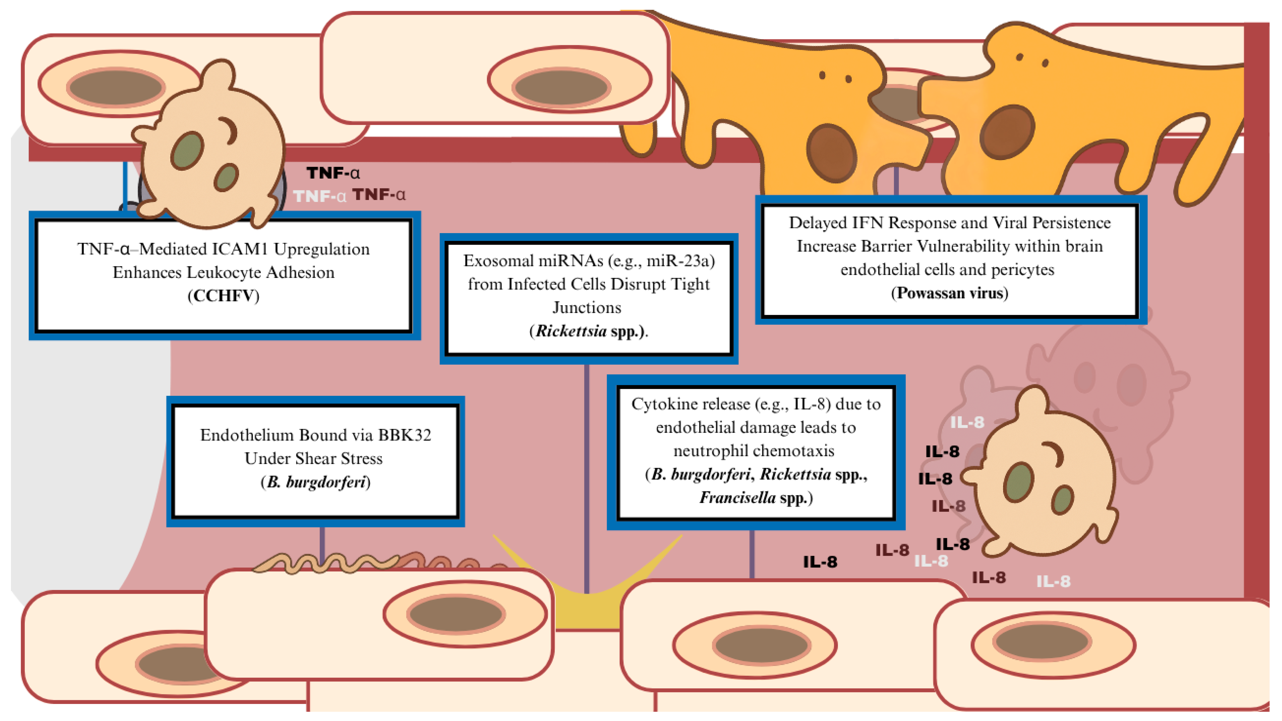
B. burgdorferi also stimulates endothelial cells to produce immune chemoattractants, which not only promotes localized inflammation but also facilitates neutrophil transmigration across the endothelium. In the context of B. burgdorferi infection, IL-8 plays a central role as the key chemotactic factor driving neutrophil recruitment. Endothelial cells rapidly produce IL-8 in response to spirochete exposure, and this response is independent of IL-1 or TNF-α signaling, indicating a direct or alternative pathway of endothelial activation by the pathogen [44]. Together, these interactions suggest that B. burgdorferi not only exploits vascular adhesion to disseminate but also actively modifies the endothelial environment to enhance immune cell recruitment, potentially disrupting barrier integrity and promoting access to protected sites such as the central nervous system
Other complex mechanisms exist between TBDs and the host endothelium. For instance, Rickettsia spp. enters host endothelial cells via Epac1-facilitated receptor-mediated invasion, involving Ku70 as a receptor and likely leveraging Epac1’s role in regulating adhesion molecules, cytoskeletal dynamics, and endocytic machinery [45]. In particular, Rickettsia employs surface proteins such as rOmpB to bind Ku70, triggering downstream signaling through c-Cbl-mediated ubiquitination, Src-family kinases, PI3-kinase, and the Arp2/3 complex to coordinate actin rearrangement and bacterial internalization via a zipper-like mechanism. This entry disrupts endothelial integrity, contributing to vascular leakage and multisystem pathologies, and underscores how targeted manipulation of host signaling pathways by Rickettsia facilitates intracellular survival and pathogenesis [46].
Moreover, in vitro studies have shown that Rickettsial infections of endothelial cells induce intercellular gap formation and altered cell morphology at later infection stages, changes that are not directly due to actin cytoskeleton disruption. Instead, these effects appear to result from a combination of endothelial injury, a reparative response, and inflammation driven by paracrine signaling, NF-κB activation, and leukocyte adhesion, highlighting the intertwined mechanisms of endothelial damage and complex host inflammatory reactions [47]. In particular, endothelial cells infected by Rickettsia rickettsii release chemoattractant cytokines such as IL-8 and MCP-1, which, similar to Borrelia burgdorferi, recruit immune cells to the site of inflammation and likely exacerbate vascular injury as graphically represented in Figure 2 [48]. Furthermore, Rickettsia spp. hijack the host’s exosomal communication system, leading to endothelial dysfunction [49]. Exosomes released from infected endothelial cells carry specific microRNAs, such as miR-23a and miR-30b, which disrupt tight junction integrity in recipient endothelial cells. This exosomal RNA-mediated mechanism contributes to increased vascular permeability and leakage, a hallmark of severe rickettsial infection.
Angiographic imaging in affected patients often reveals multifocal stenoses, segmental narrowing, or diffuse vasculopathy, particularly affecting large- and medium-sized vessels. While certain agents, such as Francisella tularensis, are less commonly associated with cerebrovascular events, their ability to trigger systemic endothelial activation and vasculitis, particularly in severe or chronic forms, suggests a plausible mechanistic overlap. Studies using the live vaccine strain (LVS) of Francisella tularensis demonstrate that both live and killed bacteria trigger proinflammatory responses in endothelial cells via atypical pathways, promoting neutrophil migration and potentially contributing to the pathogen’s high virulence [50]. Notably, IL-8 was shown to be increased, promoting neutrophil recruitment and activation, playing a key role in the early inflammatory response and highlighting a shared mechanism of pathogenesis between Francisella tularensis and other TBD infections. Moreover, in animals with tularemia, widespread necrosis has been observed, including in the brain, which further exacerbates systemic inflammation, while demonstrating the unique biochemical hallmarks of each TBD, independent of shared mechanisms [51].
The Powassan virus infects human brain endothelial cells and pericytes, key components of the blood–brain barrier. These cells act as viral reservoirs, allowing the virus to persist in the central nervous system without causing cell death. The virus avoids immune clearance by delaying interferon responses and resisting antiviral defenses, enabling continued viral spread and contributing to brain inflammation and damage [52]. Persistent glial activation, elevated M1/Th1 proinflammatory cytokine responses, and inadequate viral clearance contribute to fatal outcomes in aged mice infected with POWV, highlighting the role of host immunity in controlling viral persistence within neural and endothelial tissues [53].
The Crimean–Congo hemorrhagic fever virus directly infects and activates endothelial cells, increasing ICAM1 expression and promoting leukocyte adhesion in a dose-dependent manner. Infected endothelial cells also release proinflammatory cytokines IL-6 and IL-8, though this does not lead to paracrine activation of neighboring cells [54]. Indirectly, endothelial activation occurs via TNF-α released from infected monocyte-derived dendritic cells, as shown by ICAM1 upregulation being blocked by TNF-α neutralization. These direct and indirect effects likely contribute to the vascular leakage and inflammation seen within the virus.
Infection by TBD agents is often not confined to the endothelium, as these pathogens can also affect surrounding tissue. Rhesus monkey models of borreliosis have demonstrated perivascular inflammation with macrophage invasion during infection [55]. Although these suggestions are untested, clinical reports have shown increased inflammation in tick-borne disease exacerbating pre-existing sickle cell disease to encourage local microangiopathic thrombi formation [56]. Indeed, cerebral vasculitis is considered a focal cause of cerebral vasculopathy in neuroborreliosis [57]. These findings support a unifying vascular-centered pathogenesis in tick-borne infections with neurologic sequelae. Even so, the exact mechanism of TBD and vasculopathy is unknown. Indeed, contributions of coagulopathy seen in certain pathogens like Ehrlichia species [58] and Anaplasma species [59] have not been fully elucidated, especially in cases of co-occurring intracranial hemorrhage that are unexplained by direct endothelial damage or immune-mediated complexes.
Independent of the pathogens they carry, tick-derived toxins and salivary components disrupt host vascular function through specific molecular interactions that facilitate blood feeding. Tick saliva is inherently immunomodulatory, leading to host vasodilation and the suppression of host coagulation and inflammatory responses [38]. Following a comprehensive search among multiple relevant databases, including Scopus, PubMed, EMBASE, and Web of Science, no cases of tick-toxin-induced cerebrovascular disease were identified. This is relevant, as tick toxins are not biologically inert, as they may cause conditions such as tick paralysis [60].

5. Treatment of Cerebrovascular Disease in Tick-Borne Disease

The treatment of tick-borne bacterial diseases with cerebrovascular involvement centers on prompt antimicrobial therapy, tailored to the specific pathogen, with supportive management for associated neurologic events. For Borrelia burgdorferi infections presenting with neuroborreliosis or stroke, intravenous ceftriaxone is the preferred treatment resulting in clinical benefit. In a case report of a 74-year-old man with progressive left hemiparesis and facial palsy, treatment with IV ceftriaxone led to rapid neurologic and functional recovery [61]. This favorable response is echoed across other reports, suggesting that early recognition and antibiotic administration can mitigate vascular complications. In pediatric cases, such as a previously healthy 12-year-old boy with acute hemiparesis and imaging-confirmed multifocal cerebral vasculitis, treatment similarly focused on antibiotics, supporting the infection-driven nature of the vascular inflammation [62]. A systematic review analyzing 88 cases of Lyme neuroborreliosis with cerebrovascular events showed that most patients with imaging findings of vasculitis were treated with IV antibiotics, predominantly ceftriaxone, with variable outcomes depending on the timing of intervention [63]. Doxycycline is also a therapy of choice to address the other obligate intracellular pathogens associated with TBDs [59,64]. Nearly all cases of cerebrovascular disease secondary to TBDs treated with doxycycline resolved.
Adjunctive use of corticosteroids is not routinely recommended but may be considered in select cases with severe vasculitis or presumed immune-mediated injury. For stroke management, standard supportive measures, including antiplatelets, anticoagulation, and neurorehabilitation, may be applied in conjunction with antibiotic therapy according to current evidence in stroke management [65]. Indeed, conservative, medicinal therapies are used the majority of the time. Still, neurointerventional approaches have been found to be effective with co-antibiotherapy [66]. Other studies have suggested the use of nimodipine to reduce possible vasospasm in Lyme neuroborreliosis [67,68]. Early and appropriate antimicrobial treatment still remains the cornerstone of care and may significantly reduce neurologic morbidity.
Additionally, there have been no guidelines or investigations on stroke prevention in TBD. Indeed, many cases of TBD-derived cerebrovascular disease describe patients accessing medical therapy as a result of the event of cerebrovascular insult [56,59,69]. There is no significant evidence to date that management of comorbidities can reduce cerebrovascular disease in TBD. However, increased surveillance and precautions against contracting TBD is encouraged with appropriate population-based general prevention of stroke and intracranial hemorrhage [70,71]. Stroke due to a tick-borne disease should be considered in patients presenting with acute neurologic deficits following fever, headache, rash, or constitutional symptoms, particularly when accompanied by cytopenias, elevated liver enzymes, a history of tick exposure, or immunocompromised status. The suspicion is further heightened in the absence of conventional vascular risk factors or when imaging reveals multifocal infarcts or vasculitic patterns. Other pertinent factors include geographic locale, seasonality, cutaneous findings, and time spent outdoors.

6. Borrelia burgdorferi and Cerebrovascular Disease

Borrelia, a genus of spirochete bacteria, includes several human pathogens transmitted via ticks, most notably Borrelia burgdorferi, the causative agent of Lyme disease. While Lyme disease is classically associated with dermatologic, neurologic, and rheumatologic symptoms, emerging evidence has linked it to cerebrovascular complications, including ischemic stroke and neurovasculitis [65,72,73]. These vascular events are believed to result from a combination of direct endothelial invasion, immune-mediated injury, and inflammatory vasculitis. Case reports and observational studies have described patients presenting with large-vessel and posterior circulation strokes following active or recent Borrelia infection.
Borrelia burgdorferi has been increasingly implicated in ischemic strokes, often through a mechanism involving cerebral vasculitis. Angiographic studies and MRAs commonly reveal multifocal stenoses, arterial narrowing, or segmental obstruction of both large and small vessels [65,72,73] For instance, Almoussa and colleagues describe a case of Lyme disease-induced ischemic stroke, in which a 43-year-old previously healthy man presented with a two-week history of malaise, headache, amnestic cognitive impairment, and mild left-sided weakness [74]. He had no cardiovascular risk factors but recalled a tick bite during a trip to the Netherlands four months earlier. MRI revealed a right thalamic infarct along with other abnormal brain signals, and cerebrospinal fluid analysis confirmed active Lyme neuroborreliosis. The patient was treated with intravenous ceftriaxone for three weeks as summarized in Table 3, which led to partial recovery, although cognitive deficits persisted.
In a systematic review of 88 cases, vasculitis was identified in 52 out of 66 patients (78.8%) who underwent magnetic resonance angiography, with a predilection for large-vessel involvement [63]. The proposed pathophysiology involves two major mechanisms. First, direct bacterial invasion of the vascular endothelium by B. burgdorferi triggers localized inflammation, endothelial dysfunction, and vessel wall edema, predisposing to thrombosis. Second, in some cases, an immune complex-mediated response occurs, particularly post-infection. This is supported by reports of elevated circulating immune complexes despite no active infection, suggesting a post-infectious vasculopathy [65]. These vascular changes contribute to the clinical presentation of neuroborreliosis-associated stroke. For example, a 46-year-old male with recurrent hemiparesis was found to have multiple infarcts and widespread stenoses on imaging [62]. Similarly, in a pediatric case, a 12-year-old boy with meningeal signs had imaging findings consistent with multifocal cerebral vasculitis [62].
These findings support vasculitis as a central pathologic feature in Lyme neuroborreliosis. It should be considered in the differential diagnosis for stroke, particularly in younger patients or those living or traveling to endemic areas without clear cerebrovascular risk factors.

7. Borrelia miyamotoi and Cerebrovascular Disease

Borrelia miyamotoi is an atypical spirochete and member of the Borrelia genus [18]. Phylogenetically categorized as a relapsing fever spirochete, Borrelia miyamotoi is vectored by hard tick species such as the Ixodes species [75]. Similar to Borrelia burgdorferi, this obligate extracellular pathogen can cause significant disease, serving as the causative agent of [76] Borrelia miyamotoi disease (BMD). BMD and Borrelia miyamotoi do not have any descriptions for long-term cerebrovascular sequelae to the best of the authors’ knowledge. Although encephalopathic presentations have been noted [77,78,79,80,81,82], there are no studies on cerebrovascular complications and possible mechanisms.

8. Rickettsia Species and Cerebrovascular Disease

Rickettsia species are obligate intracellular pathogens mostly vectored by Dermacentor and Ixodes ticks that are commonly associated with Rocky Mountain Spotted Fever and related rickettsioses [83,84]. Spotted fever rickettsioses often present with a triad of fever, rash, and eschar formation. Even so, rickettsiosis is also associated with neurological symptoms, including headaches, insomnia, delirium, and encephalitis [84,85,86], and long-term sequelae, including cranial neuropathies, hemiplegias, deafness, and visual disturbances [87,88,89,90,91]. As outlined, the sequelae of Rickettsia species resemble those of cerebrovascular accidents, suggesting potential implications for future cerebrovascular health. Due to this mimicry, distinguishing between true cerebrovascular events and rickettsial mimics is essential.
Case reports have described stroke-like presentations of rickettsiosis, demonstrating the pathogen’s potential to simulate cerebrovascular events as summarized in Table 4 [64,92]. Likewise, several case reports discuss cerebrovascular disease as a rare sequela of rickettsiosis. One report by Kumar and colleagues demonstrated a 25-year-old febrile male with rickettsiosis co-presenting with right posterior cerebral, bilateral thalamopeduncular infarcts and a right posterior cerebral artery filling defect [64]. Imaging was prompted by left hemiparesis and hyperreflexia. A diagnosis of stroke secondary to cerebral vasculitis was made. Doxycycline was initiated, with treatment resulting in motor improvements [64]. An abstract by Boulahri and colleagues presented a rickettsial infection resulting in a deep left sylvian fissure ischemic stroke [93]. Another recent report by El Moussaoui et al. described nonaneurysmal subarachnoid hemorrhage secondary to encephalitis from Rickettsia rickettsii. The 23-year-old male did not present with focal neurological disease and was believed to only have meningoencephalitis before imaging. El Moussaoui et al. described resolution of intracranial pathology following doxycycline treatment as well [94]. Although not common, the correlation between Rickettsia species and vasculitis leading to cerebrovascular disease exists, with undetermined long-term effects on treated and subclinically infected patients.
Vasculitis and endothelial damage are the current paradigm for cerebrovascular risk secondary to rickettsiosis [95]. Rickettsia targets endothelial cells after bacteremia and recruits the adaptive immune system to generate an inflammatory microenvironment [96]. Once microvascular components are infected and subsequent immunity is activated, the bacteria continue to be pathogenic by increasing local coagulopathy [97] and invading surrounding non-parenchymal tissue such as neuronal or microglial cells. The subtle proinflammatory and hypercoagulable states increase the risk of cerebrovascular disease. Mouse models have shown reduced microvascular chemokine secretion with subsequent vasoconstriction as well [98].
The sequence of therapy for Rickettsia-derived acute and chronic cerebrovascular disease has not been well established, although infective therapy must include antibiotics such as doxycycline. Certainly, the effects of subclinical rickettsiosis on increasing pre-existing cerebrovascular risk have not been studied. However, vascular complications with rickettsiosis exist and improved preventive steps should be taken for patients who suffer from infection.

9. Ehrlichia chaffeensis and Cerebrovascular Disease

Ehrlichia chaffeensis is an obligate intracellular organism that is transmitted via the lone star tick [99]. In the past two decades, infections of Ehrlichia chaffeensis have increased by six-fold [100]. Infections with Ehrlichia chaffeensis and other members of the Ehrlichia family have been associated with general symptoms, including headaches, fevers, myalgias, and maculopapular rashes [101,102]. Neurological symptoms are also apparent. A retrospective study of 55 individuals with seropositive Ehrlichia species infection demonstrated that 9.1% of cases presented with neurological deficits [103]. These symptoms included seizure, dysarthria, abnormal mental status, and cranial neuropathy.
The manifestation of cerebrovascular disease in Ehrlichia species infection is not well described, but cases demonstrate this potential rare occurrence as summarized in Table 5. For example, Grant and colleagues report a case of Ehrlichia chaffeensis-induced neurologic disease in a 54-year-old man from rural Georgia who presented with fever, severe headache, meningismus, and progressive encephalopathy over the course of two weeks [104]. He had no prior cerebrovascular risk factors but reported regular tick exposure from handling his dog and working near a cow pasture. Neurologic examination revealed left arm weakness and confusion, while MRI showed a nonenhancing right frontal subcortical white matter lesion, consistent with a subacute infarction. Cerebrospinal fluid analysis demonstrated elevated protein and a significant plasmacytoid lymphocytic response. Brain and meningeal biopsies revealed perivascular and intramural lymphocytic infiltrates, implicating vasculitis-like development within the CNS. The patient was diagnosed with human monocytic ehrlichiosis due to rising antibody titers and was treated with intravenous doxycycline, resulting in gradual improvement, though residual weakness and mild confusion persisted at discharge.
Furthermore, a case report by Garc Ía-Baena and colleagues discovered a cerebral hemorrhage as an associated feature of Ehrlichia canis infection [69]. In this case, a 16-year-old male patient presented to the emergency department with new-onset seizures, mental status deterioration, and left hemiparesis. Cerebral computed tomography imaging showed right frontoparietal hemorrhage, with perilesional edema leading to mild ventricular compression without midline shift. Further work-up was performed and was negative for other explanations of idiopathic hemorrhage. CSF PCR confirmed the diagnosis. The team reported complete neurological recovery post-surgical drainage and doxycycline therapy [69]. There have been few other clear reports of cerebrovascular events as markers for Ehrlichia infection. Interestingly, canine models of Ehrlichia canis infection demonstrate clear thrombocytopenia [74] which contribute to the hemorrhagic nature of the disease. Other experimental studies and case reports point to Ehrlichia chaffeensis infection with neurological presentation as a centrally immunologically driven pathogenicity [105]. Indeed, ehrlichiosis is associated with lymphoid invasion of microvascular structures [105]. However, there are limited data on the subject, which necessitates further investigations in this area.

10. Anaplasma phagocytophilum and Cerebrovascular Disease

Anaplasma phagocytophilum is an obligate intracellular Gram-negative bacterium that is transmitted by the Ixodes scapularis in the northeast United States and by Ixodes pacificus in California [106]. Anaplasma phagocytophilum is the central cause of human granulocytic anaplasmosis (HGA) [106]. Although HGA often presents with general symptoms, 1% of cases present with focal neurological deficits, including brachial and cranial neuropathies [59,107,108,109]. Although rare, HGA has been linked to cerebrovascular disease, likely due to a combination of direct endothelial involvement and immune-mediated vascular injury. Anaplasma phagocytophilum primarily infects neutrophils but may also disrupt endothelial function, promoting a prothrombotic state, where brain imaging in such cases often demonstrates an embolic or vasculitic process as described in Table 6.
For example, Guru and colleagues describe a rare neurological manifestation of HGA in a 65-year-old woman with a history of a cerebrovascular accident who presented with gastrointestinal symptoms and altered mental status during the summer in Pennsylvania [110]. Initial work-up revealed pancytopenia, elevated liver enzymes, and worsening renal function, prompting hemodialysis and intubation. Despite broad-spectrum antibiotics, her condition deteriorated, and brain imaging revealed multiple acute infarcts, most notably a 3 cm lesion in the posterior right corona radiata, consistent with embolic stroke. Peripheral smear demonstrated morulae, and the diagnosis of HGA was confirmed by PCR for Anaplasma phagocytophilum. CSF analysis showed mild lymphocytic pleocytosis with normal glucose and protein, consistent with prior reports of anaplasmosis-related meningitis. Following initiation of doxycycline, the patient’s mental status improved and fevers resolved. The authors note that this represents only the third reported case of stroke associated with HGA, and propose that endothelial involvement or a vasculitic mechanism may underlie cerebrovascular complications in this infection. Given the increasing incidence of anaplasmosis and limitations in diagnostic timing, the authors emphasize heightened clinical suspicion for stroke presentations in patients from endemic areas during tick season.
Moreover, a case report identified by Kim and colleagues describes infarction of the basal ganglia with co-occurring thrombocytopenia due to Anaplasma phagocytophilum infection. The 70-year-old female presented to the emergency department three days post-tick bite with fever and disequilibrium. The patient presented at admission with thrombocytopenia and elevated D-dimer. Magnetic resonance imaging (MRI) confirmed lacunar infarction without changes in vascular permeability on angiography. Interestingly, co-therapeutic treatment with doxycycline was found to be effective in symptom resolution [59]. The exact mechanism of action is unclear, although dose-dependent platelet dysfunction was noted in mouse models [111]. The formation of thrombi within the hypocoagulable state with systemic inflammation is likely to increase the risk of cerebrovascular accidents. In fact, a case report by Herbst and colleagues demonstrated stroke induced by anaplasmosis-driven inflammation and sickle cell disease [56]. The 26-year-old patient presented to the emergency department recurrently for the past two weeks for shortness of breath and diffuse pain. On admission, she had acute respiratory distress syndrome, acute renal failure, and bone marrow failure, with admission MRI demonstrating diffuse scattered areas of restricted diffusion throughout the cerebellum and bilateral cortical gray matter. Anaplasmosis and co-occurring babesiosis was confirmed via peripheral blood smear and bone marrow biopsy, respectively. Despite efforts for resuscitation, the patient ultimately continued to deteriorate and was discharged to a skilled nursing facility. Consequently, the authors encourage doxycycline initiation at suspicion on high-risk patients, such as with sickle cell disease.

11. Francisella tularensis and Cerebrovascular Disease

Francisella tularensis is an intracellular, Gram-negative facultative bacteria vectored by several tick species [112]. Infection with Francisella tularensis often presents with general symptoms, with 20% presenting with dermatological findings [113]. Neurological presentations of Francisella tularensis are rare. One case report by Coban and colleagues describes co-occurring pontine ischemia and demyelinating disease secondary to Francisella tularensis infection in an endemic area [113]. As summarized in Table 7, the 42-year-old female presented to the emergency department with internuclear ophthalmoplegia and left hemiparesis. MRI confirmed the diagnosis of pontine infarction with co-occurring hyperintense non-contrast-enhancing lesions in bilateral periventricular, peritrigonal, and pericallosal regions, centrum semiovale, and corona radiata. The patient recovered but was found to have lymphadenopathy with necrotizing granulomas at follow-up. Francisella tularensis microagglutination test was positive. She was started on streptomycin and doxycycline. Interestingly, this case report did not demonstrate complete resolution with doxycycline therapy, although there was some regression of lesions on MRI. The exact mechanism for tularemic encephalitis and vasculitis is unknown but is likely attributed to the toxic effects of lipopolysaccharides [114]. Indeed, immunological dysregulation is the likely culprit for ischemic disease secondary to tularemiosis.

12. Powassan Virus and Cerebrovascular Disease

The Powassan virus (POWV) is a tick-borne flavivirus endemic to Russia and North America [23]. The Powassan virus has likely emerged in the northeastern United States following the mid-20th century resurgence of Ixodes scapularis ticks, driven by reforestation and the rebound of white-tailed deer populations. These conditions reversed earlier declines in tick numbers caused by 19th century deforestation and reduced deer density [115].
POWV results in a clinical spectrum ranging from asymptomatic infection to severe neurological disease, including focal neurologic deficits, cerebral edema, meningitis, and encephalitis. Although rare, POWV has been associated with cerebrovascular complications, particularly ischemic stroke. Much of this evidence centers on a widely cited case from New York involving a patient with a history of right putamen infarct, hypertension, hepatitis C, and substance abuse [116]. He presented with altered mental status, left facial droop, and dysarthria following multiple tick bites; his hospital course was complicated by recurrent strokes, and he was ultimately discharged with global aphasia as a residual deficit as seen in Table 8.
POWV exhibits a tropism for central nervous system cells, which may underlie its association with cerebrovascular disease. As noted, the virus is able to infect cells relevant to the BBB, resulting in residual viral particles within the brain. For instance, in animal models, infection results in perivascular mononuclear cell infiltration and microglial activation, along with a poliomyelitis-like syndrome marked by high levels of POWV antigen in the ventral horn of the spinal cord [117]. These findings further support the ability of the virus to accumulate in cerebrovascular regions of the brain and spinal cord, locally activating the host immune response.

13. Crimean–Congo Hemorrhagic Fever Virus and Cerebrovascular Disease

The Crimean–Congo hemorrhagic fever virus (CCHFV), a member of the Nairoviridae family within the Bunyavirales order, is a widespread pathogen that causes hemorrhagic fever across Africa, Southern and Eastern Europe, the Middle East, India, and Asia [118]. CCHFV is often vectored by ticks of the Hyalomma genus and ranges in clinical severity from mild to lethal. CCHFV is commonly associated with diffuse hemorrhaging, with alterations in hematological stability and reported cases of cranial implications as seen in Table 9. A report by Kleib and colleagues describes a subdural hematoma secondary to CCHFV infection. A 58-year-old male patient presented with fever and epistaxis. The patient was treated for malaria until real-time PCR and CCHF virus-specific IgM by ELISA confirmed CCHFV infection. The patient was treated with platelet transfusions and supportive therapy until the patient presented with obtunded mental status and a Glasgow Coma Scale of 13. The patient had confirmed subdural hematoma with midline shift on CT imaging. The patient was treated with corticosteroids and saline, allowing for eventual subdural hematoma resolution and viral recovery [119]. A case report by Ulusoy discusses a 60-year-old male presenting with fever, abdominal pain, nausea, vomiting, and disequilibrium. The patient had an ischemic stroke of the cerebral peduncle, confirmed by MRI. As CCHFV was considered and the patient presented with thrombocytopenia, only supportive measures were given without antiplatelet therapy. The patient was able to recover neurological function after 10 days and was prescribed an antiaggregant at discharge [120].
The exact pathogenesis of cerebral vasculopathy in CCHFV remains unclear. Studies have shown that the blood–brain barrier and neurons are disrupted by increasing excitatory amino acids in CCHFV. Indeed, uncontrolled release of cytokines can cause intravascular coagulation [121,122]. In the hemorrhagic stage of CCHFV, thrombocytopenia is notable, with changes in prothrombin and partial thromboplastin time, which could encourage intracranial hemorrhaging [123].

14. Babesia Species as Parasitic Tick-Borne Pathogens Implicated in Cerebrovascular Disease

Babesia species are intraerythrocytic protozoa vectored by the ixodid tick that can cause mild to severe multiorgan disease [124]. Serious infection has been linked to cardiopulmonary failure and hematological disease [125]. Little is known, however, about the intracranial implications of babesiosis. Nonetheless, a retrospective review by Locke and colleagues found that 59.5% of cases presented with a neurological deficit. Two of the one hundred sixty-three cases presented with cerebrovascular disease, including both stroke and SAH [124]. Another report by Herbst and colleagues presented with diffuse ischemic stroke and multiorgan failure secondary to co-infection of Anaplasma species and Babesia species in a patient with sickle cell disease [56]. However, the exact mechanism for rare stroke and hemorrhage from Babesia species has not been elucidated. Indeed, the furthest evidence connects babesiosis-induced cerebrovascular disease and developing theories of cerebral malaria [56].

15. Diagnostic and Therapeutic Decision Making

The clinical algorithm for diagnosing, prognosticating, and providing therapy for tick-borne-disease-caused cerebrovascular events have not been well-described in the literature. Indeed, the nebulous, heterogenous, and rare nature of TBD-caused cerebrovascular disease (CVD) prohibits any high-level evidence for TBD-caused CVD. Even so, much like any sophisticated infectious process, the authors agree that the diagnosis and resulting therapy of TBD-caused CVD begins with a detailed patient history and additive convincing physical exam. Historical details, such as recent exposure to the outdoors, work experience with animals [11,26,104,105], or trips out of the country of origin or to other endemic areas [37,58,74], are considered. Additionally, signs of cerebrovascular injury without significant past medical history, and historical details, including pre-incident coryza, rash, or tick bite, should lower the threshold for infectious process work-up. Indeed, the majority of cases presented with co-occurring symptoms of neurological disease, gastrointestinal processes, and general symptoms such as malaise.
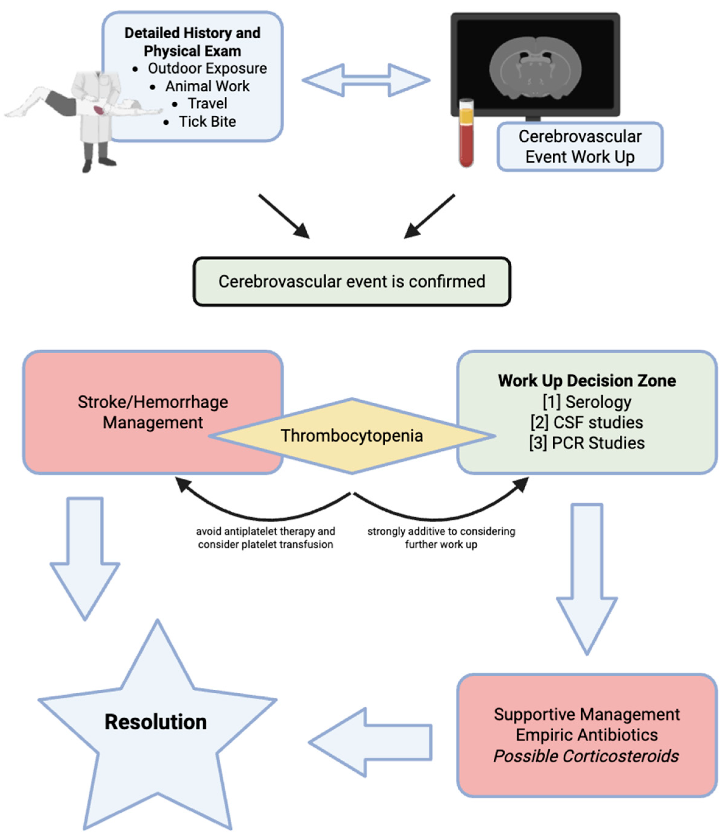
After a thorough history and physical exam is conducted, neuroimaging should occur before any further work-up is warranted as portrayed in Figure 3. Neuroimaging was the primary clinical tool to confirm CVD across cases and the initial step after all history-taking in every case. The debate between CT alone versus MRI alone versus CT then MRI remains unclear [126]; however, MRI was used in the majority of cases and has been shown to be more effective at detecting intracerebral hemorrhage [127] than CT alone. As such, resource stewardship and neurological exam findings should be considered before pursuing neuroimaging options with a lower threshold for MRI alone if imaging is urgent. If imaging confirms cerebrovascular injury, supportive care should be performed immediately, focusing on neurological and hemodynamic stability before any rigorous work-up. Conducting basic serum/blood laboratory tests can help with managing and diagnosing TBD-caused CVD. A complete blood count (CBC), complete metabolic panel (CMP), and other components of a basic stroke panel should be obtained alongside neuroimaging. Indeed, thrombocytopenia detected on the CBC can warrant more aggressive investigation for other etiologies for CVD. Immediate stroke care should be administered after neuroimaging; antithrombotic agents, however, should be held in the case of severe thrombocytopenia, and platelet transfusion should be considered instead [104,105,109]. Moreover, if intracerebral hemorrhage is present, surgical drainage is warrant per the discretion of neurosurgery and the nature of the causative agent behind the TBD.
The clinical picture of combined elements from the patient history, physical exam, neuroimaging results, and CBC/CMP should warrant further work-up if the clinical picture supports the possibility of an infectious agent. Elements that would largely lower the threshold for serology, cerebrospinal fluid (CSF), or PCR studies for further work-up include recent tick bite, local endemic disease, thrombocytopenia, or CVD in a young patient with co-occurring infectious signs and symptoms (rash, fever, or gastrointestinal upset). Moreover, if a patient recedes or does not improve following stroke/hemorrhage management, and autoimmune processes are excluded, infectious etiologies should be considered. Empiric doxycycline with or without ceftriaxone, depending on endemic TBDs, should be given while serological, CSF, and PCR studies are pending. If the etiological agent is considered or confirmed as viral, supportive management is critical. The use of high-dose corticosteroids for the management of TBD-caused CVD is not supported by any evidence, including the rationale for improving encephalitis [128]. The use of corticosteroids depends solely on the clinical provider and the possibility of autoimmune processes.

16. Limitations

This review employed a structured and transparent narrative approach. As with all narrative reviews, the potential for selection bias exists; however, this was mitigated through the use of predefined databases, targeted search terms, and explicit inclusion and exclusion criteria. While no formal risk-of-bias tool was applied, study quality and relevance were assessed collaboratively, with emphasis on clinical detail and diagnostic rigor. Conflicting findings were reviewed by the author team and interpreted within the context of the most current and robust evidence.
Given the qualitative nature of the synthesis, conclusions are intended as thematic insights rather than quantitative generalizations. Although the review did not follow systematic review protocols such as PRISMA, the methodology was intentionally designed to support a broad and integrative exploration of the topic. Despite these inherent limitations, the review offers a focused synthesis of emerging evidence linking tick-borne diseases and cerebrovascular pathology, and highlights key areas for future investigation.

17. Conclusions

In conclusion, tick-borne cerebrovascular disease presents with a remarkably heterogeneous clinical spectrum that can cause a wide array of cerebrovascular diseases. Bacterial spirochetes, like Borrelia burgdorferi and Rickettsia species, target and invade the endothelium directly, precipitating focal vessel wall inflammation, intimal proliferation, and segmental stenoses, whereas obligate intracellular bacteria such as Ehrlichia and Anaplasma invoke predominantly immune-mediated endothelial injury with circulating immune complexes and cytokine-driven vasculitis. Viral agents exhibit neurotropism and perivascular mononuclear infiltrates, resulting in both ischemic and hemorrhagic events, while protozoa, like Babesia species, contribute via hemolysis, coagulopathy, and microvascular sludging. Clinically, patients may present without traditional stroke risk factors, often with prodromal fever, headache, or neurological prodromes that predate ischemic symptoms by days to weeks. High-resolution vessel wall imaging and angiography frequently reveal multifocal stenoses or vessel wall enhancement, underscoring the value of advanced neuroimaging in diagnosis. Therapeutic insights emphasize the primacy of targeted antimicrobial therapy, intravenous ceftriaxone for neuroborreliosis, doxycycline for rickettsial and intracellular bacterial infections, and pathogen-specific antivirals or supportive care for viral encephalitides. Early empiric treatment in endemic settings often leads to rapid clinical and radiological improvement, highlighting the reversibility of infection-induced vascular lesions when diagnosed promptly. Supportive stroke care, including neurorehabilitation and secondary prevention with antithrombotics, must be tailored to each pathogen’s coagulopathic profile; for example, avoiding aggressive anticoagulation in Ehrlichia-associated thrombocytopenia or aggressive neurointervention in neuroborreliosis. These insights reinforce that, although tick-borne pathogens leverage diverse molecular mechanisms, they ultimately converge on an endothelial-driven pathway of vascular injury that is largely reversible with prompt, targeted antimicrobial and tailored neurovascular care. Moving forward, integrating rapid diagnostics, early pathogen-directed therapy, and coordinated cerebrovascular management will be essential to minimize long-term neurologic sequelae in these patients.
Future research should prioritize prospective studies to determine the anticipated incidence of tick-borne cerebrovascular events in endemic regions. Standardized diagnostic criteria, including consistent use of clinical, serologic, and imaging markers, are needed to differentiate infectious vasculitis from non-infectious etiologies. Comparative studies should further elucidate overlapping mechanisms of endothelial dysfunction, such as cytokine-driven inflammation, immune complex deposition, and direct endothelial invasion, observed across Borrelia, Rickettsia, Ehrlichia, and other agents. Furthermore, population-based and epidemiological studies can help identify related variables, including seasonality, geographic distribution, and host behaviors, such as hiking or camping, which may influence exposure risk. Clarifying these shared pathways and risk factors may support the development of cross-pathogen therapeutic approaches and effective public health interventions.

Author Contributions

Conceptualization, D.D., M.E., T.R., A.P., M.B., N.P. and S.K. (Samuel Kim); methodology, M.E., D.D. and S.K. (Samuel Kim); software, D.D.; validation, D.D.; formal analysis, M.B., A.B. and S.K. (Samuel Kim); investigation, D.D., S.K. (Samuel Kim), S.K. (Shawn Kaura) and A.B.; resources, D.D.; data curation, A.B.; writing—original draft preparation, D.D., M.B., S.K. (Samuel Kim) and A.B.; writing—review and editing, N.P., S.K. (Samuel Kim), S.K. (Shawn Kaura) and M.B.; visualization, D.D.; supervision, M.E., A.P. and T.R.; project administration, A.B., D.D., M.E. and T.R. All authors have read and agreed to the published version of the manuscript.

Funding

This research received no external funding.

Institutional Review Board Statement

Not applicable.

Informed Consent Statement

Not applicable.

Data Availability Statement

No new data were created or analyzed in this study. Data sharing is not applicable to this article.

Conflicts of Interest

The authors declare no conflicts of interest.

Abbreviations

The following abbreviations are used in this manuscript:
TBDTick-Borne Disease
CSFCerebrospinal Fluid
RMSFRocky Mountain Spotted Fever
HMEHuman Monocytic Ehrlichiosis
HGAHuman Granulocytic Anaplasmosis
POWVPowassan Virus
LVSLive Vaccine Strain
CCHFVCrimean–Congo Hemorrhagic Fever Virus
BMDBorrelia miyamotoi Disease
ELISAEnzyme-Linked Immunosorbent Assay
MRIMagnetic Resonance Imaging
MRAMagnetic Resonance Angiography
CTComputed Tomography
DICDisseminated Intravascular Coagulation
SAHSubarachnoid Hemorrhage
TIATransient Ischemic Attack

References

  1. Merino, O.; Alberdi, P.; Pérez de la Lastra, J.M.; de la Fuente, J. Tick vaccines and the control of tick-borne pathogens. Front. Cell. Infect. Microbiol. 2013, 3, 30. [Google Scholar] [CrossRef] [PubMed]
  2. Smith, R.P.; McCarthy, C.A.; Elias, S.P. Increasing actual and perceived burden of tick-borne disease in Maine. J. Maine Med. Cent. 2019, 1, 13. [Google Scholar] [CrossRef]
  3. Rosenberg, R.; Lindsey, N.P.; Fischer, M.; Gregory, C.J.; Hinckley, A.F.; Mead, P.S.; Paz-Bailey, G.; Waterman, S.H.; Drexler, N.A.; Kersh, G.J.; et al. Vital Signs: Trends in Reported Vectorborne Disease Cases—United States and Territories, 2004–2016. MMWR Morb. Mortal. Wkly. Rep. 2018, 67, 496–501. [Google Scholar] [CrossRef] [PubMed]
  4. Zaborowski, R.; Daszkiewicz, P. About the Greek origin of acarology: A short note on Argas and the Acari. Biol. Lett. 2016, 53, 3–7. [Google Scholar] [CrossRef]
  5. Dhooria, M.S. Historical Account of Acarology. In Fundamentals of Applied Acarology; Springer: Singapore, 2016. [Google Scholar] [CrossRef]
  6. Kean, W.F.; Tocchio, S.; Kean, M.; Rainsford, K.D. The musculoskeletal abnormalities of the Similaun Iceman (“ÖTZI”): Clues to chronic pain and possible treatments. Inflammopharmacology 2013, 21, 11–20. [Google Scholar] [CrossRef]
  7. Huchet, J.B.; Callou, C.; Lichtenberg, R.; Dunand, F. The dog mummy, the ticks and the louse fly: Archaeological report of severe ectoparasitosis in Ancient Egypt. Int. J. Paleopathol. 2013, 3, 165–175. [Google Scholar] [CrossRef]
  8. Arthur, D. Ticks in Egypt in 1500 B.C.? Nature 1965, 206, 1060–1061. [Google Scholar] [CrossRef]
  9. Anderson, J.F. The natural history of ticks. Med. Clin. N. Am. 2002, 86, 205–218. [Google Scholar] [CrossRef]
  10. Walker, D.; Ismail, N. Emerging and re-emerging rickettsioses: Endothelial cell infection and early disease events. Nat. Rev. Microbiol. 2008, 6, 375–386. [Google Scholar] [CrossRef]
  11. Margini, C.; Maldonado, R.; Keller, P.; Banz, Y.; Escher, R.; Waldegg, G. Fever of Unknown Origin, a Vascular Event, and Immunosuppression in Tick-Endemic Areas: Think About Neoehrlichiosis. Cureus 2023, 15, e40617. [Google Scholar] [CrossRef]
  12. Vannier, E.G.; Diuk-Wasser, M.A.; Ben Mamoun, C.; Krause, P.J. Babesiosis. Infect. Dis. Clin. N. Am. 2015, 29, 357–370. [Google Scholar] [CrossRef] [PubMed]
  13. Ismail, N.; Bloch, K.C.; McBride, J.W. Human ehrlichiosis and anaplasmosis. Clin. Lab. Med. 2010, 30, 261–292. [Google Scholar] [CrossRef] [PubMed]
  14. Underwood, J.; Harvey, C.; Lohstroh, E.; Pierce, B.; Chambers, C.; Guzman Valencia, S.; Oliva Chávez, A.S. Anaplasma phagocytophilum Transmission Activates Immune Pathways While Repressing Wound Healing in the Skin. Life 2022, 12, 1965. [Google Scholar] [CrossRef] [PubMed]
  15. Miura KMatsuo, J.; Rahman, M.A.; Kumagai, Y.; Li, X.; Rikihisa, Y. Ehrlichia chaffeensis Induces Monocyte Inflammatory Responses through MyD88, ERK, and NF-κB but Not through TRIF, Interleukin-1 Receptor 1 (IL-1R1)/IL-18R1, or Toll-Like Receptors. Infect. Immun. 2011, 79, 4947–4956. [Google Scholar] [CrossRef]
  16. Tominello, T.R.; Oliveira, E.R.A.; Hussain, S.S.; Elfert, A.; Wells, J.; Golden, B.; Ismail, N. Emerging Roles of Autophagy and Inflammasome in Ehrlichiosis. Front. Immunol. 2019, 10, 1011. [Google Scholar] [CrossRef]
  17. Steere, A.C.; Strle, F.; Wormser, G.P.; Hu, L.T.; Branda, J.A.; Hovius, J.W.; Li, X.; Mead, P.S. Lyme borreliosis. Nat. Rev. Dis. Primers 2016, 2, 16090. [Google Scholar] [CrossRef]
  18. Cleveland, D.W.; Anderson, C.C.; Brissette, C.A. Borrelia miyamotoi: A Comprehensive Review. Pathogens 2023, 12, 267. [Google Scholar] [CrossRef]
  19. Blanton, L.S. The Rickettsioses: A Practical Update. Infect. Dis. Clin. N. Am. 2019, 33, 213–229. [Google Scholar] [CrossRef]
  20. Paddock, C.D.; Childs, J.E. Ehrlichia chaffeensis: A prototypical emerging pathogen. Clin. Microbiol. Rev. 2003, 16, 37–64. [Google Scholar] [CrossRef]
  21. Dumic, I.; Jevtic, D.; Veselinovic, M.; Nordstrom, C.W.; Jovanovic, M.; Mogulla, V.; Veselinovic, E.M.; Hudson, A.; Simeunovic, G.; Petcu, E.; et al. Human Granulocytic Anaplasmosis-A Systematic Review of Published Cases. Microorganisms 2022, 10, 1433. [Google Scholar] [CrossRef]
  22. Degabriel, M.; Valeva, S.; Boisset, S.; Henry, T. Pathogenicity and virulence of Francisella tularensis. Virulence 2023, 14, 2274638. [Google Scholar] [CrossRef]
  23. Khan, M.; Beckham, J.D.; Piquet, A.L.; Tyler, K.L.; Pastula, D.M. An Overview of Powassan Virus Disease. Neurohospitalist 2019, 9, 181–182. [Google Scholar] [CrossRef]
  24. Hawman, D.W.; Feldmann, H. Crimean–Congo haemorrhagic fever virus. Nat. Rev. Microbiol. 2023, 21, 463–477. [Google Scholar] [CrossRef] [PubMed]
  25. Ord, R.L.; Lobo, C.A. Human Babesiosis: Pathogens, Prevalence, Diagnosis and Treatment. Curr. Clin. Microbiol. Rep. 2015, 2, 173–181. [Google Scholar] [CrossRef] [PubMed]
  26. Parola, P.; Raoult, D. Ticks and tickborne bacterial diseases in humans: An emerging infectious threat. Clin. Infect. Dis. Off. Publ. Infect. Dis. Soc. Am. 2001, 32, 897–928. [Google Scholar] [CrossRef] [PubMed]
  27. Singh, S.K.; Girschick, H.J. Tick-host interactions and their immunological implications in tick-borne diseases. Curr. Sci. 2003, 85, 1284–1298. [Google Scholar]
  28. Brites-Neto, J.; Duarte, K.M.; Martins, T.F. Tick-borne infections in human and animal population worldwide. Vet. World 2015, 8, 301–315. [Google Scholar] [CrossRef]
  29. McCoy, K.D.; Léger, E.; Dietrich, M. Host specialization in ticks and transmission of tick-borne diseases: A review. Front. Cell. Infect. Microbiol. 2013, 3, 57. [Google Scholar] [CrossRef]
  30. Carroll, J.F.; Schmidtmann, E.T. Dispersal of blacklegged tick (Acari:Ixodidae) nymphs and adults at the woods-pasture interface. J. Med. Entomol. 1996, 33, 554–558. [Google Scholar] [CrossRef]
  31. Estrada-Peña, A. The relationships between habitat topology, critical scales of connectivity and tick abundance Ixodes ricinus in a heterogeneous landscape in northern Spain. Ecography 2003, 26, 661–671. [Google Scholar] [CrossRef]
  32. Šimo, L.; Kazimirova, M.; Richardson, J.; Bonnet, S.I. The Essential Role of Tick Salivary Glands and Saliva in Tick Feeding and Pathogen Transmission. Front. Cell. Infect. Microbiol. 2017, 7, 281. [Google Scholar] [CrossRef]
  33. Nasirian, H. Detailed new insights about tick infestations in domestic ruminant groups: A global systematic review and meta-analysis. J. Parasit. Dis. Off. Organ Indian Soc. Parasitol. 2022, 46, 526–601. [Google Scholar] [CrossRef]
  34. Ogden, N.H.; Ben Beard, C.; Ginsberg, H.S.; Tsao, J.I. Possible Effects of Climate Change on Ixodid Ticks and the Pathogens They Transmit: Predictions and Observations. J. Med. Entomol. 2021, 58, 1536–1545. [Google Scholar] [CrossRef] [PubMed]
  35. Sonenshine, D.E. Range Expansion of Tick Disease Vectors in North America: Implications for Spread of Tick-Borne Disease. Int. J. Environ. Res. Public Health 2018, 15, 478. [Google Scholar] [CrossRef] [PubMed]
  36. İnci, A.; Yıldırım, A.; Düzlü, Ö. The Current Status of Ticks in Turkey: A 100-Year Period Review from 1916 to 2016. Turkiye Parazitol. Derg. 2016, 40, 152–157. [Google Scholar] [CrossRef] [PubMed]
  37. Estrada-Peña, A.; Ostfeld, R.S.; Peterson, A.T.; Poulin, R.; de la Fuente, J. Effects of environmental change on zoonotic disease risk: An ecological primer. Trends Parasitol. 2014, 30, 205–214. [Google Scholar] [CrossRef]
  38. Kleissl, L.; Weninger, S.; Winkler, F.; Ruivo, M.; Wijnveld, M.; Strobl, J. Ticks’ tricks: Immunomodulatory effects of ixodid tick saliva at the cutaneous tick-host interface. Front. Immunol. 2025, 16, 1520665. [Google Scholar] [CrossRef]
  39. Weninger, S.; Müller, S.; Markowicz, M.; Schötta, A.-M.; Unterluggauer, L.; Kleissl, L.; Redl, A.; Stockinger, H.; Strobl, J.; Stary, G. Borrelia burgdorferi spirochetes cause vascular damage and neurogenic inflammation in Erythema migrans. J. Investig. Dermatol. 2023, 143, S350. [Google Scholar] [CrossRef]
  40. Ebady, R.; Niddam, A.F.; Boczula, A.E.; Kim, Y.R.; Gupta, N.; Tang, T.T.; Odisho, T.; Zhi, H.; Simmons, C.A.; Skare, J.T.; et al. Biomechanics of Borrelia burgdorferi Vascular Interactions. Cell Rep. 2016, 16, 2593–2604. [Google Scholar] [CrossRef]
  41. Fischer, J.R.; LeBlanc, K.T.; Leong, J.M. Fibronectin binding protein BBK32 of the Lyme disease spirochete promotes bacterial attachment to glycosaminoglycans. Infect. Immun. 2006, 74, 435–441. [Google Scholar] [CrossRef]
  42. Steere, A.C.; Coburn, J.; Glickstein, L. The emergence of Lyme disease. J. Clin. Investig. 2004, 113, 1093–1101. [Google Scholar] [CrossRef]
  43. Ma, Y.; Sturrock, A.; Weis, J.J. Intracellular localization of Borrelia burgdorferi within human endothelial cells. Infect. Immun. 1991, 59, 671–678. [Google Scholar] [CrossRef]
  44. Burns, M.J.; Sellati, T.J.; Teng, E.I.; Furie, M.B. Production of interleukin-8 (IL-8) by cultured endothelial cells in response to Borrelia burgdorferi occurs independently of secreted [corrected] IL-1 and tumor necrosis factor alpha and is required for subsequent transendothelial migration of neutrophils. Infect. Immun. 1997, 65, 1217–1222. [Google Scholar] [CrossRef] [PubMed]
  45. Gong, B.; Shelite, T.; Mei, F.C.; Ha, T.; Hu, Y.; Xu, G.; Chang, Q.; Wakamiya, M.; Ksiazek, T.G.; Boor, P.J.; et al. Exchange protein directly activated by cAMP plays a critical role in bacterial invasion during fatal rickettsioses. Proc. Natl. Acad. Sci. USA 2013, 110, 19615–19620. [Google Scholar] [CrossRef]
  46. Chan, Y.G.-Y.; Riley, S.P.; Martinez, J.J. Adherence to and invasion of host cells by spotted fever group Rickettsia species. Front. Microbiol. 2010, 1, 139. [Google Scholar] [CrossRef] [PubMed]
  47. Valbuena, G.; Walker, D.H. Changes in the adherens junctions of human endothelial cells infected with spotted fever group rickettsiae. Virchows Arch. Int. J. Pathol. 2005, 446, 379–382. [Google Scholar] [CrossRef] [PubMed]
  48. Clifton, D.R.; Rydkina, E.; Huyck, H.; Pryhuber, G.; Freeman, R.S.; Silverman, D.J.; Sahni, S.K. Expression and secretion of chemotactic cytokines IL-8 and MCP-1 by human endothelial cells after Rickettsia rickettsii infection: Regulation by nuclear transcription factor NF-kappaB. Int. J. Med. Microbiol. 2005, 295, 267–278. [Google Scholar] [CrossRef]
  49. Liu, Y.; Zhou, C.; Su, Z.; Chang, Q.; Qiu, Y.; Bei, J.; Gaitas, A.; Xiao, J.; Drelich, A.; Khanipov, K.; et al. Endothelial Exosome Plays a Functional Role during Rickettsial Infection. mBio 2021, 12, e00769-21. [Google Scholar] [CrossRef]
  50. Forestal, C.A.; Benach, J.L.; Carbonara, C.; Italo, J.K.; Lisinski, T.J.; Furie, M.B. Francisella tularensis Selectively Induces Proinflammatory Changes in Endothelial Cells. J. Immunol. 2003, 171, 2563–2570. [Google Scholar] [CrossRef]
  51. Hestvik, G.; Uhlhorn, H.; Södersten, F.; Åkerström, S.; Karlsson, E.; Westergren, E.; Gavier-Widén, D. Tularaemia in European Brown Hares (Lepus europaeus) and Mountain Hares (Lepus timidus) Characterized by Histopathology and Immunohistochemistry: Organ Lesions and Suggestions of Routes of Infection and Shedding. J. Comp. Pathol. 2017, 157, 103–114. [Google Scholar] [CrossRef]
  52. Conde, J.N.; Sanchez-Vicente, S.; Saladino, N.; Gorbunova, E.E.; Schutt, W.R.; Mladinich, M.C.; Himmler, G.E.; Benach, J.; Kim, H.K.; Mackow, E.R. Powassan Viruses Spread Cell to Cell during Direct Isolation from Ixodes Ticks and Persistently Infect Human Brain Endothelial Cells and Pericytes. J. Virol. 2022, 96, e0168221. [Google Scholar] [CrossRef]
  53. Mladinich, M.C.; Himmler, G.E.; Conde, J.N.; Gorbunova, E.E.; Schutt, W.R.; Sarkar, S.; Tsirka, S.-A.E.; Kim, H.K.; Mackow, E.R. Age-dependent Powassan virus lethality is linked to glial cell activation and divergent neuroinflammatory cytokine responses in a murine model. J. Virol. 2024, 98, e0056024. [Google Scholar] [CrossRef] [PubMed]
  54. Connolly-Andersen, A.M.; Moll, G.; Andersson, C.; Akerström, S.; Karlberg, H.; Douagi, I.; Mirazimi, A. Crimean-Congo hemorrhagic fever virus activates endothelial cells. J. Virol. 2011, 85, 7766–7774. [Google Scholar] [CrossRef] [PubMed]
  55. Ramesh, G.; Didier, P.J.; England, J.D.; Santana-Gould, L.; Doyle-Meyers, L.A.; Martin, D.S.; Jacobs, M.B.; Philipp, M.T. Inflammation in the pathogenesis of lyme neuroborreliosis. Am. J. Pathol. 2015, 185, 1344–1360. [Google Scholar] [CrossRef] [PubMed]
  56. Herbst, J.; Crissinger, T.; Baldwin, K. Diffuse ischemic strokes and sickle cell crisis induced by disseminated anaplasmosis: A case report. Case Rep. Neurol. 2020, 11, 271–276. [Google Scholar] [CrossRef]
  57. Back, T.; Grünig, S.; Winter, Y.; Bodechtel, U.; Guthke, K.; Khati, D.; von Kummer, R. Neuroborreliosis-associated cerebral vasculitis: Long-term outcome and health-related quality of life. J. Neurol. 2013, 260, 1569–1575. [Google Scholar] [CrossRef]
  58. Rar, V.; Golovljova, I. Anaplasma, Ehrlichia, and ‘Candidatus Neoehrlichia’ bacteria: Pathogenicity, biodiversity, and molecular genetic characteristics, a review. Infect. Genet. Evol. 2011, 11, 1842–1861. [Google Scholar] [CrossRef]
  59. Kim, S.W.; Kim, C.M.; Kim, D.M.; Yun, N.R. Manifestation of anaplasmosis as cerebral infarction: A case report. BMC Infect. Dis. 2018, 18, 409. [Google Scholar] [CrossRef]
  60. Diaz, J.H. 298-Ticks, Including Tick Paralysis. Mand. Douglas Bennett’s Princ. Pract. Infect. Dis. 2015, 2, 3266–3279.e1. [Google Scholar]
  61. Zhang, Y.; Lafontant, G.; Bonner, F.J., Jr. Lyme neuroborreliosis mimics stroke: A case report. Arch. Phys. Med. Rehabil. 2000, 81, 519–521. [Google Scholar] [CrossRef]
  62. Kurian, M.; Pereira, V.M.; Vargas, M.I.; Fluss, J. Stroke-like Phenomena Revealing Multifocal Cerebral Vasculitis in Pediatric Lyme Neuroborreliosis. J. Child. Neurol. 2015, 30, 1226–1229. [Google Scholar] [CrossRef]
  63. Garkowski, A.; Zajkowska, J.; Zajkowska, A.; Kułakowska, A.; Zajkowska, O.; Kubas, B.; Jurgilewicz, D.; Hładuński, M.; Łebkowska, U. Cerebrovascular Manifestations of Lyme Neuroborreliosis-A Systematic Review of Published Cases. Front. Neurol. 2017, 8, 146. [Google Scholar] [CrossRef]
  64. Kumar, P.; Pramod, K. Neurorickettsioses: A rare presentation with stroke in a young adult. J. Clin. Diagn. Res. 2014, 8, MD03–MD04. [Google Scholar] [CrossRef] [PubMed]
  65. Powers, W.J.; Rabinstein, A.A.; Ackerson, T.; Adeoye, O.M.; Bambakidis, N.C.; Becker, K.; Biller, J.; Brown, M.; Demaerschalk, B.M.; Hoh, B.; et al. Guidelines for the Early Management of Patients With Acute Ischemic Stroke: 2019 Update to the 2018 Guidelines for the Early Management of Acute Ischemic Stroke: A Guideline for Healthcare Professionals From the American Heart Association/American Stroke Association. Stroke 2019, 50, e344–e418. [Google Scholar] [CrossRef] [PubMed]
  66. Akkurt, B.H.; Kraehling, H.; Nacul, N.G.; Elsharkawy, M.; Schmidt-Pogoda, A.; Minnerup, J.; Stracke, C.P.; Schwindt, W. Vasculitis and Ischemic Stroke in Lyme Neuroborreliosis-Interventional Management Approach and Literature Review. Brain Sci. 2023, 13, 1388. [Google Scholar] [CrossRef] [PubMed]
  67. Topakian, R.; Stieglbauer, K.; Aichner, F.T. Unexplained cerebral vasculitis and stroke: Keep Lyme neuroborreliosis in mind. Lancet Neurol. 2007, 6, 756–757. [Google Scholar] [CrossRef]
  68. Palleis, C.; Forbrig, R.; Lehner, L.; Quach, S.; Albert, N.L.; Brendel, M.; Schöberl, F.; Straube, A. Lyme neuroborreliosis: An unusual case with extensive (peri)vasculitis of the middle cerebral artery. Eur. J. Neurol. 2023, 30, 785–787. [Google Scholar] [CrossRef]
  69. Garc Ía-Baena, C.; Cárdenas, M.F.; Ramón, J.F. Cerebral haemorrhage as a clinical manifestation of human ehrlichiosis. BMJ Case Rep. 2017, 2017, bcr2016219054. [Google Scholar] [CrossRef]
  70. Bushnell, C.; Kernan, W.N.; Sharrief, A.Z.; Chaturvedi, S.; Cole, J.W.; Cornwell, W.K., III; Cosby-Gaither, C.; Doyle, S.; Goldstein, L.B.; Lennon, O.; et al. 2024 Guideline for the Primary Prevention of Stroke: A Guideline From the American Heart Association/American Stroke Association. Stroke 2024, 55, e344–e424. [Google Scholar] [CrossRef]
  71. Greenberg, S.M.; Ziai, W.C.; Cordonnier, C.; Dowlatshahi, D.; Francis, B.; Goldstein, J.N.; Hemphill, J.C., III; Johnson, R.; Keigher, K.M.; Mack, W.J.; et al. 2022 Guideline for the Management of Patients with Spontaneous Intracerebral Hemorrhage: A Guideline from the American Heart Association/American Stroke Association. Stroke 2022, 53, e282–e361. [Google Scholar] [CrossRef]
  72. Moreno Legast, G.; Schnider, A.; Nicastro, N. Ischemic Stroke: Do Not Forget Lyme Neuroborreliosis. Case Rep. Neurol. Med. 2018, 2018, 1720725. [Google Scholar] [CrossRef]
  73. Li, S.; Vytopil, M.; Hreib, K.; Craven, D.E. Lyme disease presenting as multiple ischaemic strokes. Pract. Neurol. 2015, 15, 284–288. [Google Scholar] [CrossRef]
  74. Almoussa, M.; Goertzen, A.; Fauser, B.; Zimmermann, C.W. Stroke as an unusual first presentation of Lyme disease. Case Rep. Neurol. Med. 2015, 2015, 389081. [Google Scholar] [CrossRef]
  75. Fukunaga, M.; Takahashi, Y.; Tsuruta, Y.; Matsushita, O.; Ralph, D.; McClelland, M.; Nakao, M. Genetic and Phenotypic Analysis of Borrelia Miyamotoi Sp. Nov., Isolated from the Ixodid Tick Ixodes Persulcatus, the Vector for Lyme Disease in Japan. Int. J. Syst. Bacteriol. 1995, 45, 804–810. [Google Scholar] [CrossRef] [PubMed]
  76. Platonov, A.E.; Karan, L.S.; Kolyasnikova, N.M.; Makhneva, N.A.; Toporkova, M.G.; Maleev, V.V.; Fish, D.; Krause, P.J. Humans Infected with Relapsing Fever Spirochete Borrelia Miyamotoi, Russia. Emerg. Infect. Dis. 2011, 17, 1816–1823. [Google Scholar] [CrossRef]
  77. Hovius, J.W.; de Wever, B.; Sohne, M.; Brouwer, M.C.; Coumou, J.; Wagemakers, A.; Oei, A.; Knol, H.; Narasimhan, S.; Hodiamont, C.J.; et al. A case of meningoencephalitis by the relapsing fever spirochaete Borrelia miyamotoi in Europe. Lancet 2013, 382, 658. [Google Scholar] [CrossRef] [PubMed]
  78. Henningsson, A.J.; Asgeirsson, H.; Hammas, B.; Karlsson, E.; Parke, Å.; Hoornstra, D.; Wilhelmsson, P.; Hovius, J.W. Two Cases of Borrelia miyamotoi Meningitis, Sweden, 2018. Emerg. Infect. Dis. 2019, 25, 1965–1968. [Google Scholar] [CrossRef] [PubMed]
  79. Mukerji, S.S.; Ard, K.L.; Schaefer, P.W.; Branda, J.A. Case 32-2020: A 63-year-old man with confusion, fatigue, and garbled speech. N. Engl. J. Med. 2020, 383, 1578–1586. [Google Scholar] [CrossRef]
  80. Gandhi, S.; Narasimhan, S.; Workineh, A.; Mamula, M.; Yoon, J.; Krause, P.J.; Farhadian, S.F. Borrelia Miyamotoi Meningoencephalitis in an Immunocompetent Patient. Open Forum Infect. Dis. 2022, 9, ofac295. [Google Scholar] [CrossRef]
  81. Gugliotta, J.L.; Goethert, H.K.; Berardi, V.P.; Telford, S.R. Meningoencephalitis from Borrelia Miyamotoi in an Immunocompromised Patient. N. Engl. J. Med. 2013, 368, 240–245. [Google Scholar] [CrossRef]
  82. Boden, K.; Lobenstein, S.; Hermann, B.; Margos, G.; Fingerle, V. Borrelia Miyamotoi-Associated Neuroborreliosis in Immunocompromised Person. Emerg. Infect. Dis. 2016, 22, 1617–1620. [Google Scholar] [CrossRef]
  83. Sekeyová, Z.; Danchenko, M.; Filipčík, P.; Fournier, P.E. Rickettsial infections of the central nervous system. PLoS Negl. Trop. Dis. 2019, 13, e0007469. [Google Scholar] [CrossRef] [PubMed]
  84. Horney, L.F.; Walker, D.H. Meningoencephalitis as a major manifestation of Rocky Mountain spotted fever. South Med. J. 1988, 81, 915–918. [Google Scholar] [CrossRef] [PubMed]
  85. Walker, D.H.; Raoult, D. Rickettsia rickettsii and other spotted fever group rickettsiae (Rocky Mountain spotted fever and other spotted fevers). In Principles and Practice of Infectious Diseases, 5th ed.; Mandell, G.L., Bennett, J.E., Dolin, R., Eds.; Churchill Livingstone: New York, NY, USA, 2000; Volume 2, pp. 2035–2045. [Google Scholar]
  86. Baganz, M.D.; Dross, P.E.; Reinhardt, J.A. Rocky Mountain spotted fever encephalitis: MR findings. AJNR Am. J. Neuroradiol. 1995, 16, 919–922. [Google Scholar] [PubMed]
  87. Massey, E.W.; Thames, T.; Coffey, C.E.; Gallis, H.A. Neurologic complications of Rocky Mountain spotted fever. South Med. J. 1985, 78, 1288–1290, 1303. [Google Scholar] [CrossRef]
  88. Steinfeld, H.J.; Silverstein, J.; Weisburger, W.; Rattner, F. Deafness associated with Rocky Mountain spotted fever. Md. Med. J. 1988, 37, 287–288. [Google Scholar]
  89. Kirk, J.L.; Fine, D.P.; Sexton, D.J.; Muchmore, H.G. Rocky Mountain spotted fever. A clinical review based on 48 confirmed cases, 1943–1986. Medicine 1990, 69, 35–45. [Google Scholar] [CrossRef]
  90. Archibald, L.K.; Sexton, D.J. Long-term sequelae of Rocky Mountain spotted fever. Clin. Infect. Dis. 1995, 20, 1122–1125. [Google Scholar] [CrossRef]
  91. Sexton, D.J.; Kirkland, K.B. Rickettsial infections and the central nervous system. Clin. Infect. Dis. 1998, 26, 247–248. [Google Scholar] [CrossRef]
  92. Srinivasa Murthy, C.L.; Namitha, P.; Raghavendra, K.; Kumar, N.; Pejaver, R. An unusual case of typhus group rickettsial infection presenting as cerebrovascular stroke. Pediatr. Infect. Dis. 2015, 7, 74–77. [Google Scholar] [CrossRef]
  93. Boulahri, T.; Taous, A.; Berri, M.A.; Traibi, I.; Elbenaye, J.; Rouimi, A. Rickettsiosis associated with cerebral infarction: A new case study. Pan Afr. Med. J. 2017, 26, 80. [Google Scholar] [CrossRef] [PubMed]
  94. El Moussaoui, Z.N.; Saker, Z.; Rahhal, H.; Nasserddine, A.; Younes, M. Rickettsial infection causing non-aneurysmal subarachnoid hemorrhage with transient corpus callosum lesion. J. Med. Surg. Public Health 2024, 2, 100093. [Google Scholar] [CrossRef]
  95. Laura, D.; Quynh, V.; Muhammad, A. Microvessel Cerebral Vasculitis and Peripheral Neuropathy as Neurologic Manifestations in Rocky Mountain Spotted Fever (P5.9-008). Neurology 2019, 92, P5.9-008. [Google Scholar] [CrossRef]
  96. Feng, H.; Popov, V.L.; Yuoh, G.; Walker, D.H. Role of T lymphocyte subsets in immunity to spotted fever group rickettsiae. J. Immunol. 1997, 158, 5314–5320. [Google Scholar] [CrossRef] [PubMed]
  97. Elghetany, M.T.; Walker, D.H. Hemostatic changes in Rocky Mountain spotted fever and Mediterranean spotted fever. Am. J. Clin. Pathol. 1999, 112, 159–168. [Google Scholar] [CrossRef]
  98. Rydkina, E.; Turpin, L.C.; Sahni, S.K. Rickettsia rickettsii Infection of Human Macrovascular and Microvascular Endothelial Cells Reveals Activation of Both Common and Cell Type-Specific Host Response Mechanisms. Infect. Immun. 2010, 78, 2599–2606. [Google Scholar] [CrossRef]
  99. Hamilton, K.S.; Standaert, S.M.; Kinney, M.C. Characteristic peripheral blood findings in human ehrlichiosis. Mod. Pathol. 2004, 17, 512–517. [Google Scholar] [CrossRef]
  100. Epidemiology and Statistics: Centers for Disease Control and Prevention. 2022. Available online: https://www.cdc.gov/ehrlichiosis/data-research/facts-stats/?CDC_AAref_Val=https://www.cdc.gov/ehrlichiosis/stats/index.html (accessed on 1 May 2025).
  101. Abernathy, H.; Alejo, A.; Arahirwa, V.; Mansour, O.; Brown-Marusiak, A.; Giandomenico, D.; Boyce, R.M. Leopards do not change their spots: Tick borne disease symptomology case report. BMC Infect. Dis. 2022, 22, 699. [Google Scholar] [CrossRef]
  102. Hongo, I.; Bloch, K.C. Ehrlichia infection of the central nervous system. Curr. Treat. Opt. Neurol. 2006, 8, 179–184. [Google Scholar] [CrossRef]
  103. Iyamu, O.; Ciccone, E.J.; Schulz, A.; Sung, J.; Abernathy, H.; Alejo, A.; Tyrlik, K.; Arahirwa, V.; Mansour, O.; Giandomenico, D. Neurological manifestations of ehrlichiosis among a cohort of patients: Prevalence and clinical symptoms. BMC Infect. Dis. 2024, 24, 701. [Google Scholar] [CrossRef]
  104. Grant, A.C.; Hunter, S.; Partin, W.C. A case of acute monocytic ehrlichiosis with prominent neurologic signs. Neurology 1997, 48, 1619–1623. [Google Scholar] [CrossRef]
  105. Dumler, J.S.; Walker, D.H. Tick-borne ehrlichioses. Lancet Infect. Dis. 2001, 1, 21–28. [Google Scholar] [CrossRef]
  106. Guzman, N.; Yarrarapu, S.N.S.; Beidas, S.O. Anaplasma Phagocytophilum. In StatPearls [Internet]; Updated 8 August 2023; StatPearls Publishing: Treasure Island, FL, USA, 2025. Available online: https://www.ncbi.nlm.nih.gov/books/NBK513341/ (accessed on 1 May 2025).
  107. Guo, W.P.; Huang, B.; Zhao, Q.; Xu, G.; Liu, B.; Wang, Y.H.; Zhou, E.M. Human-pathogenic Anaplasma spp., and Rickettsia spp. in animals in Xi’an, China. PLoS Negl. Trop. Dis. 2018, 12, e0006916. [Google Scholar] [CrossRef] [PubMed]
  108. Goel, R.; Westblade, L.F.; Kessler, D.A.; Sfeir, M.; Slavinski, S.; Backenson, B.; Gebhardt, L.; Kane, K.; Laurence, J.; Scherr, D.; et al. Death from Transfusion-Transmitted Anaplasmosis, New York, USA, 2017. Emerg. Infect. Dis. 2018, 24, 1548–1550. [Google Scholar] [CrossRef] [PubMed]
  109. Kobayashi, K.J.; Weil, A.A.; Branda, J.A. Case 16-2018: A 45-Year-Old Man with Fever, Thrombocytopenia, and Elevated Aminotransferase Levels. N. Engl. J. Med. 2018, 378, 2023–2029. [Google Scholar] [CrossRef]
  110. Guru, S.; Mahar, M.; Guru, N.; Parent, L. A Neurological Manifestation of Anaplasmosis: A Case Report. Cureus 2025, 17, e77877. [Google Scholar] [CrossRef]
  111. Schmaier, A.H.; Srikanth, S.; Elghetany, M.T.; Normolle, D.; Gokhale, S.; Feng, H.M.; Walker, D.H. Hemostatic/fibrinolytic protein changes in C3H/HeN mice infected with rickettsia conorii--a model for Rocky Mountain spotted fever. Thromb. Haemost. 2001, 86, 871–879. [Google Scholar]
  112. Zellner, B.; Huntley, J.F. Ticks and Tularemia: Do We Know What We Don’t Know? Front. Cell Infect. Microbiol. 2019, 9, 146. [Google Scholar] [CrossRef]
  113. Çoban, E.; Serindağ, H.C.; Kara, E.S.; Selcuk, H.H.; Eren, F.; Albay, V.B.; Soysal, A. Central Nervous System Vasculitis Due to an Endemic Zoonosis in Turkey; Tularemia. Noro Psikiyatr. Ars. 2019, 58, 73–76. [Google Scholar] [CrossRef]
  114. Kalyan, M.; Tousif, A.H.; Sonali, S.; Vichitra, C.; Sunanda, T.; Praveenraj, S.S.; Ray, B.; Gorantla, V.R.; Rungratanawanich, W.; Mahalakshmi, A.M. Role of endogenous lipopolysaccharides in neurological disorders. Cells 2022, 11, 4038. [Google Scholar] [CrossRef]
  115. Vogels, C.B.F.; Brackney, D.E.; Dupuis, A.P., 2nd; Robich, R.M.; Fauver, J.R.; Brito, A.F.; Williams, S.C.; Anderson, J.F.; Lubelczyk, C.B.; Lange, R.E.; et al. Phylogeographic reconstruction of the emergence and spread of Powassan virus in the northeastern United States. Proc. Natl. Acad. Sci. USA 2023, 120, e2218012120. [Google Scholar] [CrossRef]
  116. Bazer, D.A.; Orwitz, M.; Koroneos, N.; Syritsyna, O.; Wirkowski, E. Powassan Encephalitis: A Case Report from New York, USA. Case Rep. Neurol. Med. 2022, 2022, 8630349. [Google Scholar] [CrossRef]
  117. Santos, R.I.; Hermance, M.E.; Gelman, B.B.; Thangamani, S. Spinal Cord Ventral Horns and Lymphoid Organ Involvement in Powassan Virus Infection in a Mouse Model. Viruses 2016, 8, 220. [Google Scholar] [CrossRef] [PubMed]
  118. Hawman, D.W.; Feldmann, H. Recent advances in understanding Crimean-Congo hemorrhagic fever virus. F1000Research 2018, 7, F1000 Faculty Rev-1715. [Google Scholar] [CrossRef] [PubMed]
  119. Kleib, A.S.; Salihy, S.M.; Ghaber, S.M.; Sidiel, B.W.; Sidiya, K.C.; Bettar, E.S. Crimean-Congo Hemorrhagic Fever with Acute Subdural Hematoma, Mauritania, 2012. Emerg. Infect. Dis. 2016, 22, 1305–1306. [Google Scholar] [CrossRef]
  120. Ulusoy, E.K. Association of ischemic brainstem stroke following tick bite and Crimean-Congo hemorrhagic fever: A rare presentation. J. Clin. Res. Bioeth. 2018, 9, 1000319. [Google Scholar] [CrossRef]
  121. Metanat, M.; Sharifi-Mood, B.; Alavi-Naini, R.; Kermansaravi, F.; Hamzehnezhad, M. Crimean-Congo Hemorrhagic Fever and Hypertension: A Case Report. Arch. Clin. Infect. Dis. 2014, 9, e20696. [Google Scholar]
  122. Meisel, C.; Prass, K.; Braun, J.; Victorov, I.; Wolf, T.; Megow, D.; Halle, E.; Volk, H.D.; Dirnagl, U.; Meisel, A. Preventive antibacterial treatment improves the general medical and neurological outcome in a Mouse model of stroke. Stroke 2004, 35, 2–6. [Google Scholar] [CrossRef]
  123. Ozkurt, Z.; Ozden, K.; Kiki, I.; Usanmaz, M. Prognostic significance of antithrombin activity in patients with crimean-congo hemorrhagic Fever. Eurasian J. Med. 2011, 43, 83–86. [Google Scholar] [CrossRef]
  124. Locke, S.; O’Bryan, J.; Zubair, A.S.; Rethana, M.; Moffarah, A.S.; Krause, P.J.; Farhadian, S.F. Neurologic Complications of Babesiosis, United States, 2011–2021. Emerg. Infect. Dis. 2023, 29, 1127–1135. [Google Scholar] [CrossRef]
  125. Vannier, E.; Krause, P.J. Human babesiosis. N. Engl. J. Med. 2012, 366, 2397–2407. [Google Scholar] [CrossRef] [PubMed]
  126. Holick, M.F. Evolution, Biologic Functions, and Recommended Dietary Allowances for Vitamin D. In Nutrition and Health; Holick, M.F., Ed.; Humana Press: Totowa, NJ, USA, 1999; Available online: https://link.springer.com/chapter/10.1007/978-1-4757-2861-3_1#citeas (accessed on 1 May 2025).
  127. Kidwell, C.S.; Chalela, J.A.; Saver, J.L.; Starkman, S.; Hill, M.D.; Demchuk, A.M.; Butman, J.A.; Patronas, N.; Alger, J.R.; Latour, L.L.; et al. Comparison of MRI and CT for detection of acute intracerebral hemorrhage. JAMA 2004, 292, 1823–1830. [Google Scholar] [CrossRef] [PubMed]
  128. Dos, A.H.; Stenor, C. Exploring corticosteroid therapy in tick-borne encephalitis: A case report and rationale for future research. Diagn. Microbiol. Infect. Dis. 2025, 22, 116964. [Google Scholar] [CrossRef]
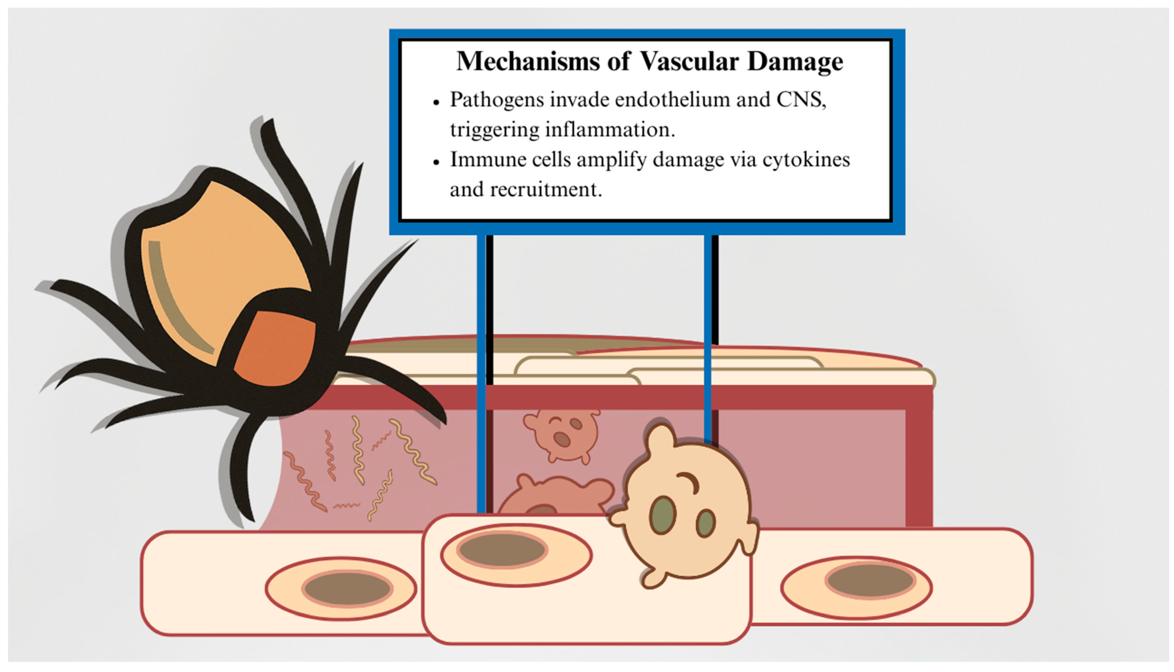
Figure 1. Simplified Mechanisms of Vascular Injury in Tick-Borne Disease: This schematic illustrates the two primary pathways by which tick-borne pathogens cause vascular damage. On the left, spirochetes (e.g., Borrelia burgdorferi) infiltrate the endothelium following tick transmission, initiating localized inflammation. On the right, recruited immune cells promote further endothelial injury through cytokine-mediated signaling and cell infiltration. Endothelial cells line the vasculature and play essential roles in regulating vascular tone, permeability, hemostasis, angiogenesis, and immune responses. Tick-borne pathogens can directly disrupt these functions through mechanisms such as phospholipase A2 activation and lipid peroxidation, leading to increased vascular permeability and endothelial damage. Immune-mediated injury, although less well understood, may involve complement activation, immune complex deposition, and recruitment of cytotoxic immune cells, further compromising endothelial integrity. The relative contribution of direct and immune-mediated mechanisms to cerebrovascular injury in tick-borne disease remains unclear and is likely pathogen-dependent.
Jvd 04 00033 g001
Figure 2. Example Mechanisms of Endothelial and Barrier Dysfunction in Tick-Borne Disease: This image illustrates selected pathogen-specific strategies contributing to endothelial injury, immune activation, and blood–brain barrier compromise in tick-borne disease. Borrelia burgdorferi binds endothelium under shear stress via BBK32 and induces IL-8-mediated neutrophil recruitment. Rickettsia spp. release exosomal miRNAs that disrupt junctions and promote vascular instability. Powassan virus persists in pericytes and brain endothelial cells by delaying interferon signaling. CCHFV enhances leukocyte adhesion through TNF-α-driven ICAM1 upregulation. These mechanisms underscore the multifaceted nature of vascular injury in TBD.
Jvd 04 00033 g002
Figure 3. Graphical representation of a reasonable algorithm for diagnostic and therapeutic decision making for tick-borne-disease-caused cerebrovascular events. This schematic algorithm is a basic decision-making visual aid for clinicians considering stroke or intracerebral hemorrhage secondary to tick-borne disease. The schematic begins with a detailed physical and history (top left) working in tandem with ongoing neuroimaging (top right). The exact neuroimaging modality encouraged is still contested in the literature. The algorithm continues with the verification of cerebrovascular injury through neuroimaging and patient history.
Jvd 04 00033 g003
Table 1. Relevant organisms and related generalized hallmarks of infection.
Organism Classification Core Associated Condition Hallmarks of Infection
Borrelia burgdorferiSpirochete BacteriaLyme Disease [17]
  • Erythema migrans
  • Flu-like symptoms
  • Neurologic, cardiac, or joint complications (late stage)
Borrelia miyamotoiSpirochete BacteriaRelapsing Fever [18]
  • Recurrent fever
  • Chills, headache, fatigue, and myalgia
  • Meningoencephalitis (in immunocompromised patients)
Rickettsia speciesObligate Intracellular BacteriaSpotted Fever Rickettsioses (e.g., RMSF) [19]
  • Fever, headache, and myalgia
  • Rash and gastrointestinal symptoms
  • Vascular injury and neurologic deficits
  • Multiorgan failure or death (if untreated)
Ehrlichia chaffeensisObligate Intracellular BacteriaHuman Monocytic Ehrlichiosis (HME) [20]
  • Fever, headache, malaise, and myalgia
  • Cytopenias
  • Respiratory failure and meningoencephalitis (severe cases)
  • Organ dysfunction or death (especially in immunocompromised)
Anaplasma phagocytophilumObligate Intracellular BacteriaHuman Granulocytic Anaplasmosis (HGA) [21]
  • Fever, headache, and myalgia
  • Cytopenias
  • Elevated liver enzymes
  • Organ failure (especially in immunocompromised)
Francisella tularensisGram-negative CoccobacillusTularemia [22]
  • Fever
  • Lymphadenopathy
  • Ulceroglandular, glandular, oculoglandular, oropharyngeal, pneumonic, or typhoidal forms
  • Pneumonia or sepsis (severe cases)
Powassan VirusFlavivirus (RNA Virus)Powassan Encephalitis [23]
  • Often asymptomatic
  • Fever and headache
  • Encephalitis, seizures, and coma
  • Death or long-term neurological deficits
Crimean–Congo Hemorrhagic Fever VirusNairovirus (RNA Virus, Bunyaviridae family)Crimean-Congo Hemorrhagic Fever [24]
  • Sudden fever and myalgia
  • Hemorrhage
  • Multiorgan failure
  • High fatality in severe cases (influenced by viral load, immunity, and comorbidities)
Babesia SpeciesProtozoa (Apicomplexan Parasite)Babesiosis [25]
  • Asymptomatic to severe
  • Fever and anemia
  • Organ failure (in severe cases)
  • Severity influenced by species, age, immune status, and spleen function
Table 2. Summary of discussed tick-borne organisms and their endothelial/immune effects.
OrganismRepresentative Mechanisms
Borrelia burgdorferi
  • Endothelial adhesion via BBK32 under shear stress
  • IL-8-driven neutrophil chemotaxis and inflammation
  • Immune complex-mediated vasculitis
  • Heparan sulfate binding and endothelial internalization
  • vWF reduction and persistent vascular colonization
Rickettsia spp.
  • Receptor-mediated entry via Ku70/rOmpB and Epac1
  • Disruption of tight junctions via exosomal miRNAs (miR-23a, miR-30b)
  • Promotes IL-8 and MCP-1 cytokine release
  • Paracrine inflammation via NF-κB activation
Francisella tularensis
  • Neutrophil recruitment via IL-8 elevation
  • Endothelial activation by live/killed forms
Powassan virus
  • Infects pericytes and endothelial cells of BBB
  • Delayed interferon response facilitates viral persistence
  • Chronic glial activation and immune evasion
Crimean–Congo hemorrhagic fever virus
  • Direct endothelial infection and ICAM1 upregulation
  • Promotes IL-6/IL-8 cytokine release
  • TNF-α-dependent leukocyte adhesion
Tick saliva components
  • Vasodilation and inhibition of coagulation
  • Suppression of host inflammatory responses
  • No direct cerebrovascular involvement observed
Table 3. Example cerebrovascular cases in Lyme neuroborreliosis.
CasePatient DemographicsCerebrovascular EventImaging FindingsTherapy and Outcome
Kurian et al., 2014 [62]12-year-old boy
Previously healthy
Tick bite history
Multifocal cerebral vasculitis with stroke-like symptoms (no infarct).Multifocal arterial stenoses (MCA, ACA, basilar), vessel wall enhancement (MRI)IV ceftriaxone (4 wks), steroids, aspirin → full recovery.
Almoussa et al., 2015 [74]43-year-old man
No known risk factors
Tick bite 4 months prior
Right thalamic infarct
Cognitive deficits, hemiparesis
Right thalamic infarct
Hyperintense signals in periventricular, periaqueductal areas
Bilateral vascular abnormalities (MRI)
IV ceftriaxone (3 wks) → motor recovery, persistent amnesia.
Table 4. Example cerebrovascular cases in Rickettsia species.
CasePatient DemographicsCerebrovascular EventImaging FindingsTherapy and Outcome
Kumar et al., 2014 [64]25-year-old male from India with no comorbiditiesLeft MCA territory infarctLeft frontotemporal infarct (CT)Doxycycline → motor recovery
Srinivasa Murthy et al., 2015 [92]18-month-old male child with no prior conditionsRight MCA territory multifocal infarcts, left hemiplegia and UMN facial palsyInfarct of the right corona radiata, right basal ganglia, right frontal gyri, insular cortex, and right anterior temporal lobe (MRI)Doxycycline → rapid recovery
Boulahri et al., 2017 [93]50-year-old female from Morocco without any cerebrovascular risk factorsDeep left MCA infarct, aphasia, hemiplegiaLeft sylvian infarct (MRI), normal CTADoxycycline + ciprofloxacin (10 days) → marked recovery
El Moussaoui et al., 2024 [94]23-year-old male from Lebanon with no prior conditionsNon-aneurysmal SAH with splenial lesionTransient splenial lesion, no aneurysm (CTA)Doxycycline (2 wks) → full recovery
Table 5. Example cerebrovascular cases in Ehrlichia chaffeensis.
CasePatient DemographicsCerebrovascular EventImaging FindingsTherapy and Outcome
García-Baena et al., 2017 [69]16-year-old male who was previously healthyIntracerebral hemorrhageRight frontoparietal intracerebral hemorrhage with surrounding edema causing mild ventricular compression, without midline shift (CT)Surgical drainage + oral doxycycline (6 months) → complete recovery
Grant et al., 1997 [104]54-year-old male from Georgia with no previous cardiovascular risk factorsSubacute ischemic strokeNonenhancing right frontal subcortical white matter lesion (MRI)Intravenous doxycycline → partial recovery
Table 6. Examples of cerebrovascular cases in Anaplasma phagocytophilum.
CasePatient DemographicsCerebrovascular EventImaging FindingsTherapy and Outcome
Herbst et al., 2020 [56]26-year-old female with HbSC sickle-cell diseaseDiffuse bilateral ischemic strokesWidespread acute + subacute cortical and subcortical ischemic lesions in both hemispheres (MRI)Doxycycline + clindamycin + atovaquone + eculizumab → poor recovery
Kim et al., 2018 [59] 70-year-old female from Korea with no comorbiditiesLacunar cerebral infarction in the left basal gangliaSmall lacunar infarct in left basal ganglia (MRI); normal MRADoxycycline → complete recovery
Guru et al., 2025 [110]65-year-old female with past medical history of hypertension, type II diabetes, and previous cerebrovascular accidentAcute infarction likely due to embolismIll-defined hypodensity in posterior right corona radiata (CT); 3 cm area of restricted diffusion between subcortical white matter and lateral wall of right lateral ventricle ± additional watershed lesions (MRI)Oral doxycycline (14 days) → complete recovery
Table 7. Example of cerebrovascular case in Francisella tularensis.
CasePatient DemographicsCerebrovascular EventImaging FindingsTherapy and Outcome
Çoban et al., 2019 [113]42-year-old female, previously healthyAcute pontine infarction with central nervous-system vasculitisHyperintense non-contrast enhancing lesions in bilateral periventricular, peritrigonal, and pericallosal regions, centrum semiovale, and corona radiata (MRI); narrowing and irregularities of bilateral medial and anterior cerebral arteries (MRA)Streptomycin and doxycycline (3 weeks + 5 days) → regression of lesions on imaging
Table 8. Example of cerebrovascular case in Powassan virus.
CasePatient DemographicsCerebrovascular EventImaging FindingsTherapy and Outcome
Bazer et al., 2022 [116]62-year-old male from USA with previous right putamen infarct, hepatitis C, hypertension, and substance abuseAcute diffuse infarctionAcute left putamen infarct (MRI); normal MRA; small foci of acute infarct involving cerebellum, left basal ganglia, and splenium of corpus callosum (2-week in-patient MRI)Ceftriaxone + acyclovir → IVIG → no recovery
Table 9. Example cerebrovascular cases in Crimean–Congo hemorrhagic fever virus.
CasePatient DemographicsCerebrovascular EventImaging FindingsTherapy and Outcome
Kleib et al., 2016 [119]58-year-old male shepherd with no previous historyLeft subdural hematomaLeft subdural hematoma without midline shift (CT); left subdural hematoma with midline shift (repeat 2-week CT)Corticosteroids + supportive care → complete recovery
Ulosoy, 2018 [120]60-year-old male livestock worker without previous historyIschemic strokeRestricted diffusion in anterior left hemi-pons compatible with acute infarct (MRI)Ribavirin + platelet transfusion → complete recovery
Disclaimer/Publisher’s Note: The statements, opinions and data contained in all publications are solely those of the individual author(s) and contributor(s) and not of MDPI and/or the editor(s). MDPI and/or the editor(s) disclaim responsibility for any injury to people or property resulting from any ideas, methods, instructions or products referred to in the content.