Abstract
The purpose of this study was to verify the inter-rater agreement level as a means of obtaining an efficiency measure of a standard mastication evaluation through video recordings. The studied population included oral breathing children and teenagers with maxillary atresia. The chewing aspects studied were mode of chewing and preferential chewing side. A white tag was placed on half the subjects´ chins while the recordings were made. Two expert evaluators analyzed 54 video recordings at regular viewing speed. The lead author analyzed the same video recordings both at reduced speed and at reduced speed linked through graphical computing techniques. The analysis was conducted on chewing cycles with the viewing angle of the frontal plane. Findings indicated that when comparing the data for the three manners of watching the video recordings, the agreement level was higher for videos with the tag on the chin watched at reduced speed. It was also determined that alternating and bilateral mastication modes were prevalent (64.7%) in this sample.
INTRODUCTION
Mastication can be considered the most relevant stomatognatc function (Andrada e Silva, Natalini, Ramires, & Ferreira, 2007) because it prepares food for ingestion and influences dentofacial growth and development (Pastana, Costa, & Chiappetta, 2007). Physiologically, mastication is characterized by: cutting the food with the incisors; labial occlusion; predominance of rotational mandibular movements, with a curve in the shape of a drop, i.e. tear-drop shape, and with the mandibular movement to the side in which the food is located (Douglas, 1998a, 1998b); alternating sides of chewing; synchronic bilateral muscular activity, and uniform pressure on tissues supporting the teeth (Pignataro Neto, Bérzin, & Rontani, 2004; Rodrigues, Lefèvre, Mott, Tugumia, & Pena, 2003).
Mastication directly influences facial growth and development, both saggital and transverse mandibular and maxillary development (Pignataro Neto et al., 2004), which helps to prevent facial asymmetries (Bianchini, 1998). Mastication also influences facial muscle and bone development, the maintenance of dental arches, occlusion stability, the proper condition of the temporomandibular joints, and muscle movement integration (Motta, 2004). Mastication is the stomatognatic function that requires great strength during the performance of complex, coordinated and highly precise movements (Piancino MG, Farina D, Talpone, Merlo, & Bracco, 2009).
Alterations in mastication may be due to the lack of harmony between the relationship of maxilla and mandible, lack of muscle tone, malocclusion and/or decreased quantity of saliva in the oral cavity (Pereira, Gavião, Engelen, & Van Der Bilt, 2007). Lack of saliva may be due to drying (Cintra, 2003) or diminished production of saliva, which is a common effect of anti-histamine use (Assencio-Ferreira, 2003). These alterations are commonly found in oral breathers.
According to Andrade e Silva (2007), in oral breathing cases the need to breathe is greater than the need to masticate. Depending on the severity of nasal obstruction, mastication may occur in a shorter period of time and with fewer mastication strokes. Some features of the inadequacy of the mastication function in individuals who are oral breathers include: alteration of food cutting; diminished unilateral or bilateral mastication strength, unilateral mastication (Felício, 1999; Terra, 2004); reduced or even absent amplitude of rotation movements, or reversed cycles, that is mandibular movement to the side without food (Piancino et al., 2009; Saitoh, Yamada, Hayasaki, Maruyama, Iwase, & Yamasaki, 2010), and diminished food crushing, fast mastication cycles, and a reduced number of cycles (Coutinho, Abath, Campos, Antunes, & Carvalho, 2009).
Ahlgren (1966) described three types of mastication cycles in subjects with normal occlusion and four additional types in subjects with malocclusions derived from the mandible trajectory and direction. This suggests that in the cases of functional immaturity, as seen in subjects who are mouth breathers due to variable mandibular movements, exaggerated participation of the perioral muscles, head projection for swallowing, difficulty of cutting the food, and difficulty with labial closure (Lowe and Tanaka, 1984; Whitaker, 2005). It is more difficult to assess only mastication. Tay (1994) proposed a classification for mastication manner. He provided guidelines regarding the mastication side. He established that for bilateral alternated mastication the number of mastication cycles on the most used side occurred under 66.1% of the total of cycles; for individuals with a unilateral preference the number of mastication cycles on the most used side was between 66.1% and 95.0% of the total of cycles; and for individuals with a chronic unilateral preference the number of mastication cycles on the preferred side was above 95.0% of the total of cycles.
Video recording is an easily accessible resource and it allows for repeated viewings as many times as necessary. No standards were presented in previously conducted research that used video recordings. (Lima, Freire, Nepomuceno Filho, Stampford, Cunha, & Silva, 2006; Andrada e Silva et al., 2007; Pastana et al., 2007; Pignataro Neto, 2000; Silva, Natalie, Ramires, & Ferreira, 2007; Withaker, 2005).
Due to its importance, mastication has been studied by diverse health related areas. Speech pathologists are the professionals responsible for the assessment and treatment of stomatognatic functions. Sophisticated instrumentation, such as electrognatography and electromyography, is being used in research studies (Casselli, Landulpho, Silva, & Silva, 2007; Coelho-Ferraz, Berzin, Amorim, Romano, & Queluz, 2010; Gomes, Custodio, Jufer, Del Bel Cury, & Garcia, 2010; Nakata, Ueda, Kato, Tabe , Shikata-Wakisaka, Matsumoto, Koh, Tanaka, & Tanne, 2007; Qiong, Zuisei, Tianmin, Jiuxiang, & Kunimichi, 2010; Pereira et al., 2007).
Studies have been conducted which increase of understanding of the physiological and pathological processes. While sophisticated instrumentation is important for research, such equipment is not financially feasible for use in daily clinical practice. Therefore, it is also important to identify efficient and effective tools which are financially feasible for use in daily practice which improve the clinician’s ability to observe and assess disorders. Video recording is an easily accessible resource and it allows for repeated viewings as many times as necessary. No standards were presented in previously conducted research that used video recordings. (Lima, Freire, Nepomuceno Filho, Stampford, Cunha, & Silva, 2006; Andrada e Silva et al., 2007; Pastana et al., 2007; Pignataro Neto, 2000; Silva, Natalie, Ramires, & Ferreira, 2007; Withaker, 2005).
A positive aspect of the development of protocols and standardization for the analysis of mastication by using video recording is that the procedure is easily able to be integrated into daily clinical practice. In this study, video recording was the selected tool to be used in the observation of the side in which the food was placed in each mastication cycle and classification of manner of mastication. In developing an accessible technique that could be reproduced and allow for comparative analyses, it was necessary to establish a standard protocol for capturing images. It was felt that video recording analyses would allow the extraction of the maximum amount of information on mastication function. It was also felt that video recording of individuals who demonstrated oral breathing would provide the greatest amount of variance in mastication cycles and manner of mastication, which would provide adequate trials to determine the potential for inter-rater reliability using a standardized analysis protocol.
METHODS
This study was approved by the Ethics in Research Committee of UNIFESP-EPM, Hospital São Paulo, under the process number 1186/05. All individuals responsible for the subjects in this study signed a free and informed term of agreement.
This research was conducted on 34 subjects, aged from 5 to 12 who were patients at the Center for the Mouth Breather at UNIFESP. Half of those subjects were males and the other half, females. Criteria for inclusion of individuals in this study was: (1) a history of mouth breathing for at least 6 months as documented in a multidisciplinary evaluation with an otolaryngologist, dentist, allergist, and speech pathologist; (2) the patient had a complaint of nasal obstruction, runny nose, rhinorea, nasal itching, sneezing in bunches, (3) a skin test of immediate positive hyper-sensibility to at least one air allergen (Lemos, Wilhelmsen, Mion, & Mello Júnior, 2009); (4) show no obstructive hypertrophy of adenoids or amygdale, nor nasal septum deviation, or nasal tumors; (5) posterior teeth needed to be well maintained, with no lesion in the oral cavity; (6) the presence of maxillary atresia. The14-Cybershot, Sony®, 3.0 megapixels digital camera was placed on a tripod for stabilization. The camera was positioned in front of an ordinary chair with the front feet of the tripod in contact with the front feet of the chair. In order to make the image fit adequately the middle third of the face was positioned between the lines that limit the central recording area. Subjects were then seated with their feet on the floor and without any head support. The food used was French bread from the same supplier and always freshly baked on the same day the recording was made. Enough bread was offered independently of bite size so that there would be 50 seconds of video recording. Subject´s were asked to move as little as possible while looking straight into the camera. They were also instructed to eat and masticate in their usual manner.
A white tag was placed on the subject´s chin to help the visualization of mandibular movement. The white color was suggested by computer graphic technicians. The placement of a self-adhesive white tag on the subject´s chin was a resource to aid both image capture and the placement of the camera.
Initially, 14 subjects were video recorded only with the white tag while the remaining 20 subjects were video recorded with and without the white tag providing a total of 54 video recordings. Those 20 subjects were video recorded in two different but sequential trials on the same day. The first trial was completed without the tag, and the second trial was completed with the tag. These video recordings were randomly saved on CDs.
The assessment conducted by speech pathologists consisted of clinical observation of aspect, tonus, and mobility of the orofacial structures, and of the functions of breathing, mastication, swallowing, and speech. The subjects who presented with favorable nasal ventilation at the time of assessment had their mastication recorded.
Three different manners of watching the video recordings and assessing mastication were conducted. The goal was to identify which procedure would prove to be the most helpful for use in the professionals’ clinical practice.
The first manner of assessing the video recordings was completed by two experienced speech pathologists (SP1 and SP2), who were certified specialists in orofacial disorders. They watched the video recordings independently of one another, in the normal viewing speed. They registered the number of mastication cycles observed on each side of the oral cavity, during the 50 seconds of each video recording. The number of times the professionals watched each recording was not controlled.
The second manner of assessing the video recordings was completed by the lead author (SFH), who watched the same CD of video recordings but at a reduced speed (RS) of 0.1seconds (frame by frame).
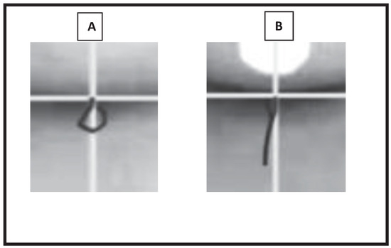
An analysis of the number of mastication cycles observed on each side of the oral cavity was registered. A third analysis was performed by SFH who watched the recordings at a RS of 0.1seconds (frame by frame) accompanied by the observation of traces of mastication cycles by computer graphics (VFWRS). This was considered the standard because it registers mandibular movements using a computer and therefore was considered an objective method. The use of this analysis allows for the differentiation of the direction, shape and type of mandibular movement. Tracings of the VFWRS were obtained from the intersection of the inferior line of the mandible plane and the saggital plane. One point was established on each frame and the points were then manually united creating the trace of the mastication cycles (Figure 1).

Figure 1.
Sample of the image obtained by computer graphics for each mastication cycle in video recordings without the tag (A) and with the tag (B), starting at the intersection of the inferior line of the mandible plane and the saggital plane, one point was established on each frame.
Each subject was classified according to their mastication manner by each one of the methods described earlier, that is, SP1 and SP2, independently registered the number of mastication cycles observed on each side of the oral cavity, determining manner of mastication and preferred mastication side. The same occurred with SFH and with the analysis SFH made using slow motion and the images obtained by computer graphics (VFWRS).
The statistical analysis of data was performed using the coefficient Kappa. The level of agreement between the findings of each of the analysis described earlier was calculated. The following interpretation was applied: Kappa lower than 20% = irrelevant; 21% to 40 %= minimal; 41% to 60% = average; 61% to 80%=good; and, higher than 81%=very good (Conover, 1999). Significance was set at p ≤ 0.05.
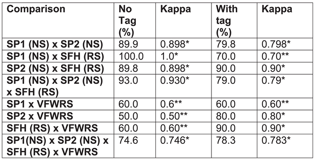
The rejection level for the null hypothesis that the use of the tag would not improve the agreement level between the raters was established at p ≤ 0.05 The significant levels are identified with a * on Table 1.

Table 1.
Agreement levels found between the comparison of raw data obtained by SP1, SP2, SFH, and SFH with VFWRS.
RESULTS
Table 1 displays the agreement levels found between the comparisons of all the raw data obtained for the multiple viewers with and without the white tag on the chin. All the results were of high inter-rater agreement levels and were statistically significant. The agreement levels were numerically higher when the comparison was made between video analyses of recordings with and without the tag on the chin.
However, when the analyses obtained by SP1, SP2 and SFH (SM) were compared to those completed in slow motion with the computer tracings obtained from the mandible movements (VFWRS), the results indicated a higher agreement level between the items compared when the tag was on the chin.

Table 2.
Distribution of mastication manner in oral breathing subjects (N=34) video recorded with a tag and analyzed by VFWRS.
Table 2.
Distribution of mastication manner in oral breathing subjects (N=34) video recorded with a tag and analyzed by VFWRS.
 |
Equality coefficient between two proportions: p=0.015. The most frequent mastication mode in this sample of oral breathing subjects was the alternate bilateral mode, noted at 64.7% of the sample.
DISCUSSION
A literature review revealed that video recording had been used in various researches. Pastana et al. (2007) assessed mastication in children with posterior cross bites using video recording with the analyses of two speech pathologists trained to verify food cutting, mastication manner, and mandible movements when it was asked of them to chew unilaterally or bilaterally, on the right and/or left side.
Gomes, Melo, & Chiappetta (2006), used video recordings to verify aspects of mastication pattern in deciduous and mixed dentitions in three to nine year olds. They concluded that the mastication patterns that presented as significant factors between the types of dentition studied were mandible movements, posture of the lips and mastication speed. It was noted that in both deciduous and mixed dentitions bilateral alternate mastication was the most frequent mastication manner. The mandibular movements most often noted were different for each dentition. In the deciduous dentition the most frequently observed mandible movements were vertical and in the mixed dentition, they were rotational.
Andrada e Silva (2007) assessed mastication in oral and nasal breathing children using direct observation and video recording with an established distance from the camera. A single observer noted the type of cut (frontal, lateral, fronto-lateral or manual), mastication manner (alternate bilateral, preferentially unilateral or chronic), shape of mandible movements (only vertical, predominantly vertical or vertical and rotational), and the duration of mastication, when the amount and quantity of bread were controlled. They observed that the frontal bite was present in 82.6% of their sample; bilateral alternate mastication in 87.0% and vertical and rotator in 100% of the oral breathers, with mean mastication duration of 15.92 seconds. It was concluded that the respiratory manner had a negative influence in those subjects regarding the duration of mastication and food residue in the oral cavity, lips posture, and production of noise during mastication.
In this study, video recording was the selected tool to be used in the observation of the side in which the food was placed in each mastication cycle and classification of manner of mastication. A white tag was placed on the chin of some subjects and recordings and analyses were conducted in these two situations, with and without the tag. These video recordings were analyzed by three speech pathologists (SP1, SP2 and SFH) who independently watched the recorded videos randomly saved on a CD in different speeds (NS and RS). An objective computer analysis method (VFWRS) was also completed and the inter-rater agreement levels were obtained. The highest inter-rater agreement levels were obtained when all the analyses were compared to VFWR, obtained in RS and when the white tag was on the chin.
It is believed that the presence of the tag on the chin may have made it easier for the observers to note in RS two aspects that take place physiologically at the same time, in the same visual stimulus, by enhancing mandible movements. That occurred for SP1 and SP2 when compared to VFWRS. This comparison involved a person watching a video recording in NS and the objective, computer analyses, in RS. Therefore, the gain noted in the inter-rater agreement level was obtained by SP1 and SP2.
However that was incorrect. What the analyses of images in SM allowed for was the visualization of each of the two aspects individually: location of food and mandibular movements. The easiness in the separation of these aspects provided by the slow motion method of analysis during mastication occurs because it allows the observation that the jaw does not always lateralize to the side on which the food is located. The reverse cycle is an example of that. The reverse cycle occurs when the subject moves the mandible to the side opposite to that where the food is located. (Douglas, 1998a, 1998b; Saitho et al., 2010).
The possibility of separating these aspects during the analyses is fundamental to the adequate diagnosis of mastication function. Therefore, the improved inter-rater agreement level displayed in the comparison among the results of the three professionals SP1, SP2, and SFH and the most objective analyses mean (VFWRS) obtained when the tag was placed on the chin reveals an important quality gain that is quite relevant in a private clinic situation, where high cost and state of the art devices are hardly ever found.
In accordance with previous findings of Andrada e Silva, (2007) and Motta et al. (2003), the results of the current research indicate that the prevalent manner of mastication was alternated bilateral, which was observed in 64.7% of the mouth breathing subjects. A study that reported somewhat contradictory findings was reported by Ferla, Silva & Corrêa (2008), who indicated that using electromyography noted a higher muscle activity in one of the sides of the oral cavity in mouth breathers which could suggest preferential use of one of the sides for chewing.
In the present study the subjects presented atresia of the maxilla without a cross bite. This may explain why this research did not identify preferential or chronic unilateral mastication manner in the studied sample. Camargo, Santana, Cara, Roda, Melo, Mandetta & Capp (2008) indicated that the preference of a determined mastication side occurs depending on the occlusion relationship on that side. Their finding is supported by Pastana (2007), who observed unilateral mastication in subjects with a posterior cross bites.
Based on the results of the present study, it may be said that video recording should be adopted as a method of assessing the function of mastication. The presented findings also led to the recommendation of the use of the tag on the chin when video recording is to take place in an evaluation as a protocol for the registration of the mastication aspects of manner of mastication and preferred mastication side.
CONCLUSIONS
The NS method of watching the videos enables clear visualization of mandible movements with the tag for every cycle and more precise assessment of where the positioning of the food occurs, allowing for a faster classification of the mastication manner for each subject in a clinic situation It is suggested that video recordings be used as the basis for discussion of cases in a multi-disciplinary approach to help in providing information on the progress of treatment. It is felt that video recordings would also be useful for the orientation of the patient themselves or their families.
The ease with which this technique can be added to the daily speech therapy clinical practice provides another dimension for the rehabilitation of this highly fundamental function of mastication. According to studies of Sever, Marion & Ovsenik (2010) a mastication disorder, if prolonged, may cause disruption of the integration of skeletal, occlusion and facial muscles. Hence, they recommended early treatment to normalize the mastication cycle pattern to ensure normal growth and development of the orofacial system.
Based on the results of the present study, the techniques described may provide a basis for future research. It would be of interest to determine if the methods of assessing mastication presented in this study are useful in the assessment of mastication for other samples of subjects, such as: individuals who use nasal when compared with individuals who use oral breathing methods; individuals with diverse occlusion alterations; oral breathers with hypertrophy of tonsils; individuals with temporomandibular joint disorders. A variety of additional pathologies may also be of interest for future research. The more detailed the knowledge of mastication the more likely the contribution to a more efficient diagnostic and rehabilitation process for individuals experiencing difficulties.
Acknowledgments
I would like to thank my personal friend, Speech-Language Pathologist Carla Nechar de Queiroz, Ph.D. for her help in the translation and adaptation of this article from Portuguese into English.
References
- Ahlgren, J. 1966. The mechanism of mastication. Scandinavia: Acta Odontologica 24 suppl.: 44. [Google Scholar]
- Andrada e Silva, M. A., V. Natalini, R. R. Ramires, and L. P. Ferreira. 2007. Análise comparativa da mastigação de crianças respiradoras nasais e orais com dentição decídua. Revista CEFAC 9: 190–198. [Google Scholar] [CrossRef]
- Andrade, F. V., D. V. Andrade, A. S. Araujo, A. C. C. Ribeiro, L. D. G. Deccax, and K. Nemr. 2005. Alterações estruturais de órgãos fonoarticulatórios e más oclusões dentárias em respiradores orais de 6 a 10 anos. Revista CEFAC 7, 3: 318–325. [Google Scholar]
- Assencio-Ferreira, V. J. 2003. Edited by L. Krakauer, R. Di Francesco and I. Q. Marchesan. Alterações dos pares cranianos devido á respiração oral. In Respiração oral. São José dos Campos, SP: Pulso Editorial, pp. 37–45. [Google Scholar]
- Bianchini, E. M. G. 1998. Edited by I. Q. Marchesan. Mastigação e ATM: Avaliação e terapia. In Fundamentos em fonoaudiologia: Aspectos clínicos da motricidade oral. Rio de Janeiro, RJ: Guanabara Koogan, pp. 37–49. [Google Scholar]
- Camargo, M. A., A. C. Santana, A. A. Cara, M. I. Roda, R. O. D. N. Melo, S. Mandetta, and C. I. Capp. 2008. Lado preferido da mastigação. Acaso ou oclusão? Revista de Odontologia da Universidade Cidade de São Paulo 20: 82–86. [Google Scholar] [CrossRef]
- Casselli, H., A. B. Landulpho, W. A. B. Silva, and F. A. Silva. 2007. Electrognathographic evaluations of rehabilitated edentulous patients. Brazilian Oral Research 21, 4: 355–361. [Google Scholar] [CrossRef]
- Cintra, C. F. S. C. 2003. A rinite como fator complicador das alterações buco-faciais. Master’s thesis, Faculdade de Medicina da Universidade de São Paulo. [Google Scholar]
- Coelho-Ferraz, M. J. P., F. Berzin, C. F. Amorim, F. L. Romano, and D. P. Queluz. 2010. Electromyographic and cephalometric correlation with the predominant masticatory movement. Stomatologija, Baltic Dental and Maxillofacial Journal 12: 51–55. [Google Scholar] [PubMed]
- Conover, W. J. 1999. Practical nonparametric statistics, 3rd ed. Austin, TX: Wiley. [Google Scholar]
- Coutinho, T. A., M. B. Abath, G. J. L. Campos, A. A. Antunes, and R. W. F. Carvalho. 2009. Adaptações do sistema estomatognático em indivíduos com desproporções maxilo-mandibulares: Revisão da literatura. Revista da Sociedade Brasileira de Fonoaudiologia 14, 2: 275–279. [Google Scholar] [CrossRef]
- Douglas, C. R. 1998a. Edited by C. R. Douglas. Fisiologia geral do sistema estomatognático. In Patofisiologia oral: Vol. 1. São Paulo, SP: Pancast, pp. 197–224. [Google Scholar]
- Douglas, C. R. 1998b. Edited by C. R. Douglas. Fisiologia do ato mastigatório. In Patofisiologia oral: Vol 1. São Paulo, SP: Pancast, pp. 245–271. [Google Scholar]
- Felício, C. M. 1999. Edited by C. M. Felício. Sistema estomatognático e funções. In Fonoaudiologia aplicada a casos odontológicos. São Paulo, SP: Pancast, pp. 15–48. [Google Scholar]
- Ferla, A., A. M. T. Silva, and E. C. R. Corrêa. 2008. Atividade eletromiográfica dos músculos temporal anterior e masseter em crianças respiradoras bucais e em respiradoras nasais. Revista Brasileira de Otorrinolaringologia 74: 588–595. [Google Scholar] [CrossRef]
- Gomes, S. G. F., W. Custodio, J. S. M. Jufer, A. A. Del Bel Cury, and R. C. M. R. Garcia. 2010. Correlation of mastication and masticatory movements and effect of chewing side preference. Brazilian Dental Journal 21: 351–355. [Google Scholar] [CrossRef]
- Gomes, F. C. S., L. F. Melo, and A. L. M. L. Chiappetta. 2006. Aspectos do padrão mastigatório na dentição decídua e mista em crianças de três a nove anos. Revista CEFAC 8, 3: 313–319. [Google Scholar]
- Lemos, C. M., N. S. W. Wilhelmsen, O. G. Mion, and J. F. Mello Júnior. 2009. Alterações funcionais do sistema estomatognático em pacientes com rinite alérgica: Estudo caso-controle. Brazilian Journal of Otorhinolaryngology 75, 2: 268–274. [Google Scholar] [CrossRef] [PubMed]
- Lima, R. M. F., O. C. B. Freire, J. L. Nepomuceno Filho, S. Stampford, D. A. Cunha, and H. J. Silva. 2006. Padrão mastigatório em crianças de 5 a 7 anos: Suas relações com o crescimento craniofacial e hábitos alimentares. Revista CEFAC 8, 2: 205–215. [Google Scholar]
- Lowe, A. A., and K. Takada. 1984. Association between anterior temporal, masseter, and orbicularis oris muscle activity and craniofacial morphology in children. American Journal of Orthodology 86, 4: 319–330. [Google Scholar] [CrossRef] [PubMed]
- Motta, A. R. 2004. Mastigação e pesquisa: Uma parceria necessária. In Comitê de motricidade orofacial-SBFa. Motricidade orofacial: Como atuam os especialistas. São José dos Campos, SP: Pulso, pp. 61–66. [Google Scholar]
- Nakata, Y., H. M. Ueda, M. Kato, H. Tabe, N. Shikata-Wakisaka, E. Matsumoto, M. Koh, E. Tanaka, and K. Tanne. 2007. Changes in stomatognathic function induced by orthognathic surgery in patients with mandibular prognathism. Journal of Oral Maxillofacial Surgery 65: 444–451. [Google Scholar] [CrossRef]
- Pastana, S. G., S. M. Costa, and A. L. M. L. Chiappetta. 2007. Análise da mastigação de indivíduos que apresentam mordida cruzada unilateral na faixa etária de 07 a 12 anos. Revista CEFAC 9, 3: 351–357. [Google Scholar] [CrossRef]
- Pereira, L. J., M. B. D. Gavião, L. Engelen, and A. Van Der Bilt. 2007. Mastication and swallowing: Influence of fluid addition to foods. Journal of Applied Oral Science 15, 1: 55–60. [Google Scholar] [CrossRef]
- Piancino, M. G., D. Farina, F. Talpone, A. Merlo, and P. Bracco. 2009. Muscular activation during reverse and non-reverse chewing cycles in unilateral posterior cross bite. European Journal of Oral Science 117: 122–128. [Google Scholar] [CrossRef]
- Pignataro Neto, G., F. Bérzin, and R. M. P. Rontani. 2004. Identificação do lado de preferência mastigatória através de exame eletromiográfico comparado ao visual. Revista Dental Press Ortodontia Ortopedia Facial 9, 4: 77–85. [Google Scholar] [CrossRef]
- Qiong, N., K. Zuisei, X. Tianmin, L. Jiuxiang, and S. Kunimichi. 2010. Clinical study of frontal chewing patterns in various cross bite malocclusions. American Journal of Orthodontics and Dentofacial Orthopedic 138, 3: 323–329. [Google Scholar]
- Rodrigues, K. A., A. P. Lefèvre, L. Mott, D. Tugumia, and P. L. Pena. 2003. Análise comparativa entre o lado de predominância mastigatória e medidas da mandíbula por meio do paquímetro. Revista CEFAC 5: 347–351. [Google Scholar]
- Saitoh, I., C. Yamada, H. Hayasaki, T. Maruyama, T. Iwase, and Y. Yamasaki. 2010. Is the reverse cycle during chewing abnormal in children with primary dentition? Journal of Oral Rehabilitation 37: 26–33. [Google Scholar] [CrossRef] [PubMed]
- Sever, E., L. Marion, and M. Ovsenik. 2010. Relationship between masticatory cycle morphology and unilateral cross bite in the primary dentition. European Journal of Orthodontics. http://ejo.oxfordjournals.org/content/early/2010/11/30/ejo.cjq070.long.
- Silva, M. A. A., V. Natalie, R. R. Ramires, and L. P. Ferreira. 2007. Análise da mastigação de crianças respiradoras nasais e orais com dentição decídua. Revista CEFAC 9, 2: 190–198. [Google Scholar] [CrossRef]
- Tay, D. K. L. 1994. Physiognomy in the classification of individuals with a lateral preference in mastication. Journal of Orofacial Pain 8: 61–72. [Google Scholar] [PubMed]
- Terra, V. Mastigação–abordagens terapêuticas. In Comitê de motricidade orofacial-SBFa. Motricidade orofacial: Como atuam os especialistas. São José dos Campos, SP: Pulso, pp. 47–56.
- Whitaker, M. E. 2005. Função mastigatória: Proposta de protocolo de avaliação clínica. Master’s thesis, Universidade de São Paulo, Bauru, SP. [Google Scholar]
© 2011 by the author. 2011 Silvia Fernandes Hitos, Dirceu Solé, Maria Cecília Periotto, Maria Lúcia T. N. Fernandes, Luc L. M. Weckx, Zelita C. F. Guedes.
