Abstract
The search for new antifungal substances is increasingly relevant due to growing antifungal resistance. Candida albicans is the most common pathogen yeast in humans, primarily in immuno-compromised individuals. Isothiocyanates, derived from glucosinolates, are compounds with an antimicrobial effect at low concentrations. The purpose of this study was to analyse the ultrastructural changes in three C. albicans isolates after exposure to benzyl isothiocyanate (BITC) at different lengths of exposure time (2.5, 5 and 24 h). Before exposure to BITC, cells presented a regular round or oval shape, with a uniform cell wall. After exposure to BITC, cell wall damage and loss occurred in the three strains. The cells developed extensive indentations, and a band of electrodense material was formed in the cortical cytoplasm. Although, for one isolate, no intact cells were detected, at the highest exposure time, two of the isolates showed a relevant response, regaining almost normal cell shape with nearly complete cell wall recovery. Cell lysis led to the deposition of a melted and unmixed mass with two apparently distinct fractions, the cell wall fraction and the cytoplasmic fraction. The present work demonstrates that, through targeting the C. albicans cell wall, BITC may prove to be a promising antifungal compound.
1. Introduction
Most of the antifungals, such as polyenes, azoles and allylamine/thiocarbamates, target ergosterol, the major lipid component of yeast membrane [1]. In fighting fungal infections, some antifungal substances have three main disadvantages: a limited range of action, self-medication, which may interact negatively with different types of antifungal agents, and resistance to microorganisms [2,3]. Furthermore, despite the improvements in antifungal therapies over the last 30 years, antifungal resistance is still of major concern in clinical practice [4]. The structure and biosynthesis of a fungal cell wall is unique and is therefore an excellent target for the development of new antifungal drugs. ITCs are an important class of compounds derived from glucosinolates, secondary plant compounds present mainly in the Brassicaceae family. They are volatile substances with an inhibitory effect on a variety of microorganisms at low concentrations and appear to be potential antimicrobial agents [5]. In the current study, the ultrastructure of three C. albicans isolates was analyzed before and after incubation in BITC and a comparative morphological study was performed in order to identify the progressive morphological changes induced throughout over time.
2. Materials and Methods
Three C. albicans isolates (two oral isolates, O33 and O5, and the collection culture strain C. albicans ATCC 90028, used for quality control purposes when chemical compounds are tested against yeast) were used in this study, based on their susceptibilities to BITC (range 1.43–143.0 μg/mL) determined by the disk diffusion method (DD) [6]. All the isolates were susceptible for fluconazole (FLU) (breakpoint ≥ 19 mm) and, at 4.3 μg of BITC/mL, had inhibition zone diameters below the FLU breakpoint [6]. The isolates were maintained at −80 °C until needed. Cultures of C. albicans grew on Mueller–Hinton (MH) agar and, after 24 h, a loopfull of each was collected for Transmission Electron Microscope (TEM). For the TEM samples exposed to BITC, the same cultures were inoculated in 50 mL of MH broth, supplemented with glucose (0.9% w/v). After incubation (3 h) in an orbital shaker (Certomat S, B. Braun Biotech International) at 150 rpm at 35 °C, the cultures were inoculated in 250 mL of MH supplemented with BITC to a final concentration of 0.004 M. Several samples were collected at different times of incubation (2.5, 5 and 24 h) for cell fixation. The samples were washed with a solution of polyphosphate buffer saline (PBS) at pH 7.3 and centrifuged at 16.000 rpm for 4 min.
The isolate samples were subjected to double fixation at 4 °C with OsO4, dehydrated, infiltrated and embedded in Epon. Ultrathin sections were examined and the images captured by a TEM at Electron Microscopy Unit (UME) UTAD.
3. Results
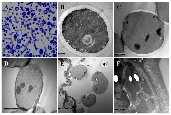
C. albicans cells presented a round or oval shape and were covered by a uniform cell wall firmly attached to the plasma membrane. The presence of two to three cell wall sublayers with a smooth appearance was evident. Cells possessed a well-developed intracytoplasmic cistern membrane system closely associated with the plasmatic membrane (Figure 1A,B). After exposure to BITC for 2.5 h, most of the C. albicans cells maintained their round shape, despite the identification of bevels or indentations on the cell surface. Cells revealed a heterogenous wall with a notorious thickness decrease. The cell wall sublayers were not identifiable, as they were before BITC exposure. The plasmatic membrane presented fewer invaginations, without connections to intracellular canaliculi, when compared to unexposed cells (Figure 1C). C. albicans cells exposed to BITC for 5 h revealed a very irregular contour with cell surface damage, forming pronounced bevels and indentations. Cells accumulated dense vesicles and vacuoles and, in some portions along the cell surface, there was complete cell wall loss. In these situations, cortical cytoplasm, devoid of cell wall, accumulated an electron-dense material that was irregular in shape and size (Figure 1D). After 24 h of exposure to BITC, most of the cells recovered a round shape, tending to a regular outline. Dense intracytoplasmic inclusions remained visible, but no accumulation of dense material in the cell periphery was observed. The most remarkable change was the almost total recovery of the cell wall structural integrity (Figure 1E). There was also a decrease in the volume of the accumulated extracellular material, although it seemed denser, more heterogeneous and granular in appearance. In one isolate, only vestigial cells were found in the semithin sections and no cells are found in the ultrathin sections. We found an extraordinary amount of melted cells and unmixed debris, compatible with the cell wall semblance, while the other fraction had a cytoplasmic appearance (Figure 1F).

Figure 1.
(A) Semithin section of C. albicans before exposure to BITC, stained with toluidine blue 1%. (B) TEM image of C. albicans before exposure to BITC. (C) TEM image of C. albicans after an exposure of 2.5 h to BITC. (D) TEM image of C. albicans after an exposure of 5 h to BITC. (E) TEM image of C. albicans after an exposure of 24 h to BITC. (F) TEM image of debris obtained after C. albicans exposure of 24 h to BITC.
4. Discussion
The present work revealed the drastic changes in C. albicans cellular morphology and in cell wall removal caused by BITC. Because the cell wall structure of yeast is unique, it is an important target for pharmaceutical research. Although we do not know the action mechanism of BITC at the level of the cell wall, the present work presents images that undoubtedly demonstrate the effect of this agent at this level, constituting, alone or combined, a promising antifungal.
Author Contributions
Conceptualization, A.C.S. and A.C.; methodology and data analysis, C.P. and A.C.; validation, A.C. and A.C.S.; resources and funding acquisition, A.C. and A.C.S.; writing—original draft preparation, A.C.; writing—review and editing, A.C. and A.C.S. All authors have read and agreed to the published version of the manuscript.
Funding
The authors are grateful for the financial support of Fundação para a Ciência e a Tecnologia (FCT) to CITAB (UID/AGR/04033/2020) and CECAV (UID/AGR/04033/2020). This work was supported by the projects UIDB/00772/2020 (Doi:10.54499/UIDB/00772/2020) funded by the Portuguese Foundation for Science and Technology (FCT).
Institutional Review Board Statement
Not applicable.
Informed Consent Statement
Not applicable.
Data Availability Statement
Data are contained within the article.
Conflicts of Interest
The authors declare no conflicts of interest.
References
- Cassell, G.; Mekalanos, J. Development of antimicrobial agents in the era of new and reemerging infectious diseases and increasing antibiotic resistance. J. Am. Med. Assoc. 2001, 285, 601–605. [Google Scholar] [CrossRef] [PubMed]
- Espinel-Ingroff, A.; Canton, E.; Peman, J.; Rinaldi, M.G.; Fothergill, A.W. Comparison of 24-Hour and 48-Hour voriconazole MICs as determined by the Clinical and Laboratory Standards Institute broth microdilution method (M27-A3 document) in three laboratories: Results obtained with 2162 clinical isolates of Candida spp. and other yeasts. J. Clin. Microbiol. 2009, 47, 2766–2771. [Google Scholar] [CrossRef] [PubMed]
- Carrillo-Muñoz, A.J.; Giusiano, G.; Ezkurra, P.A.; Quindós, G. Antifungal agents: Mode of action in yeast cells. Rev. Esp. Quimioter. 2006, 19, 130–139. [Google Scholar] [PubMed]
- Pfaller, M.; Diekema, D. Epidemiology of invasive candidiasis: A persistent public health problem. Clin. Microbiol. Rev. 2007, 20, 133–163. [Google Scholar] [CrossRef] [PubMed]
- Dufour, V.; Stahl, M.; Baysse, C. The antibacterial properties of isothiocyanates. Microbiology 2015, 161, 229–243. [Google Scholar] [CrossRef] [PubMed]
- Pereira, C.; Calado, A.M.; Sampaio, A.C. The effect of benzyl isothiocyanate on Candida albicans growth, cell size, morphogenesis, and ultrastructure. World J. Microbiol. Biotechnol. 2020, 36, 153. [Google Scholar] [CrossRef] [PubMed]
Disclaimer/Publisher’s Note: The statements, opinions and data contained in all publications are solely those of the individual author(s) and contributor(s) and not of MDPI and/or the editor(s). MDPI and/or the editor(s) disclaim responsibility for any injury to people or property resulting from any ideas, methods, instructions or products referred to in the content. |
© 2023 by the authors. Licensee MDPI, Basel, Switzerland. This article is an open access article distributed under the terms and conditions of the Creative Commons Attribution (CC BY) license (https://creativecommons.org/licenses/by/4.0/).