Review

Inclusion Bodies in Ionic Liquids

by
András Szabadi
1,2,
Robert Klausser
3,4,
Oliver Spadiut
3,4 and
Christian Schröder
1,*
1
Faculty of Chemistry, Institute for Computational Biological Chemistry, University of Vienna, Währingerstr. 17, 1090 Vienna, Austria
2
Doctoral School in Chemistry (DoSChem), University of Vienna, Währingerstr. 42, 1090 Vienna, Austria
3
Integrated Bioprocess Development, Technische Universität Wien, Gumpendorferstrasse 1A/5th Floor, 1060 Vienna, Austria
4
Christian Doppler Laboratory IB Processing 4.0, Technische Universität Wien, Gumpendorferstrasse 1A/5th Floor, 1060 Vienna, Austria
*
Author to whom correspondence should be addressed.
Liquids 2024, 4(1), 1-31; https://doi.org/10.3390/liquids4010001
Submission received: 24 October 2023 / Revised: 30 November 2023 / Accepted: 13 December 2023 / Published: 22 December 2023
(This article belongs to the Section Chemical Physics of Liquids)

Abstract

:
The pivotal role of proteins in pharmaceuticals is challenged by stability issues, making the study of inclusion bodies—a source of insoluble protein aggregates—increasingly relevant. This review outlines the critical procedures in inclusion body processing, focusing on ’mild solubilization concepts’ and refolding methodologies. Attention is afforded to the emerging role of ionic liquids with unique and tunable physicochemical properties in optimizing protein unfolding and refolding processes. The review critically assesses the existing literature at the intersection of inclusion bodies and ionic liquids, identifying recent advancements, potential applications, and avenues for future research. This comprehensive analysis aims to elucidate the complexities in efficient protein processing from inclusion bodies.

1. Introduction

Scientific advancement in the 21st century has entailed an increasing focus on exploring alternative solvents and innovative approaches to handling recalcitrant biological entities. The current review seeks to dissect the intertwined relationship between inclusion bodies and ionic liquids, emphasizing their potential implications for various industries and scientific disciplines.

1.1. Inclusion Bodies

The role of proteins in pharmaceuticals is undeniably significant, constituting 10% of pharmaceutical products. These pharmaceutical proteins are largely produced recombinantly in biotechnological processes. The best-understood microbial organism is Escherichia coli, which still serves widely as a host organism for recombinant protein production due to its fast growth rate and ease of genetic engineering. However, overexpression of difficult-to-fold proteins often results in their production as misfolded protein aggregates, known as inclusion bodies. These aggregates may also contain host proteins, lipids, and nucleic acids, indicative of a sophisticated interplay among genetic, environmental, and physiological factors. They were found across various cellular types, including neurons, various blood cells, bacteria, viruses, and plant cells. In neuronal cells, inclusion bodies may localize within the cytoplasm or nucleus, where they are implicated in many neurodegenerative diseases. Similarly, in muscle cells, inclusion bodies composed of multiple protein aggregations are observed in conditions like inclusion body myositis and hereditary inclusion body myopathy. In bacterial contexts, inclusion bodies appear as dense, aggregated protein particles, often indicative of viral replication sites within bacterial or eukaryotic cells, predominantly comprising viral capsid proteins.
Misfolded protein structures within inclusion bodies can include amyloid structures, amorphous regions, and incorrect disulfide bonds. Nevertheless, inclusion bodies maintain correctly folded secondary and tertiary protein structures [1]. For example, the fluorescence of green fluorescent protein was still observed within inclusion bodies, suggesting structural similarities with their native conformation [2,3].
Despite their insoluble and frequently non-functional nature, the potential of inclusion bodies in protein production, particularly for proteins challenging to express in soluble, correctly folded forms, has been increasingly recognized. This recognition broadened the scope of research on inclusion bodies and led to novel strategies for refolding processes, their roles as protein storage mediums, and their direct applications in nanotechnology and biotechnology. In the context of inclusion body processing, the workflow can be broadly categorized into two main segments: upstream and downstream processing [4,5]. The upstream segment encompasses microbial fermentation [6] and subsequent cellular harvesting. After cells are harvested using centrifugation or filtration methods, cell lysis is then performed. This can be achieved through mechanical techniques such as high-pressure homogenization, acoustic cavitation, bead milling, and chemical approaches like enzymatic lysis. While the desired protein is mainly found within the insoluble inclusion bodies, impurities, such as host cell proteins and nucleic acids, are in a soluble state after cell lysis. Consequently, the inclusion bodies can be selectively isolated using filtration or centrifugation procedures and subsequently purified through resuspension in aqueous solutions or specialized wash buffers [5,7].
Conversely, the downstream segment can be dissected into a series of discrete unit operations, as illustrated in Figure 1. Solubilization (blue sphere) represents a pivotal step toward extracting the target protein from the inclusion bodies and its subsequent transition into a soluble state for further processing. Commonly utilized agents for this purpose include urea and guanidinium hydrochloride (GndHCl). In instances where the protein configuration includes disulfide bridges, a reducing agent such as dithiothreitol may be incorporated to facilitate their cleavage.
These solubilizing agents effectively induce protein unfolding (yellow sphere in Figure 1) as water molecules within the protein’s hydration shell are displaced by the chaotropic molecules. Given the preferential affinity of these chaotropic agents for the hydrophobic regions of the protein, it becomes thermodynamically favorable for the protein to adopt an unfolded conformation. For proteins that are initially present in highly misfolded aggregate forms, this induced unfolding serves as a prerequisite to facilitate subsequent correct refolding.

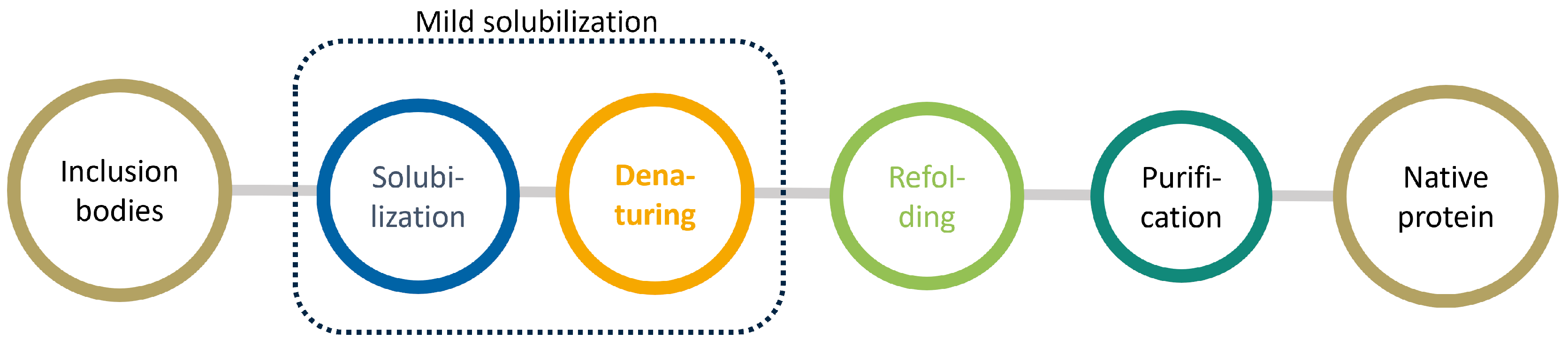
1.2. Mild Solubilization Concept

Solubilization is followed by segregating oligomeric structures into dissolved monomers, which must be executed without substantial protein denaturation. This methodology, referred to as the “mild solubilization concept” [1], employs urea concentrations below the threshold of 2–3 m urea to unfold the protein only partially. The approach necessitates a finely calibrated equilibrium of the denaturant concentration. The concentration needs to be increased enough to unfold the misfolded structures, but it must also be controlled to maintain the properly folded structures. Under favorable conditions, the correctly folded portions of the protein exhibit higher stability than the misfolded regions, highlighting the critical balance needed in this process [8]. The mild solubilization concept necessitates optimizing chemical and physical conditions [1] and limiting the use of traditional solubilization agents. Physical methods may include the application of high pressure up to 2.4   k bar . Chemical conditions to increase protein solubility might encompass maintaining an alkaline pH and employing organic solvents. Additionally, the technique may involve using surfactants to form micelles or reverse micelles [8,9].

1.3. Refolding and Purification
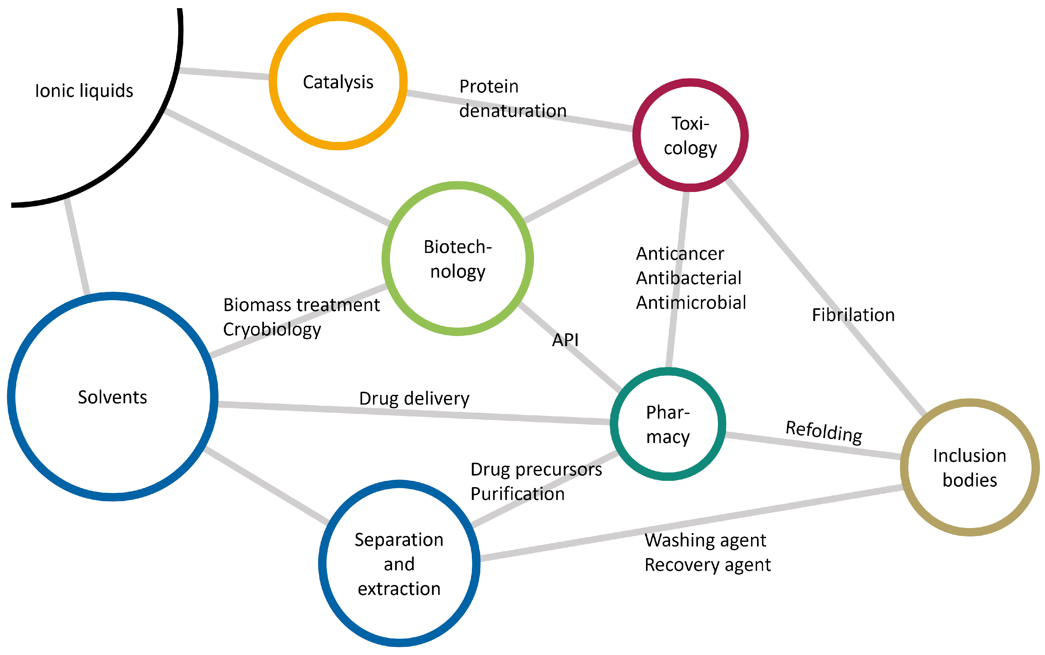
The next stage of the downstream processing is refolding (green sphere in Figure 1), wherein the target protein undergoes a transition from a (partially) unfolded state to its native functional conformation [4,10]. Initiation of the folding reaction is primarily achieved by removing chaotropic agents by diluting the solution with an appropriate refolding buffer [11]. However, the dilution approach necessitates large processing volumes with low product concentrations. This increases operational costs attributable to chemical reagents and demands sizable chromatography columns for subsequent capture and purification steps. In light of these considerations, alternative methods for denaturant removal have been explored, including dialysis and on-column refolding strategies. Nonetheless, due to its operational simplicity, dilution remains the predominant methodology employed for this stage of the process. The concluding step encompasses the isolation and extraction of the renatured protein.
The existing ambiguity surrounding the precise mechanisms and pathways renders the determination of conditions for the entire process—from the inclusion body to the native state—a complex and intricate task. This lack of clarity currently necessitates an approach based on trial and error, a method that is often associated with substantial expenditures in time and resources. Advancing the development of more effective additives for both the unfolding and refolding stages is imperative for future progress in this field [12]. Here, ionic liquids come into play [13].

1.4. Ionic Liquids

Ionic liquids are organic salts that exist in a liquid state at temperatures below 100 ° C . Due to their unique physical and chemical properties, these substances have captured widespread attention in both academic and industrial contexts.
Characterized by negligible vapor pressure, high thermal stability, and versatile solvation capability, ionic liquids promise to be “green” solvents that could reduce the environmental impact of various industrial processes by replacing conventional volatile organic solvents [14,15,16]. The uniqueness of ionic liquids lies in their tunability. By altering the cationic and anionic components, the physicochemical properties of the resulting ionic liquid can be adjusted, providing a vast and adaptable toolkit for various applications as electrolytes in electrochemical applications [17], lubricants [18,19], or solvents [15,20,21]. In this review, we focus only on a small part of these applications using ionic liquids as solvents for biotechnology, as illustrated in Figure 2. Here, several properties of the ionic liquids are exploited: Their broad liquid range as a solvent can be used in cryobiology [13,22,23], and their solvation properties are useful for biomass treatment, separations, and extractions of valuable compounds [20,24,25]. The biotechnological applications also concern toxicology and pharmacy. Ionic liquids can be active pharmaceutical ingredients [26,27] that may have anticancer, antibacterial, and antimicrobial effects [27,28,29,30]. The efficiency of a drug depends strongly on its bioavailability, which, in the case of the human organism, relates directly to drug permeability and solubility. Lower solubility results in lower dissolution and absorption rates. Therefore, higher doses are required to reach a therapeutic effect [27]. In contrast, active pharmaceutical ionic liquids are more soluble in water, facilitating drug delivery due to their charged nature. If the ionic liquid is not the active pharmaceutical ingredient, it can still support the drug delivery as a co-solvent, co-polymer, or emulsifier [27]. Their amphiphilic nature may also threaten proteins [31]. Both the charged core and the hydrophobic tails may catalyze protein unfolding. Ionic liquids may also extract drug precursors from biomass or purify active pharmaceutical ingredients in general. Abbreviations for the ionic liquids discussed in this work are summarized in Table 1.
The combination of inclusion bodies and ionic liquids represents a novel and promising direction in both academic and industrial contexts. This intersection offers opportunities for both the solubilization and refolding of inclusion bodies into active proteins, as well as the prevention of subsequent unfolding or amyloid fibrillation of the native protein [30,32,33]. However, imidazolium-based and ammonium-based ionic liquids have the potential to promote amyloidogenesis as well [33]. Moreover, ionic liquids can be used as washing and recovery agents for proteins produced in the form of inclusion bodies. This review, therefore, sets out to critically analyze the current knowledge landscape at the intersection of inclusion bodies and ionic liquids, providing a comprehensive account of recent research developments, potential applications, and future perspectives.

2. Theoretical Understanding

Apart from the polypeptide sequence, i.e., the primary protein structure, the stability of proteins is intrinsically linked to various solvation and reaction conditions, including temperature, pressure, ionic strength, and pH levels. When considering the factors that influence the formation of secondary structures, it is important to carefully analyze their impact on the rugged energetic landscape in these external conditions.

2.1. From Inclusion Bodies Back to Native State

Numerous computational methodologies have been employed to elucidate the competitive kinetics governing protein refolding and aggregation. One especially impactful model, suggested by Kiefhaber [34], has gained considerable recognition in the scientific community because of its balance between simplicity and predictive precision. In this model, the refolding pathway is conceptualized as a series of transitions from an unfolded state through intermediate states, culminating in the native protein conformation, as shown in Figure 3. The imposition of stress on proteins, achieved by altering their optimal conditions, can lead to various consequences. For example, partial unfolding leads to intermediate denatured states [30,32,35]. This process may be initiated on purpose to transform misfolded proteins in inclusion bodies into intermediate (=partially unfolded) states. The formation of intermediate states is assumed to be rapid, rendering the corresponding rate constant as non-limiting. The rate-limiting step is the conversion from the intermediate to the native structure, which includes the formation of disulfide bonds, for example. Importantly, this transition obeys first-order kinetics governed by intramolecular interactions.
Competition exists between this rate-limiting refolding process and the kinetic pathway, leading to the formation of unfolded proteins forming insoluble disordered aggregates within seconds. These aggregation processes are driven by intermolecular interactions following higher-order kinetics. Hence, increasing the protein concentration during the refolding process exponentially amplifies the rate of aggregate formation, thereby inversely affecting the refolding yield [34].
In the native state, monomers may form dimers, tetramers, or hexamers to stabilize the secondary structure [36]. The reverse transition from the native state back to the intermediate state is considered negligible given the thermodynamic stability of the native form. The aggregates of intermediate states can be inclusion bodies or prefibrillar species. For example, prefibrillar species may turn into amyloid fibrils containing high cross β -sheet contents, making this aggregation irreversible. These amyloids are associated with neurodegenerative diseases such as Alzheimer’s and Parkinson’s, and their dissolution is considered essential for novel biomedical treatments and the preservation of recombinant proteins [35].

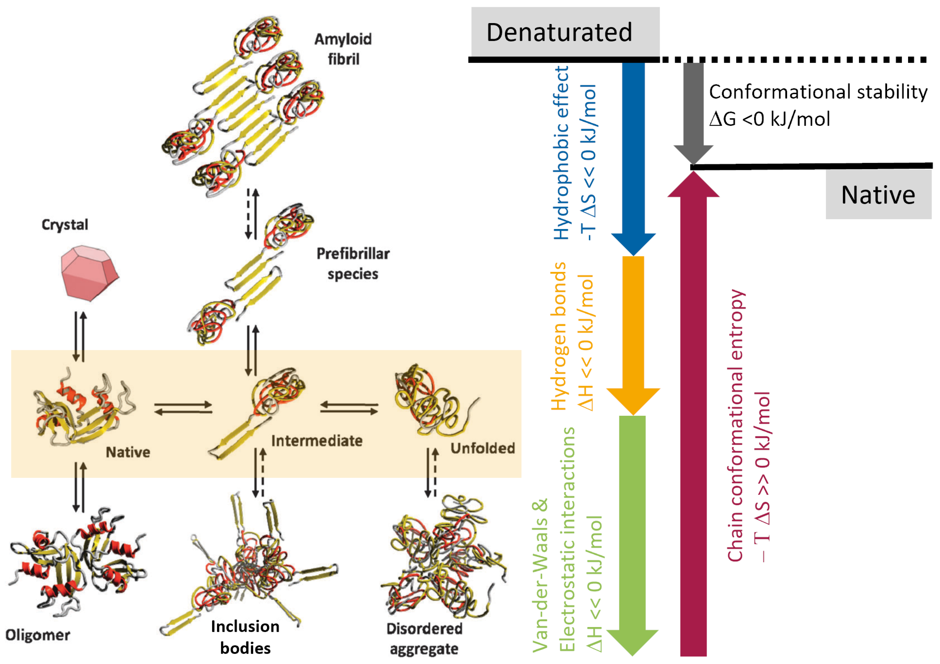
2.1.1. Protein States and Their Aggregation

The difference in protein states in inclusion bodies, fibrils, and disordered aggregates in Figure 3 primarily relates to their structural characteristics, formation processes, and biological implications:
  • Inclusion bodies are dense aggregates of partially misfolded proteins that may still contain a high percentage of α -helices and β -sheets. They form due to high local concentrations of a specific protein.
  • Fibrils, and in particular amyloid fibrils, are highly ordered, stable structures composed of β -sheet-rich protein aggregates. These fibrils are characterized by their elongated, fibrous nature. Fibrillation is a specific type of protein aggregation often associated with pathological conditions.
  • Disordered aggregates are irregular, amorphous conglomerations of proteins. Unlike proteins in inclusion bodies or fibrils, they lack a defined secondary or tertiary structure and are more heterogeneous. These aggregates form when proteins misfold or (partially) unfold, leading to exposure of hydrophobic residues that facilitate aggregation.
Oligomers and aggregates gradually become insoluble as they grow. The aggregation process involves a lag phase (rapid assembly of large aggregates), elongation phase, and final plateau. An energy barrier, the size of the aggregates, and factors like temperature, ionic strength, and interfacial exposure influence the lag phase. Various interactions control their size, including electrostatic repulsions, attractions, hydrophobic interactions, hydrogen bonds, and van der Waals forces. The dominant force depends on protein structure, solvent nature, and other experimental conditions [35]. Aggregation may lead to lower diffusivity, gelation, and phase separation, decreasing the pharmaceutical quality. Consequently, there is a need to prevent unwanted denaturation and aggregation. The protein concentration should be lower than 0.1 mg mL 1 to minimize aggregation to oligomers, which can be realized by continuous dilution. High temperatures, hydrophobic interactions, and hydrogen bonds promote aggregation, whereas specific pH ranges may suppress aggregation [36]. Aggregation can also be controlled by selecting solvents for conformational stability and balancing intermolecular forces [35]. The pH of the solvent affects the distribution of charged amino acids at the surface as well as the interaction between them. This is also true for ions dissolved in the solvent that may promote or prohibit aggregation, i.e., salting in and salting out. The aggregation of the protein has a strong influence on the (re-)folding process. Oligomers usually stabilize their secondary structure, e.g., the helical alignment. Consequently, the protein should be monomeric for the (re-)folding process.

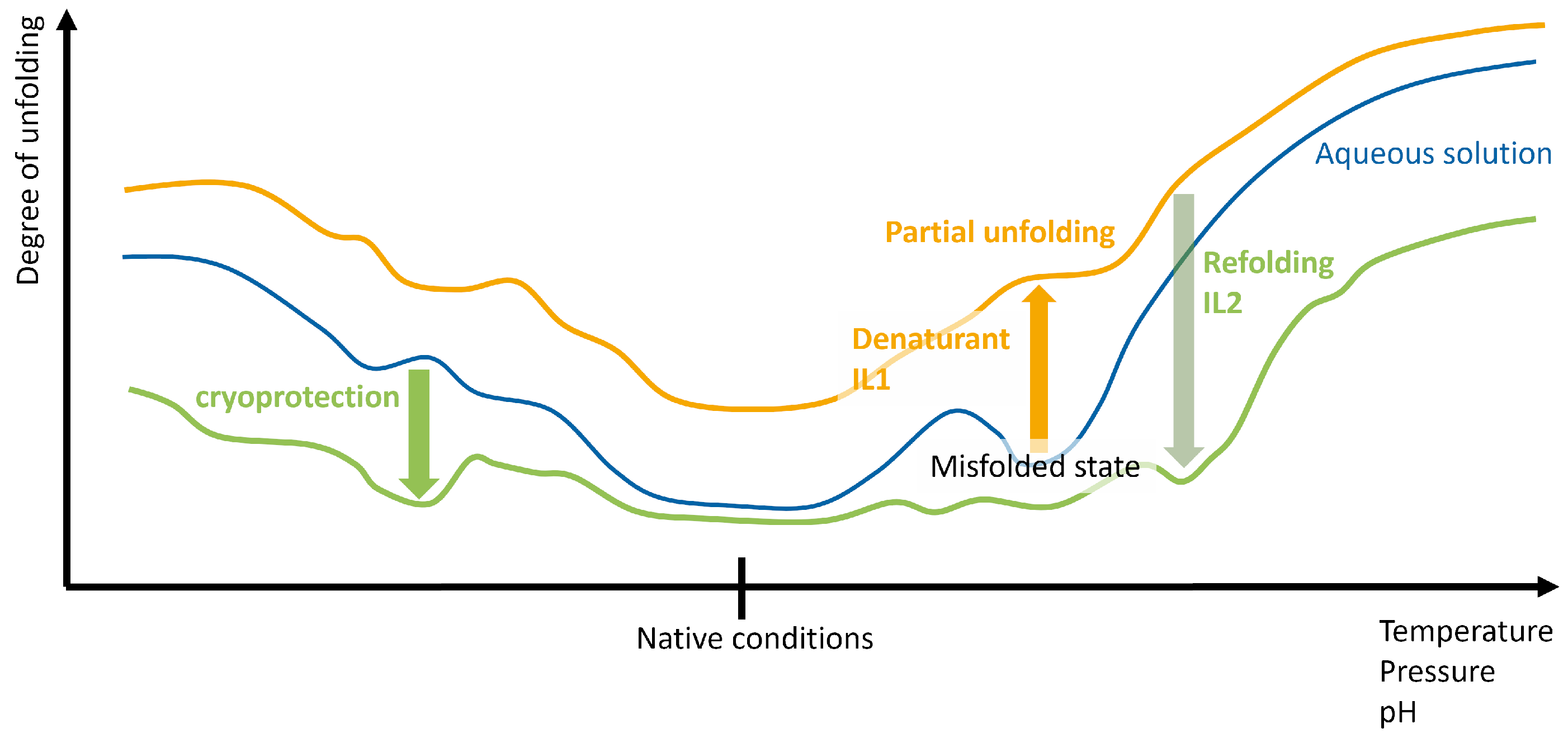
2.1.2. Pathways of (Re-)Folding

Levinthal [37] hypothesized that a stochastic search for the native state conformation of a medium-sized protein could potentially exceed the universe’s age. This finding underscores the necessity for well-defined folding pathways (illustrated by arrows in Figure 3) to facilitate refolding within a practical time frame. In living organisms (in vivo), proteins can fold into their native form through intermediates with the help of molecular chaperones. In laboratory conditions (in vitro), folding can be assisted by chemical chaperones like osmolytes and salts. Surface-exposed hydrophobic amino acid residues can interact with solvents, resulting in conformational changes [35]. Fundamental knowledge of these pathways is of incredible value as they can increase refolding yields from inclusion bodies to the native state of the protein. This is facilitated by unfolding the misfolded secondary structure while keeping the correctly folded parts of the protein. Also, preventing aggregation is crucial for optimizing recombinant protein production in bacterial systems.
Recent advancements in understanding protein folding have incorporated the “free energy funnel” approach [35,38], which asserts that unfolded conformations are likely to possess higher Gibbs energies in comparison to intermediate or native conformations. Within the proximity of the native state, several localized energy minima (intermediate states) are associated with modestly deformed native structures. These instances are significantly outnumbered by the plethora of misfolded structures, and their corresponding entropies are lower but compensated by the enthalpy originating from interactions such as van der Waals forces, electrostatic relationships, or the creation of hydrogen and disulfide bonds, as illustrated in Figure 3. Also, the entropic hydrophobic effect stabilizes the folded native state. This intricate interplay between entropy and enthalpy is responsible for the unexpectedly small Gibbs free energy difference Δ G 0 between the folded native and (partially) unfolded, denatured states and explains the concurrent existence of denatured states with the native state at ambient temperature [38,39], characterized by the chemical equilibrium
K = Π i a i ν i = e Δ G 0 k B T
The chemical equilibrium constant K depends on the chemical activity a i and their stoichiometric coefficient ν i , which are positive for denatured states and negative for the native state. Since the Gibbs energy Δ G is a function of temperature and pressure, their changes [40] may profoundly impact the stability of particular protein conformations i, as sketched in Figure 4.
However, Δ G is not a monotonic function of temperature T and pressure p. Both increasing and lowering T or p destabilize the native state [40]. Changing the temperature immediately affects all entropic contributions in Figure 3 and may lead to positive conformational stability Δ G > 0 k J mol−1 pushing a conformational change, as depicted in Figure 4. Unfolding proteins under high hydrostatic pressure has been known for over 90 years [42]. Applying pressures between 1 and 3 k bar promotes the dissociation of oligomers. To completely unfold a protein, pressures of more than 8 k bar are necessary [42]. The pressure dependence of Δ G corresponds to specific volume differences induced by internal cavities, voids, and perturbations in the electrostatic energy [42]. High pressure, although altering specific volume by less than 1%, exposes hydrophobic patches during unfolding. The synergistic application of high pressure in conjunction with an alkaline pH environment increases the solubilization of inclusion bodies under mild denaturing conditions without an actual denaturant [43]. Subsequently, decreasing the pH stops the unfolding [44], thereby stabilizing the protein as sketched in Figure 4.

2.2. Interaction with the Refolding Buffer

Once misfolded states like inclusion bodies are formed, refolding to the native state under mild solubilization conditions in a refolding buffer is desirable [1,42]. The buffer should be cost-effective, compatible with purification, and provide high yields of the native protein. Buffer molecules generally have a low molecular weight and reduce aggregation, enhancing protein solubility. Afterwards, they should be removable from the final product.

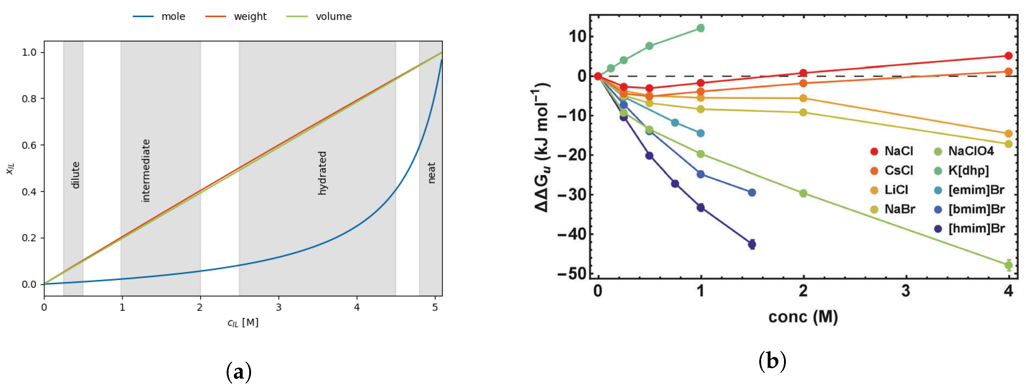
2.2.1. Gibbs Free Energy as a Function of Concentration

Gentle solubilization methods preserve the native-like folded secondary structures. This can be realized by small contents of chaotropes as co-solvents to unfold the misfolded parts of the proteins. Quite generally, the co-solvents in the refolding buffer change the Gibbs free energy [38,45,46,47]:
Δ G c s = Δ G + i n i · α i · δ g i , c s
= Δ G m · k B T · c c s
The changes depend on the number n i of the interacting group i, their average surface accessibility α i , and the energy transfer δ g i , c s of these groups to water and the co-solvent. Experimentally, a linear relationship between the energy transfer and the co-solvent concentration was found [46,48,49]. The ability of the co-solvent to (de-)stabilize the protein can be characterized by the so-called m value [40,45,46,47,49,50]. Positive m-values of the co-solvent (green in Figure 4) indicate protein stabilization, whereas negative values denote the co-solvent as a denaturant (orange in Figure 4). However, the transition from Equation (2) to (3) is a step back for the studies on inclusion bodies as the last equation only characterizes the overall unfolding. For the pathway back to the native state, region-specific m-values would be more appropriate since correctly folded regions should be untouched (low local m-value), but misfolded regions should be unfolded (high local m-value). However, the m-value in Equation (3) is only an average of all local m-values.
According to Schellman theoretical thermodynamic analysis of the interactions [51], the m-value should be proportional to the protein surface area exposed to the solvent upon unfolding, which was confirmed by simulations [52] and experiment [53]. Consequently, this value is not only a function of the solvent but also the protein [50]. Nevertheless, m-values may still serve to compare the capabilities of different denaturating molecules [47]. m-values are also accessible from molecular dynamics simulations via the “molecular transfer model” [49,52,54] on the basis of Equation (2).
Furthermore, this linear relationship in Equation (3) is only valid for a limited concentration range, usually below 0.5 M to 1 M depending on the protein and co-solvent [48,55,56], which corresponds to the ’dilute regime’ (see Figure 5a) according to Ref. [35]. Beyond this concentration threshold, the free energy Δ G starts to be a non-linear function of the concentration. This deviation from linearity manifests in Figure 5a, showing that the mole fraction likewise becomes non-linear as a function of the co-solvent concentration in the “intermediate regime”. The curvature of Δ G c s ( c c s ) is predominantly positive [45,55,57] (see Figure 5b), which may induce protein stabilization even when the co-solvent initially appears to be destabilizing [55]. This tunable interplay between stabilization and destabilization in the current state of the protein as a function of co-solvent concentration may be exploited to facilitate the protein’s transition from the inclusion body state to its native conformation.

2.2.2. Hofmeister Series

The original experiments conducted by Lewith and Hofmeister spanned several days, indicating that the mentioned protein processes are likely thermodynamic in nature [59]. The Pitzer method [60] has become increasingly popular to determine the thermodynamic properties of electrolytes and to predict ion activity coefficients, although standard electrolyte theories cannot explain salting in and salting out [61]. Given that ions must be in direct contact with the protein to interact [62], their surface activity undoubtedly influences the solubilization process, as, for example, modeled by a cavity model [63,64]:
Δ Δ G = A · γ
using the surface area A of the cavity and the actual surface tension γ of water influenced by the ions. The Hofmeister series offers a systematic classification of ion surface activity based on their lyotropic properties [59,65], specifically their capacity to either salt out or salt in proteins [59,66] with consistent trends for various proteins. Although Pearson’s hard and soft acids and bases theory [67] has arguably stronger theoretical foundations than the Hofmeister series and offers similar ion rankings [59,62,68], it is a less popular reference for the interpretation. A table of hard/soft and acidic/basic ions is provided in Ref. [59].
The ion ranking within the Hofmeister series resembles the lyotropic series, which is predicated on the heat of hydration of the ions [59,62,69]. This similarity initiated discussions concerning the competitive interactions between proteins and ions with water. Hofmeister postulated that each ion’s behavior is a manifestation of its ability to adsorb water, leading to a categorization of ions as kosmotropes, characterized by strong hydration due to high charge density, or chaotropes, which disrupt the water network [35,59,70]. Hence, the Hofmeister series methodically orders ions from kosmotropes to chaotropes. Leontidis extends this order by hydrophobic ions, hydrotropes, and surfactants [62]. The Hofmeister ranking agrees with experimental m-values [56,71] in the dilute regime (see Figure 5a). At very low concentrations below 0.1 M, the corresponding Coulomb potentials dominate the ion interactions with the protein [68] and may lead to an inverse Hofmeister ranking [72]. Above 0.1 M in the dilute regime, the dispersion of the ions competes with the electrostatic interactions, resulting in the classical Hofmeister series [68,73]. In fact, Hofmeister behavior is usually expected at intermediate concentrations between 0.5 and 3 M [61]. Higher ion concentrations ensure that the ions influence significant amounts of water as their structure-making/breaking impact seems very local [62,74,75,76,77,78]. Nevertheless, Hofmeister’s initial idea of water-absorbing ions comes close to modern interpretations of competition between the ions and the protein for the hydrogen bond network in water [32].
The kosmotropic and chaotropic ions interact with the less hydrated chaotropic amide and the hydrated kosmotropic carboxylate groups of the protein. The chaotropic anions have a greater affinity towards the chaotropic amides than the kosmotropic cations with the kosmotropic carboxylate group as, in the latter case, both kosmotropes compete in their interactions with water [79]. This explains the opposite Hofmeister trend of cations and anions in influencing the protein stability and also the stronger effect of anions than cations [32,61,68,79,80,81,82].
The properties behind the Hofmeister ranking of the ions are displayed in Figure 6, which is a simplification of Figure 3 in Ref. [59]. The primary properties of the ions are size, shape, charge, dipole (if not symmetric or monoatomic), and polarizability. The charge and the dipole determine the charge distribution of the ion. Here, the charge distribution of the anions plays a more important role than that of the cations [79]. In the case of a symmetric distribution, for example, in BF 4 , PF 6 or NO 3 , the dipole vanishes. The permanent dipoles are accompanied by induced dipoles, which depend on the ion’s polarizability. Both dipoles change the electrostatic interactions of the molecule with the surrounding molecules and may counteract simple charge–charge interactions. As the local environment varies with time, these interactions are summarized as charge fluctuations, which may also result in partial charge transfer [83,84]. Small permanent dipoles but significant interactions between induced dipoles lead to dispersion [73,85], which is usually strong in hydrophobic molecules [32,61,86,87]. The dispersion depends on the molecular polarizability, which scales with the size of the ions [61,88].
The charge distribution is also a crucial property for ion solvation [32,35,59,62]. Based on the law of matching affinities [70,89,90], the ions will predominantly pair with each other or be independently solvated by water molecules. Of course, this behavior influences not only the interaction with water but also the interactions with proteins. However, the solvation is not only characterized by the coordination of water molecules but also by the fluctuation of neighboring water molecules. Small ions with high charge density will keep their hydration shell for a very long time, whereas large ions with low charge density allow for rapid exchange. Leontidis [62] calls this phenomenon “stickiness” of the hydration [59,62]. In addition to the charge distribution, the size and shape of the ions also play a role in the solvation [62,70,73] as the ions have to fit into the water network. Kosmotropic ions are strongly hydrated, whereas chaotropic ions are weakly hydrated [62,70,90]. If the ions are too large or their shape is significantly anisotropic, they do not fit in the tetrahedral network of water [70]. Consequently, these ionic species will start to aggregate in ion clusters or accumulate at the protein surface [70]. This is also important for the micellization of ions [91] and for hydrotropy [92,93].
A tentative Hofmeister ranking for common ionic liquid anions has been gathered from several resources [62,68,70,81,94] and is depicted in red in Figure 6. Due to the manifold of influencing factors depicted in Figure 6, several authors suggest arguing in terms of “specific ion effects (SIE)” [35,59,61,68,95] instead of a Hofmeister or lyotropic series to pronounce the individual interactions of the ions with the protein. In addition, the competition between all these properties to be responsible for the most significant interaction with the protein results in partial reversals of the series for some proteins. Nevertheless, the somewhat ’outdated’ Hofmeister series still serves as a first evaluation when comparing various ions in their capability to solubilize or (de-)stabilize proteins.

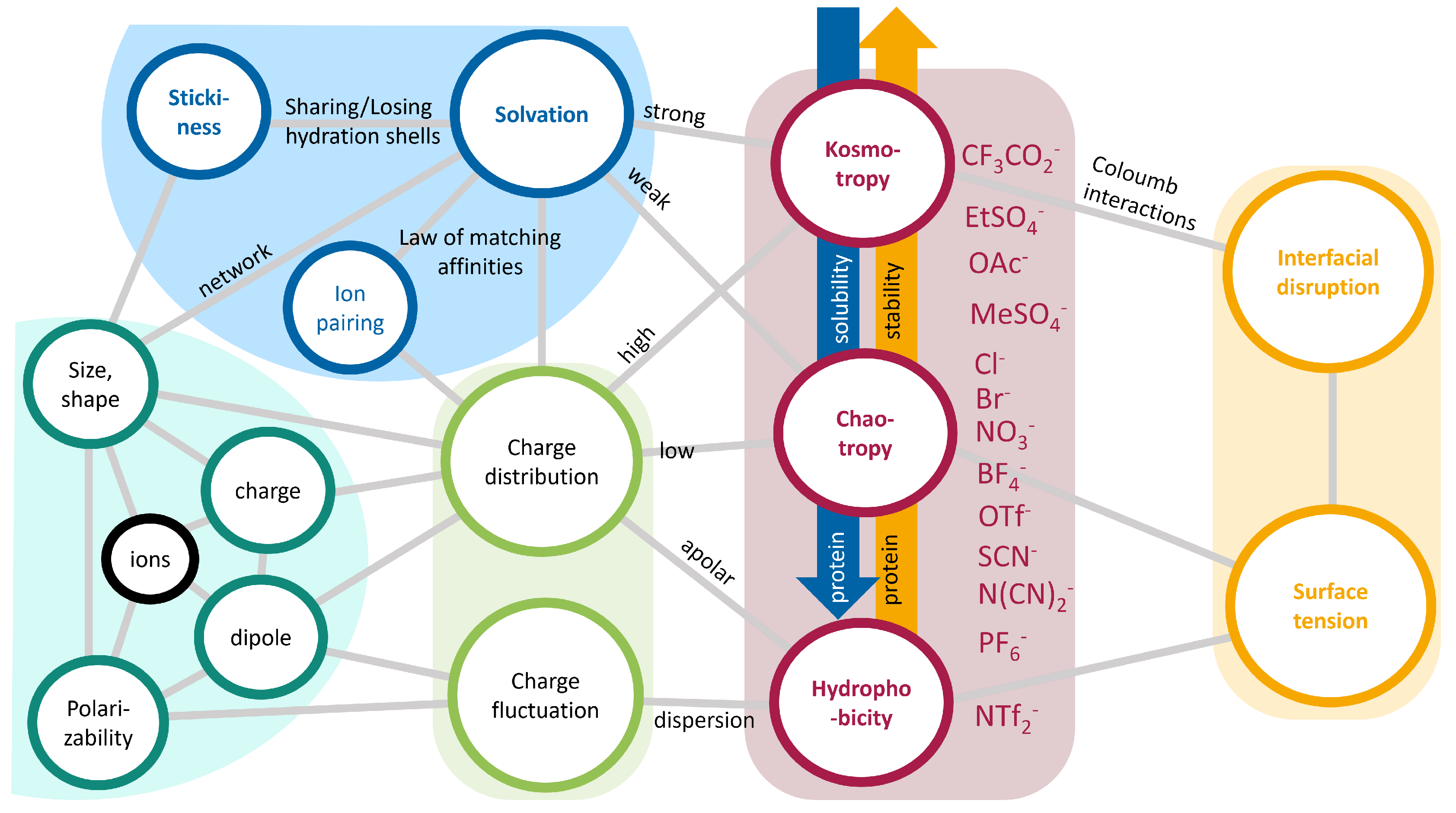
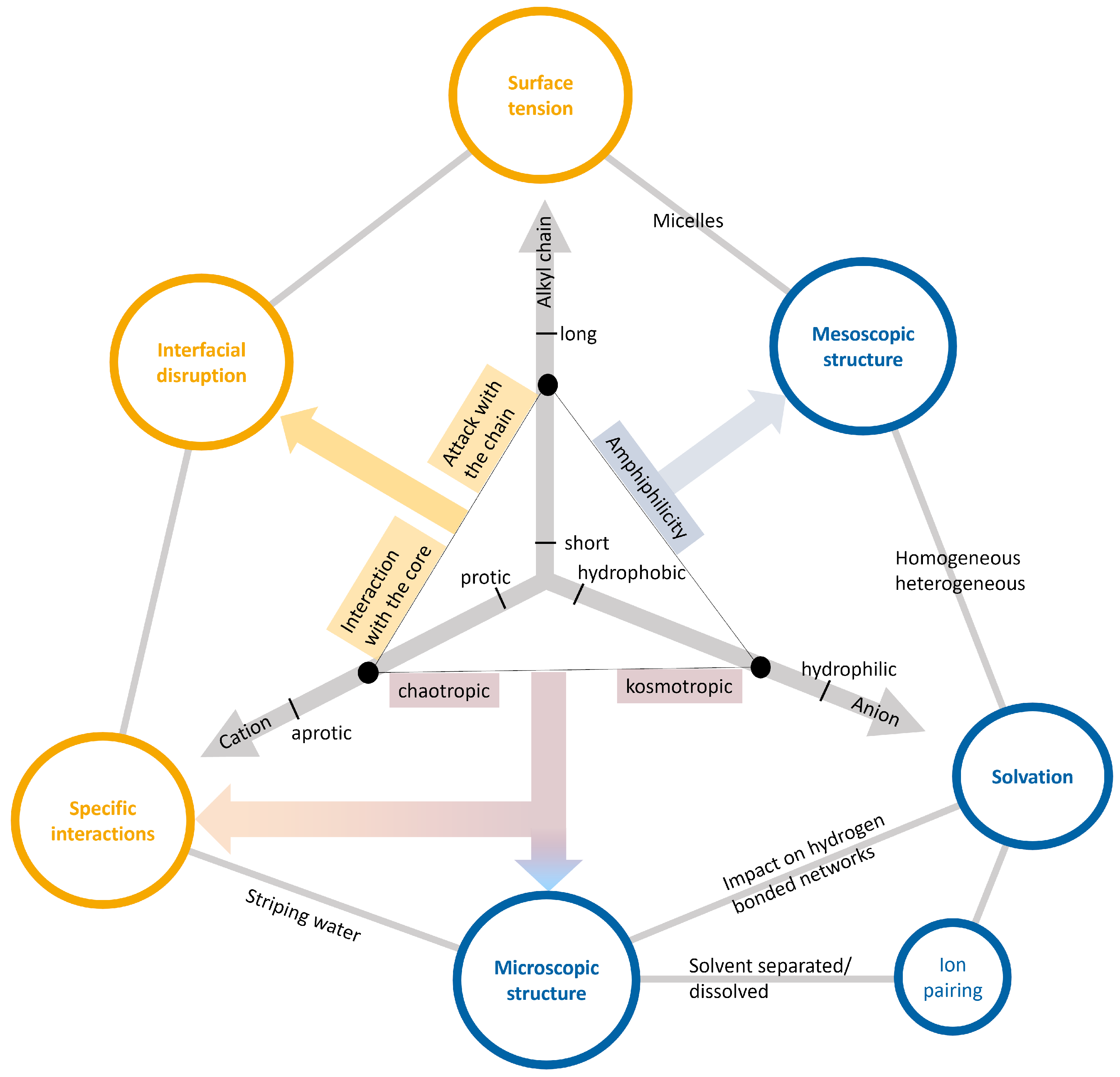
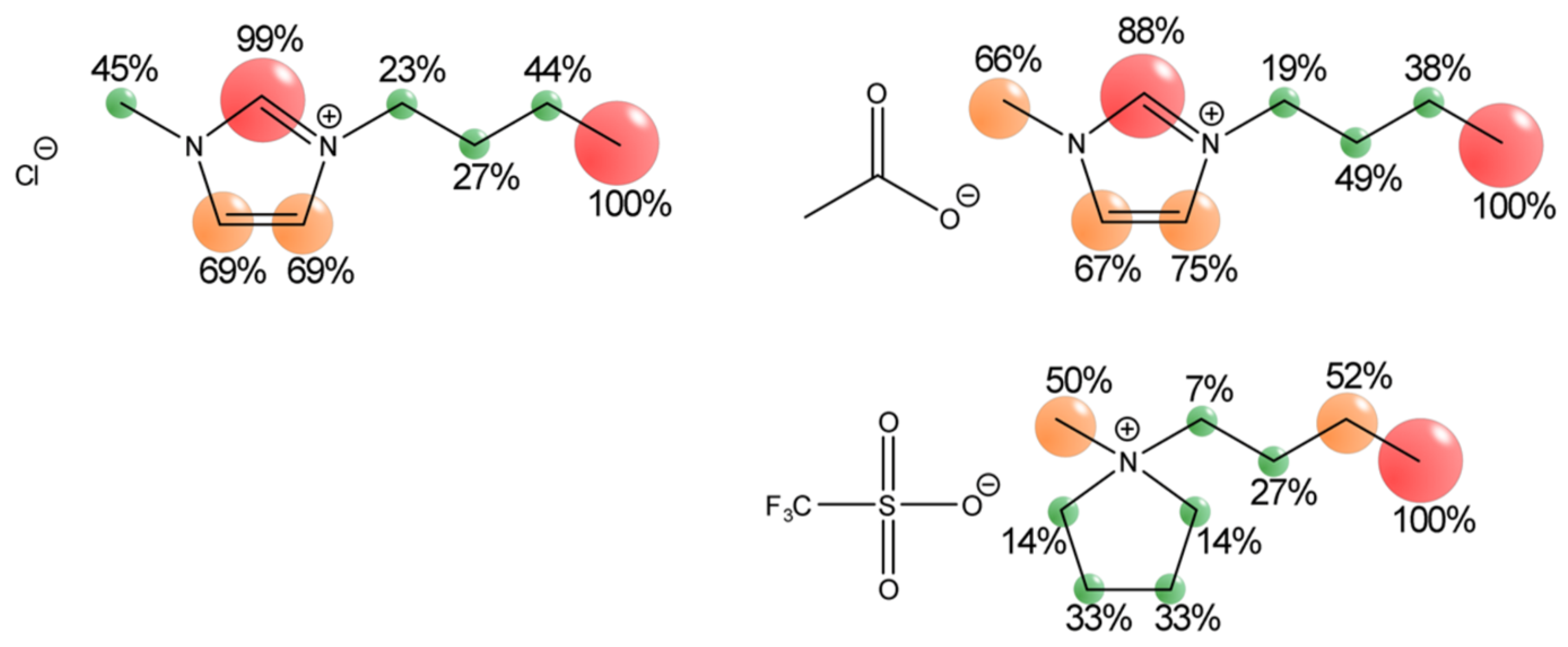
2.3. Ionic Liquids as Co-Solvents

The manifestations of Hofmeister effects in non-aqueous solvents [61,96], such as ionic liquids [97], have garnered significant attention in recent literature. Contrary to the classical paradigm, which portrays ionic liquids as inert, non-interactive solvents [14,27,98], emerging evidence challenges this notion by demonstrating their activity in chemical processes [15,27,39,94]. The mechanisms underlying such participatory roles are multifaceted and include the stabilization of charged reaction intermediates, selective solvation around either hydrophilic or hydrophobic moieties, and significant alterations in the local electrostatic environment. Due to these attributes, ionic liquids emerge as viable co-solvents in biomolecular systems, notably in stabilizing proteins within aqueous matrices [70,99,100]. A salient case in point is the variable influence of ionic liquids on the melting point of ribonuclease A, illustrating the capacity to fine-tune solvent properties to suit particular solutes or applications [94].
Historically, the academic focus has been predominantly on ionic liquids bearing imidazolium, pyrrolidinium, or phosphonium cations. The advantage of these specific cations lies in the extensive experimental and computational data available, thereby simplifying the interpretation of novel findings. Conversely, although ammonium-based ionic liquids represent one of the earliest known categories [101], they have gradually receded in terms of prevalence in contemporary research. In biological contexts, however, ammonium-based constituents present distinct advantages. They emulate some of the key physicochemical properties of water, such as facilitating proton transfers between acidic and alkaline functionalities and participating in hydrogen bonding networks [30]. Consequently, these species exhibit higher biocompatibility by minimally perturbing the structure of aqueous matrices. Ethylammonium nitrate is an excellent medium for recovering denatured/reduced hen egg white lysozyme [102]. In particular, the denatured protein is prevented from forming aggregates, thus enabling gentle refolding, resulting in recovery rates of up to 90%. A significant advantage of such additives is that these can often be easily separated from the active protein by simple desalting methods. Interestingly, the same strategy can also be used to gain X-ray diffraction quality protein crystals [103]. This is based on the non-monotonic solubility behavior of lysozyme, which shows a minimum at intermediate ethylammonium nitrate concentrations and a maximum at high ionic liquid content.
Particularly noteworthy is the cholinium cation, distinguished by its bioavailability, economic viability, and facile synthetic procedures. Furthermore, cholinium-based ionic liquids demonstrate remarkable efficacy in stabilizing proteins under extremely acidic conditions. The judicious selection of ionic liquids can thus counter multiple stressors that adversely affect proteins, including hostile chemical environments and temperature oscillations. Such attributes render them indispensable as co-solvents, especially in extending the shelf-life of peptides and proteins by mitigating undesirable denaturation and aggregation phenomena [30].

Properties of Ionic Liquids

The unique physicochemical attributes of ionic liquids offer a wide scope of customization for specialized applications. While the archetypal ionic liquids predominantly consist of commonly used cations such as imidazolium, ammonium, and pyridinium, the theoretical combinatorial possibilities for generating binary mixtures extend into the millions [104]. This array of combinations contrasts sharply with the limited spectrum of conventionally employed molecular solvents and the finite set of systematically explored ionic liquids [105] using the anions depicted in Figure 6. The formidable challenge in biomolecular interactions is the identification of suitable ionic liquid mixtures with the requisite properties. While heuristic guidelines may offer some direction, the precise prediction of the properties of a given mixture remains an intricate endeavor. A quintessential characteristic of biomolecules, particularly proteins, is the simultaneous existence of both polar and apolar domains. This complex structure enables an array of interaction modalities with polar solvents, most notably water, and the hydrophobic core functional groups [35]. Chirality presents another critical aspect for ionic liquids, especially considering the inherent chirality of biomolecules such as sugars, DNA, and amino acids. Chiral ionic liquids have been effectively employed in various applications, ranging from capillary electrophoresis of proteins to separation techniques employing aqueous biphasic systems [106,107,108]. While cationic chirality is more prevalent due to the frequent use of symmetric inorganic anions, instances of anionic chirality have also been reported as early as 1999 [14,109].
Ionic liquid cations comprise an aliphatic, hydrophobic tail and a polar, hydrophilic head group [110] as shown in Figure 7. Such amphiphilic moieties undergo self-assembly on the nanoscale, forming mesoscopic regions (blue sphere) that dictate the homogeneity or heterogeneity of the resulting solutions, e.g., one-phase systems or micellar or biphasic systems [35,39,111]. The amphiphilic nature of the cation can be systematically modulated by adjusting the length of the alkyl chain. For example, the solubility of 1-alkyl-3-methyl-imidazolium hexafluorophosphate in water diminishes from methyl to nonyl chains, eventually leading to complete immiscibility [112]. The alkyl chain itself is the major source [91,113] of the surface tension of the liquid solution sketched by the chain arrow pointing to the surface tension in Figure 7. If the mesoscopic structure of the ionic liquid in water prefers micelles [91], this will significantly impact the surface tension. The anionic component often serves as a mitigating agent against the hydrophobic tendencies of the cations. Nevertheless, there are also a plethora of hydrophobic anions, i.e., PF 6 or NTf 2 . As the cations cannot reverse their hydrophobic character, the anion axis in Figure 7 points to the solvation, and the anion characterizes the hydrophilicity or hydrophobicity of an ionic liquid [31]; i.e., the chaotropic or kosmotropic character of the anions overwhelms that of the cation, as already discussed before. The combination of the kosmotropic/chaotropic character of the ions determines the microscopic structure (red sphere in Figure 7). If the anion–cation interaction is sufficiently strong, contact ion pairs will be the dominant structures, and, if the ion–solvent interaction is stronger, solvent-shared and solvent-separated ion pairs will prevail [114,115,116].
Various classification paradigms have been proposed based on ionic liquids’ molecular compositions and properties. A commonly employed distinction categorizes cations into protic and aprotic types [35], which relates to the potential for proton exchange between cations and anions. Despite this interdependence, these terms are generally applied exclusively to cations. Protic ionic liquids can be conveniently synthesized via acid–base neutralization reactions. Aqueous ionic liquid solutions often cause significant pH shifts due to the acidic or basic nature of the ions. These shifts also influence the surface charge of proteins [81,117]. To address this, most studies use ionic liquid in combination with buffers to maintain a constant pH [35,81]. Before proton exchange, intermolecular hydrogen bonds are established between cations and anions. These bonds may persist even without actual proton transfer and can significantly impact the microscopic structure (red sphere in Figure 7) of the ionic liquid, leading to various states, such as ion pairing, solvent-separated ion pairs, or single dissolved ions [70,116]. Not only do the cations compete with the anions in hydrogen bonding to the water molecules but the anions may also stripe water from the protein surface, decreasing the protein stability. Strong hydrogen bonding between the anion and water may also lead to an expulsion of the cations and their accumulation at the protein surface [70,118,119]. Hydrogen bonding can be quantitatively characterized using Kamlet–Taft parameters α , β , and π * , which denote a solvent’s hydrogen bond donating and accepting capabilities and its polarizability [35,120]. The hydrogen bond accepting parameter β is primarily dominated by the anion and affects the stability of proteins significantly [121]. β values higher than 0.8 are known to deactivate protease, which has been rationalized by the fact that the ionic liquid anions interfere with the protein’s own hydrogen bond network. This way, Kamlet–Taft parameters can indicate the biocompatibility of ionic liquid–protein mixtures.
Although the low charge density makes proteins usually hydrophobic in water, Coulombic interactions of charged amino acids with the ionic liquid ions may lead to their accumulation at the protein surface. The higher charge density of the smaller anions [122] results in their slower protein surface diffusion compared to the cations [119,123]. Competing with electrostatic forces, van der Waals interactions mainly arise due to the presence of the alkyl chains of the cation. Based on the length of their aliphatic chains, ionic liquid cations disrupt the protein interface in a different manner: Short-chain cations predominantly interact with proteins via their charged core, while long-chain analogs engage through their hydrophobic tails [70]. This dual behavior of the cations is reflected in Figure 7 as the cation arrow points to the specific interactions [39,119,124].
Using saturation transfer difference NMR experiments, Ref. [125] determined the relative frequency of the direct interactions of nitrogen-based ionic liquid cations and the green fluorescent protein. According to the NMR signals of Figure 8, the terminal methyl group of the butyl chain is consistently the most likely site to interact with the protein. Interestingly, the proton of the C2 carbon of the imidazolium ring is the second most favored spot, surpassing even the methyl group of the imidazolium ring. This indicates that structural similarity translates to similar interaction sites. The influence of the anion appears to be moderate as acetate and chloride do not significantly change the relative signal intensities. Specific ion effects (SIE) of ionic liquids with proteins are summarized in Table 2 of Ref. [39].

3. Experimental and Theoretical Studies

Inclusion bodies represent heterogeneous entities characterized by significant variations in their morphology, architecture, organizational features, and functional roles [4]. Given this inherent complexity, a comprehensive array of analytical techniques is frequently employed to decipher their intricate structural and functional fingerprints. As several experimental techniques to promote the theoretical understanding of inclusion bodies have been reviewed by others [4,7], we briefly mention the most important methods here.

3.1. Analytical Techniques to Study Folding States

NMR spectroscopy [4,7,126] is a valuable tool for monitoring the real-time structural changes in protein refolding, especially for smaller proteins (<40 kDa). However, it requires high-purity protein samples at concentrations ≥0.5 mM. In some cases, NMR analysis of inclusion bodies may be complicated by the presence of contaminants, such as phospholipids or other proteins, necessitating prior purification. Isotope labeling, using 13C and 15N, helps simplify spectra and enables the study of intermediate conformers. Saturation transfer difference NMR experiments [125] can identify cationic moieties interacting with specific protein sites for potential denaturing/refolding agent design. Kamatari et al. [127] employed NMR to investigate partially folded protein structures under varying pressure conditions. Additionally, water proton NMR, based on water–protein interactions, is sensitive to protein aggregate formation and can detect inclusion bodies.
Infrared [128,129,130,131] and Raman [129,132] spectroscopy can be applied to detect secondary structure elements as the amide I and II bands (1700–1600 cm−1) are the most prominent and sensitive vibrational bands of the protein backbone. Even membrane proteins’ unfolding and refolding pathways can be studied [130].
Circular dichroism spectroscopy [4,130,133,134] is vital for non-destructively distinguishing between refolded and denatured proteins by assessing conformational dynamics. This technique examines changes in both secondary structure (in the far-ultraviolet range, 190–250 n m ) and tertiary structure (in the near-ultraviolet range, 260–300 n m ) [134]. The far-UV region primarily reveals information about peptide bonds, while the near-UV region pertains to aromatic amino acids within the protein core. Far-UV circular dichroism is particularly useful for discerning α -helices and β -sheets, making it effective for distinguishing refolded proteins enriched in α -helices from denatured ones. However, it requires high sample concentrations, i.e., 0.1   m g / m L for far-UV and 1 m g / m L for near-UV studies. Recent advancements allow real-time in situ analysis of protein aggregation and conformational changes. Nevertheless, certain proteins’ insolubility can limit far-UV circular dichroism, leading to increased light scattering and compromised signal-to-noise ratios [135].
Fluorescence spectroscopy [4,136] is a non-destructive analytical method, enabling real-time assessment of aggregation kinetics, ranging from monomeric states to forming sizable aggregates. Additionally, it possesses the capability to monitor the self-assembly processes of proteins. The technique’s sensitivity—often impeded by chaotropes—can be increased by employing either intrinsic or extrinsic fluorophores. In the case of intrinsic fluorophores, the fluorescence characteristics of aromatic amino acids are harnessed for detection. Conversely, extrinsic fluorophores rely on externally applied fluorescent probes that either covalently bind to hydrophobic regions or chemically attach to the target molecule. The elevated sensitivity of extrinsic fluorescence renders it particularly suited for high-throughput screening applications in the context of protein refolding. Variations in surface hydrophobicity are concomitantly reflected by increments in fluorescence intensity during the refolding process. However, the utilization of extrinsic dyes can potentially perturb the native protein structure, thereby influencing the integrity of the detection process. Moreover, the quantum yield associated with intrinsic fluorescence methodologies is markedly inferior compared to that of extrinsic fluorescence approaches.
Dynamic light scattering [129,137,138] assesses protein surface charge properties, making it adept at quantifying electrostatic interactions among molecules. This renders it suitable for sizing protein aggregates ranging from 1 nm to 5 μm [138]. To ensure optimal sensitivity, it is crucial to maintain a stable experimental environment, including constant temperature, viscosity, and interparticle interactions, while avoiding excessively high protein concentrations. Dynamic light scattering offers high-throughput capabilities and characterizes both reversible and irreversible aggregates. Unlike fluorescence-based methods, dynamic light scattering is non-invasive, reducing the risk of assay-induced aggregation and measurement artifacts. However, its data are often semi-quantitative, and its high sensitivity can also lead to false positives, detecting extraneous particles like dust.
Atomic force microscopy (AFM) offers a nuanced approach for characterizing complex and heterogeneous conformational states [129,139]. Achieving high-resolution AFM imaging hinges on two pivotal parameters: vertical and lateral resolution. The former is principally susceptible to environmental factors such as thermal and acoustic noise, typically rendering a resolution in the order of 1 Å. Conversely, the lateral resolution is constrained by the finite geometric dimensions of the AFM tip and commonly varies between 1 and 10 n m . These variables are paramount for ensuring the fidelity and reproducibility of AFM data, particularly in examining monomolecular and aggregated protein structures.

3.2. Molecular Dynamics Simulations to Study (Re-)Folding

All-atom molecular dynamics is a computational methodology that solves Newton’s equations of motion under stipulated temperature and pressure conditions using a classical force field [140]. Investigating the spontaneous time-dependent dynamics of biomolecular systems provides atomistic insights into relatively transient physicochemical phenomena, but its applicability becomes constrained for larger molecular assemblies or for processes that occur over physiologically relevant timescales. Despite advancements in computational hardware and algorithmic optimization, the method’s efficacy diminishes when probing phenomena like molecular self-assembly that require exhaustive phase space exploration, particularly for complex biomolecular systems with high energy barriers and protracted equilibrium timescales. Insights at the molecular level have proven invaluable in interpreting experimental findings [141]. In the realm of self-assembled peptide-based nanostructured materials, it is often desirable to employ a “coarse-grained” representation [142,143,144], which entails a significant reduction in atomic-level details. This simplification consequently diminishes the molecular flexibility of the solute, thereby facilitating larger temporal integration steps and longer simulation periods [140]. Enhanced conformational sampling algorithms, such as “umbrella sampling” or “steered molecular dynamics”, possess the capability to augment the traditional brute-force molecular dynamics simulations in modeling protein folding. These specialized algorithms are designed to facilitate the exploration of molecular conformations that are separated by high energy barriers, all within computationally feasible timeframes. The inherent biasing parameters in these algorithms permit an expanded sampling of the conformational space, thereby enabling a more detailed understanding of the specific molecular interactions [140]. “Replica exchange molecular dynamics” simulations are a sophisticated sampling technique characterized by the concurrent execution of multiple replicated simulations under varying temperature conditions or other control variables. These simulations operate in parallel, and random exchanges of atomic coordinates between them are facilitated at predetermined time intervals. Crucially, to maintain the integrity of each system’s Hamiltonian—defined as the sum of the average potential and kinetic energy—coordinate exchanges are permitted solely when the potential energies of the two interacting replicated systems overlap. In temperature-variable replica exchange studies, introducing higher temperature conditions augments the frequency at which energy barriers separating kinetically trapped microstates are crossed. Consequently, replica exchange MD has found valuable applications in studying spontaneous self-assembly mechanisms by enabling these aggregated systems to surmount energy barriers and thus explore a broader conformational space. “Metadynamics” [145,146] is another enhanced sampling technique employing one or more collective variables to characterize movement through multidimensional phase space. These collective variables can range from simple geometric parameters like distance, angle, or dihedral angle between atoms to more complex descriptors derived from techniques such as principal component analysis.
Also, artificial intelligence may contribute to predicting the folded structure of proteins [147]. In contrast to the interaction-based models described before, programs like AlphaFold follow an evolutionary-based approach. The evolutionary paradigm leverages bioinformatics tools to analyze evolutionary constraints on protein structure, benefiting from the expanding repository of experimentally solved structures and advances in genomic sequencing and deep learning. AlphaFold [147] represents a significant advancement in protein structure prediction as it features a unique architecture designed to integrate multiple sequence alignments, an innovative output representation coupled with an associated loss function for accurate end-to-end prediction, and the application of equivariant attention mechanisms. Additional refinements include using intermediate losses for iterative refinement, masked multiple sequence alignment loss for joint training, and self-distillation techniques to learn from unlabeled protein sequences.

3.3. Screening of Key Parameters

The application of both computational and experimental methodologies has substantially enhanced our theoretical comprehension, facilitating the development of models for the solubilization and refolding of proteins [1]. While there are substantial data under conventional conditions, the limited data availability necessitates a systematic examination of the critical parameters in relation to the specific ionic liquid and its concentration.
Smiatek and co-workers [12] investigated the influence of various process parameters on the classical solubilization of inclusion bodies without ionic liquids through explainable machine learning. The final yield of solubilization is predominantly determined by three key factors: the total protein concentration, urea concentration, and the fraction of protonated titrable groups influenced by the actual pH level. Boosting ensemble-based models, characterized by normalized root mean square errors around 0.19, have been identified as the most accurate for predictive analysis. Through the application of “Shapley Additive Explanations” values, they observed that these three variables considerably outweigh other factors, such as dithiothreitol and GndHCl concentration, in their effect on the final yield. Detailed “Shapley Additive Explanations” analysis further illuminates an inverse relationship between total protein concentration and yield values, which can be understood via surface solvation effects. Conversely, increasing urea concentrations and fractions of protonated titrable groups facilitate higher yields, likely through preferential binding and exclusion mechanisms and electrostatic repulsion, respectively.
While there are some experimental studies [148] that directly investigate the potential of ionic liquids for the refolding of solubilized proteins (see Table 2), the field is still young, and most of the research is aimed at understanding neurodegenerative diseases, like Parkinson’s or Alzheimer’s disease. Thus, the dissolution and formation of amyloid protein aggregates have been investigated more frequently than inclusion bodies. Much of the published literature concerning the interaction of ionic liquids with protein folding and aggregation can be categorized into three types of studies according to the purpose of the used ionic liquids:
  • Studies that use ionic liquids to dissolve aggregated or otherwise difficult-to-solubilize proteins;
  • Studies that add ionic liquids to solutions of denatured protein in order to induce the (re)folding of the previously unfolded protein;
  • Studies in which ionic liquids act as protein structure stabilizers that protect dissolved native proteins against unfolding and aggregation.
Table 2 depicts a list of experimental studies from the last two decades, sorted into these three categories. Recovering the active protein from inclusion bodies requires two steps: the dissolution of insoluble protein aggregates and the subsequent refolding into the native protein structure. Therefore, the experimental results are reviewed from these three perspectives in the next three sections on solubilization, refolding, and stabilization using the same subdivision as Table 2.

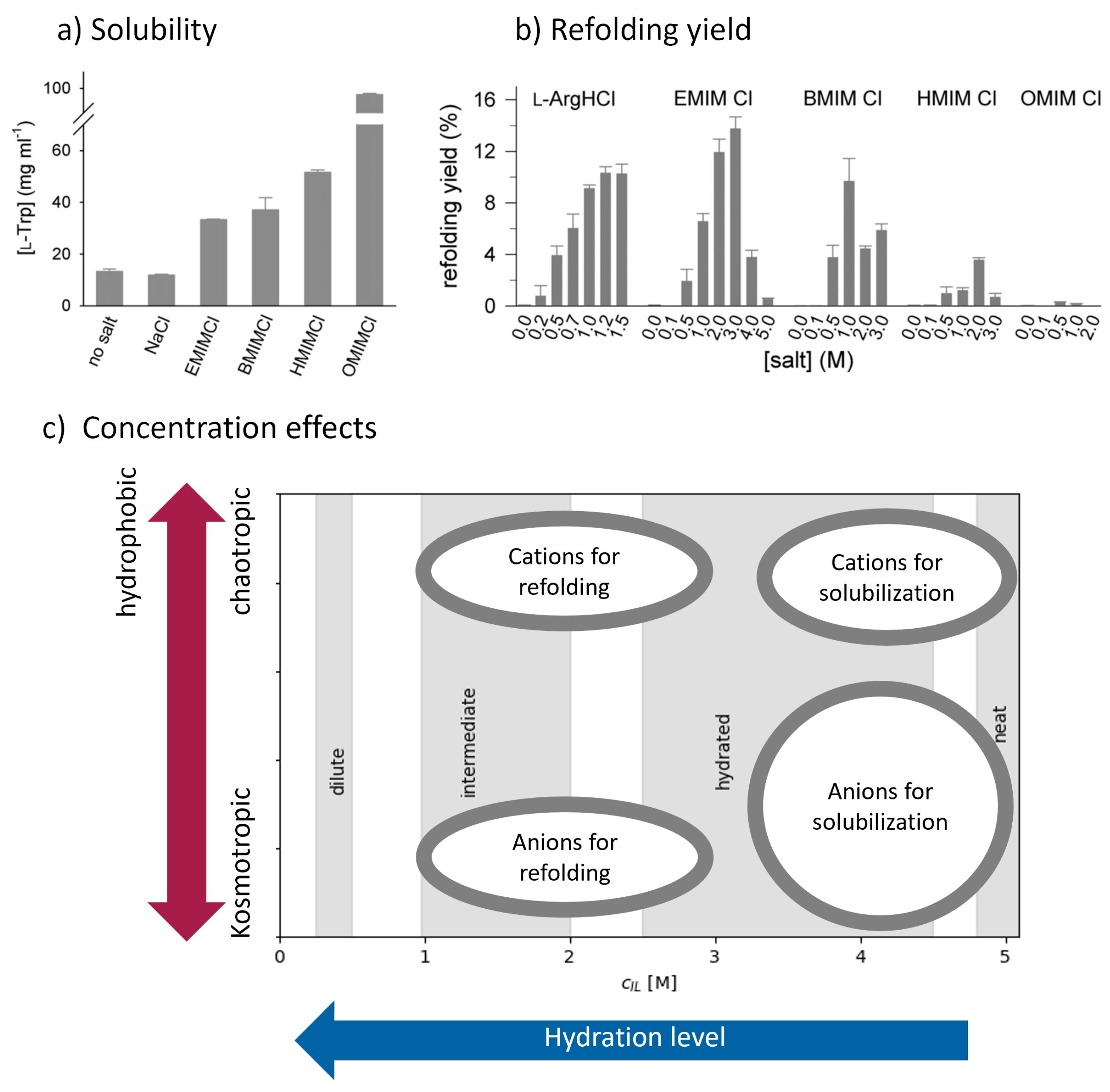
3.3.1. Ionic Liquids as Solubilizing Agents

To date, publications on the direct solubilization of inclusion bodies in ionic liquids are limited, with seminal contributions primarily stemming from the work of Fujita et al. [154,155]. Ref. [154] demonstrated the capacity of the hydrated ionic liquid [chol][dhp] to not only dissolve inclusion bodies but also to concomitantly refold the protein into its native conformation, thereby restoring its biological activity. Despite this potential as a solubilization agent, the solubility of the protein was very low, similarly observed for other native proteins in ionic liquids [152]. However, an economically feasible inclusion body process requires a high target protein solubility. Hence, understanding how the solubility of proteins in ionic liquids can be adjusted is crucial for their use as inclusion body solubilization agents.
In several studies, Fujita et al. observed that the hydration level of ionic liquids strongly affects protein solubility [150,171,172]. While neat ionic liquids can show low protein solubility, excessive hydration has also been shown to diminish solubility [150]. Hydration levels between three and seven water molecules seem to be the optimal range for protein solubilization [150,155,173]. Other research groups [23,159] have dissolved amyloid insulin fibrils in ionic liquids with hydration levels of five water molecules per ion pair, further supporting these findings. Like classical inclusion body solubilization, the chaotropicity and hydrophobicity of the ionic liquids’ cations also greatly impact protein solubility. In their follow-up study to Ref. [152], Fujita et al. [155] improved the inclusion body solubility by using tetraalkyl-ammonium and -phosphonium cations over -cholinium. Overall, they observed the best solubility for cations with butyl chains. Similar results were observed for the dissolution of amyloid fibrils, where BMIM + -based ionic liquids resulted in more efficient amyloid dissolution than other cations [23,159]. At low concentrations, the ionic liquids’ chaotropic effect was consistent with the Hofmeister effects of their respective ions. At high ionic liquid concentrations, the Kamlet–Taft parameters α and β , corresponding to the solvent’s hydrogen bonding abilities, were highly correlated with the amyloid dissolution properties. Thus, Takekiyo et al. [159] suggested the combined α β parameter as a quantitative indicator for the amyloid dissolution ability of concentrated ionic liquids. However, Spange et al. [174] reported a linear relationship between α and β , casting doubt on the meaning of the difference between these parameters.
The influence of the ionic liquids’ anion on protein solubility is less consistent across the literature. For native cytochrome C, anions containing oxo acid residues were reported to vastly increase solubility over anions without this moiety [171]. However, a similar comparison for aggregated protein is missing. While the very chaotropic anion SCN was found to be better at dissolving amyloids than NO 3 and Cl at mole fractions x I L < 15%, the order changed for x I L > 15% [23,158,175]. Similarly, the dhp anion seemed to increase the solubilization of inclusion bodies and heat-aggregated protein over halogen anions Br and Cl . However, the effect was inconsistent across all tested ionic liquids and less significant than the effect of the cation [150,155]. Combining ionic liquids with other protein-solubilizing conditions, like alkaline pH, is very poorly explored yet but might be essential to solve the current challenges in inclusion body processing. Ref. [149] fully dissolved heat-denatured hen egg whites using a mixture of [BMIM][SCN] (40% v/v) and 0.5 NaOH, which was not possible with 8 M urea, 7 M GndHCl, formic acid, 10% SDS, or the ionic liquid and alkaline components alone. A microscopic study on the dissolution of wool and silk in ionic liquids [156] revealed that reducing agents can also greatly impact protein solubility. It is likely that adding a reducing agent might be crucial for proteins containing disulfide bridges. Regrettably, to our knowledge, there are no practical studies on this topic yet.
Compared to conventional solubilizing agents such as urea and guanidinium chloride, ionic liquids have been shown to be more effective in the solubilization of protein aggregates [175], attributable largely to their inherent hydrophobic and chaotropic properties. However, these attributes also tend to destabilize or denature protein structures [94,124,176]. Moreover, unambiguous evidence supporting the restoration of native protein structures, as validated by enzymatic activity, has predominantly been obtained following buffer exchange or dilution into aqueous media [154,155,157,171]. Apparently, such a step might still be necessary to achieve fully refolded protein. Hence, finding a balance between the solubility of the aggregates and the disruption of protein structures is important, similar to the well-known “mild solubilization approach” [1].
Another issue analogous to traditional inclusion body solubilization is the reaggregation of the solubilized protein (see Figure 3). While the secondary structure content of amyloid aggregates treated with EAN was shifted from β -sheet structures toward helical structures [158], the other ionic liquids investigated in the same study seemed to further increase the β -sheet content of the treated amyloid fibrils. Takekiyo et al. [166] hypothesized that the helical structure induced by EAN might also be a polyproline II structure rather than α -helices. This polyproline II structure can easily transform into intermolecular β -sheets and thus promote reaggregation.
To summarize, when ionic liquids are used as solubilizing agents for inclusion bodies, the protein solubility must be increased, while, ideally, the secondary structures of the protein are preserved, or at least not fully denatured. The main factors that influence solubility seem to be the amount of hydration of the ionic liquid and the hydrophilicity as well as chaotropicity (i.e., increasing chain length) of the respective cation, as shown in Figure 9a. This finding is supported by the fact that the addition of sodium chloride has no beneficial effect on solubility since neither sodium nor chloride is hydrophobic. However, hydrophilic and/or chaotropic cations can destabilize or unfold the solubilized protein structure and thus must be chosen carefully. In contrast to the solubility in Figure 9a, short chain cations and protonated L-arginine have higher refolding yields, as depicted in Figure 9b. As the anion seems to have a lower impact on solubility but a strong influence on the protein structure (as discussed below), ionic liquids composed of a chaotropic cation and a kosmotropic anion with a hydration level between three and five water molecules per ion pair seem to be the best choice as a solubilizing agent (see Figure 9c).

3.3.2. Ionic Liquids as Refolding Additives

The standard refolding method of diluting the solubilizate with aqueous buffer, thereby replacing the denaturant with mainly water molecules, is simple. However, it also reduces the product concentration immensely. While this has the beneficial side effect of decreasing the chance of proteins to aggregate, it is ecologically and economically disadvantageous due to the vast amounts of buffer, the required large-scale equipment, and the subsequent need to concentrate the product. Refolding additives can reduce required dilution by either directly inducing a structural shift in the protein towards the native structure or by preventing reaggregation. Due to their preferential interaction with the protein, ionic liquids can influence the folding of denatured protein even without dilution. Singh and Patel observed a partial refolding of alkali-denatured cytochrome C by long alkyl chain imidazolium chlorides [OMIM]Cl and [DMIM]Cl [164]. However, not all unfolding stress was counteracted equally. For urea- and GndHCl-denatured proteins, the ionic liquids only had a marginal effect. Comparable results were observed for cationic gemini surfactants with a similar structure to tetra alkyl-ammonium, a cation often found in ionic liquids [177]. In both cases, stabilizing a native-adjacent folding intermediate was more pronounced for a longer alkyl chain. These interesting results contrast the effects observed in screening studies of ionic liquids used as buffer additives for dilution refolding. In these cases, less hydrophobic cations, such as EMIM + and sometimes BMIM + , were more effective refolding enhancers than their counterparts with increased hydrophobicity, as shown in Figure 9b and reported by several authors [160,161,163]. [EMIM]Cl and [BMIM]Cl show their largest impact on the refolding at concentrations from 0.5 M–3 M, which is in the intermediate to hydrated solvation regime (see Figure 5a and Figure 9c). Long alkyl chains on organic cations and anions prevented refolding as they even unfolded the protein via hydrophobic interactions [161,163]. The choice among the ionic liquids’ anion seems to have a greater impact on the refolding of the protein than in the solubilization step. Generally, more kosmotropic anions improved refolding yields over chaotropic anions [161,163]. However, kosmotropicity is not the only important factor for anions. In one example [162], [EMIM]Cl resulted in higher refolding yields than [EMIM][EtSO 4 ] or [EMIM][OAc] despite OAc and EtSO 4 being more kosmotropic than the Cl anion. The Cl-ion was also found to stabilize native protein structures slightly or at least destabilize them less than other anions [57]. Since this study did not include OAc or EtSO 4 anions, it is difficult to say if a Hofmeister or another type of effect is responsible for this observation. Despite their reported tendency to destabilize the native protein structure [57,94], many ionic liquids still enhance the refolding yield when employed as an additive in the refolding buffer [160,161,163]. This increase in refolding yield is usually attributed to their ability to prevent protein reaggregation. However, the mechanistic explanation for this effect still needs to be clarified. For low ion concentrations (< 50 m M ), Kutsch et al. [169] found the aggregation of insulin to be determined by Coulomb interactions and therefore to be independent of the ion properties. Up to salt concentrations of 100 mM, aggregation kinetics accelerated with the ion concentration. At higher concentrations, ion-specific effects dominated, and aggregation generally slowed down again. The extent of this inhibition was ion-specific and followed a Hofmeister ranking of the tested cations (the anion was chloride for all the tested salts). A recent study [33] observed that the growth among amyloid fibrils in different ionic liquids and aqueous buffers led to significant morphological differences between the fibers. This difference was attributed to changes within the protein monomer structure, which led to a different oligomerization pathway. This link between secondary structure shifts and aggregation is supported by other reports [166,168], but further studies are necessary to fully understand which ionic liquids are suitable to prevent aggregation of a specific protein.
The recovery of the refolded protein from concentrated ionic liquids is often achieved by incubation with ethanol at −80 °C, after which the precipitated native protein can be dried and reconstituted in aqueous buffer [155,170]. Recovering the protein in crystallized form might be beneficial for storing and shipping the product. However, separation of the protein via chromatography using a phenyl column was also reported [155] and led to a higher recovery of protein activity. Since chromatography steps are the state-of-the-art purification steps after protein refolding, they could be well-suited to ionic-liquid-assisted refolding. Again, this would match the already established workflow of refolding processes.
In summary, ionic liquids that have been effectively used as refolding additives can be described as containing a chaotropic cation with moderate hydrophobicity and a kosmotropic anion. The main difference to ionic liquids that were reported as effective in solubilizing aggregates is the hydration level and slightly lower hydrophobicity (cf. Figure 9c). They were typically used at concentrations between 0.5 M and 3 M, corresponding to a mole fraction of x I L < 0.1. Therefore, refolding might be possible by solubilizing the aggregates with hydrated ionic liquids and diluting the solubilizate with a much smaller dilution factor than traditional refolding processes. Since this approach matches the already established workflow of traditional inclusion body processes, it could easily be integrated into industrial processes.

3.3.3. Ionic Liquids as Stabilizers

Regarding the stability of non-enzymatic proteins, Veríssimo et al. [111] investigated four different parameters, including structural and thermal stability, as well as activity and aggregation, a summary of which is provided in Figure 10. The most promising category is aggregation (see Figure 10D), where all the investigated types of ionic liquids have decreased aggregation in more than 50% of the cases. Since inclusion bodies need to be solubilized before partial unfolding can occur, deaggregation is a crucial step in recovering the native structure. Intriguingly, consistent structural and thermal stability trends were observed for ionic liquid cations (see Figure 10A,B). Ammonium-based ionic species were the most conducive to stabilizing protein structures, followed by cholinium and imidazolium, with pyridinium trailing behind. Possibly, ammonium ions are less disruptive to the hydrogen bonding network of an aqueous medium owing to their capability to engage in hydrogen bonding themselves. With respect to bioactivity in Figure 10C, the trends diverged significantly. While ammonium-based ions appeared less effective in stabilization, cholinium, attributed to its biocompatibility, positively influenced bioactivity in approximately two-thirds of the investigated scenarios. Intriguingly, imidazolium ions also enhanced bioactivity in over 60% of the protein–ionic liquid combinations studied. Conversely, neither pyridinium nor pyrrolidinium exhibited any positive impact on bioactivity. This may be because these cations are less studied, and, consequently, the corresponding statistical values in Figure 10 are less reliable.

4. Challenges and Limitations

Although ionic liquids present a promising alternative for the treatment of inclusion bodies, some fundamental challenges and limitations remain, as addressed in the SWOT (Strengths, Weaknesses, Opportunities, and Threats) analysis in Table 3.
The major strength of ionic liquids lies in their advantageous properties for hydrophobic protein solubilization [111,148,175]. Their superior efficacy in dissolving protein aggregates, as compared to traditional chaotropic agents such as urea and GndHCl, stems from their unique hydrophobic and chaotropic properties [94,175,176]. Moreover, their customizability allows for molecular-level tailoring to specific proteins, enhancing their performance as stabilizers [70,99,111], which is impossible for traditional solubilizers. They also offer a wide range of hydrophilic to hydrophobic ion combinations. Furthermore, ionic liquids often facilitate higher protein recovery rates, countering the limitations of traditional solubilization methods, which tend to yield low recovery [40,42,111].
However, several limitations temper the enthusiasm for applying ionic liquids in inclusion body treatment. Notably, the production costs associated with these solvents can be prohibitively high [111], particularly when considering large-scale applications. The effectiveness of ionic liquids can also exhibit significant variability depending on the specific protein target, necessitating case-by-case optimization [12,32,111]. Moreover, incorporating ionic liquids typically necessitates subsequent purification steps to remove residual amounts, thus introducing complexities into the protein recovery process [42]. In addition, some of the ionic liquids may destabilize proteins and are consequently toxic and raise human health concerns.
Beyond immediate practical applications, the use of ionic liquids also presents several intriguing research opportunities. Their unique stabilizing effects on proteins offer the potential for the development of new protein-based products with enhanced stability [40,111]. The improved solubilization capabilities of these agents could notably increase the overall yields in protein production processes. Additionally, their complex physicochemical properties could drive advancement in novel protein purification methodologies. Furthermore, ionic liquids may act as multipurpose co-solvents as their properties concerning proteins strongly depend on their concentration. This may reduce purification steps as the salt concentration is usually changed between the solubilization/denaturing and the refolding step.
Nevertheless, several threats, challenges, and considerations must not be overlooked [111]. Environmental pollution remains a worry, particularly at large industrial scales. The health effects of ionic liquids are not yet fully understood, and initial research suggests they could be harmful. Regulatory barriers could also arise due to their unique chemical properties, especially in places with strict safety laws [42]. Intellectual property issues in protein refolding could further complicate their use. Lastly, the inconsistency in their dissolving and stabilizing abilities poses risks, particularly when dealing with different proteins. Consequently, there is no “the best ionic liquid” for a larger variety of proteins.

5. Conclusions

In summary, the recent incorporation of ionic liquids in the biotechnological realm, especially concerning protein solubilization and refolding, is a remarkable development. Ionic liquids are lauded for their unique solubilizing capabilities and the flexibility they offer in terms of chemical customization. However, they introduce a set of challenges, such as heightened costs, complex purification protocols, and unresolved questions related to environmental and health impacts, as well as regulatory compliance. The mild solubilization strategy aims to maintain the protein structures inherent in inclusion bodies, thereby enhancing refolding yields and minimizing protein reaggregation. This approach involves reducing concentrations of urea and GndHCl during the solubilization process while also deploying strategies such as the use of alkaline pH levels, high pressure, detergents, and organic solvents, along with freeze–thaw cycles to amplify protein solubility. Ionic liquids emerge as a potent addition in this setting, serving dual roles as agents that can solubilize protein aggregates without disrupting their native-like structures and as refolding additives that counteract the traditional denaturants. However, the wide variety of ion combinations makes empirical screening of buffer components a daunting task.
Current modeling approaches for the solubilization and refolding of proteins would benefit from a consistent and transferable thermodynamic understanding of the processes. At present, the absence of comprehensive databases containing systematically screened parameters hampers the utilization of artificial intelligence in model development, thereby preventing a holistic approach. Empirical rankings, such as the Hofmeister series, provide valuable qualitative insights but fall short of offering a comprehensive mechanistic understanding. These rankings are further limited in their applicability due to their basis on single-ion effects, which are most relevant at low concentrations. However, many practical applications necessitate considerably higher concentrations, thereby questioning the relevance of such rankings due to higher-order effects. Specific ion effects may describe the interactions with the protein better but fail the goal of elucidating the general pathways for protein refolding.
To move beyond these limitations, there is an increasing recognition of the potential utility of molecular dynamics simulations paired with mechanistic models to elucidate the thermodynamics of protein (un)folding. Such a rigorous science-based approach could lend physical parameters to guide experimental designs and thereby deepen our understanding of the process. Advanced machine learning techniques, particularly as embodied in algorithms like AlphaFold, are making headway in sequence-based protein structure prediction. These computational advancements could eliminate the need for resource-heavy structural analytics, offering a detailed three-dimensional structure derived from the protein’s sequence instead. This innovative integration could potentially allow for a complete predictive model for the refolding process based on the protein sequence alone.
In light of these considerations, while ionic liquids offer tantalizing possibilities for future research, they come appended with many challenges and considerations that must not be sidestepped. A multidisciplinary approach that harnesses both empirical experimentation and computational modeling could pave the way for more efficient, economically viable, and environmentally friendly applications of ionic liquids in protein refolding.

Funding

This research was funded by Christian Doppler Research Association grant number Inclusion Body Prozessierung 4.0.

Acknowledgments

R.K. and O.S. want to thank the Christian Doppler Forschungsgesellschaft (CDG) and Boehringer Ingelheim RCV for their funding of the “CD-Labor für Inclusion Body Prozessierung 4.0” facilitating this study.

Conflicts of Interest

The authors declare no conflict of interest.

References

  1. Klausser, R.; Kopp, J.; Brichtova, E.P.; Gisperg, F.; Elshazly, M.; Spadiut, O. State-of-the-art and novel approaches to mild solubilization of inclusion bodies. Front. Bioeng. Biotechnol. 2023, 11, 1249196. [Google Scholar] [CrossRef] [PubMed]
  2. Tsumoto, K.; Umetsu, M.; Kumagai, I.; Ejima, D.; Arakawa, T. Solubilization of active green fluorescent protein from insoluble particles by guanidine and arginine. Biochem. Biophys. Res. Commun. 2003, 312, 1383–1386. [Google Scholar] [CrossRef] [PubMed]
  3. Umetsu, M.; Tsumoto, K.; Nitta, S.; Adschiri, T.; Ejima, D.; Arakawa, T.; Kumagai, I. Nondenaturing solubilization of β2 microglobulin from inclusion bodies by L-arginine. Biochem. Biophys. Res. Commun. 2005, 328, 189–197. [Google Scholar] [CrossRef] [PubMed]
  4. Kachhawaha, K.; Singh, S.; Joshi, K.; Nain, P.; Singh, S.K. Bioprocessing of recombinant proteins from Escherichia coli inclusion bodies: Insights from structure-function relationship for novel applications. Prep. Biochem. Biotechnol. 2022, 53, 728–752. [Google Scholar] [CrossRef] [PubMed]
  5. Buscajoni, L.; Martinetz, M.C.; Berkemeyer, M.; Brocard, C. Refolding in the modern biopharmaceutical industry. Biotechnol. Adv. 2022, 61, 108050. [Google Scholar] [CrossRef]
  6. Slouka, C.; Kopp, J.; Hutwimmer, S.; Strahammer, M.; Strohmer, D.; Eitenberger, E.; Schwaighofer, A.; Herwig, C. Custom made inclusion bodies: Impact of classical process parameters and physiological parameters on inclusion body quality attributes. Microb. Cell Fact. 2018, 17, 148. [Google Scholar] [CrossRef]
  7. Humer, D.; Spadiut, O. Wanted: More monitoring and control during inclusion body processing. World J. Microbiol. Biotechnol. 2018, 34, 158. [Google Scholar] [CrossRef]
  8. Singh, A.; Upadhyay, V.; Upadhyay, A.K.; Singh, S.M.; Panda, A.K. Protein recovery from inclusion bodies of Escherichia coli using mild solubilization process. Microb. Cell Fact 2015, 14, 41. [Google Scholar] [CrossRef]
  9. Burgess, R.R. Purification of overproduced Escherichia coli RNA polymerase σ factors by solubilizing inclusion bodies and refolding from Sarkosyl. In RNA Polymerase and Associated Factors Part A; Methods in Enzymology; Elsevier: Amsterdam, The Netherlands, 1996; Volume 273, pp. 145–149. [Google Scholar] [CrossRef]
  10. Gani, K.; Bhambure, R.; Deulgaonkar, P.; Mehtaa, D.; Kamble, M. Understanding unfolding and refolding of the antibody fragment (Fab). I. In-vitro study. Biochem. Eng. J. 2020, 164, 107764. [Google Scholar] [CrossRef]
  11. Khan, S.; Siraj, S.; Shahid, M.; Haque, M.M.; Islam, A. Osmolytes: Wonder molecules to combat protein misfolding against stress conditions. Int. J. Biol. Macromol. 2023, 234, 123662. [Google Scholar] [CrossRef]
  12. Martinetz, M.; Friedrich, A.; Walther, C.; Tscheließnig, A.L.; Voigtmann, M.; Jung, A.; Brocard, C.; Bluhmki, E.; Smiatek, J. Solubilization of Inclusion Bodies: Insights from Explainable Machine Learning Approaches. Front. Chem. Eng. 2023, 5, 1227620. [Google Scholar] [CrossRef]
  13. Guncheva, M. Role of ionic liquids on stabilization of therapeutic proteins and model proteins. Protein J. 2022, 41, 369–380. [Google Scholar] [CrossRef] [PubMed]
  14. Javed, F.; Ullah, F.; Zakaria, M.R.; Akil, H.M. An approach to classification and hi-tech applications of room- temperature ionic liquids (RTILs): A review. J. Mol. Liq. 2018, 271, 403–420. [Google Scholar] [CrossRef]
  15. Hallett, J.P.; Welton, T. Room-Temperature Ionic Liquids: Solvents for Synthesis and Catalysis. 2. Chem. Rev. 2011, 111, 3508–3576. [Google Scholar] [CrossRef] [PubMed]
  16. Kaur, G.; Kumar, H.; Singla, M. Diverse applications of ionic liquids: A comprehensive review. J. Mol. Liq. 2022, 351, 118556. [Google Scholar] [CrossRef]
  17. Watanabe, M.; Thomas, M.L.; Zhang, S.; Ueno, K.; Yasuda, T.; Dokko, K. Application of Ionic Liquids to Energy Storage and Conversion Materials and Devices. Chem. Rev. 2017, 117, 7190–7239. [Google Scholar] [CrossRef] [PubMed]
  18. Somers, A.E.; Howlett, P.C.; MacFarlane, D.R.; Forsyth, M. A review of ionic liquid lubricants. Lubricants 2013, 1, 3–21. [Google Scholar] [CrossRef]
  19. Zhou, F.; Liang, Y.; Liu, W. Ionic liquid lubricants: Designed chemistry for engineering applications. Chem. Soc. Rev. 2009, 38, 2590–2599. [Google Scholar] [CrossRef]
  20. Mohd, N.; Draman, S.F.S.; Salleh, M.S.N.; Yusof, N.B. Dissolution of cellulose in ionic liquid: A review. AIP Conf. Proc. 2017, 1809, 020035. [Google Scholar] [CrossRef]
  21. Eyckens, D.J.; Henderson, L.C. A Review of Solvate Ionic Liquids: Physical Parameters and Synthetic Applications. Front. Chem. Green Sustain. Chem. 2019, 7, 1. [Google Scholar] [CrossRef]
  22. Takekiyo, T.; Ishikawa, Y.; Yoshimura, Y. Cryopreservation of Proteins Using Ionic Liquids: A Case Study of Cytochrome c. J. Phys. Chem. B 2017, 121, 7614–7620. [Google Scholar] [CrossRef] [PubMed]
  23. Ishikawa, Y.; Takekiyo, T.; Yoshimura, Y. Recovery and cryopreservation of insulin amyloid using ionic liquids. J. Mol. Liq. 2018, 272, 1019–1024. [Google Scholar] [CrossRef]
  24. Zaijun, L.; Jie, C.; Haixia, S.; Jiaomai, P. Advance of Room Temperature Ionic Liquid as Solvent for Extraction and Separation. Rev. Analy. Chem. 2007, 26, 109–153. [Google Scholar] [CrossRef]
  25. Ventura, S.P.; e Silva, F.A.; Quental, M.V.; Mondal, D.; Freire, M.G.; Coutinho, J.A. Ionic-Liquid-Mediated Extraction and Separation Processes for Bioactive Compounds: Past, Present, and Future Trends. Chem. Rev. 2017, 117, 6984–7052. [Google Scholar] [CrossRef] [PubMed]
  26. Shamshina, J.L.; Kelley, S.P.; Gurau, G.; Rogers, R.D. Chemistry: Develop ionic liquid drugs. Nature 2015, 528, 188–189. [Google Scholar] [CrossRef] [PubMed]
  27. Egorova, K.S.; Gordeev, E.G.; Ananikov, V.P. Biological Activity of Ionic Liquids and Their Application in pharmaceutics and Medicine. Chem. Rev. 2017, 117, 7132–7189. [Google Scholar] [CrossRef] [PubMed]
  28. Ventura, S.P.M.; de Barros, R.L.; Sintra, T.; Soares, C.M.F.; Lima, A.S.; Coutinho, J.A.P. Simple screening method to identify toxic/non-toxic ionic liquids: Agar diffusion test adaptation. Ecotoxicol. Environ. Saf. 2012, 83, 55–62. [Google Scholar] [CrossRef] [PubMed]
  29. Benedetto, A.; Ballone, P. Room Temperature Ionic Liquids Meet Biomolecules: A Microscopic View of Structure and Dynamics. ACS Sustain. Chem. Eng. 2016, 4, 392–412. [Google Scholar] [CrossRef]
  30. Kumar, A.; Bhakuni, K.; Venkatesu, P. Strategic planning of proteins in ionic liquids: Future solvents for the enhanced stability of proteins against multiple stresses. Phys. Chem. Chem. Phys. 2019, 21, 23269–23282. [Google Scholar] [CrossRef]
  31. Egorova, K.S.; Ananikov, V.P. Fundamental importance of ionic interactions in the liquid phase: A review of recent studies of ionic liquids in biomedical and pharmaceutical applications. J. Mol. Liq. 2018, 272, 271–300. [Google Scholar] [CrossRef]
  32. Weingärtner, H.; Cabrele, C.; Herrmann, C. How ionic liquids can help to stabilize native proteins. Phys. Chem. Chem. Phys. 2012, 14, 415–426. [Google Scholar] [CrossRef]
  33. Pillai, V.V.S.; Kumari, P.; Kolagatla, S.; Sakai, V.G.; Rudić, S.; Rodriguez, B.J.; Rubini, M.; Tych, K.M.; Benedetto, A. Controlling Amyloid Fibril Properties Via Ionic Liquids: The Representative Case of Ethylammonium Nitrate and Tetramethylguanidinium Acetate on the Amyloidogenesis of Lysozyme. J. Phys. Chem. Lett. 2022, 13, 7058–7064. [Google Scholar] [CrossRef] [PubMed]
  34. Kiefhaber, T.; Rudolph, R.; Kohler, H.H.; Buchner, J. Protein Aggregation in vitro and in vivo: A Quantitative Model of the Kinetic Competition between Folding and Aggregation. Nat. Biotechnol. 1991, 9, 825–829. [Google Scholar] [CrossRef]
  35. Han, Q.; Brown, S.J.; Drummond, C.J.; Greaves, T.L. Protein aggregation and crystallization with ionic liquids: Insights into the influence of solvent properties. J. Colloid Interface Sci. 2022, 608, 1173–1190. [Google Scholar] [CrossRef] [PubMed]
  36. Schröder, C.; Steinhauser, O.; Sasisanker, P.; Weingärtner, H. Orientational alignment of amyloidogenic proteins in pre-aggregated solutions. Phys. Rev. Lett. 2015, 114, 128101. [Google Scholar] [CrossRef] [PubMed]
  37. Levinthal, C. Are there pathways for protein folding? J. Chim. Phys. 1968, 65, 44–45. [Google Scholar] [CrossRef]
  38. Smiatek, J. Aqueous ionic liquids and their effects on protein structures: An overview on recent theoretical and experimental results. J. Phys. Condens. Matter. 2017, 29, 233001. [Google Scholar] [CrossRef] [PubMed]
  39. Shukla, S.K.; Mikkola, J.P. Use of Ionic Liquids in Protein and DNA Chemistry. Front. Chem. Phys. Chem. Chem. Phys. 2020, 8, 1. [Google Scholar] [CrossRef]
  40. Wakayama, R.; Uchiyama, S.; Hall, D. Ionic liquids and protein folding—old tricks for new solvents. Biophys. Rev. 2019, 11, 209–225. [Google Scholar] [CrossRef]
  41. Sindhu, A.; Kumar, S.; Venkatesu, P. Contemporary Advancement of Cholinium-Based Ionic Liquids for Protein Stability and Long-Term Storage: Past, Present, and Future Outlook. ACS Sustain. Chem. Eng. 2022, 10, 4323–4344. [Google Scholar] [CrossRef]
  42. Jungbauer, M.K.E.A. Technical refolding of proteins: Do we have freedom to operate? Biotechnol. J. 2010, 5, 547–559. [Google Scholar] [CrossRef]
  43. Morganti, L.; Chura-Chambi, R.M. Using High Pressure and Alkaline pH for Refolding; Springer: New York, NY, USA, 2023; pp. 177–187. [Google Scholar] [CrossRef]
  44. Coutard, B.; Danchin, E.G.J.; Oubelaid, R.; Canard, B.; Bignon, C. Single pH buffer refolding screen for protein from inclusion bodies. Protein Expr. Purif. 2012, 82, 352–359. [Google Scholar] [CrossRef] [PubMed]
  45. Ibarra-Molero, B.; Sanchez-Riuz, J.M. A Model-Independent, Nonlinear Extrapolation Procedure for the Characterization of Protein Folding Energetics from Solvent-Denaturation Data. Biochemistry 1996, 35, 14689–14702. [Google Scholar] [CrossRef] [PubMed]
  46. Canchi, D.R.; García, A.E. Cosolvent effects on protein stability. Annu. Rev. Phys. Chem. 2013, 64, 273–293. [Google Scholar] [CrossRef] [PubMed]
  47. Pierce, V.; Kang, M.; Aburi, M.; Weerasinghe, S.; Smith, P.E. Recent applications of Kirkwood-Buff theory to biological systems. Cell Biochem. Biophys. 2008, 50, 1–22. [Google Scholar] [CrossRef]
  48. Tadeo, X.; Blanca López-Méndez, D.C.n.; Trigueros, T.; Millet, O. Protein Stabilization and the Hofmeister Effect: The Role of Hydrophobic Solvation. Biophys. J. 2009, 97, 2595–2603. [Google Scholar] [CrossRef]
  49. Bolen, D.W.; Rose, G.D. Structure and Energetics of the Hydrogen-Bonded Backbone in Protein Folding. Annu. Rev. Biochem. 2008, 77, 339–362. [Google Scholar] [CrossRef]
  50. Myers, J.K.; Pace, C.N.; Scholtz, J.M. Denaturant m values and heat capacity changes: Relation to changes in accessible surface areas of protein folding. Protein Sci. 1995, 4, 2138–2148. [Google Scholar] [CrossRef]
  51. Schellman, J.A. Solvent denaturation. Biopolymers 1978, 17, 1305–1322. [Google Scholar] [CrossRef]
  52. O’Brien, E.P.; Brooks, B.R.; Thirumalai, D. Molecular origin of constant m-values, denatured state collapse, and residue-dependent transition midpoints in globular proteins. Biochemistry 2009, 48, 3743–3754. [Google Scholar] [CrossRef]
  53. Guinn, E.J.; Kontur, W.S.; Tsodikov, O.V.; Record, M.T., Jr. Probing the protein-folding mechanism using denaturant and temperature effects on rate constants. Proc. Natl. Acad. Sci. USA 2013, 110, 16784–16789. [Google Scholar] [CrossRef] [PubMed]
  54. O’Brien, E.P.; Ziv, G.; Haran, G.; Thirumalai, D. Effects of denaturants and osmolytes on proteins are accurately predicted by the molecular transfer model. Proc. Natl. Acad. Sci. USA 2008, 105, 13403–13408. [Google Scholar] [CrossRef] [PubMed]
  55. Senske, M.; Constantinescu-Aruxandei, D.; Havenith, M.; Herrmann, C.; Weingärtner, H.; Ebbinghaus, S. The temperature dependence of the Hofmeister series: Thermodynamic fingerprints of cosolute–protein interactions. Phys. Chem. Chem. Phys. 2016, 18, 29698–29708. [Google Scholar] [CrossRef] [PubMed]
  56. Pegram, L.M.; Wendorff, T.; Erdmann, R.; Shkel, I.; Bellisssimo, D.; Felitsky, D.J.; Record, M.T., Jr. Why Hofmeister effects of many salts favor protein folding but not DNA helix formation. Proc. Natl. Acad. Sci. USA 2010, 107, 7716–7721. [Google Scholar] [CrossRef] [PubMed]
  57. Canstantinescu, D.; Weingärtner, H.; Herrmann, C. Protein Denaturation by Ionic Liquids and the Hofmeister Series: A case study of aqueous solutions of ribonuclease A. Angew. Chem. Int. Ed. 2007, 46, 8887–8889. [Google Scholar] [CrossRef] [PubMed]
  58. Lia, Z.; Han, Q.; Wang, K.; Song, S.; Xue, Y.; Ji, X.; Zhai, J.; Huang, Y.; Zhang, S. Ionic liquids as a tunable solvent and modifier for biocatalysis. Catal. Rev. 2022, 1–47. [Google Scholar] [CrossRef]
  59. Gregory, K.P.; Elliott, G.R.; Robertson, H.; Kumar, A.; Wanless, E.J.; Webber, G.B.; Craig, V.S.J.; Andersson, G.G.; Page, A.J. Understanding specific ion effects and the Hofmeister series. Phys. Chem. Chem. Phys. 2022, 24, 12682–12718. [Google Scholar] [CrossRef]
  60. Pitzer, K.S. Thermodynamics of electrolytes. I. Theoretical basis and general equations. J. Phys. Chem. 1973, 77, 268–277. [Google Scholar] [CrossRef]
  61. Salis, A.; Ninham, B.W. Models and mechanism of Hofmeister effects in electrolyte solutions, and colloid and protein systems revisted. Chem. Soc. Rev. 2014, 43, 7358–7377. [Google Scholar] [CrossRef]
  62. Leontidis, E. Investigations of the Hofmeister series and other specific ion effects using lipid model systems. Adv. Colloid Interface Sci. 2017, 243, 8–22. [Google Scholar] [CrossRef]
  63. Sharp, K.A.; Nicholls, A.; Fine, R.F.; Honig, B. Reconciling the Magnitude of the Microscopic and Macroscopic Hydrophobic Effects. Science 1991, 252, 106–109. [Google Scholar] [CrossRef] [PubMed]
  64. Baldwin, R.L. How Hofmeister Ion Interactions affect Protein Stability. Biophys. J. 1996, 71, 2056–2063. [Google Scholar] [CrossRef] [PubMed]
  65. Collins, K.D.; Washabaugh, M.W. The Hofmeister Effect and the Behaviour of Water at Interfaces. Q. Rev. Biophys. 1985, 18, 323–422. [Google Scholar] [CrossRef] [PubMed]
  66. Hyde, A.M.; Zultanski, S.L.; Waldman, J.H.; Zhong, Y.L.; Shevlin, M.; Peng, F. General Principles and Strategies for Salting-Out Informed by the Hofmeister Series. Org. Process Res. Dev. 2017, 21, 1355–1370. [Google Scholar] [CrossRef]
  67. Pearson, R.G. Hard and soft acids and bases. J. Am. Chem. Soc. 1963, 85, 3533–3539. [Google Scholar] [CrossRef]
  68. Zhao, H. Protein stabilization and enzyme activation in ionic liquids: Specific ion effects. J. Chem. Technol. Biotechnol. 2016, 91, 25–50. [Google Scholar] [CrossRef]
  69. Voet, A. Quantative Lyotropy. Chem. Rev. 1937, 20, 169–179. [Google Scholar] [CrossRef]
  70. Schröder, C. Proteins in Ionic Liquids: Current Status of Experiments and Simulations. Top. Curr. Chem. 2017, 375, 25. [Google Scholar] [CrossRef]
  71. Record, M.T., Jr.; Guinn, E.; Pegram, L.; Cappb, M. Interpreting and Predicting Hofmeister Salt Ion and Solute Effects on Biopolymer and Model Processes Using the Solute Partitioning Model. Faraday Discuss. 2013, 160, 9–120. [Google Scholar] [CrossRef]
  72. Zhang, Y.; Cremer, P.S. The inverse and direct Hofmeister series for lysozyme. Proc. Natl. Acad. Sci. USA 2009, 106, 15249–15253. [Google Scholar] [CrossRef]
  73. Boström, M.; Williams, D.R.M.; Ninham, B.W. Why the properties of proteins in salt solutions follow a Hofmeister series. Curr. Opin. Colloid. Interface Sci. 2004, 9, 48–52. [Google Scholar] [CrossRef]
  74. Ball, P. Water as a biomolecule. ChemPhysChem 2008, 9, 2677–2685. [Google Scholar] [CrossRef] [PubMed]
  75. Omta, A.W.; Kropman, M.F.; Woutersen, S.; Bakker, H.J. Negligible Effect of Ions on the Hydrogen-Bond Structure in Liquid Water. Science 2003, 301, 347–349. [Google Scholar] [CrossRef] [PubMed]
  76. Omta, A.W.; Kropman, M.F.; Woutersen, S.; Bakker, H.J. Influence of ions on thehydrogen-bond structure in liquid water. J. Chem. Phys. 2003, 119, 12457–12461. [Google Scholar] [CrossRef]
  77. Waluyo, I.; Huang, C.; Nordlund, D.; Bergmann, U.; Weiss, T.M.; Pettersson, L.G.M.; Nilsson, A. The structure of water in the hydration shell of cations from x-ray Raman and small angle x-ray scattering measurements. J. Chem. Phys. 2011, 134, 064513. [Google Scholar] [CrossRef] [PubMed]
  78. Schienbein, P.; Schwaab, G.; Forbert, H.; Havenith, M.; Marx, D. Correlations in the Solute–Solvent Dynamics Reach Beyond the First Hydration Shell of Ions. J. Phys. Chem. Lett. 2017, 8, 2373–2380. [Google Scholar] [CrossRef] [PubMed]
  79. Sedlák, E.; Stagg, L.; Wittung-Stafshede, P. Effect of Hofmeister ions on protein thermal stability: Roles of ion hydration and peptide groups? Arch. Biochem. Biophys. 2008, 479, 69–73. [Google Scholar] [CrossRef]
  80. Zhang, Y.; Cremer, P.S. Chemistry of Hofmeister Anions and Osmolytes. Annu. Rev. Phys. Chem. 2010, 61, 63–83. [Google Scholar] [CrossRef]
  81. Yang, Z. Hofmeister effects: An explanation for the impact of ionic liquids on biocatalysis. J. Biotechnol. 2009, 144, 12–22. [Google Scholar] [CrossRef]
  82. Sheldon, R.A. Biocatalysis in ionic liquids: State-of-the-union. Green Chem. 2021, 23, 8406–8427. [Google Scholar] [CrossRef]
  83. Rick, S.W.; Stuart, S.J.; Bader, S.; Berne, B.J. Fluctuating charge force fields for aqueous solutions. J. Mol. Liq. 1995, 65–66, 31–40. [Google Scholar] [CrossRef]
  84. Mancini, G.; Brancato, G.; Barone, V. Combining the Fluctuating Charge Method, Non-periodic BoundaryConditions and Meta-dynamics: Aqua Ions as Case Studies. J. Chem. Theory Comput. 2014, 10, 1150–1163. [Google Scholar] [CrossRef] [PubMed]
  85. Kunz, W.; Belloni, L.; Bernard, O.; Ninham, B.W. Osmotic coefficients and surface tensions of aqueous electrolyte solutions: Role of dispersion forces. J. Phys. Chem. B 2004, 108, 2398–2404. [Google Scholar] [CrossRef]
  86. Wiggins, P.M. Hydrophobic hydration, hydrophobic forces and protein folding. Phys. A 1997, 238, 113–128. [Google Scholar] [CrossRef]
  87. Némethy, G. Hydrophobic interactions. Angew. Chem. Int. Ed. 1967, 6, 195–206. [Google Scholar] [CrossRef] [PubMed]
  88. Bica, K.; Deetlefs, M.; Schröder, C.; Seddon, K.R. Polarisabilities of alkylimidazolium ionic liquids. Phys. Chem. Chem. Phys. 2013, 15, 2703–2711. [Google Scholar] [CrossRef] [PubMed]
  89. Mazzini, V.; Craig, V.S.J. Volcano Plots Emerge from a Sea of Nonaqueous Solvents: The Law of Matching Water Affinities Extends to All Solvents. ACS Cent. Sci. 2018, 4, 1056–1064. [Google Scholar] [CrossRef]
  90. Collins, K.D. The behavior of ions in water is controlled by their water affinity. Q. Rev. Biophys. 2019, 52, e11. [Google Scholar] [CrossRef]
  91. Buettner, C.; Cognigni, A.; Schröder, C.; Schröder, K. Surface-active ionic liquids; A review. J. Mol. Liq. 2022, 21, 118160. [Google Scholar] [CrossRef]
  92. Booth, J.; Abbott, S.; Shimizu, S. Mechanism of hydrophobic drug solubilization by small molecule hydrotropes. Phys. Chem. Chem. Phys. 2012, 116, 14915–14921. [Google Scholar] [CrossRef]
  93. Zeindlhofer, V.; Khlan, D.; Bica, K.; Schröder, C. Computational analysis of the solvation of coffee ingredients in aqueous ionic liquid mixtures. RSC Adv. 2017, 7, 3495–3504. [Google Scholar] [CrossRef] [PubMed]
  94. Constatinescu, D.; Herrmann, C.; Weingärtner, H. Patterns of protein unfolding and protein aggregation in ionic liquids. Phys. Chem. Chem. Phys. 2010, 12, 1756–1763. [Google Scholar] [CrossRef] [PubMed]
  95. Okur, H.; Hladílková, J.; Rembert, K.; Cho, Y.; Heyda, J.; Dzubiella, J.; Cremer, P.; Jungwirth, P. Beyond the Hofmeister Series: Ion-Specific Effects on Proteins and Their Biological Functions. J. Phys. Chem. B 2017, 121, 1997–2014. [Google Scholar] [CrossRef] [PubMed]
  96. Peruzzi†, N.; Ninham, B.W.; Lo Nostro, P.; Baglioni, P. Hofmeister Phenomena in Nonaqueous Media: The Solubility of Electrolytes in Ethylene Carbonate. J. Phys. Chem. B 2012, 116, 14398–14405. [Google Scholar] [CrossRef]
  97. Wang, Y.; Zhang, L.; Guo, X.; Wu, S. Salting-in counter-current chromatography separation of tanshinones based on room temperature ionic liquids. J. Chromat. A 2018, 1559, 149–155. [Google Scholar] [CrossRef] [PubMed]
  98. Chowdhury, S.; Mohan, R.S.; Scott, J.L. Reactivity of ionic liquids. Tetrahedron 2007, 63, 2363–2389. [Google Scholar] [CrossRef]
  99. Reslan, M.; Kayser, V. Ionic liquids as biocompatible stabilizers of proteins. Biophys. Rev. 2018, 10, 781–793. [Google Scholar] [CrossRef]
  100. Hagen, M.L.; Harper, J.B.; Croft, A.K. Recent advances in the use of ionic liquids as solvents for protein-based materials and chemistry. Curr. Opin. Green Sustain. Chem. 2022, 36, 100637. [Google Scholar] [CrossRef]
  101. Walden, P. Molecular weights and electrical conductivity of several fused salts. Bull. Acad. Imper. Sci. 1914, 1800. [Google Scholar]
  102. Summers, C.A.; Flowers, R.A. Protein renaturation by the liquid organic salt ethylammonium nitrate. Prot. Sci. 2000, 9, 2001–2008. [Google Scholar] [CrossRef]
  103. Byrne, N.; Angell, C.A. The solubility of hen lysozyme in ethylammonium nitrate/H2O mixtures and a novel approach to protein crystallization. Molecules 2010, 15, 793–803. [Google Scholar] [CrossRef] [PubMed]
  104. Rogers, R.D.; Seddon, K.R. Ionic liquids–solvents of the future? Science 2003, 302, 792–793. [Google Scholar] [CrossRef] [PubMed]
  105. Rogers, R.D. Reflections on ionic liquids. Nature 2007, 447, 917–918. [Google Scholar] [CrossRef] [PubMed]
  106. Bica, K.; Gaertner, P. Applications of chiral ionic liquids. Eur. J. Org. Chem. 2008, 2008, 3235–3250. [Google Scholar] [CrossRef]
  107. Yanes, E.G.; Gratz, S.R.; Baldwin, M.J.; Robison, S.E.; Stalcup, A.M. Capillary electrophoretic application of 1-alkyl-3-methylimidazolium-based ionic liquids. Anal. Chem. 2001, 73, 3838–3844. [Google Scholar] [CrossRef] [PubMed]
  108. Hatti-Kaul, R. Aqueous Two-Phase Systems: Methods and Protocols; Springer Science & Business Media: Berlin/Heidelberg, Germany, 2008; Volume 11. [Google Scholar]
  109. Earle, M.J.; McCormac, P.B.; Seddon, K.R. Diels–Alder reactions in ionic liquids. A safe recyclable alternative to lithium perchlorate–diethyl ether mixtures. Green Chem. 1999, 1, 23–25. [Google Scholar] [CrossRef]
  110. Canongia Lopes, J.N.; Pádua, A.A. Nanostructural organization in ionic liquids. J. Phys. Chem. B 2006, 110, 3330–3335. [Google Scholar] [CrossRef]
  111. Veríssimo, N.V.; Vicente, F.A.; de Oliveira, R.C.; Likozar, B.; de Souza Oliveira, R.P.; Pereira, J.F.B. Ionic liquids as protein stabilizers for biological and biomedical applications: A review. Biotechnol. Adv. 2022, 61, 108055. [Google Scholar] [CrossRef]
  112. Han, D.; Tang, B.; Ri Lee, Y.; Ho Row, K. Application of ionic liquid in liquid phase microextraction technology. J. Sep. Sci. 2012, 35, 2949–2961. [Google Scholar] [CrossRef]
  113. Almeida, H.F.D.; Freire, M.G.; Fernandes, A.M.; da Silva, J.A.L.; Morgado, P.; Shimizu, K.; Filipe, E.J.M.; Lopes, J.N.C.; Santos, L.M.N.B.F.; Coutinho, J.A.P. Cation Alkyl Side Chain Length and Symmetry Effects on the Surface Tension of Ionic Liquids. Langmuir 2014, 30, 6408–6418. [Google Scholar] [CrossRef]
  114. Marcus, Y.; Hefter, G. Ion Pairing. Chem. Rev. 2006, 106, 4585–4621. [Google Scholar] [CrossRef] [PubMed]
  115. Stange, P.; Fumino, K.; Ludwig, R. Ion Speciation of Protic Ionic Liquids in Water: Transition from Contact to Solvent-Separated Ion Pairs. Angew. Chem. Int. Ed. 2013, 52, 2990–2994. [Google Scholar] [CrossRef] [PubMed]
  116. Kirchner, B.; Malberg, F.; Firaha, D.S.; Hollóczki, O. Ion pairing in ionic liquids. J. Phys. Condens. Matter 2015, 27, 463002. [Google Scholar] [CrossRef] [PubMed]
  117. Pedersen, J.N.; Zhou, Y.; Guo, Z.; Pérez, B. Genetic and chemical approaches for surface charge engineering of enzymes and their applicability in biocatalysis: A review. Biotechnol. Bioeng. 2019, 116, 1795–1812. [Google Scholar] [CrossRef] [PubMed]
  118. Diddens, D.; Lesch, V.; Heuer, A.; Smiatek, J. Aqueous ionic liquids and their influence on peptide conformations: Denaturation and dehydration mechanisms. Phys. Chem. Chem. Phys. 2017, 19, 20430–20440. [Google Scholar] [CrossRef]
  119. Okur, H.I.; Kherb, J.; Cremer, P.S. Cations Bind Only Weakly to Amides in Aqueous Solutions. J. Am. Chem. Soc. 2013, 135, 5062–5067. [Google Scholar] [CrossRef]
  120. Oehlke, A.; Hofmann, K.; Spange, S. New aspects on polarity of 1-alkyl-3-methylimidazolium salts as measured by solvatochromic probes. New J. Chem. 2006, 30, 533–536. [Google Scholar] [CrossRef]
  121. Han, Q.; Wang, X.; Byrne, N. Understanding the influence of key ionic liquid properties on the hydrolytic activity of thermomyces lanuginosus lipase. ChemCatChem 2016, 8, 1551–1556. [Google Scholar] [CrossRef]
  122. Lim, G.S.; Klähn, M. On the stability of proteins solvated in imidazolium-based ionic liquids studied with replica exchange molecular dynamics. J. Phys. Chem. B 2018, 122, 9274–9288. [Google Scholar] [CrossRef]
  123. Tomé, L.I.; Jorge, M.; Gomes, J.R.; Coutinho, J.A. Molecular dynamics simulation studies of the interactions between ionic liquids and amino acids in aqueous solution. J. Phys. Chem. B 2012, 116, 1831–1842. [Google Scholar] [CrossRef]
  124. Figueiredo, A.M.; Sardinha, J.; Moore, G.R.; Cabrita, E.J. Protein destabilisation in ionic liquids: The role of preferential interactions in denaturation. Phys. Chem. Chem. Phys. 2013, 15, 19632–19643. [Google Scholar] [CrossRef] [PubMed]
  125. Bui-Le, L.; Clarke, C.J.; Bröhl, A.; Brogan, A.P.S.; Arpino, J.A.J.; Polizzi, K.M.; Hallett, J.P. Revealing the complexity of ionic liquid–protein interactions through a multi-technique investigation. Commun. Chem. 2020, 3, 55. [Google Scholar] [CrossRef] [PubMed]
  126. Tokunaga, Y.; Takeuchi, K. Role of NMR in High Ordered Structure Characterization of Monoclonal Antibodies. Int. J. Mol. Sci. 2021, 22, 46. [Google Scholar] [CrossRef] [PubMed]
  127. Kamatari, Y.O.; Kitahara, R.; Yamada, H.; Yokoyama, S.; Akasaka, K. High-pressure NMR spectroscopy for characterizing folding intermediates and denatured states of proteins. Methods 2004, 34, 133–143. [Google Scholar] [CrossRef] [PubMed]
  128. Schwaighofer, A.; Lendl, B. Infrared Spectroscopy for Structure Analysis of Protein Inclusion Bodies. In Inclusion Bodies. Methods in Molecular Biology; Kopp, J., Spadiut, P., Eds.; Humana: New York, NY, USA, 2023; Volume 2617. [Google Scholar] [CrossRef]
  129. Tatkiewicz, W.; Elizondo, E.; Moreno, E.; Díez-Gil, C.; Ventosa, N.; Veciana, J.; Ratera, I. Methods for Characterization of Protein Aggregates. In Insoluble Proteins. Methods in Molecular Biology; García-Fruitós, E., Ed.; Humana Press: New York, NY, USA, 2014; Volume 1258. [Google Scholar] [CrossRef]
  130. Sukumaran, S.; Hauser, K.; Maier, E.; Benz, R.; Mäntele, W. Tracking the Unfolding and Refolding Pathways of Outer Membrane Protein PorinfromParacoccus denitrificans. Biochemistry 2006, 45, 3972–3980. [Google Scholar] [CrossRef]
  131. Jackson, M.; Mantsch, H.H. The Use and Misuse of FTIR Spectroscopy in the Determination of Protein Structure. Crit. Rev. Biochem. Mol. Biol. 1995, 30, 95–120. [Google Scholar] [CrossRef]
  132. Ami, D.; Natalello, A.; Taylor, G.; Tonon, G.; Doglia, S.M. Structural analysis of protein inclusion bodies by Fourier transform infrared microspectroscopy. Biochim. Biophys. Acta: Proteins Proteom. 2006, 1764, 793–799. [Google Scholar] [CrossRef]
  133. Sheli, K.B.; Ghorbani, M.; Hekmat, A.; Soltanian, B.; Mohammadian, A.; Jalalirad, R. Structural characterization of recombinant streptokinase following recovery from inclusion bodies using different chemical solubilization treatments. Biotechnol. Rep. 2018, 19, e00259. [Google Scholar] [CrossRef]
  134. Miles, A.J.; Janes, R.W.; Wallace, B.A. Tools and Methods for Circular Dichroism Spectroscopy of Proteins: A Tutorial Review. Chem. Soc. Rev. 2021, 50, 8400–8413. [Google Scholar] [CrossRef]
  135. Gatti-Lafranconi, P.; Natalello, A.; Ami, D.; Doglia, S.M.; Lotti, M. Concepts and Tools to Exploit the Potential of Bacterial Inclusion Bodies in Protein Science and Biotechnology. FEBS J. 2011, 278, 2408–2418. [Google Scholar] [CrossRef]
  136. Pignataro, M.F.; Herrera, M.G.; Dodero, V.I. Evaluation of Peptide/Protein Self-Assembly and Aggregation by Spectroscopic Methods. Molecules 2020, 25, 4854. [Google Scholar] [CrossRef] [PubMed]
  137. Stetefeld, J.; McKenna, S.A.; Patel, T.R. Dynamic Light Scattering: A Practical Guide and Applications in Biomedical Sciences. Biophys. Rev. 2016, 8, 409–427. [Google Scholar] [CrossRef] [PubMed]
  138. Bansal, R.; Gupta, S.; Rathore, A.S. Analytical Platform for Monitoring Aggregation of Monoclonal Antibody Therapeutics. Pharma. Res. 2019, 36, 152. [Google Scholar] [CrossRef] [PubMed]
  139. Ruggeri, F.S.; Šneideris, T.; Vendruscolo, M.; Knowlesa, T.P. Atomic force microscopy for single molecule characterization of protein aggregation. Arch. Biochem. Biophys. 2019, 664, 134–148. [Google Scholar] [CrossRef] [PubMed]
  140. Williams-Noonan, B.J.; Kamboukos, A.; Todorova, N.; Yarovsky, I. Self-assembling peptide biomaterials: Insights from spontaneous and enhanced sampling molecular dynamics simulations. Chem. Phys. Rev. 2023, 4, 021304. [Google Scholar] [CrossRef]
  141. Lin, Y.; Penna, M.; Spicer, C.D.; Higgins, S.G.; Gelmi, A.; Kim, N.; Wang, S.T.; Wojciechowski, J.P.; Pashuck, E.T.; Yarovsky, I.; et al. High-Throughput Peptide Derivatization toward Supramolecular Diversification in Microtiter Plates. ACS Nano 2021, 15, 4034–4044. [Google Scholar] [CrossRef]
  142. Thota, N.; Luo, Z.; Hu, Z.; Jiang, J. Self-Assembly of Amphiphilic Peptide (AF)6H5K15: Coarse-Grained Molecular Dynamics Simulation. J. Phys. Chem. B 2013, 117, 9690–9698. [Google Scholar] [CrossRef]
  143. Fu, I.W.; Markegard, C.B.; Chu, B.K.; Nguyen, H.D. Role of Hydrophobicity on Self-Assembly by Peptide Amphiphiles via Molecular Dynamics Simulations. Langmuir 2014, 30, 7745–7754. [Google Scholar] [CrossRef]
  144. Joshi, S.Y.; Deshmukh, S.A. A review of advancements in coarse-grained molecular dynamics simulations. Mol. Sim. 2021, 47, 786–803. [Google Scholar] [CrossRef]
  145. Bussi, G.; Laio, A.; Parrinello, M. Equilibrium Free Energies from Nonequilibrium Metadynamics. Phys. Rev. Lett. 2006, 96, 090601. [Google Scholar] [CrossRef]
  146. Laio, A.; Gervasio, F.L. Metadynamics: A method to simulate rare events and reconstruct the free energy in biophysics, chemistry and material science. Rep. Prog. Phys. 2008, 71, 126601. [Google Scholar] [CrossRef]
  147. Jumper, J.; Evans, R.; Pritzel, A.; Green, T.; Figurnov, M.; Ronneberger, O.; Tunyasuvunakool, K.; Bates, R.; Žídek, A.; Potapenko, A.; et al. Highly accurate protein structure prediction with AlphaFold. Nature 2021, 596, 583–592. [Google Scholar] [CrossRef] [PubMed]
  148. Sivapragasam, M.; Moniruzzaman, M.; Goto, M. Recent advances in exploiting ionic liquids for biomolecules: Solubility, stability and applications. Biotechnol. J. 2016, 11, 1000–1013. [Google Scholar] [CrossRef] [PubMed]
  149. Taoka, M.; Horita, K.; Takekiyo, T.; Uekita, T.; Yoshimura, Y.; Ichimura, T. An ionic liquid-based sample preparation method for next-stage aggregate proteomic analysis. Anal. Chem. 2019, 91, 13494–13500. [Google Scholar] [CrossRef] [PubMed]
  150. Fujita, K.; Nakano, R.; Nakaba, R.; Nakamura, N.; Ohno, H. Hydrated ionic liquids enable both solubilisation and refolding of aggregated concanavalin A. Chem. Commun. 2019, 55, 3578–3581. [Google Scholar] [CrossRef] [PubMed]
  151. Biswas, A.; Shogren, R.; Stevenson, D.; Willett, J.; Bhowmik, P.K. Ionic liquids as solvents for biopolymers: Acylation of starch and zein protein. Carbohydr. Polym. 2006, 66, 546–550. [Google Scholar] [CrossRef]
  152. Fujita, K.; Ohno, H. Enzymatic activity and thermal stability of metallo proteins in hydrated ionic liquids. Biopolymers 2010, 93, 1093–1099. [Google Scholar] [CrossRef]
  153. Fujita, K.; MacFarlane, D.R.; Forsyth, M. Protein solubilising and stabilising ionic liquids. Chem. Commun. 2005, 38, 4804–4806. [Google Scholar] [CrossRef]
  154. Fujita, K.; Kajiyama, M.; Liu, Y.; Nakamura, N.; Ohno, H. Hydrated ionic liquids as a liquid chaperon for refolding of aggregated recombinant protein expressed in Escherichia coli. Chem. Commun. 2016, 52, 13491–13494. [Google Scholar] [CrossRef]
  155. Fujita, K.; Kobayashi, K.; Ito, A.; Yanagisawa, S.; Ichida, K.; Takeda, K.; Nakamura, N.; Ohno, H. Improved renaturation process of aggregated recombinant proteins through the design of hydrated ionic liquids. J. Mol. Liq. 2023, 377, 121440. [Google Scholar] [CrossRef]
  156. Chen, J.; Vongsanga, K.; Wang, X.; Byrne, N. What happens during natural protein fibre dissolution in ionic liquids. Materials 2014, 7, 6158–6168. [Google Scholar] [CrossRef] [PubMed]
  157. Byrne, N.; Angell, C.A. Formation and dissolution of hen egg white lysozyme amyloid fibrils in protic ionic liquids. Chem. Commun. 2009, 9, 1046–1048. [Google Scholar] [CrossRef] [PubMed]
  158. Mangialardo, S.; Gontrani, L.; Leonelli, F.; Caminiti, R.; Postorino, P. Role of ionic liquids in protein refolding: Native/fibrillar versus treated lysozyme. RSC Adv. 2012, 2, 12329–12336. [Google Scholar] [CrossRef]
  159. Takekiyo, T.; Kato, M.; Yoshimura, Y. Pressure-induced structural changes of alanine oligopeptides in aqueous solutions. High Press. Res. 2019, 39, 202–209. [Google Scholar] [CrossRef]
  160. Yamamoto, E.; Yamaguchi, S.; Nagamune, T. Protein refolding by N-alkylpyridinium and N-alkyl-N-methylpyrrolidinium ionic liquids. Appl. Biochem. Biotechnol. 2011, 164, 957–967. [Google Scholar] [CrossRef] [PubMed]
  161. Bae, S.W.; Chang, W.J.; Koo, Y.M.; Ha, S.H. Enhanced refolding of lysozyme with imidazolium-based room temperature ionic liquids: Effect of hydrophobicity and sulfur residue. Sci. China Chem. 2012, 55, 1657–1662. [Google Scholar] [CrossRef]
  162. Bae, S.W.; Eom, D.; Mai, N.L.; Koo, Y.M. Refolding of horseradish peroxidase is enhanced in the presence of metal cofactors and ionic liquids. Biotechnol. J. 2016, 11, 464–472. [Google Scholar] [CrossRef]
  163. Buchfink, R.; Tischer, A.; Patil, G.; Rudolph, R.; Lange, C. Ionic liquids as refolding additives: Variation of the anion. J. Biotechnol. 2010, 150, 64–72. [Google Scholar] [CrossRef]
  164. Singh, U.K.; Patel, R. Dynamics of ionic liquid-assisted refolding of denatured cytochrome c: A study of preferential interactions toward renaturation. Mol. Pharm. 2018, 15, 2684–2697. [Google Scholar] [CrossRef]
  165. Castro, L.S.; Pereira, P.; Passarinha, L.A.; Freire, M.G.; Pedro, A.Q. Enhanced performance of polymer-polymer aqueous two-phase systems using ionic liquids as adjuvants towards the purification of recombinant proteins. Sep. Purif. Technol. 2020, 248, 117051. [Google Scholar] [CrossRef]
  166. Takekiyo, T.; Yamada, N.; Nakazawa, C.T.; Amo, T.; Asano, A.; Yoshimura, Y. Formation of α-synuclein aggregates in aqueous ethylammonium nitrate solutions. Biopolymers 2020, 111, e23352. [Google Scholar] [CrossRef] [PubMed]
  167. Byrne, N.; Wang, L.M.; Belieres, J.P.; Angell, C.A. Reversible folding–unfolding, aggregation protection, and multi-year stabilization, in high concentration protein solutions, using ionic liquids. Chem. Commun. 2007, 26, 2714–2716. [Google Scholar] [CrossRef] [PubMed]
  168. Sankaranarayanan, K.; Sathyaraj, G.; Nair, B.; Dhathathreyan, A. Reversible and irreversible conformational transitions in myoglobin: Role of hydrated amino acid ionic liquid. J. Phys. Chem. B. 2012, 116, 4175–4180. [Google Scholar] [CrossRef] [PubMed]
  169. Kutsch, M.; Hortmann, P.; Herrmann, C.; Weibels, S.; Weingärtner, H. Dissecting ion-specific from electrostatic salt effects on amyloid fibrillation: A case study of insulin. Biointerphases 2016, 11, 019008. [Google Scholar] [CrossRef] [PubMed]
  170. Belchior, D.C.; Quental, M.V.; Pereira, M.M.; Mendonça, C.M.; Duarte, I.F.; Freire, M.G. Performance of tetraalkylammonium-based ionic liquids as constituents of aqueous biphasic systems in the extraction of ovalbumin and lysozyme. Sep. Purif. Technol. 2020, 233, 116019. [Google Scholar] [CrossRef]
  171. Fujita, K.; MacFarlane, D.R.; Forsyth, M.; Yoshizawa-Fujita, M.; Murata, K.; Nakamura, N.; Ohno, H. Solubility and stability of cytochrome c in hydrated ionic liquids: Effect of oxo acid residues and kosmotropicity. Biomacromolecules 2007, 8, 2080–2086. [Google Scholar] [CrossRef] [PubMed]
  172. Fujita, K.; Ohno, H. Hydrated Ionic Liquids: Perspective for Bioscience. Chem. Rec. 2023, e202200282. [Google Scholar] [CrossRef]
  173. Ohno, H.; Fujita, K.; Kohno, Y. Is seven the minimum number of water molecules per ion pair for assured biological activity in ionic liquid-water mixtures? Phys. Chem. Chem. Phys. 2015, 17, 14454–14460. [Google Scholar] [CrossRef]
  174. Spange, S.; Lungwitz, R.; Schade, A. Correlation of molecular structure and polarity of ionic liquids. J. Mol. Liq. 2014, 192, 137–143. [Google Scholar] [CrossRef]
  175. Mester, P.; Wagner, M.; Rossmanith, P. Ionic liquids designed as chaotrope and surfactant for use in protein chemistry. Sep. Purif. Technol. 2012, 97, 211–215. [Google Scholar] [CrossRef]
  176. Lange, C.; Patil, G.; Rudolph, R. Ionic liquids as refolding additives: N’-alkyl and N’-(ω-hydroxyalkyl) N-methylimidazolium chlorides. Protein Sci. 2005, 14, 2693–2701. [Google Scholar] [CrossRef] [PubMed]
  177. Patel, R.; Mir, M.U.H.; Singh, U.K.; Beg, I.; Islam, A.; Khan, A.B. Refolding of urea denatured cytochrome c: Role of hydrophobic tail of the cationic gemini surfactants. J. Colloid Interface Sci. 2016, 484, 205–212. [Google Scholar] [CrossRef] [PubMed]
Figure 1. Downstream processing of inclusion bodies [5].
Liquids 04 00001 g001
Figure 2. Particular applications of ionic liquids relevant to inclusion bodies. APIs are active pharmaceutical ingredients.
Liquids 04 00001 g002
Figure 3. Various states of proteins and some possible reaction pathways between them governed by the thermodynamics of (re-)folding. Adapted from Refs. [32,35].
Liquids 04 00001 g003
Figure 4. The native state of a protein can be destabilized by temperature, pressure, and pH changes. Various ionic liquids may stabilize or destabilize the protein at the given reaction conditions [22,30,41].
Liquids 04 00001 g004
Figure 5. Concentration-dependent behavior of some select properties. (a) Theoretical mole, weight, and volume fraction of 1-butyl-3-methylimidazolium dicyanamide in water. The characterization of the aqueous solutions is according to Refs. [35,58]. (b) Co-solute induced changes Δ Δ G u in the Gibbs free energy of unfolding [55] for various simple salts and imidazolium-based ionic liquids.
Liquids 04 00001 g005
Figure 6. Properties influencing the Hofmeister series of ions from kosmotropic to chaotropic character (indicated in red). Leontidis [62] added hydrophobic ions according to the lyotropic series. The position of each ion within this series is indicative of its relative proficiency in protein solubilization (denoted by the blue arrow) and its influence on protein (de-)stabilization (highlighted by the yellow arrow). Two factors predominantly influence the ion placement: ion solvation (illustrated in blue) and charge distribution and fluctuation (represented in green). These two secondary characteristics stem from fundamental ion properties (depicted in mint), encompassing aspects such as size, shape, charge, dipole moment, and polarizability. The interaction of ions with proteins is visually represented by yellow spheres.
Liquids 04 00001 g006
Figure 7. The properties of an ionic liquid can be interpreted in three dimensions: the nature of the cation (protic/aprotic), the nature of the anion (hydrophobic/hydrophilic), and the alkyl chain length (short/long). Cations and anions can be characterized by their chaotropic or kosmotropic behavior, leading to a microscopic structure (law of matching affinities). In accordance with the color scheme established in Figure 6, the ramifications of the Hofmeister series (in red) on ion solvation (in blue) lead to the solvent structure surrounding the protein. The interaction of the protein with the ions is again indicated in yellow.
Liquids 04 00001 g007
Figure 8. Cation binding interactions with the protein surface determined by saturation transfer difference NMR. The percentages denote relative signal intensities. Taken from [125] with permission by Creative Commons license 4.0.
Liquids 04 00001 g008
Figure 9. (a) The equilibrium solubility of amino acid L-Tryptophan in buffers containing 1 M of chloride-based salts increases with the alkyl chain length due to the hydrophilicty [163]. (b) In contrast, the yield of oxidative refolding of recombinant plasminogen activator rPA in the presence of ionic liquids decreases with increasing alkyl chain length [163]. (c) As a result, the optimal concentration and degree of kosmotropicity/chaotropicity/hydrophobicity for solubilization and refolding using ionic liquids differ significantly. Printed with permission from Elsevier.
Liquids 04 00001 g009
Figure 10. Percentage of reported ionic liquid solutions that maintained or increased non-enzymatic proteins’ stability parameters. Taken from Ref. [111] with permission by Creative Commons license 4.0.
Liquids 04 00001 g010
Table 1. (Ionic liquid) ions and their abbreviations in this review.
CationsAnions
1-ethyl-3-methylimidazoliumEMIM + triflateOTf
1-butyl-3-methylimidazoliumBMIM + bis(trifluoromethylsulfonyl)imideNTf 2
1-hexyl-3-methylimidazoliumHMIM + dihydrogen phosphatedhp
1-methyl-3-octylimidazoliumOMIM + acetateOAc
1-decyl-3-methylimidazoliumDMIM + methylsulfateMeSO 4
choliniumchol + ethyl sulfateEtSO 4
hexylsulfateHexSO 4
Ethyl-ammonium nitrateEAN
Propyl-ammonium nitratePAN
Butyl-ammonium nitrateBAN
Sodium dodecylsulfateSDS
guanidinium chlorideGndHCl
L-Arginine monohydrochloride[L-Arg][HCl]
Table 2. Overview of some experimental studies on proteins and ionic liquids. A more detailed list is provided in Ref. [148]. Cations and anions are ranked by their kosmotropy↔chaotropy. However, the alkyl chain length has a significant influence on the cation position in this ranking. The corresponding studies may contain more uncommon cations or anions not listed here.
Protein StateCholiniumAmmoniumImidazoliumPyrrolidiniumPhosphoniumSulfatesPhosphatesCarboxylatesHalidesNitrate BF 4 SCN Protein
Solubilizationheat aggregated egg white protein [149]
heat aggregated concanavalin A [150]
native Zein [151]
native metalloproteins [152]
native cytochrome C [153]
inclusion body Coprinopsis cinerea cellulase 6A [154]
inclusion body Coprinopsis cinerea cellulase 6A [155]
natural fibres wool, silk [156]
amyloid fibrils lysozyme [157,158]
amyloid fibrils bovine insulin amyloid [23,159]
Refoldingchemically denatured lysozyme [160]
chemically denatured lysozyme [161]
chemically denatured horseradish peroxidase [162]
chemically denatured ScFvOx, lysozyme, recomb. plasminogen activator [163]
chemically denatured cytochrome C [164]
chemically denatured interferon alpha 2b [165]
Stabilizationnative>fibrillation alpha-synuclein [166]
native>fibrillation lysozyme [33]
native ribonuclease A [55,94]
native lysozyme [167]
native horse heart myoglobin [168]
native insulin [169]
native metalloproteins [152]
native ovalbumin, lysozyme [170]
native cytochrome C [153]
native cytochrome C [171]
Table 3. SWOT analysis of using ionic liquids for refolding.
StrengthsWeaknesses
effective solubilizereffectiveness depends on protein
customizabilitycomplex synthesis and high costs
protein activity enhancertoxicity
hydrophilic↔hydrophobic
multipurpose co-solvent
OpportunitiesThreats
alternative solventsother biocompatible solvents
enhanced protein yieldslow reproducibility
advanced purificationenvironmental impact
protein-based productsregulatory challenges
concentration-dependent effects
Disclaimer/Publisher’s Note: The statements, opinions and data contained in all publications are solely those of the individual author(s) and contributor(s) and not of MDPI and/or the editor(s). MDPI and/or the editor(s) disclaim responsibility for any injury to people or property resulting from any ideas, methods, instructions or products referred to in the content.

