

Discriminative Validity and Reliability of the Single-Leg Squat and Single-Leg Landing Frontal Plane Kinematics in Individuals with Lower Limb Functional Deficits †

 , , and
Abstract
1. Introduction
2. Materials and Methods
2.1. Participants
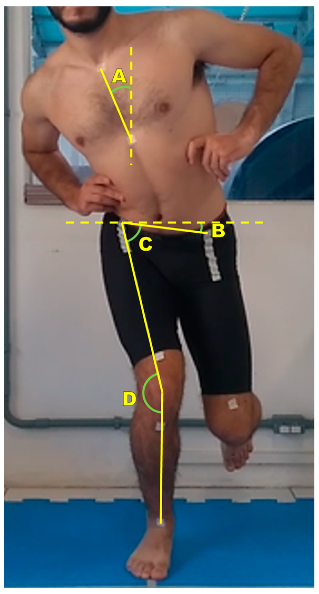
2.2. Measures
2.3. Design and Procedures
2.4. Data Analysis
2.5. Statistical Analysis
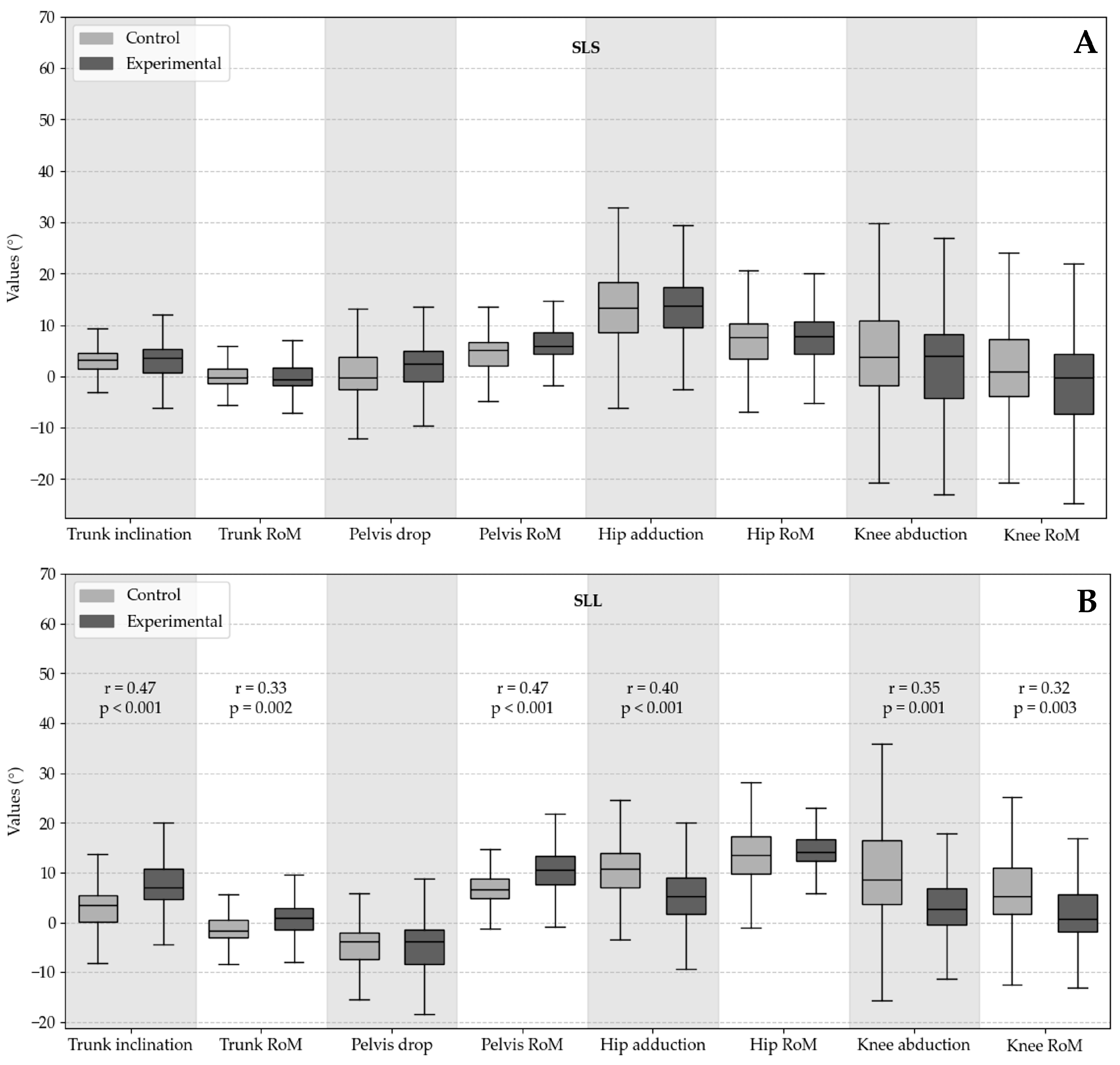
3. Results
4. Discussion
Study Limitations
5. Conclusions
Author Contributions
Funding
Institutional Review Board Statement
Informed Consent Statement
Data Availability Statement
Conflicts of Interest
Abbreviations
| ACL | Anterior cruciate ligament |
| BMI | Body mass index |
| CI | Confidence interval |
| COSMIN | Consensus-based standards for the selection of health measurement instruments |
| ES | Effect size |
| FPPA | Frontal plane projection angle |
| ICC | Intraclass correlation coefficient |
| LEFS | Lower Extremity Functional Scale |
| MCL | Medial collateral ligament |
| MDD | Minimal detectable difference |
| MPFL | Medial patellofemoral ligament |
| RoM | Range of motion |
| SD | Standard deviation |
| SEM | Standard error of measurement |
| SLL | Single-leg landing |
| SLS | Single-leg squat |
| TT | Tibial tuberosity |
References
- Poston, G.R.; Schmitt, L.C.; Ithurburn, M.P.; Hugentobler, J.A.; Thomas, S.; Paterno, M. V Reduced 2-d Frontal Plane Motion during Single-Limb Landing Is Associated with Risk of Future Anterior Cruciate Ligament Graft Rupture after Anterior Cruciate Ligament Reconstruction and Return to Sport: A Pilot Study. J. Orthop. Sports Phys. Ther. 2021, 51, 82–87. [Google Scholar] [CrossRef] [PubMed]
- Harris-Hayes, M.; Hillen, T.J.; Commean, P.K.; Harris, M.D.; Mueller, M.J.; Clohisy, J.C.; Salsich, G.B. Hip Kinematics during Single-Leg Tasks in People with and without Hip-Related Groin Pain and the Association among Kinematics, Hip Muscle Strength, and Bony Morphology. J. Orthop. Sports Phys. Ther. 2020, 50, 243–251. [Google Scholar] [CrossRef] [PubMed]
- Wilczyński, B.; Zorena, K.; Ślęzak, D. Dynamic Knee Valgus in Single-Leg Movement Tasks. Potentially Modifiable Factors and Exercise Training Options. A Literature Review. Int. J. Environ. Res. Public Health 2020, 17, 8208. [Google Scholar] [CrossRef] [PubMed]
- Willson, J.D.; Davis, I.S. Lower Extremity Mechanics of Females with and without Patellofemoral Pain across Activities with Progressively Greater Task Demands. Clin. Biomech. 2008, 23, 203–211. [Google Scholar] [CrossRef]
- Bailey, R.W.; Richards, J.; Selfe, J. A Biomechanical Investigation of Selected Lumbopelvic Hip Tests: Implications for the Examination of Walking. J. Manip. Physiol. Ther. 2016, 39, 411–419. [Google Scholar] [CrossRef]
- Scholtes, S.A.; Salsich, G.B. Consistency of Dynamic Knee Valgus Kinematics and Pain across Functional Tasks in Females with Patellofemoral Pain: A Cross-Sectional Study. Int. J. Sports Phys. Ther. 2020, 15, 985–994. [Google Scholar] [CrossRef]
- Rees, D.; Younis, A.; MacRae, S. Is There a Correlation in Frontal Plane Knee Kinematics between Running and Performing a Single Leg Squat in Runners with Patellofemoral Pain Syndrome and Asymptomatic Runners? Clin. Biomech. 2019, 61, 227–232. [Google Scholar] [CrossRef]
- Jones, P.A.; Herrington, L.C.; Munro, A.G.; Graham-Smith, P. Is There a Relationship between Landing, Cutting, and Pivoting Tasks in Terms of the Characteristics of Dynamic Valgus? Am. J. Sports Med. 2014, 42, 2095–2102. [Google Scholar] [CrossRef]
- Herrington, L. Knee Valgus Angle during Single Leg Squat and Landing in Patellofemoral Pain Patients and Controls. Knee 2014, 21, 514–517. [Google Scholar] [CrossRef]
- Nakagawa, T.H.; dos Santos, A.F.; Lessi, G.C.; Petersen, R.S.; Silva, R.S. Y-Balance Test Asymmetry and Frontal Plane Knee Projection Angle During Single-Leg Squat as Predictors of Patellofemoral Pain in Male Military Recruits. Phys. Ther. Sport 2020, 44, 121–127. [Google Scholar] [CrossRef]
- Warner, M.B.; Wilson, D.A.; Herrington, L.; Dixon, S.; Power, C.; Jones, R.; Heller, M.O.; Carden, P.; Lewis, C.L. A Systematic Review of the Discriminating Biomechanical Parameters during the Single Leg Squat. Phys. Ther. Sport 2019, 36, 78–91. [Google Scholar] [CrossRef] [PubMed]
- Räisänen, A.M.; Pasanen, K.; Krosshaug, T.; Vasankari, T.; Kannus, P.; Heinonen, A.; Kujala, U.M.; Avela, J.; Perttunen, J.; Parkkari, J. Association between Frontal Plane Knee Control and Lower Extremity Injuries: A Prospective Study on Young Team Sport Athletes. BMJ Open Sport Exerc. Med. 2018, 4, e000311. [Google Scholar] [CrossRef] [PubMed]
- Gagnier, J.J.; Lai, J.; Mokkink, L.B.; Terwee, C.B. COSMIN Reporting Guideline for Studies on Measurement Properties of Patient-Reported Outcome Measures. Qual. Life Res. 2021, 30, 2197–2218. [Google Scholar] [CrossRef]
- Kingston, B.; Murray, A.; Norte, G.E.; Glaviano, N.R. Validity and Reliability of 2-Dimensional Trunk, Hip, and Knee Frontal Plane Kinematics during Single-Leg Squat, Drop Jump, and Single-Leg Hop in Females with Patellofemoral Pain. Phys. Ther. Sport 2020, 45, 181–187. [Google Scholar] [CrossRef]
- Herrington, L.; Alenezi, F.; Alzhrani, M.; Alrayani, H.; Jones, R. The Reliability and Criterion Validity of 2D Video Assessment of Single Leg Squat and Hop Landing. J. Electromyogr. Kinesiol. 2017, 34, 80–85. [Google Scholar] [CrossRef]
- Gwynne, C.R.; Curran, S.A. Quantifying Frontal Plane Knee Motion during Single Limb Squats: Reliability and Validity of 2-Dimensional Measures. Int. J. Sports Phys. Ther. 2014, 9, 898–906. [Google Scholar]
- Jamaludin, N.I.; Sahabuddin, F.N.A.; Rasudin, N.S.; Shaharudin, S. The Concurrent Validity and Reliability of Single Leg Squat Among Physically Active Females with and without Dynamic Knee Valgus. Int. J. Sports Phys. Ther. 2022, 17, 574–584. [Google Scholar] [CrossRef]
- Alrayani, H.; Herrington, L.; Liu, A.; Jones, R. Frontal Plane Projection Angle Predicts Patellofemoral Pain: Prospective Study in Male Military Cadets. Phys. Ther. Sport 2023, 59, 73–79. [Google Scholar] [CrossRef]
- Levinger, P.; Gilleard, W.; Coleman, C. Femoral Medial Deviation Angle during a One-Leg Squat Test in Individuals with Patellofemoral Pain Syndrome. Phys. Ther. Sport 2007, 8, 163–168. [Google Scholar] [CrossRef]
- Scholtes, S.A.; Salsich, G.B. A Dynamic Valgus Index That Combines Hip and Knee Angles: Assessment of Utility in Females with Patelofemoral Pain. Int. J. Sports Phys. Ther. 2017, 12, 333–340. [Google Scholar]
- Willson, J.D.; Davis, I.S. Utility of the Frontal Plane Projection Angle in Females with Patellofemoral Pain. J. Orthop. Sports Phys. Ther. 2008, 38, 606–615. [Google Scholar] [CrossRef] [PubMed]
- Charlton, P.C.; Bryant, A.L.; Kemp, J.L.; Clark, R.A.; Crossley, K.M.; Collins, N.J. Single-Leg Squat Performance Is Impaired 1 to 2 Years After Hip Arthroscopy. PM&R 2016, 8, 321–330. [Google Scholar] [CrossRef]
- Werner, D.M.; Stasi, S.; Lewis, C.L.; Barrios, J.A. Test-Retest Reliability and Minimum Detectable Change for Various Frontal Plane Projection Angles during Dynamic Tasks. Phys. Ther. Sport 2019, 40, 169–176. [Google Scholar] [CrossRef] [PubMed]
- Munro, A.; Herrington, L.; Carolan, M. Reliability of 2-Dimensional Video Assessment of Frontal-Plane Dynamic Knee Valgus during Common Athletic Screening Tasks. J. Sport Rehabil. 2012, 21, 7–11. [Google Scholar] [CrossRef]
- DiCesare, C.A.; Bates, N.A.; Myer, G.D.; Hewett, T.E. The Validity of 2-Dimensional Measurement of Trunk Angle during Dynamic Tasks. Int. J. Sports Phys. Ther. 2014, 9, 420–427. [Google Scholar]
- Donohue, M.R.; Ellis, S.M.; Heinbaugh, E.M.; Stephenson, M.L.; Zhu, Q.; Dai, B. Differences and Correlations in Knee and Hip Mechanics during Single-Leg Landing, Single-Leg Squat, Double-Leg Landing, and Double-Leg Squat Tasks. Res. Sports Med. 2015, 23, 394–411. [Google Scholar] [CrossRef]
- Yamazaki, J.; Muneta, T.; Ju, Y.J.; Sekiya, I. Differences in Kinematics of Single Leg Squatting between Anterior Cruciate Ligament-Injured Patients and Healthy Controls. Knee Surg. Sports Traumatol. Arthrosc. 2010, 18, 56–63. [Google Scholar] [CrossRef]
- Dingenen, B.; Malfait, B.; Vanrenterghem, J.; Verschueren, S.M.P.; Staes, F.F. The Reliability and Validity of the Measurement of Lateral Trunk Motion in Two-Dimensional Video Analysis during Unipodal Functional Screening Tests in Elite Female Athletes. Phys. Ther. Sport 2014, 15, 117–123. [Google Scholar] [CrossRef]
- Furlan, L.; Sterr, A. The Applicability of Standard Error of Measurement and Minimal Detectable Change to Motor Learning Research—A Behavioral Study. Front. Hum. Neurosci. 2018, 12, 95. [Google Scholar] [CrossRef]
- Nakagawa, T.H.; Moriya, E.T.U.; MacIel, C.D.; Serrão, F. V Trunk, Pelvis, Hip, and Knee Kinematics, Hip Strength, and Gluteal Muscle Activation during a Single-Leg Squat in Males and Females with and without Patellofemoral Pain Syndrome. J. Orthop. Sports Phys. Ther. 2012, 42, 491–501. [Google Scholar] [CrossRef]
- Pereira, L.M.; Dias, J.M.; Mazuquin, B.F.; Castanhas, L.G.; Menacho, M.O.; Cardoso, J.R. Translation, Cross-Cultural Adaptation and Analysis of the Psychometric Properties of the Lower Extremity Functional Scale (LEFS): LEFS- BRAZIL. Braz. J. Phys. Ther. 2013, 17, 272–280. [Google Scholar] [CrossRef] [PubMed]
- Dingemans, S.A.; Kleipool, S.C.; Mulders, M.A.M.; Winkelhagen, J.; Schep, N.W.L.; Goslings, J.C.; Schepers, T. Normative Data for the Lower Extremity Functional Scale (LEFS). Acta Orthop. 2017, 88, 422–426. [Google Scholar] [CrossRef] [PubMed]
- Mehta, S.P.; Fulton, A.; Quach, C.; Thistle, M.; Toledo, C.; Evans, N.A. Measurement Properties of the Lower Extremity Functional Scale: A Systematic Review. J. Orthop. Sports Phys. Ther. 2016, 46, 200–216. [Google Scholar] [CrossRef]
- Springate, S.D. The Effect of Sample Size and Bias on the Reliability of Estimates of Error: A Comparative Study of Dahlberg’s Formula. Eur. J. Orthod. 2012, 34, 158–163. [Google Scholar] [CrossRef]
- Machado, J.M.; de Souza, A.; Castro, M.P.d.; Pierri, C.A.A.; Ruschel, C. Repetibilidade e Capacidade Discriminativa Da Análise Cinemática Bidimensional Durante o Agachamento e Aterrissagem Unilaterais Em Indivíduos Com e Sem Disfunção Nos Membros Inferiores. In Proceedings of the XX Brazilian Congress of Biomechanics, Bauru, SP, Brazil, 18–22 April 2023; p. 42. [Google Scholar]
- Sakurai, A.; Harato, K.; Morishige, Y.; Kobayashi, S.; Niki, Y.; Nagura, T. Effects of Toe Direction on Biomechanics of Trunk, Pelvis, and Lower-Extremity during Single-Leg Drop Landing. J. Sport Rehabil. 2020, 29, 1069–1074. [Google Scholar] [CrossRef]
- Johnston, P.T.; McClelland, J.A.; Webster, K.E. Lower Limb Biomechanics During Single-Leg Landings Following Anterior Cruciate Ligament Reconstruction: A Systematic Review and Meta-Analysis. Sports Med. 2018, 48, 2103–2126. [Google Scholar] [CrossRef]
- Agostinone, P.; Di Paolo, S.; Grassi, A.; Pinelli, E.; Bontempi, M.; Bragonzoni, L.; Zaffagnini, S. ACL Deficiency Influences Medio-Lateral Tibial Alignment and Knee Varus–Valgus during in Vivo Activities. Knee Surg. Sports Traumatol. Arthrosc. 2020, 29, 389–397. [Google Scholar] [CrossRef]
- Markström, J.L.; Tengman, E.; Häger, C.K. ACL-Reconstructed and ACL-Deficient Individuals Show Differentiated Trunk, Hip, and Knee Kinematics during Vertical Hops More than 20 Years Post-Injury. Knee Surg. Sports Traumatol. Arthrosc. 2018, 26, 358–367. [Google Scholar] [CrossRef]
- Talarico, M.K.; Lynall, R.C.; Mauntel, T.C.; Wasserman, E.B.; Padua, D.A.; Mihalik, J.P. Effect of Single-Leg Squat Speed and Depth on Dynamic Postural Control Under Single-Task and Dual-Task Paradigms. J. Appl. Biomech. 2019, 35, 272–279. [Google Scholar] [CrossRef]
- Lopes, T.J.A.; Ferrari, D.; Ioannidis, J.; Simic, M.; Azevedo, F.M.; Pappas, E. Reliability and Validity of Frontal Plane Kinematics of the Trunk and Lower Extremity Measured with 2-Dimensional Cameras during Athletic Tasks: A Systematic Review with Meta-Analysis. J. Orthop. Sports Phys. Ther. 2018, 48, 812–822. [Google Scholar] [CrossRef]
- Lu, Z.; Nazari, G.; MacDermid, J.C.; Modarresi, S.; Killip, S. Measurement Properties of a 2-Dimensional Movement Analysis System: A Systematic Review and Meta-Analysis. Arch. Phys. Med. Rehabil. 2020, 101, 1603–1627. [Google Scholar] [CrossRef] [PubMed]

| Variables | Control (n = 43) | Experimental (n = 43) | p-Value |
|---|---|---|---|
| Age, y, mean (SD) | 31 (8) | 32 (9) | 0.441 b |
| BMI, kg/m2, mean (SD) | 23.4 (2.2) | 24.4 (3.9) | 0.129 b |
| LEFS, median [25th to 75th percentile] | 80 [80 to 80] | 65 [60 to 70] | <0.001 c |
| Sex, n (%) | |||
| Male | 23 (53.5) | 23 (53.5) | |
| Female | 20 (46.5) | 20 (46.5) | |
| Lower limb, n (%) | |||
| Dominant | 24 (55.8) | 24 (55.8) | |
| Non-dominant | 19 (44.2) | 19 (44.2) | |
| Joint involved, n (%) | |||
| Knee | 29 (67.4) | ||
| Hip | 13 (30.2) | ||
| Both | 1 (2.3) | ||
| Sides involved, n (%) | |||
| Unilateral | 30 (69.8) | ||
| Bilateral | 13 (30.2) | ||
| Surgery in the last 2 years, n (%) | |||
| 0 | 18 (41.9) | ||
| 1 | 18 (41.9) | ||
| 2 | 7 (16.3) | ||
| Surgical procedure, n (%) d | |||
| ACL reconstruction | 13 (30.2) | ||
| MPFL Reconstruction | 1 (2.3) | ||
| MCL Reconstruction | 2 (4.6) | ||
| Meniscal suture | 12 (27.9) | ||
| Chondral procedure—knee | 3 (7) | ||
| TT osteotomy | 3 (7) | ||
| Femoral osteochondroplasty | 13 (30.2) |
| Kinematic Variables | Control (n = 30) | ||
|---|---|---|---|
| ICC (95% CI) | SEM | MDD | |
| SLS | |||
| Trunk inclination | 0.41 (0.07 to 0.67) | 3° | 8° |
| Trunk RoM | 0.2 (−0.17 to 0.52) | 2° | 6° |
| Pelvis drop | 0.5 (0.17 to 0.73) | 4° | 11° |
| Pelvis RoM | 0.34 (−0.02 to 0.63) | 4° | 11° |
| Hip adduction | 0.68 (0.44 to 0.84) | 3° | 8° |
| Hip RoM | 0.57 (0.27 to 0.77) | 3° | 8° |
| Knee abduction | 0.74 (0.52 to 0.87) | 5° | 14° |
| Knee RoM | 0.7 (0.45 to 0.84) | 4° | 11° |
| SLL | |||
| Trunk inclination | 0.28 (−0.05 to 0.57) | 5° | 14° |
| Trunk RoM | 0.47 (0.14 to 0.70) | 4° | 11° |
| Pelvis drop | 0.53 (0.22 to 0.74) | 3° | 8° |
| Pelvis RoM | 0.51 (0.20 to 0.73) | 2° | 6° |
| Hip adduction | 0.54 (0.24 to 0.75) | 5° | 14° |
| Hip RoM | 0.47 (0.14 to 0.71) | 5° | 14° |
| Knee abduction | 0.57 (0.26 to 0.77) | 7° | 19° |
| Knee RoM | 0.51 (0.18 to 0.73) | 7° | 19° |
| Kinematic Variables | Control (n = 30) | Experimental (n = 30) | ||||
|---|---|---|---|---|---|---|
| ICC (95% CI) | SEM | MDD | ICC (95% CI) | SEM | MDD | |
| SLS | ||||||
| Trunk inclination | 0.98 (0.96 to 0.99) | 1° | 3° | 0.88 (0.76 to 0.94) | 1° | 3° |
| Trunk RoM | 0.82 (0.65 to 0.91) | 1° | 3° | 0.69 (0.43 to 0.84) | 2° | 6° |
| Pelvis drop | 0.66 (0.41 to 0.83) | 3° | 8° | 0.9 (0.80 to 0.95) | 1° | 3° |
| Pelvis RoM | 0.52 (0.20 to 0.74) | 3° | 8° | 0.83 (0.66 to 0.92) | 1° | 3° |
| Hip adduction | 0.98 (0.92 to 0.99) | 1° | 3° | 0.93 (0.85 to 0.96) | 2° | 6° |
| Hip RoM | 0.96 (0.92 to 0.98) | 1° | 3° | 0.93 (0.85 to 0.97) | 1° | 3° |
| Knee abduction | 0.95 (0.86 to 0.98) | 2° | 6° | 0.93 (0.83 to 0.97) | 3° | 8° |
| Knee RoM | 0.95 (0.90 to 0.98) | 2° | 6° | 0.93 (0.83 to 0.97) | 2° | 6° |
| SLL | ||||||
| Trunk inclination | 0.9 (0.81 to 0.95) | 1° | 3° | 0.63 (0.35 to 0.80) | 3° | 8° |
| Trunk RoM | 0.8 (0.62 to 0.90) | 2° | 6° | 0.83 (0.67 to 0.91) | 2° | 6° |
| Pelvis drop | 0.4 (0.06 to 0.65) | 3° | 8° | 0.62 (0.35 to 0.80) | 3° | 8° |
| Pelvis RoM | 0.47 (0.16 to 0.71) | 3° | 8° | 0.31 (−0.05 to 0.6) | 4° | 11° |
| Hip adduction | 0.82 (0.55 to 0.92) | 2° | 6° | 0.8 (0.62 to 0.90) | 3° | 8° |
| Hip RoM | 0.8 (0.62 to 0.90) | 3° | 8° | 0.6 (0.31 to 0.79) | 3° | 8° |
| Knee abduction | 0.82 (0.64 to 0.91) | 4° | 11° | 0.66 (0.40 to 0.82) | 4° | 11° |
| Knee RoM | 0.76 (0.54 to 0.88) | 4° | 11° | 0.68 (0.43 to 0.83) | 4° | 11° |
| Kinematic Variables | Control (n = 30) | Experimental (n = 30) | ||||
|---|---|---|---|---|---|---|
| ICC (95% CI) | SEM | MDD | ICC (95% CI) | SEM | MDD | |
| SLS | ||||||
| Trunk inclination | 0.99 (0.98 to 1.00) | 1° | 3° | 0.99 (0.99 to 1.00) | 1° | 3° |
| Trunk RoM | 0.99 (0.97 to 0.99) | 1° | 3° | 0.9 (0.80 to 0.95) | 1° | 3° |
| Pelvis drop | 0.74 (0.52 to 0.86) | 3° | 8° | 0.98 (0.97 to 0.99) | 1° | 3° |
| Pelvis RoM | 0.63 (0.35 to 0.80) | 3° | 8° | 0.98 (0.97 to 0.99) | 1° | 3° |
| Hip adduction | 0.99 (0.98 to 1.00) | 1° | 3° | 0.99 (0.98 to 1.00) | 1° | 3° |
| Hip RoM | 0.97 (0.94 to 0.99) | 1° | 3° | 0.57 (0.28 to 0.77) | 4° | 11° |
| Knee abduction | 0.99 (0.98 to 1.00) | 1° | 3° | 0.99 (0.98 to 1.00) | 1° | 3° |
| Knee RoM | 0.98 (0.96 to 0.99) | 1° | 3° | 0.99 (0.96 to 0.99) | 1° | 3° |
| SLL | ||||||
| Trunk inclination | 0.97 (0.94 to 0.99) | 1° | 3° | 0.94 (0.88 to 0.97) | 1° | 3° |
| Trunk RoM | 0.95 (0.90 to 0.98) | 1° | 3° | 0.83 (0.67 to 0.91) | 1° | 3° |
| Pelvis drop | 0.93 (0.85 to 0.96) | 1° | 3° | 0.92 (0.83 to 0.96) | 1° | 3° |
| Pelvis RoM | 0.89 (0.78 to 0.95) | 1° | 3° | 0.82 (0.66 to 0.91) | 2° | 6° |
| Hip adduction | 0.93 (0.87 to 0.97) | 1° | 3° | 0.97 (0.93 to 0.98) | 1° | 3° |
| Hip RoM | 0.92 (0.84 to 0.96) | 2° | 6° | 0.86 (0.72 to 0.93) | 2° | 6° |
| Knee abduction | 0.94 (0.89 to 0.97) | 2° | 6° | 0.91 (0.83 to 0.96) | 2° | 6° |
| Knee RoM | 0.91 (0.81 to 0.95) | 3° | 8° | 0.87 (0.75 to 0.94) | 2° | 6° |
Disclaimer/Publisher’s Note: The statements, opinions and data contained in all publications are solely those of the individual author(s) and contributor(s) and not of MDPI and/or the editor(s). MDPI and/or the editor(s) disclaim responsibility for any injury to people or property resulting from any ideas, methods, instructions or products referred to in the content. |
© 2025 by the authors. Licensee MDPI, Basel, Switzerland. This article is an open access article distributed under the terms and conditions of the Creative Commons Attribution (CC BY) license (https://creativecommons.org/licenses/by/4.0/).
Share and Cite
Machado, J.M.; Peduzzi de Castro, M.; Souza, A.d.; Pierri, C.A.A.; Araujo, F.X.d.; de Brito Fontana, H.; Ruschel, C. Discriminative Validity and Reliability of the Single-Leg Squat and Single-Leg Landing Frontal Plane Kinematics in Individuals with Lower Limb Functional Deficits. Biomechanics 2025, 5, 20. https://doi.org/10.3390/biomechanics5020020
Machado JM, Peduzzi de Castro M, Souza Ad, Pierri CAA, Araujo FXd, de Brito Fontana H, Ruschel C. Discriminative Validity and Reliability of the Single-Leg Squat and Single-Leg Landing Frontal Plane Kinematics in Individuals with Lower Limb Functional Deficits. Biomechanics. 2025; 5(2):20. https://doi.org/10.3390/biomechanics5020020
Chicago/Turabian StyleMachado, Jean Marlon, Marcelo Peduzzi de Castro, Amandda de Souza, Carlos Alberto Atherinos Pierri, Francisco Xavier de Araujo, Heiliane de Brito Fontana, and Caroline Ruschel. 2025. "Discriminative Validity and Reliability of the Single-Leg Squat and Single-Leg Landing Frontal Plane Kinematics in Individuals with Lower Limb Functional Deficits" Biomechanics 5, no. 2: 20. https://doi.org/10.3390/biomechanics5020020
APA StyleMachado, J. M., Peduzzi de Castro, M., Souza, A. d., Pierri, C. A. A., Araujo, F. X. d., de Brito Fontana, H., & Ruschel, C. (2025). Discriminative Validity and Reliability of the Single-Leg Squat and Single-Leg Landing Frontal Plane Kinematics in Individuals with Lower Limb Functional Deficits. Biomechanics, 5(2), 20. https://doi.org/10.3390/biomechanics5020020