
The “Scrubbing Brush Technique” for Access to Tight Lateral Recess of the Sphenoid Sinus: A Single Case Report

Abstract
1. Introduction
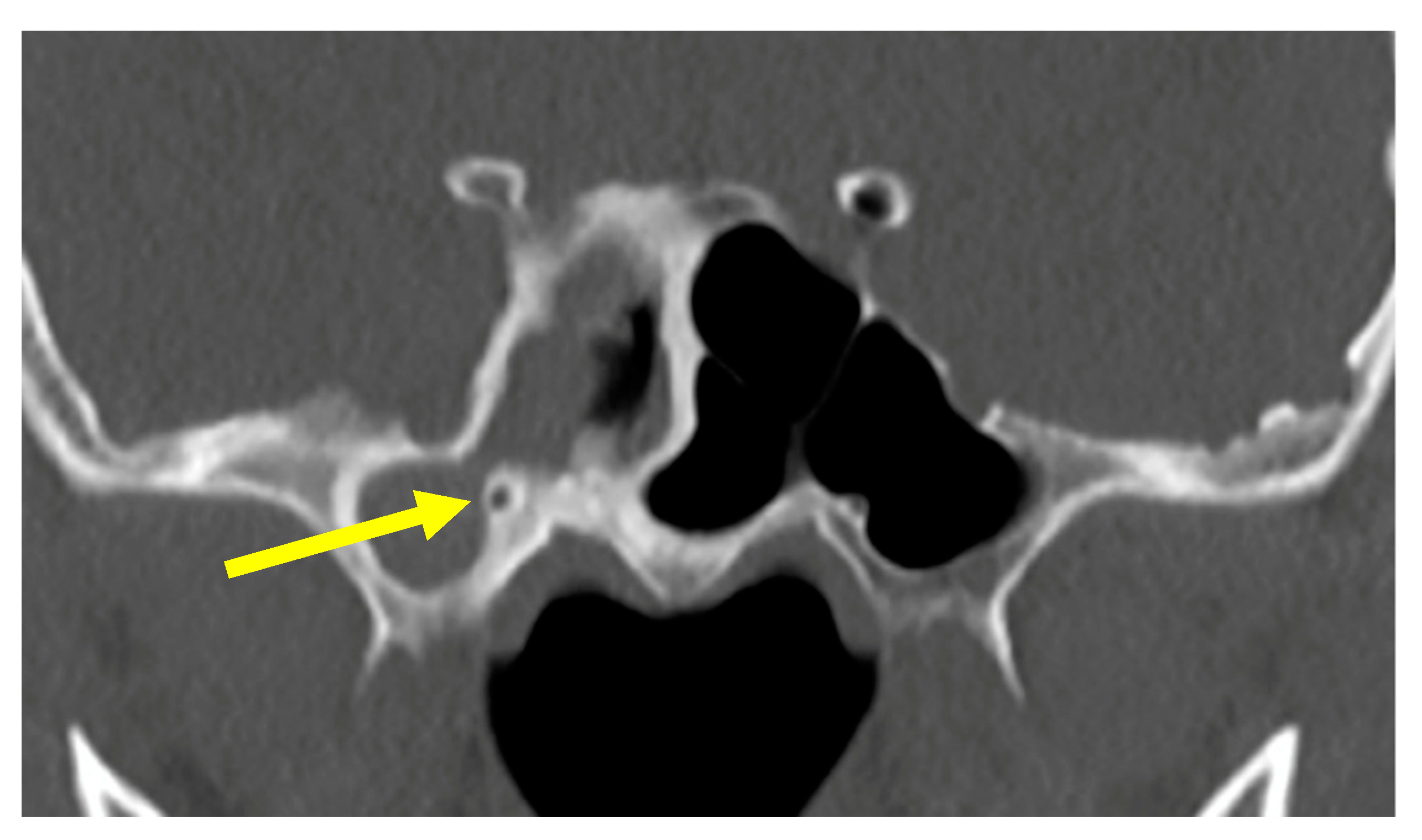
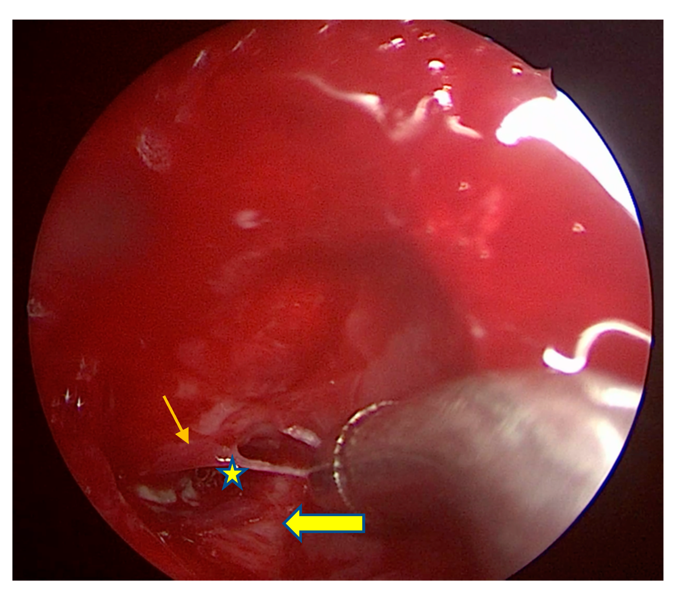
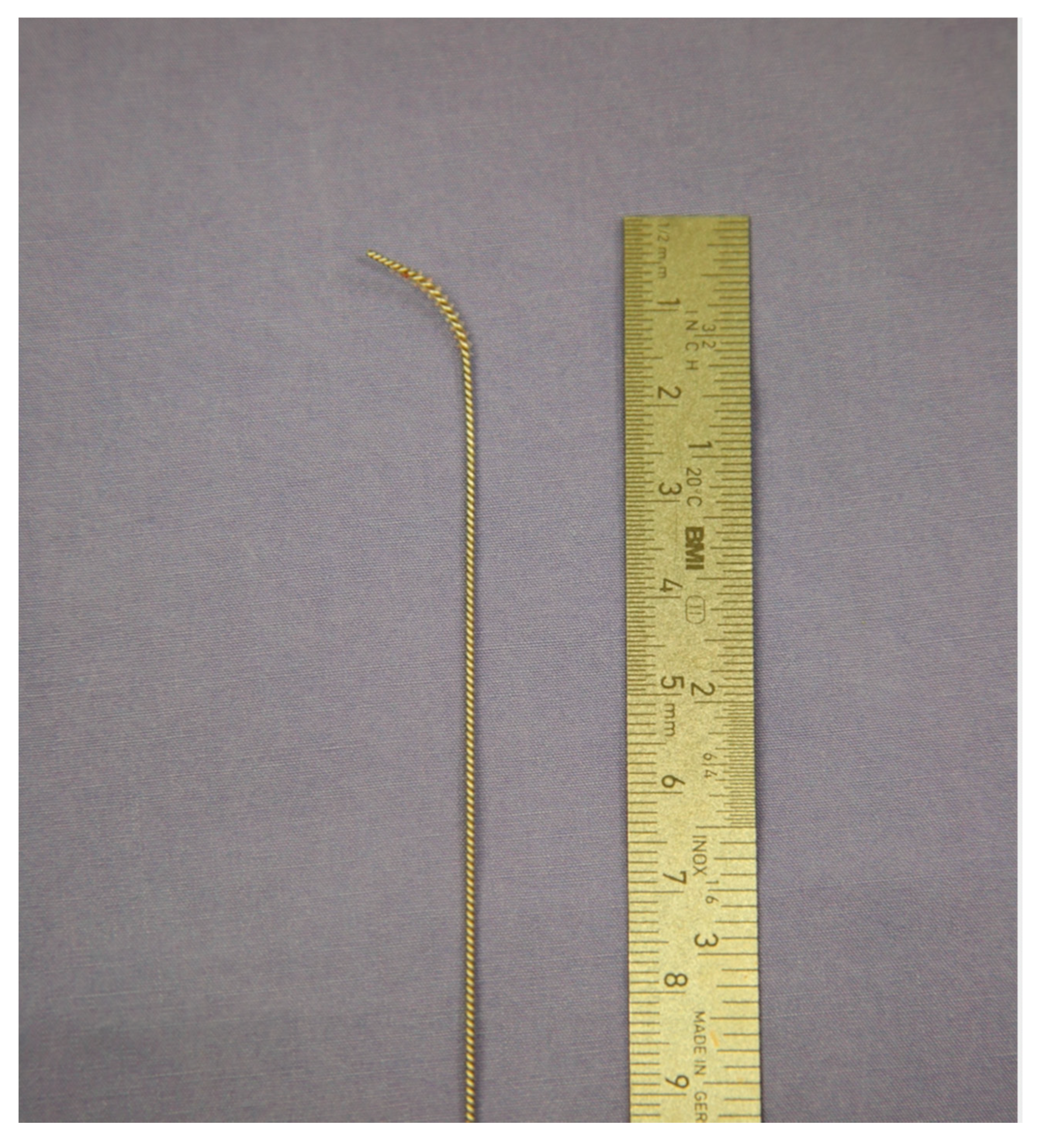
2. Method
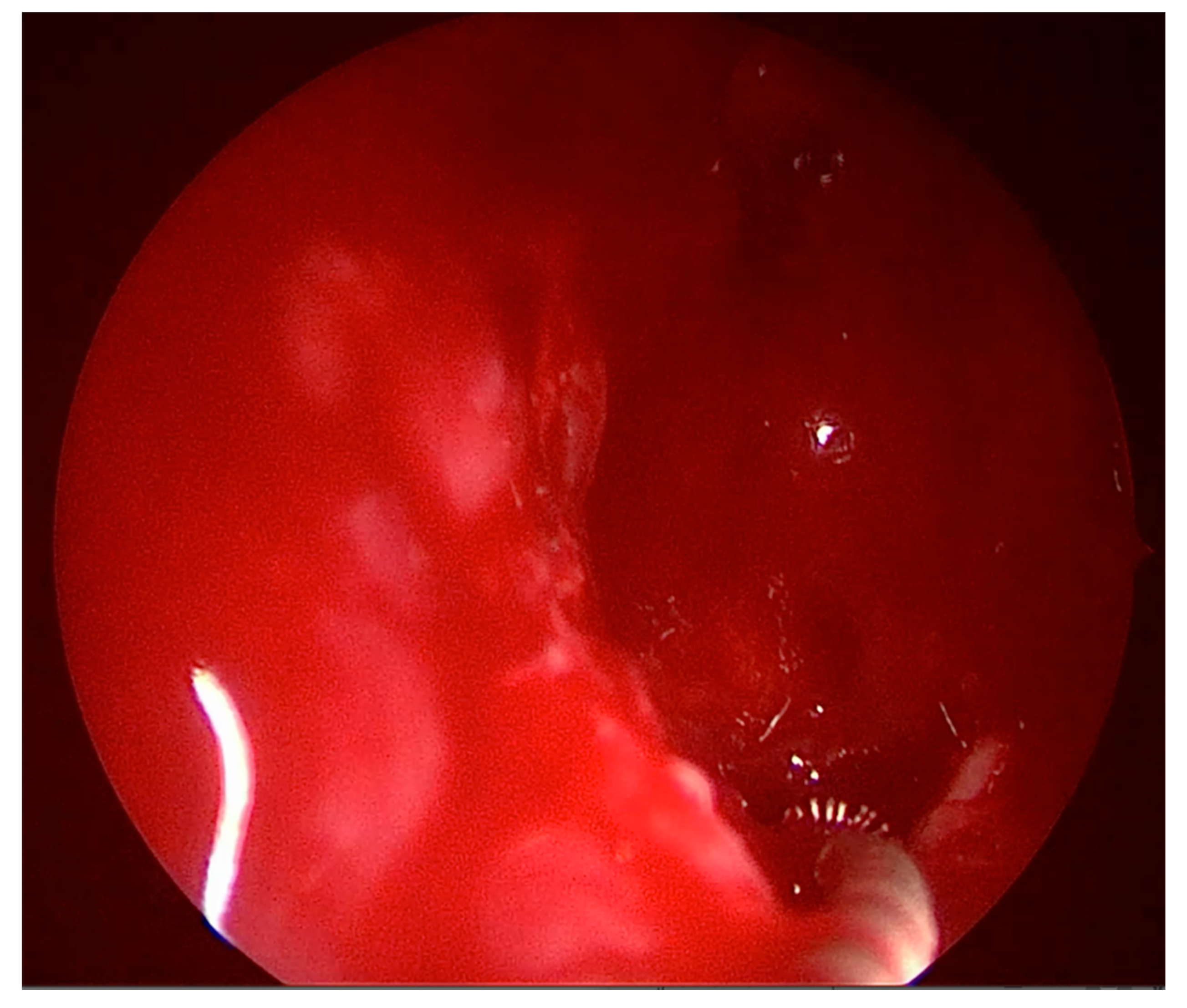
3. Results
4. Discussion
5. Conclusions
Author Contributions
Funding
Informed Consent Statement
Data Availability Statement
Conflicts of Interest
References
- Jaworek-Troć, J.; Zarzecki, M.; Zamojska, I.; Iwanaga, J.; Przybycień, W.; Mazur, M.; Chrzan, R.; Walocha, J.A. The dimensions of the sphenoid sinuses–evaluation before the functional endoscopic sinus surgery. Folia Morphol. 2020. [Google Scholar] [CrossRef] [PubMed]
- Jaworek-Troć, J.; Iwanaga, J.; Chrzan, R.; Zarzecki, J.J.; Żmuda, P.; Pękala, A.; Tomaszewska, I.M.; Tubbs, R.S.; Zawiliński, J.; Zarzecki, M.P. Anatomical variations of the main septum of the sphenoidal sinus and its importance during transsphenoidal approaches to the sella turcica. Transl. Res. Anat. 2020, 21, 100079. [Google Scholar] [CrossRef]
- Govindaraju, R.; Tang, I.P.; Prepageran, N. Management of sphenoid lateral recess encephalocoeles. Curr. Opin. Otolaryngol. Head Neck Surg. 2019, 27, 37–46. [Google Scholar] [CrossRef] [PubMed]
- Wang, J.; Bidari, S.; Inoue, K.; Yang, H.; Rhoton Jr, A. Extensions of the sphenoid sinus: A new classification. Neurosurgery 2010, 66, 797–816. [Google Scholar] [CrossRef] [PubMed]
- Li, L.; London, N.R.; Prevedello, D.M.; Carrau, R.L. Endoscopic prelacrimal approach to lateral recess of sphenoid sinus: Feasibility study. Int. Forum Allergy Rhinol. 2020, 10, 103–109. [Google Scholar] [CrossRef] [PubMed]
- Bolger, W.E. Endoscopic Transpterygoid Approach to the Lateral Sphenoid Recess: Surgical Approach and Clinical Experience. Otolaryngol. Neck Surg. 2005, 133, 20–26. [Google Scholar] [CrossRef] [PubMed]
- Al-Nashar, I.S.; Carrau, R.L.; Herrera, A.; Snyderman, C.H. Endoscopic Transnasal Transpterygopalatine Fossa Approach to the Lateral Recess of the Sphenoid Sinus. Laryngoscope 2004, 114, 528–532. [Google Scholar] [CrossRef] [PubMed]
- Lageju, N.; Pradhan, B.; Thapa, N. Endoscopic Sinus Surgery for Sinonasal Polyposis: Microdebrider or Conventional Instru-ments. JNMA J. Nepal. Med. Assoc. 2017, 56, 447–450. [Google Scholar] [CrossRef] [PubMed]


Publisher’s Note: MDPI stays neutral with regard to jurisdictional claims in published maps and institutional affiliations. |
© 2021 by the authors. Licensee MDPI, Basel, Switzerland. This article is an open access article distributed under the terms and conditions of the Creative Commons Attribution (CC BY) license (https://creativecommons.org/licenses/by/4.0/).
Share and Cite
Zhang, M.; Subramaniam, S.; Ng, C.L. The “Scrubbing Brush Technique” for Access to Tight Lateral Recess of the Sphenoid Sinus: A Single Case Report. Sinusitis 2021, 5, 67-70. https://doi.org/10.3390/sinusitis5010008
Zhang M, Subramaniam S, Ng CL. The “Scrubbing Brush Technique” for Access to Tight Lateral Recess of the Sphenoid Sinus: A Single Case Report. Sinusitis. 2021; 5(1):67-70. https://doi.org/10.3390/sinusitis5010008
Chicago/Turabian StyleZhang, Margaret, Somasundram Subramaniam, and Chew Lip Ng. 2021. "The “Scrubbing Brush Technique” for Access to Tight Lateral Recess of the Sphenoid Sinus: A Single Case Report" Sinusitis 5, no. 1: 67-70. https://doi.org/10.3390/sinusitis5010008
APA StyleZhang, M., Subramaniam, S., & Ng, C. L. (2021). The “Scrubbing Brush Technique” for Access to Tight Lateral Recess of the Sphenoid Sinus: A Single Case Report. Sinusitis, 5(1), 67-70. https://doi.org/10.3390/sinusitis5010008