Case Report

Current Status of Digital Complete Dentures Technology

by
Aristeidis Villias
1,2,*,
Hercules Karkazis
1,
Stavros Yannikakis
2,
Antonios Theocharopoulos
2,
Nikitas Sykaras
1 and
Gregory Polyzois
1
1
Department of Prosthodontics, School of Dentistry, National and Kapodistrian University of Athens, 11527 Athens, Greece
2
Division of Dental Technology, Department of Biomedical Sciences, School of Health and Care Sciences, University of West Attica, 12243 Athens, Greece
*
Author to whom correspondence should be addressed.
Prosthesis 2021, 3(3), 229-244; https://doi.org/10.3390/prosthesis3030023
Submission received: 7 June 2021 / Revised: 28 August 2021 / Accepted: 30 August 2021 / Published: 7 September 2021
(This article belongs to the Section Prosthodontics)

Abstract

:
Fabrication of complete dentures (CDs) utilizing computer-aided design and computer-aided manufacturing (CAD/CAM) methods has attracted a lot of attention. The purpose of this paper was to summarize current knowledge about digital CDs and the relevant technology, and to present the application of the new technology in a dental geriatrics case. Initially, some of the challenges regarding digitization of the oral mucosa as a supporting surface of the CDs’ intaglio surface are listed. Next, a brief introduction of the CAD software capabilities regarding CDs is presented. The latest CAM additive and subtractive techniques for CDs are following. Subsequently, the consecutive steps for the construction of a digital CD as part of the prosthodontic treatment of a 90-year-old ambulative female patient are presented. Finally, some considerations about the digital workflow in CD manufacturing are discussed. In conclusion, the new digital technology has clear advantages; however, implementation requires careful planning. The digital workflow is applicable and versatile.

1. Introduction

Although preservation of natural teeth is one of the goals of modern dentistry, as people get older, they are statistically more likely to experience tooth loss. Edentulism coupled with longer life expectancy inevitably leads to a progressively increasing demand for partial and complete dentures (CDs) [1,2].
Since its introduction in the 1980s, digital dentistry, as a spinoff of digital technology and robotics, has revolutionized many aspects of the dental practice. Fabrication of CDs is no exception. It has been more than 25 years since the first publications about the utilization of computer-aided design and computer-aided manufacturing (CAD/CAM) methods for complete denture (CD) fabrication [3,4]. Nowadays, the fabrication of CDs using CAD/CAM methods has attracted a lot of attention from the industry, resulting in breakthroughs in both the design and in the manufacturing sectors [5,6,7,8,9]. The new technology is promising quicker and better quality deliverables [10,11,12,13,14,15,16,17,18]. The objective of this article was to summarize the current knowledge about digital CDs and present the application of the new technology in a case.

1.1. Digitally Produced Dentures

There are commercially available CAD software packages that can aid in the design of CDs, either for the maxilla, for the mandible or for both. A variety of virtual tools and options for adjustments are offered from these dedicated programs. The CD modules are usually sold as extras for the basic CAD suite set-up and are regularly updated. Nevertheless, there are options available as stand-alone design programs for CDs. The software companies are putting in effort to create a friendly and simple user interface (UI), however, given the large number of digital tools and the necessary parameter adjustments, the UI might appear to be complicated.
There are numerous paths to achieve an acceptable treatment with CDs produced through a digital workflow, with the first challenge being the accurate recording of the shape and size of alveolar ridges and the hard palate, as well as the functional depth and width of the border seal and the post-palatal seal. An accurate recording of the maxillomandibular relations, the establishment of the appropriate vertical dimension of occlusion and the fulfillment of the aesthetics requirements is the next step. This information will dictate the design of the cameo surface, the ultimate space for the artificial teeth, the plan for a functional and aesthetic teeth set-up and, eventually, will contribute to the fabrication of a successful pair of dentures.
The clinical protocols for digitizing the tissue surface of a denture are still evolving and are highly dependent on the used system. Nevertheless, there are two options in general. One would be to directly scan the supporting tissues utilizing an intraoral scanner and the other to indirectly scan a stone cast, or even the impression, utilizing a laboratory desktop scanner or an intraoral scanner. Intraoral scanning shows some advantages compared with conventional impressions, such as improvement of patients’ comfort (no gag reflexes, reduced chairside time, fewer appointments) and facilitation of laboratory procedures (cast preparation, handling and shipping) without the need of physical casts [19,20,21]. Moreover, intraoral scans have been reported to be dimensionally accurate for CD fabrication [22,23]. In the absence of an intraoral scanner, scanning of physical casts or impressions is necessary.
Despite the efforts that are being made in merging intraoral scans with face scans and integrating virtual face-bows and articulators, digital denture technology is still behind as regards the registration of maxillomandibular relationships. It seems that the construction of baseplates with wax rims and recording of edentulous jaw relationships are, at the moment, unavoidable for the production of customized CDs. Then, the baseplates and wax rims with all the necessary information are scanned by the intraoral or lab scanner and the files are integrated to the digital case, properly aligned with the digitized tissue surfaces that support the dentures [21,24,25]. Once the digital casts of the maxilla and the mandible and the relations between them have been imported to the relevant CAD software, the surface of the denture is designed.
Jaw movement and the relationship between mandible and maxilla can be reproduced with dedicated computer programs, which are usually incorporated in the most recent dental CAD software packages [26]. These computer programs are called virtual articulators and the user interface usually conveniently corresponds to the features of the traditional articulator models that are combatable with the CAD software [27,28].
Efforts are being made towards a fully digital approach to evaluating the mandibular position in relation to the maxilla, utilizing the acquired data of the various imaging techniques [29]. Furthermore, there is a trend towards simplification of the complex hardware and imaging techniques; for instance, utilization of images acquired by smartphones has been reported [30]. The idea is to reproduce the function of the traditional articulator in digitally simulated movements through the development of mathematical models [31]. The new technology, in combination with cone beam computer tomography (CBCT) images, has the potential to accurately simulate the mandibular movements and condyle trajectories [32]. Regarding the fabrication of digital dentures, the most resent specialized CAD software allows tooth selection, arrangement of the artificial teeth in the three dimensions and fine-tuning of the occlusion with the virtual articulator [33,34].
However, there is still a lot of space for improvements, since only a few of the existing artificial teeth series have been digitized and, therefore, are compatible with the CAD software. On the other hand, the restorative patch of the available dental CAD software allows the design of patient-specific complete dental arches. The software might also allow changes to the shape of each tooth separately for detailed characterization.
Considering the CAM part of the new approach for CD production, currently two principal methods exist: one is additive and the other is subtractive. Nonetheless, both require the appropriate three-dimensional (3D) denture design CAD files [35].
The additive method (also called rapid prototyping) was invented in 1981 and can be encountered as 3D printing [36]. The additive manufacturing method includes a group of techniques that fabricate a 3D object in a layer-by-layer fashion based on the relevant CAD file. Rapid prototyping has many applications in the field of engineering and the greater area of medicine [37]. In short, the 3D-printed denture production concept comprises printing of the denture base in pink material, printing of the teeth individually or in complete arches in tooth shade material and processing of the printed products for removal of excess unpolymerized material, mostly utilizing ultrasonic baths in alcohol. Printed teeth are next bonded to the denture bases with liquid material from the printer tank and, finally, are light-cured for complete polymerization. The output is one of high detail and smooth surface. Cutting of the supports and final polishing of the denture is the last step. As an alternative, commercially available denture teeth selected from the digital library of CAD software may be used and bonded to the printed base.
There are numerous additive manufacturing systems, however not all of them are suitable for CD production; either due to the lack of appropriate compatible materials or due to the lack of available building volume. A scan of the relevant market revealed a list of systems that can be utilized for the production of CDs, both in a laboratory setting as well as chairside [38,39,40].
The 3D printers usually require specialized cartridges containing resins intended for denture bases and acrylic resins are mostly used for the base of dentures [41,42,43]. The offered variety of the material hue and color stability are important factors to be considered while purchasing the product, since esthetics is an important aspect for the acceptance of the final restoration by the patient. Particular attention should also be placed on the mechanical properties of the material after it has been fully polymerized, as they contribute both to longevity of the prosthesis and patient comfort. Therefore, utilization of certified materials according to manufacturer’s instructions is strongly recommended. Since the technology is evolving and incompatibilities might arise, one should carefully plan the transition to the new era [44,45,46,47,48,49].
The dedicated 3D printers utilize light sources or lasers that polymerize a liquid to produce the required objects. The production procedure requires some moving parts, which are computer-guided with high precision; specifically, a traveling printing platform that submerges in the printing material and a processing system that hardens the material layer-by-layer, forming the required shape. The most commonly used 3D printer technologies in dentistry for CD production are briefly described in Table 1 [50,51].
The subtractive method includes milling the denture base from a red pigmented industrially produced resin disk [52]. The artificial teeth are either milled or selected from a prefabricated series and bonded to the denture base [53]. There are many CNC/milling CAM systems, intended for laboratory or chairside use. However, in order to be capable of manufacturing CDs, the system has to be compatible with the relevant milling blanks. In Table 2, some of the options that are intended for CD production with the subtractive method are briefly described [54,55,56,57,58].

1.2. Comparing the Additive vs. the Subtractive Method
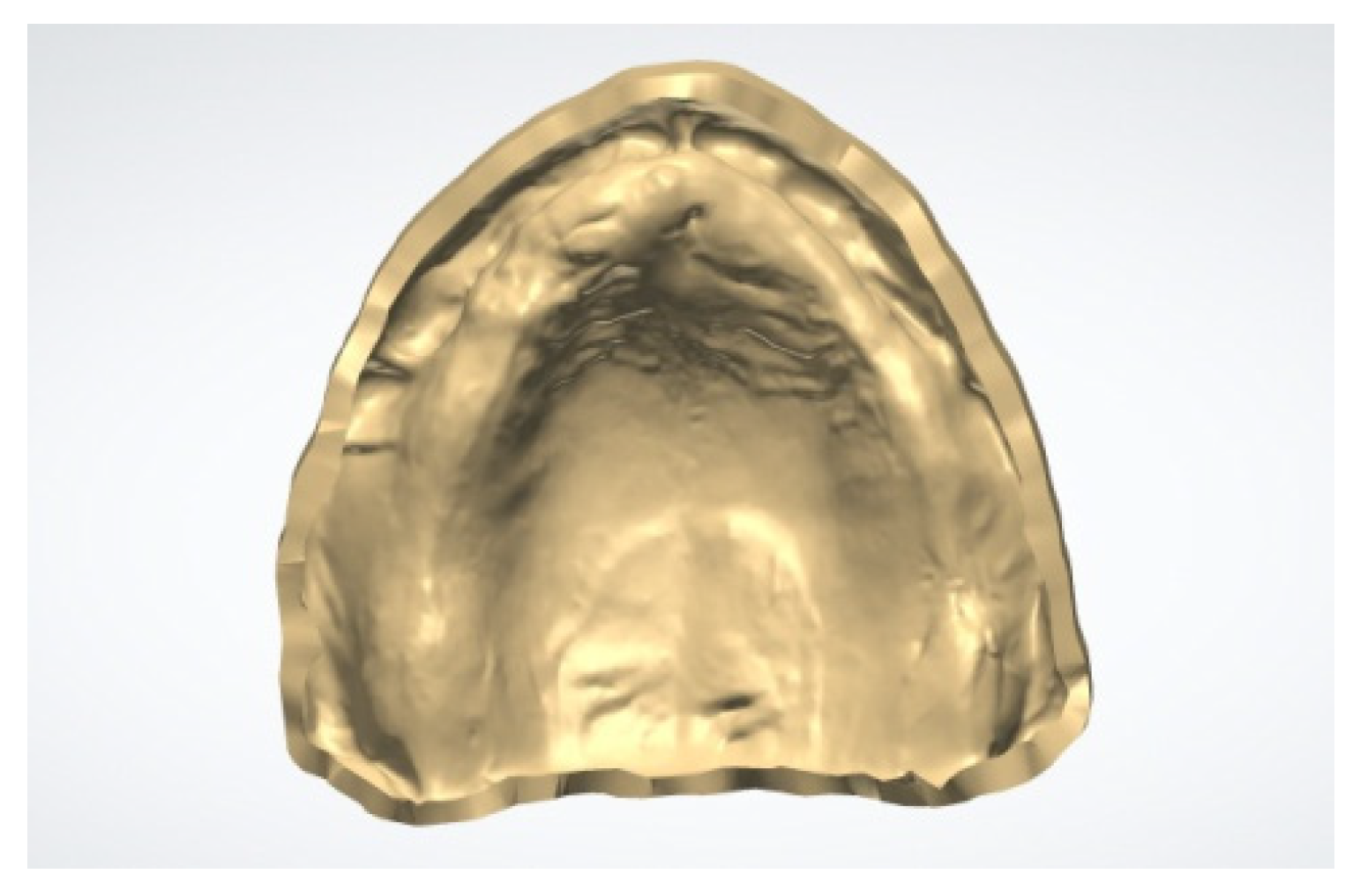
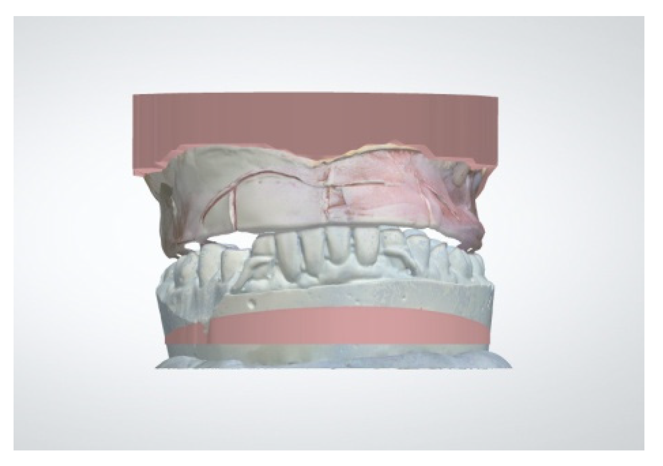
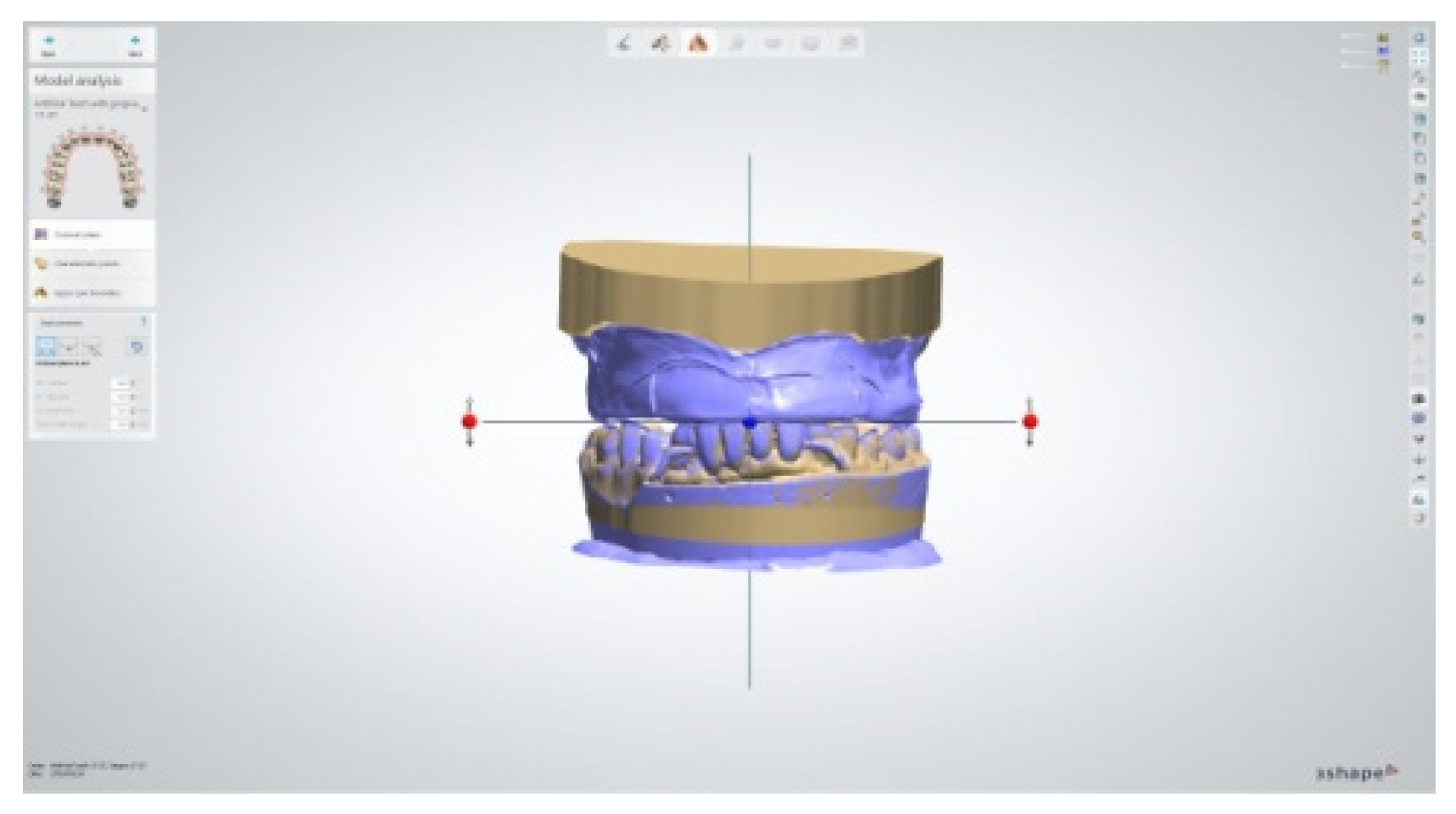
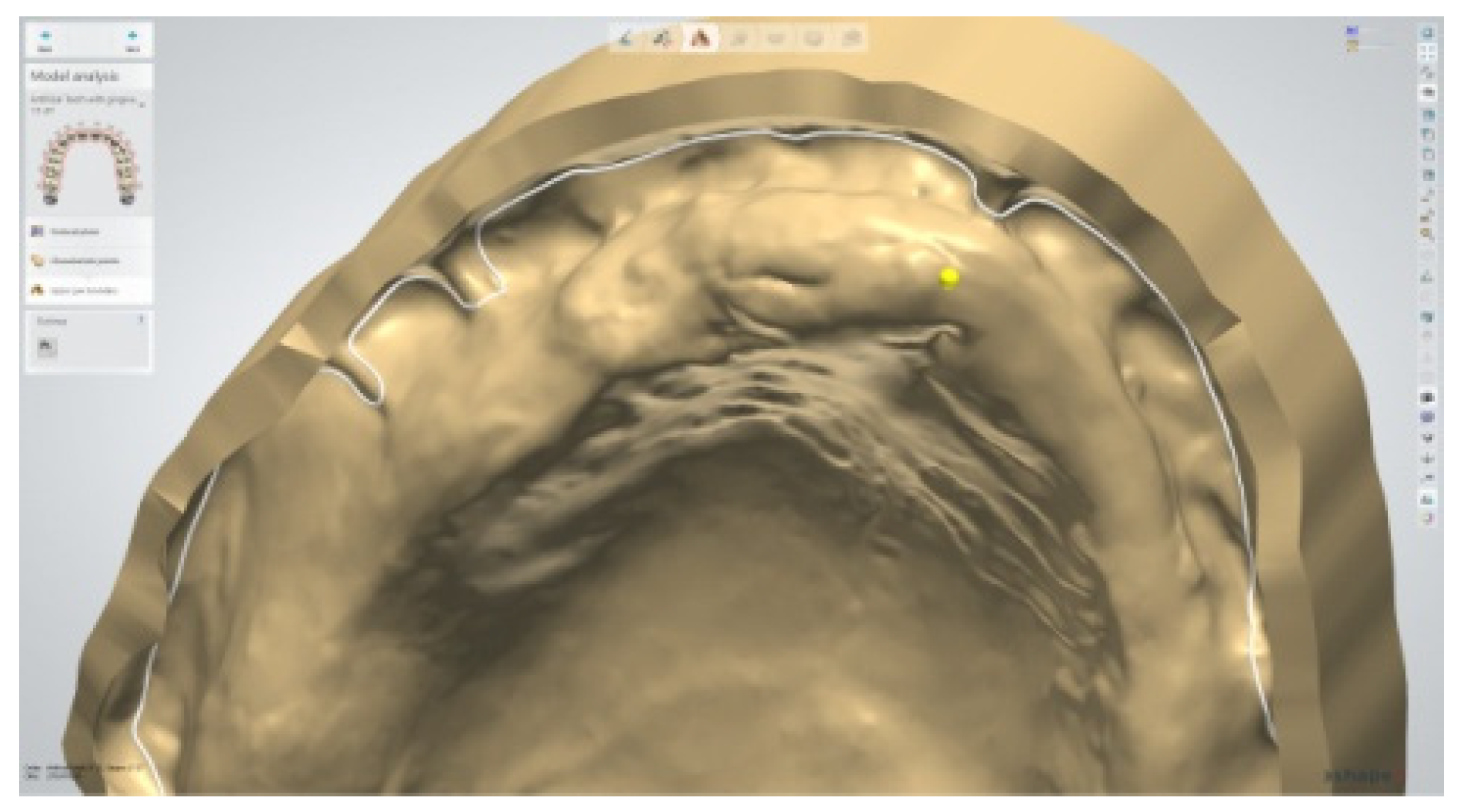
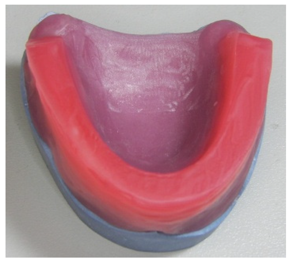
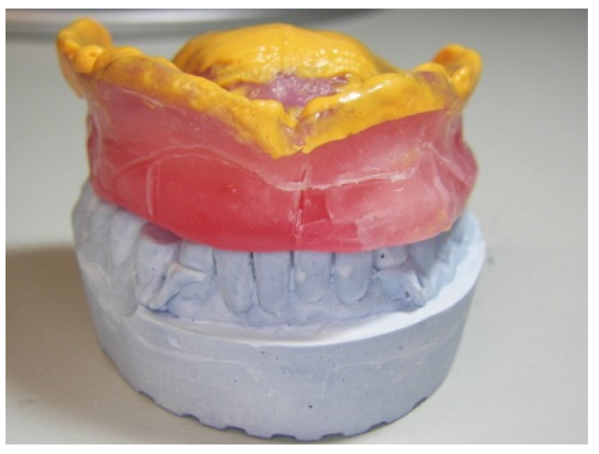
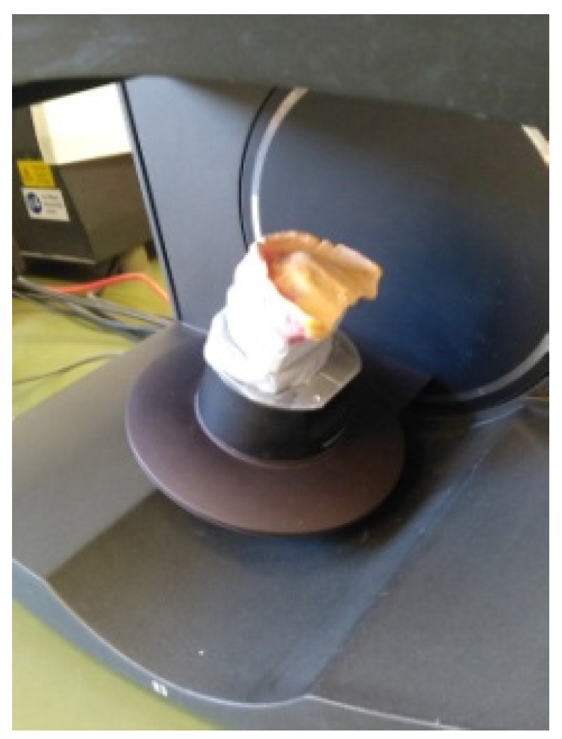
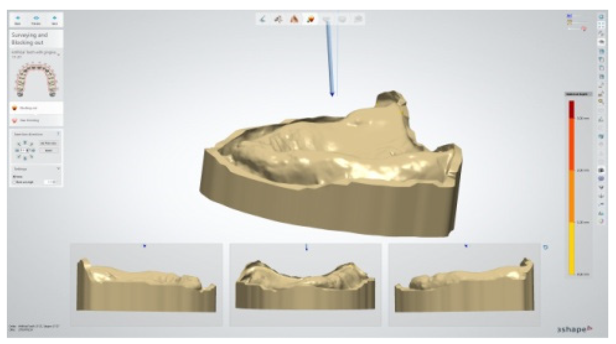
Both CAM technologies offer clinically acceptable results faster than the traditional process. The additive manufacturing technique is more recent and has the advantage that a 3D printer is more affordable than a milling machine [59]. Furthermore, the 3D printing technology is advancing rapidly, with applications in many fields, including dental laboratories. A range of materials are available for the manufacturing of a plethora of products, such as models, parts and complete restorations [60].
Additive manufacturing methods are more efficient than the subtractive ones and are available for a range of tasks, output volumes and laboratory sizes. Selection of the appropriate 3D printer is a complicated task that includes considerations about software compatibilities, printable materials and services provided by the manufacturer. The milling machines, on the other hand, have been on the market for a longer period and the customers are more likely to be familiar with the available options.
Accuracy for both techniques seem to be acceptable, however 3D printing might present a higher variance [61]. The additive manufacturing technique is evolving and several parameters that seem to affect accuracy need to be addressed. The orientation of the printed denture base, for instance, seems to affect the accuracy of the resulting product with some additive techniques [62].
Although in laboratory studies the wear resistance of the 3D-printed artificial teeth seem to be at least adequate for denture use, long duration randomized clinical trials are necessary for in depth evaluation of this aspect as well [63,64]. Regarding other mechanical properties, CDs produced with the subtractive method have been found to be superior in toughness and surface roughness than the 3D-printed ones [65]. Additionally, the CDs produced with the subtractive method have been found to have better color stability than the ones produced with the additive technique [65].
Digitally produced dentures seem to maintain the selected occlusal schemes better than conventionally produced ones [66]. However, the bond strength of denture teeth might be a concern with both CAM techniques [67].

2. Case Report

A 90-year-old ambulative female patient was presented in SY’s private practice for the replacement of the existing upper complete denture, projecting the lack of its retention as her main complaint. During her interview, she was wearing the denture and she elaborated that the problems were lack of retention during speech and chewing. She also revealed that she could use her denture only after the application of denture adhesives.
The medical record revealed excellent mental clarity, good general health in the absence of general diseases and taking medication only for hypercholesterolemia. The clinical examination showed that the upper complete denture was a repair of an old removable partial denture, which was lacking coverage of the palate and unilateral contacts with the antagonists (Figure 1). The Kennedy class I edentulism of the lower jaw had been rehabilitated with a removable partial denture (RPD), supported by a single metal–ceramic bridge from the lower left canine to the lower right one (33 to 43), with all the abutments present. The intraoral examination also revealed extensive absorption of the anterior upper and posterior lower alveolar ridge, displacement of the occlusal plane in the posterior region well below the middle of the retromolar pads, as well as a flabby ridge in the upper anterior area, between the canine regions, advocating the presence of combination syndrome.
The patient was informed that both the upper CD and the lower RPD should be replaced. Nevertheless, she agreed to proceed with the replacement of the lower partial removable denture after the upper CD was inserted.
In order to counteract the expressions of the combination syndrome, the following steps were taken: Firstly, emphasis was given to the accuracy of the impressions. Secondly, an accurate functional impression was taken, which captured the flabby ridge in a rest state. The lower RPD was relined chairside with a tissue conditioner material to facilitate the fit with the absorbed posterior mandibular alveolar ridge. The patient was also informed and agreed upon necessary occlusal adjustments. Furthermore, precautions were taken in order to avoid application of occlusal forces on the flabby ridge and an occlusion scheme was selected that would distribute forces evenly on the underlying alveolar ridge.
Considering the main complaint of the geriatric patient, execution of the treatment plan initiated with fabrication of a new upper complete denture, which was accepted by the patient. For the fabrication of the upper CD, a digital workflow was implemented utilizing CAD/CAM technology, the clinical and laboratory steps of which are presented next. The initial CD in this case was designed, for educational purposes, with two different CAD programs, namely; the Dental System 2019 program (3Shape A/S, Copenhagen, Denmark) and the DentalCAD 2.4 Plovdiv program (exocad GmbH, Darmstadt, Germany).
During the first clinical session, an initial impression was taken with putty silicone (Silagum™-Putty, DMG®, Hamburg, Germany) and a market tray for edentulous patients.
In the following step, dental stone casts were fabricated from the impressions in the laboratory and a baseplate with wax rim was produced for the upper jaw (Figure 2).
In the second clinical appointment, the upper baseplate was checked intraorally for under or overextended flanges and appropriate corrections were made. The vertical and horizontal jaw relationships were recorded, appropriate lip support was formed and the required elements’ guidelines for anterior tooth selection were carved on the wax. The existing RPD in the lower jaw was relined chairside with a tissue conditioner material (GC® tissue conditioner, GC® Corporation, Tokyo, Japan), to compensate the alveolar ridge absorption. For this step, the upper baseplate and the wax rim was in place and the newly defined vertical jaw relationship was maintained. After this step, an alginate impression of the lower jaw with the RPD was taken. Finally, with the patient being in occlusion and utilizing the upper baseplate, a wash impression with light body silicone (Honigum™-Light, DMG®, Hamburg, Germany) was taken (Figure 3).
In the following step, three scans were performed in a laboratory scanner (E3, 3Shape™ A/S, Copenhagen, Denmark), which were the tissue surface of the baseplate-wash impression, the lower jaw cast and the baseplate wax rim in relation with the mandible cast (Figure 4). The “impression scanning” option was selected in the CAD software, in order to digitally shift the maxillary tissue surface from the negative representation (impression) to a positive representation (digital cast). Finally, the digital files were imported into the CAD software for further processing (Figure 5 and Figure 6). Next, the alignment of the casts took place by selecting reference points. The occlusal plane was determined by the digitized wax rim in the Dental System 2019 program (3Shape™ A/S, Copenhagen, Denmark) (Figure 7). The functional borders of the denture were next designed (Figure 8). The marking of the appropriate reference points on the alveolar ridge (canines, incisive papilla and maxillary tuberosity) followed for tooth set-up (Figure 9). The ideal path of insertion and the degree of undercut blocking was also determined in the software (Figure 10). In the following step, parameters for the appropriate tooth selection and set-up were determined. In the present case, the option for milling teeth was preferred. Therefore, after the initial selection of size and shape, teeth were further individualized for the needs of the occlusion in this case (Figure 11 and Figure 12). Upon the completion of the tooth set-up, the design of the denture base and the “configuration of cameo surface” followed. A key parameter was the thickness of the palate for the upper denture. In this case, a thickness of 1.5 mm was chosen. Finally, a monolithic trial denture was manufactured utilizing a 3D printer (Asiga Max UV™, Sydney, Australia) designated for rapid prototypes.
At the third clinical session, evaluation of the trial denture took place. The required changes were made chairside, which included tooth arrangement by grinding or wax addition and correcting the extension and the thickness of the base. A mild overjet was created in order to avoid application of occlusal forces on the flabby ridge. Additionally, a bilaterally balanced occlusal scheme was created and carefully regulated chairside on the try-in denture.
A corrective impression with tissue conditioner material (GC® tissue conditioner, GC® Corporation, Tokyo, Japan) was taken in the same appointment. The absorbed residual ridge and the flabby mucosa in the anterior region advocated for this decision. The monolithic denture in combination with the selected material facilitated the taking of an accurate functional impression (Figure 13). The final outcome was approved by the clinician and the patient and was photographically documented (Figure 14, Figure 15 and Figure 16). Finally, the adapted trial denture and the photos were shipped to the laboratory for final adjustments and completion of the manufacturing.
Back in the laboratory, a new scan of the adapted trial denture was performed. The new scan was superimposed on the previous digital denture file, forming a new intaglio surface. The position of the anterior teeth was corrected according to the instructions and the photos sent to the laboratory. These changes were designed in the laboratory only in one of the two utilized CAD programs for time-saving purposes (DentalCAD 2.4 Plovdiv, exocad® GmbH, Darmstadt, Germany). The choice of the program was arbitrary (Figure 17). Next, the Standard Tessellation Language (STL) files of both the denture base and teeth were exported to a 5-axis milling machine (CharlyDental™ CD05-S, Mecanumeric®, Marssac-sur-Tarn, France). The base was milled in a 30 mm PMMA pink puck (IvoBase™ CAD, Ivoclar Vivadent® AG, Schaan, Liechtenstein), while teeth were milled in a 20 mm tooth-colored multilayered resin puck (SR Vivodent® CAD Multi, Ivoclar Vivadent® AG, Liechtenstein) (Figure 18 and Figure 19). The teeth sockets on the base and the cervical part of teeth were abraded with 50 μm aluminum oxide sand blasting, thoroughly cleaned, chemically activated with methyl-methacrylate monomer and bonded with a specially designed PMMA cold-curing resin for digital denture teeth bonding (IvoBase CAD Bond™, Ivoclar Vivadent® AG, Liechtenstein). The selected bonding system contained predetermined doses of a powder–liquid bonding material, special application syringes and special modeling liquid (Figure 20). After the application of the resin, the base with the teeth was placed in a heat-pressure pot to assist the polymerization (50 °C, 2.5 bar, 20 min). Finally, the standard finishing–polishing procedures were performed (Figure 21 and Figure 22). The prosthesis was kept in a water-vapor-saturated (100%RH) sealed container for 24 h before it was sent back to the dental practice [53].
Delivery to the patient was the final clinical step. No selective grinding was required at this point. The final clinical session included insertion of the CD, evaluation of the occlusion, training of the patient regarding use and maintenance of the CD and scheduling of recall appointments [68] (Figure 23).
The patient returned a week later for her first post-insertion recall appointment. Two minor sore spots were recognized. One was located in the labial sulcus to the right and the other at the buccal sulcus to the left, probably caused during insertion of the CD. After the necessary minor adjustments, the patient did not return for a second post-insertion visit, nor reported any problems on the phone, which is in accordance with the reported post-insertion management from other authors [69].

3. Discussion

The available systems for digital dentures present a variety of clinical advantages. It is obvious that the influence on the well-established clinical practices is slow as the new digital techniques need to be mastered by the dentists, the auxiliaries and the dental technicians [15]. Once the learning curve starts to flatten and the indications for the application of a digital workflow become clearer, then the community might adapt faster to the new technology [10]. Additionally, the CAD files can be easily stored for future use. However, digital dental archives are considered sensitive personal data, therefore, dedicated personnel should be adequately trained and data security policies should be implemented. File type compatibilities should also be taken into consideration whenever software is updated or hardware, such as 3D printers, is being replaced.
Among the advantages of the digital techniques are the superior mechanical properties of denture bases milled from pre-polymerized PMMA pucks, although the clinical impact of these improvements is yet to be established through future research [11,52]. Furthermore, the CDs produced from a digital workflow seem to have improved fit with the underlying tissues in comparison to the conventional ones [12,13]. The principal idea behind digital technology in removable prosthodontics is sparing chair time in the dental practice, time which could be used for other, possibly more urgent or more profitable, treatments [9,18,49]. The goal is to shorten the denture-making technique and to improve the final product [17]. However, if this is actually achieved remains to be verified with randomized long-term clinical trials. It is not clear or self-evident that incorporation of digital technology is immediately leading to faster production per case, unless otherwise stated by the system’s protocol [14,16,40]. As with so many scientific breakthroughs in the past, the rise of the digital era is also associated with enthusiasm. Nonetheless, the wide range of possible applications of the new technology and its potential value in everyday dentistry are slowly being revealed over time.
There are additional reasons to proceed with the digital transformation. Conventional removable prosthodontics require professionals with skills that develop over time, along with experience, and the outcome is technique-sensitive, since error might accumulate through the numerous manufacturing steps. It is challenging to find experienced dental technicians for high-quality removable dentures. Furthermore, the conventional technique is harder to track and record for post hoc quality control and optimization of the procedure, which can significantly benefit both the patient and the dentist [4].
Regarding the patient benefits from the digital CD technology, traditional dentures take a long time to be produced and usually require multiple appointments, which might be problematic for geriatric patients or patients with special needs. The digital transition offers the option for faster treatment, enhancing the convenience for the patients. Furthermore, the easily stored CAD files from a previous treatment can be used for the production of a new prosthesis in case of loss of the old one [65]. In such a case, the patient is only required for the insertion appointment.
The transition to the digital era requires a careful financial planning from the side of the dentist, who also gets the role of the entrepreneur. In busy practices, chair time is treated as an asset that should be optimally exploited in order to increase the business revenue. However, one has to think as a business manager in order to comprehend and value the new technology. Admittedly, diving into the digital world requires a totally different mindset than earlier, a large initial investment followed by high maintenance costs, development of new skills and recognition of previously non-existent threats [46].
Additionally, a large number of combinations between the classic treatment procedure and the digital approach can be applied [35]. Besides, the selection of a CAD/CAM denture fabrication system and the pathway combining conventional and digital workflows depends on the dentist’s prosthodontic experience and patient-specific requirements [40]. Moreover, compatibilities between CAD software and CAM systems, as well as between materials and CAM systems, are still under discussion. The technology is evolving rapidly, which is an additional point of concern.
Although the digital transformation is accompanied by many advantages, it is not, by definition, lucrative in all business models. It requires busy practices or dental technician laboratories with demand for a large number of prostheses, mature markets with developed logistic chains and an educated workforce. A quick and wide digital transformation that is not thoroughly planned and adequately supported both by the entrepreneur and the market might lead to unwanted results. Therefore, the digital transformation, despite its numerous practical advantages, requires knowledge of the market and has the characteristics of an investment that should be analyzed not only from its technical aspects, but also financially, before proceeding with a purchase [49].
Nowadays, educational institutions are in a state of incorporating the new technology in their curricula [47]. However, dental schools and dental technology schools might need to equally emphasize subjects of business administration, including business planning, development, finance, data analysis, quality assurance, sales and marketing [44,45]. A plethora of advantages derive from the digital transition and the implementation of the digital workflow in the educational field [48]. Apart from the positive effect on the reputation of the institution, the researchers and the students could benefit from the transition as well, since tracking in CAD/CAM procedures is easily performed. With the appropriate software, students can even follow up the production steps remotely or in asynchronous distance learning set-ups, the value of which is easily recognizable, especially nowadays, during the COVID-19 pandemic.

4. Conclusions

In conclusion, complete denture fabrication through digital technology seems to be versatile, enabling the combination of the well-documented traditional technique in clinical practice with the latest CAD/CAM breakthroughs in the laboratory setting. The new digital technology provides new and promising opportunities and a way to bring the dental profession in the next level. Nevertheless, this path should not be taken without consideration and careful planning.

Author Contributions

A.V., H.K., S.Y. and G.P. contributed substantially to the conception and design of the work; A.V., A.T. and N.S. contributed to the acquisition and analysis of the literature; and A.V., H.K. and S.Y. contributed to the interpretation of data for the work. Additionally, A.V., H.K., A.T. and S.Y. contributed to drafting the work, while H.K., S.Y., N.S. and G.P. revised it critically for important intellectual content. All authors finally approved the version to be published and all agreed to be accountable for all aspects of the work in ensuring that questions related to the accuracy or integrity of any part of the work are appropriately investigated and resolved. All authors have read and agreed to the published version of the manuscript.

Funding

This research received no external funding.

Institutional Review Board Statement

Not applicable. This was not an intervention study involving human subjects. The reported treatment was conducted in a private practice.

Informed Consent Statement

Informed consent was obtained from all subjects involved in the study.

Data Availability Statement

Not applicable.

Conflicts of Interest

The authors declare no conflict of interest.

References

  1. Polzer, I.; Schimmel, M.; Müller, F.; Biffar, R. Edentulism as part of the general health problems of elderly adults. Int. Dent. J. 2010, 60, 143–155. [Google Scholar] [CrossRef]
  2. Kim, J.J. Revisiting the Removable Partial Denture. Dent. Clin. N. Am. 2019, 63, 263–278. [Google Scholar] [CrossRef]
  3. Maeda, Y.; Minoura, M.; Tsutsumi, S.; Okada, M.; Nokubi, T. A CAD/CAM system for removable denture. Part. I: Fabrication of complete dentures. Int. J. Prosthodont. 1994, 7, 17–21. [Google Scholar] [PubMed]
  4. Dental Products Report. Available online: https://www.dentalproductsreport.com/view/future-dentures-digital (accessed on 1 May 2021).
  5. Goodacre, C.J.; Garbacea, A.; Naylor, W.P.; Daher, T.; Marchack, C.B.; Lowry, J. CAD/CAM fabricated complete dentures: Concepts and clinical methods of obtaining required morphological data. J. Prosthet. Dent. 2012, 107, 34–46. [Google Scholar] [CrossRef]
  6. Kattadiyil, M.T.; Goodacre, C.J.; Baba, N.Z. CAD/CAM complete dentures: A review of two commercial fabrication systems. J. Calif. Dent. Assoc. 2013, 41, 407–416. [Google Scholar] [PubMed]
  7. Infante, L.; Yilmaz, B.; McGlumphy, E.; Finger, I. Fabricating complete dentures with CAD/CAM technology. J. Prosthet. Dent. 2014, 111, 351–355. [Google Scholar] [CrossRef] [PubMed]
  8. Inokoshi, M.; Kanazawa, M.; Minakuchi, S. Evaluation of a complete denture trial method applying rapid prototyping. Dent. Mater. J. 2012, 31, 40–46. [Google Scholar] [CrossRef] [Green Version]
  9. Deak, A.; Marinello, C.P. CAD-CAM-Anwendung in der Totalprothetik CAD-CAM-Anwendung in der Totalprothetik CAD-CAM-Anwendung in der Totalprothetik. Swiss Dent. J. 2015, 125, 713–728, (In French/German). [Google Scholar] [PubMed]
  10. Bonnet, G.; Batisse, C.; Bessadet, M.; Nicolas, E.; Veyrune, J.L. A new digital denture procedure: A first practitioners appraisal. BMC Oral Health 2017, 17, 155. [Google Scholar] [CrossRef] [Green Version]
  11. Janeva, N.M.; Kovacevska, G.; Elencevski, S.; Panchevska, S.; Mijoska, A.; Lazarevska, B. Advantages of CAD/CAM versus Conventional Complete Dentures—A Review. Open Access Maced. J. Med. Sci. 2018, 6, 1498–1502. [Google Scholar] [CrossRef] [Green Version]
  12. Steinmassl, O.; Dumfahrt, H.; Grunert, I.; Steinmassl, P.A. CAD/CAM produces dentures with improved fit. Clin. Oral Investig. 2018, 22, 2829–2835. [Google Scholar] [CrossRef] [Green Version]
  13. Srinivasan, M.; Cantin, Y.; Mehl, A.; Gjengedal, H.; Müller, F.; Schimmel, M. CAD/CAM milled removable complete dentures: An in vitro evaluation of trueness. Clin. Oral Investig. 2017, 21, 2007–2019. [Google Scholar] [CrossRef] [Green Version]
  14. Schlenz, M.A.; Schmidt, A.; Wöstmann, B.; Rehmann, P. Clinical performance of computer-engineered complete dentures: A retrospective pilot study. Quintessence Int. 2019, 50, 706–711. [Google Scholar] [CrossRef] [PubMed]
  15. Schwindling, F.S.; Stober, T. A comparison of two digital techniques for the fabrication of complete removable dental prostheses: A pilot clinical study. J. Prosthet. Dent. 2016, 116, 756–763. [Google Scholar] [CrossRef]
  16. John, A.V.; Abraham, G.; Alias, A. Two-visit CAD/CAM milled dentures in the rehabilitation of edentulous arches: A case series. J. Indian Prosthodont. Soc. 2019, 19, 88–92. [Google Scholar] [CrossRef]
  17. Kattadiyil, M.T.; Jekki, R.; Goodacre, C.J.; Baba, N.Z. Comparison of treatment outcomes in digital and conventional complete removable dental prosthesis fabrications in a predoctoral setting. J. Prosthet. Dent. 2015, 114, 818–825. [Google Scholar] [CrossRef]
  18. Arakawa, I.; Al-Haj Husain, N.; Srinivasan, M.; Maniewicz, S.; Abou-Ayash, S.; Schimmel, M. Clinical outcomes and costs of conventional and digital complete dentures in a university clinic: A retrospective study. J. Prosthet. Dent. 2021, in press. [Google Scholar] [CrossRef] [PubMed]
  19. Kattadiyil, M.T.; Mursic, Z.; AlRumaih, H.; Goodacre, C.J. Intraoral scanning of hard and soft tissues for partial removable dental prosthesis fabrication. J. Prosthet. Dent. 2014, 112, 444–448. [Google Scholar] [CrossRef] [PubMed]
  20. Lo Russo, L.; Salamini, A. Single-arch digital removable complete denture: A workflow that starts from the intraoral scan. J. Prosthet. Dent. 2018, 120, 20–24. [Google Scholar] [CrossRef]
  21. Lo Russo, L.; Salamini, A.; Troiano, G.; Guida, L. Digital dentures: A protocol based on intraoral scans. J. Prosthet. Dent. 2021, 125, 597–602. [Google Scholar] [CrossRef]
  22. Chebib, N.; Kalberer, N.; Srinivasan, M.; Maniewicz, S.; Perneger, T.; Müller, F. Edentulous jaw impression techniques: An in vivo comparison of trueness. J. Prosthet. Dent. 2019, 121, 623–630. [Google Scholar] [CrossRef]
  23. Lo Russo, L.; Caradonna, G.; Troiano, G.; Salamini, A.; Guida, L.; Ciavarella, D. Three-dimensional differences between intraoral scans and conventional impressions of edentulous jaws: A clinical study. J. Prosthet. Dent. 2020, 123, 264–268. [Google Scholar] [CrossRef] [PubMed]
  24. Lo Russo, L.; Caradonna, G.; Salamini, A.; Guida, L. A single procedure for the registration of maxillo-mandibular relationships and alignment of intraoral scans of edentulous maxillary and mandibular arches. J. Prosthodont. Res. 2020, 64, 55–59. [Google Scholar] [CrossRef]
  25. Lo Russo, L.; Di Gioia, C.; Salamini, A.; Guida, L. Integrating intraoral, perioral, and facial scans into the design of digital dentures. J. Prosthet. Dent. 2020, 123, 584–588. [Google Scholar] [CrossRef]
  26. Lepidi, L.; Galli, M.; Mastrangelo, F.; Venezia, P.; Joda, T.; Wang, H.L.; Li, J. Virtual Articulators and Virtual Mounting Procedures: Where Do We Stand? J. Prosthodont. 2021, 30, 24–35. [Google Scholar] [CrossRef] [PubMed]
  27. Exocad. Available online: https://exocad.com/our-products/exocad-dentalcad/virtual-articulator (accessed on 9 May 2021).
  28. 3shape. Available online: https://www.3shape.com/en/scanners/lab (accessed on 9 May 2021).
  29. Park, J.H.; Lee, G.H.; Moon, D.N.; Kim, J.C.; Park, M.; Lee, K.M. A digital approach to the evaluation of mandibular position by using a virtual articulator. J. Prosthet. Dent. 2021, 125, 849–853. [Google Scholar] [CrossRef]
  30. Li, J.; Chen, Z.; Decker, A.M.; Wang, H.L.; Joda, T.; Mendonca, G.; Lepidi, L. Trueness and Precision of Economical Smartphone-Based Virtual Facebow Records. J. Prosthodont. 2021. [Google Scholar] [CrossRef] [PubMed]
  31. Yau, H.T.; Liao, S.W.; Chang, C.H. Modeling of digital dental articulator and its accuracy verification using optical measurement. Comput. Methods Programs Biomed. 2020, 196, 105646. [Google Scholar] [CrossRef]
  32. Li, L.; Sun, Y.; Wang, Y.; Li, W.; Dai, N.; Tian, S.; Cui, H. Accuracy of a Novel Virtual Articulator for Recording Three-Dimensional Dentition. Int. J. Prosthodont. 2020, 33, 441–451. [Google Scholar] [CrossRef]
  33. Bae, M.; Park, J.W.; Kim, N. Fully automated estimation of arch forms in cone-beam CT with cubic B-spline approximation: Evaluation of digital dental models with missing teeth. Comput. Biol. Med. 2021, 131, 104256. [Google Scholar] [CrossRef]
  34. Yu, X.; Cheng, X.; Dai, N.; Chen, H.; Yu, C.; Sun, Y. Study on digital teeth selection and virtual teeth arrangement for complete denture. Comput. Methods Programs Biomed. 2018, 155, 53–60. [Google Scholar] [CrossRef] [PubMed]
  35. Schweiger, J.; Stumbaum, J.; Edelhoff, D.; Güth, J.F. Systematics and concepts for the digital production of complete dentures: Risks and opportunities. Int. J. Comput. Dent. 2018, 21, 41–56. [Google Scholar] [PubMed]
  36. Sachs, E.; Cima, M.; Cornie, J. Three-Dimensional Printing: Rapid Tooling and Prototypes Directly from a CAD Model. CIRP Ann. 1990, 39, 201–204. [Google Scholar] [CrossRef]
  37. Shaiba, F.; Abid, H.; Shashi, B.; Mohd, J.; Sunil, K.M.; Someet, S. Exploring the significant applications of Internet of Things (IoT) with 3D printing using advanced materials in medical field. Mater. Today Proc. 2021, 45, 4844–4851. [Google Scholar] [CrossRef]
  38. DC Dentalcompare. Dental 3D Printers. Available online: https://www.dentalcompare.com/Dental-Lab-Products/24989-Dental-Laboratory-3D-Printers/ (accessed on 28 April 2021).
  39. DC Dentalcompare. Chairside 3D Printers. Available online: https://www.dentalcompare.com/Quick-Picks/348533-Chairside-3D-Printers/ (accessed on 28 April 2021).
  40. Steinmassl, P.A.; Klaunzer, F.; Steinmassl, O.; Dumfahrt, H.; Grunert, I. Evaluation of Currently Available CAD/CAM Denture Systems. Int. J. Prosthodont. 2017, 30, 116–122. [Google Scholar] [CrossRef] [Green Version]
  41. Tzeng, J.J.; Yang, T.S.; Lee, W.F.; Chen, H.; Chang, H.M. Mechanical Properties and Biocompatibility of Urethane Acrylate-Based 3D-Printed Denture Base Resin. Polymers 2021, 13, 822. [Google Scholar] [CrossRef]
  42. Chen, S.; Yang, J.; Jia, Y.-G.; Lu, B.; Ren, L. A Study of 3D-Printable Reinforced Composite Resin: PMMA Modified with Silver Nanoparticles Loaded Cellulose Nanocrystal. Materials 2018, 11, 2444. [Google Scholar] [CrossRef] [Green Version]
  43. Liao, W.; Zheng, S.; Chen, S.; Zhao, L.; Huang, X.; Huang, L.; Kang, S. Surface silanization and grafting reaction of nano-silver loaded zirconium phosphate and properties strengthen in 3D-printable dental base composites. J. Mech. Behav. Biomed. Mater. 2020, 110, 103864. [Google Scholar] [CrossRef]
  44. Badger, G.R.; Fryer, C.E.; Giannini, P.J.; Townsend, J.A.; Huja, S. Helping Dental Students Make Informed Decisions About Private Practice Employment Options in a Changing Landscape. J. Dent. Educ. 2015, 79, 1396–1401. [Google Scholar] [CrossRef]
  45. Barber, M.; Wiesen, R.; Arnold, S.; Taichman, R.S.; Taichman, L.S. Perceptions of business skill development by graduates of the University of Michigan Dental School. J. Dent. Educ. 2011, 75, 505–517. [Google Scholar] [CrossRef]
  46. Willis, D.O. Business Basics for Dentists; Wiley-Blackwell: Ames, IA, USA, 2013; pp. 161–172, 319–351. [Google Scholar]
  47. Fernandez, M.A.; Nimmo, A.; Behar-Horenstein, L.S. Digital Denture Fabrication in Pre- and Postdoctoral Education: A Survey of U.S. Dental Schools. J. Prosthodont. 2016, 25, 83–90. [Google Scholar] [CrossRef]
  48. Goodacre, C.J.; Goodacre, B.J.; Baba, N.Z. Should Digital Complete Dentures Be Part of A Contemporary Prosthodontic Education? J. Prosthodont. 2021, 30, 163–169. [Google Scholar] [CrossRef]
  49. Smith, P.B.; Perry, J.; Elza, W. Economic and Clinical Impact of Digitally Produced Dentures. J. Prosthodont. 2021, 30, 108–112. [Google Scholar] [CrossRef]
  50. Hazeveld, A.; Huddleston Slater, J.J.; Ren, Y. Accuracy and reproducibility of dental replica models reconstructed by different rapid prototyping techniques. Am. J. Orthod. Dentofac. Orthop. 2014, 145, 108–115. [Google Scholar] [CrossRef] [PubMed]
  51. Bilgin, M.S.; Baytaroğlu, E.N.; Erdem, A.; Dilber, E. A review of computer-aided design/computer-aided manufacture techniques for removable denture fabrication. Eur. J. Dent. 2016, 10, 286–291. [Google Scholar] [CrossRef] [PubMed] [Green Version]
  52. Srinivasan, M.; Gjengedal, H.; Cattani-Lorente, M.; Moussa, M.; Durual, S.; Schimmel, M.; Müller, F. CAD/CAM milled complete removable dental prostheses: An in vitro evaluation of biocompatibility, mechanical properties, and surface roughness. Dent. Mater. J. 2018, 37, 526–533. [Google Scholar] [CrossRef] [PubMed] [Green Version]
  53. Steinmassl, P.A.; Wiedemair, V.; Huck, C.; Klaunzer, F.; Steinmassl, O.; Grunert, I.; Dumfahrt, H. Do CAD/CAM dentures really release less monomer than conventional dentures? Clin. Oral Investig. 2017, 21, 1697–1705. [Google Scholar] [CrossRef] [PubMed] [Green Version]
  54. Zirkonzahn. Available online: https://zirkonzahn.com/en/products/millable-materials/tecno-med-mineral (accessed on 28 April 2021).
  55. Ivoclar Digital. Available online: https://www.ivoclardigital.com/en/laboratory/material/removable-prosthetics (accessed on 28 April 2021).
  56. Vita. Available online: https://www.vita-zahnfabrik.com/en/VITA-VIONIC-SOLUTIONS-Digital-prosthetics-are-now-available-for-practical-use-51503,57329.html (accessed on 28 April 2021).
  57. Avadent Digital Dental Solutions. Available online: https://www.avadent.com/products/solutions/ (accessed on 28 April 2021).
  58. Baltic Denture System. Available online: https://www.baltic-denture-system.de/en/ (accessed on 28 April 2021).
  59. Jurado, C.A.; Tsujimoto, A.; Alhotan, A.; Villalobos-Tinoco, J.; AlShabib, A. Digitally Fabricated Immediate Complete Dentures: Case Reports of Milled and Printed Dentures. Int. J. Prosthodont. 2020, 33, 232–241. [Google Scholar] [CrossRef] [PubMed]
  60. Anadioti, E.; Musharbash, L.; Blatz, M.B.; Papavasiliou, G.; Kamposiora, P. 3D printed complete removable dental prostheses: A narrative review. BMC Oral Health 2020, 20, 343. [Google Scholar] [CrossRef]
  61. Herpel, C.; Tasaka, A.; Higuchi, S.; Finke, D.; Kühle, R.; Odaka, K.; Rues, S.; Lux, C.J.; Yamashita, S.; Rammelsberg, P.; et al. Accuracy of 3D printing compared with milling—A multi-center analysis of try-in dentures. J. Dent. 2021, 110, 103681. [Google Scholar] [CrossRef]
  62. Unkovskiy, A.; Schmidt, F.; Beuer, F.; Li, P.; Spintzyk, S.; Kraemer Fernandez, P. Stereolithography vs. Direct Light Processing for Rapid Manufacturing of Complete Denture Bases: An In Vitro Accuracy Analysis. J. Clin. Med. 2021, 10, 1070. [Google Scholar] [CrossRef] [PubMed]
  63. Pham, D.M.; Gonzalez, M.D.; Ontiveros, J.C.; Kasper, F.K.; Frey, G.N.; Belles, D.M. Wear Resistance of 3D Printed and Prefabricated Denture Teeth Opposing Zirconia. J. Prosthodont. 2021. [Google Scholar] [CrossRef]
  64. Cha, H.S.; Park, J.M.; Kim, T.H.; Lee, J.H. Wear resistance of 3D-printed denture tooth resin opposing zirconia and metal antagonists. J. Prosthet. Dent. 2020, 124, 387–394. [Google Scholar] [CrossRef] [PubMed]
  65. Srinivasan, M.; Kamnoedboon, P.; McKenna, G.; Angst, L.; Schimmel, M.; Özcan, M.; Müller, F. CAD-CAM Removable Complete Dentures: A systematic review and meta-analysis of trueness of fit, biocompatibility, mechanical properties, surface characteristics, color stability, time-cost analysis, clinical and patient-reported outcomes. J. Dent. 2021, in press. [Google Scholar] [CrossRef]
  66. Chaturvedi, S.; Addas, M.K.; Alqahtani, N.M.; Al Ahmari, N.M.; Alfarsi, M.A. Clinical analysis of CAD-CAM milled and printed complete dentures using computerized occlusal force analyser. Technol. Health Care 2021, 29, 797–811. [Google Scholar] [CrossRef] [PubMed]
  67. Choi, J.J.E.; Uy, C.E.; Plaksina, P.; Ramani, R.S.; Ganjigatti, R.; Waddell, J.N. Bond Strength of Denture Teeth to Heat-Cured, CAD/CAM and 3D Printed Denture Acrylics. J. Prosthodont. 2020, 29, 415–421. [Google Scholar] [CrossRef]
  68. Available online: http://www.gerodontology.gr/en/public.html (accessed on 5 August 2021).
  69. Clark, W.A.; Brazile, B.; Matthews, D.; Solares, J.; De Kok, I.J. A Comparison of Conventionally Versus Digitally Fabricated Denture Outcomes in a Dental Clinic. J. Prosthodont. 2021, 30, 47–50. [Google Scholar] [CrossRef]
Figure 1. Patient’s existing upper denture.
Prosthesis 03 00023 g001
Figure 2. Baseplate-wax rim.
Prosthesis 03 00023 g002
Figure 3. Maxillomandibular records and wash impression.
Prosthesis 03 00023 g003
Figure 4. Maxillomandibular records and wash impression scanning in a lab scanner.
Prosthesis 03 00023 g004
Figure 5. Digital cast of the upper edentulous jaw.
Prosthesis 03 00023 g005
Figure 6. Digital maxillomandibular records.
Prosthesis 03 00023 g006
Figure 7. Determination of the occlusal plane in Dental System 2019 (3Shape™ A/S, Copenhagen, Denmark) software.
Prosthesis 03 00023 g007
Figure 8. Designing the functional borders of the denture.
Prosthesis 03 00023 g008
Figure 9. Reference points (canines, incisive papilla and maxillary tuberosity) are marked digitally and serve as guidelines for tooth set-up.
Prosthesis 03 00023 g009
Figure 10. Ideal denture’s path of insertion.
Prosthesis 03 00023 g010
Figure 11. Tooth selection and draft arrangement.
Prosthesis 03 00023 g011
Figure 12. Customized tooth set-up with individual corrections in size, shape and position. The intensity of the occlusal contacts and the relationship of the cusps with the occlusal plane were noted in color. Blue contacts indicated teeth that needed to be moved upward in relation to the occlusal plane or a required virtual selective grinding.
Prosthesis 03 00023 g012
Figure 13. Corrective functional impression with the trial denture.
Prosthesis 03 00023 g013
Figure 14. 3D-printed trial monolithic denture at the session of clinical evaluation. Unilateral occlusal contacts and wax corrections were detected.
Prosthesis 03 00023 g014
Figure 15. Patient’s photo in rest position with a light smile.
Prosthesis 03 00023 g015
Figure 16. The midline has to move slightly to the right, regardless of the midline below. Moreover a cervical movement of 21 to the distal and a slight movement of 22 and 23 downwards were recommended.
Prosthesis 03 00023 g016
Figure 17. Defining a new tissue surface and correcting the position of the teeth. The color contrast indicates the old and the new digital outline overlapping each other (DentalCAD 2.4 Plovdiv, exocad® GmbH, Darmstadt, Germany).
Prosthesis 03 00023 g017
Figure 18. Milled denture base with shaped teeth sockets.
Prosthesis 03 00023 g018
Figure 19. Milled denture teeth.
Prosthesis 03 00023 g019
Figure 20. Digital denture teeth bonding kit.
Prosthesis 03 00023 g020
Figure 21. Denture after teeth bonding.
Prosthesis 03 00023 g021
Figure 22. The digitally manufactured CD after the finishing and polishing procedures.
Prosthesis 03 00023 g022
Figure 23. The patient wearing her new CD.
Prosthesis 03 00023 g023
Table 1. Additive manufacturing methods for CDs.
StereolithographyDigital Light ProcessingMultiJet/PolyJet Printing
AcronymSLADLP-
Production TimeUsually longer than DLPShort print cyclesColor combinations prolong printing cycles
Light sourceUV LASER 1Specialized light projectorsUV light
Bath tankYesYesNo
Supports neededYesYesNo
1 UV: ultraviolet. LASER: light amplification by stimulated emission of radiation.
Table 2. Subtractive manufacturing methods for CDs.
Zirkonzahn Denture System™Ivoclar® Digital DentureVita Vionic™AvaDent® Digital DenturesBaltic Denture System™
Monolithic CD-Yes-Yes-
Resin puck for denture base with preformed teeth set-ups----Yes
Resin puck for denture baseYesYesYesYes-
Resin puck for denture teeth-Yes---
Whole dental arch fabrication/adjustment-Yes--Yes
Compatibility with prefabricated teethYes-YesYes-
Teeth hand bondingYesYes (if separately produced)YesYes-
Hand characterizationYes----
Hand polishingYesYesYesYesYes
Light-colored pucks for try-in denturesYes--Yes-
Appropriate for all jaw relationshipsYesYesYesYes-
Publisher’s Note: MDPI stays neutral with regard to jurisdictional claims in published maps and institutional affiliations.

