Abstract
PostCOVID-19 is a condition affecting approximately 10% of individuals infected with SARS-CoV-2, presenting significant challenges in diagnosis and clinical management. Portable neuroimaging techniques, such as functional near-infrared spectroscopy (fNIRS), offer real-time insights into cerebral hemodynamics and represent a promising tool for studying postCOVID-19 in naturalistic settings. This study investigates the integration of fNIRS with machine learning to identify neural correlates of postCOVID-19. A total of six machine learning classifiers—Random Forest, Support Vector Machine (SVM), K-Nearest Neighbors (KNNs), XGBoost, Logistic Regression, and Multi-Layer Perceptron (MLP)—were evaluated using a stratified subject-aware cross-validation scheme on a dataset comprising 29,737 time-series samples from 37 participants (9 postCOVID-19, 28 controls). Four different feature representation strategies were compared: raw time-series, PCA-based dimensionality reduction, statistical feature extraction, and a hybrid approach that combines time-series and statistical descriptors. Among these, the hybrid representation demonstrated the highest discriminative performance. The SVM classifier trained on hybrid features achieved strong discrimination ( = 0.909) under subject-aware CV5; at the default threshold, was moderate and was high, outperforming all other methods. In contrast, models trained on statistical features alone exhibited limited despite high . These findings highlight the importance of temporal information in the fNIRS signal and support the potential of machine learning combined with portable neuroimaging for postCOVID-19 identification. This approach may contribute to the development of non-invasive diagnostic tools to support individualized treatment and longitudinal monitoring of patients with persistent neurological symptoms.
1. Introduction
Long COVID, also known as postCOVID-19 condition, is defined by the World Health Organization (WHO) as a condition occurring in individuals with a history of probable or confirmed SARS-CoV-2 infection, usually three months after the onset of COVID-19, with symptoms lasting for at least two months that an alternative diagnosis cannot explain. This condition can affect multiple organ systems and has a significant impact on daily functioning []. It is associated with at least 10% of SARS-CoV-2 infections, presenting persistent symptoms that are difficult to diagnose and treat []. An estimated 7% of adults—or about 17 million people—in the United States reported having postCOVID-19 in March 2024, based on data from the Centers for Disease Control and Prevention (CDC) []. Clinical practice guidelines have been developed to help healthcare professionals manage the long-term effects of this condition, focusing on symptom monitoring, rehabilitation, and tailored interventions for affected individuals [].
Neurological symptoms associated with COVID-19 range from headaches and loss of taste and smell to sleep disorders and cognitive impairments such as difficulties in concentration, language, and executive function, as well as clinically significant depression and anxiety. Additionally, autonomic neurocirculatory abnormalities have been suggested, including orthostatic intolerance syndromes [,]. The main proposed mechanisms of neuropathology in acute COVID-19 include systemic inflammation, neuroinflammation, and microvascular injury with leakage of blood products into the parenchyma and microthrombosis. Several studies in subacute neuro-COVID patients have observed activation of innate immune responses, including autopsy studies that support this as a potential mechanism, suggesting a possible localization of autonomic symptoms in brainstem dysfunction or vascular injury. A post-viral, immune-mediated process is the most likely cause in this context, and some immunological dysregulation features have been reported in postCOVID-19 cohorts. However, the overall pathophysiology remains unclear []. Neuroimaging, combined with machine learning techniques, offers a promising approach to distinguish patients with postCOVID-19 symptoms. Studies have explored various aspects, including multivariate prediction of postCOVID-19 headache using structural MRI features [], risk predictors integrated into clinical workflows [], and precision phenotyping through machine learning []. These approaches utilize diverse data types, including CT imaging [], laboratory data, and structural MRI features [], to understand and predict postCOVID-19’s impact on patients.
Recent studies in neuroimaging have significantly advanced our understanding of the neurological consequences of postCOVID-19. Barnden et al. [] conducted a pivotal study highlighting altered brain connectivity during cognitive exertion in postCOVID-19 patients, indicating potential neural pathways affected by the virus. Parallel to this, García-González et al. [] explored the neurological implications of both acute and postCOVID-19 using brain organoids, providing valuable insights into the virus’s long-term effects on brain function. Adding to this domain, Matías-Guiu and Díez-Cirarda [] investigated cognitive and neuroimaging signatures associated with postCOVID-19, suggesting the presence of distinct neurological markers. Complementing these findings, Kim and Young [] discussed the broader neuroimaging observations in COVID-19 patients, including those suffering from long-term effects, thus broadening the perspective on COVID-19’s neurological impact. Lastly, the study by Kim, Yang, and Kim [] utilized MRI features to predict postCOVID-19 headaches in adolescents, focusing on the structural changes in gray matter, which opens new avenues for understanding and predicting postCOVID-19 symptoms.
The exploration of postCOVID-19’s neurological impacts through neuroimaging has primarily focused on traditional modalities like MRI, as evidenced by recent studies [,]. However, there is a notable gap in the use of portable functional modalities such as functional near-infrared spectroscopy (fNIRS) in this field. Functional Near-Infrared Spectroscopy (fNIRS) is a non-invasive optical imaging technique. It measures the hemodynamic responses associated with neural activity, specifically monitoring the changes in blood oxygenation and blood volume in the cerebral cortex. fNIRS utilizes near-infrared light to penetrate the skull and measure the differential absorption of light by oxy-hemoglobin and deoxy-hemoglobin, thus providing insights into brain function []. While fNIRS offers advantages like portability, lower cost, and ease of use in various settings, its application in studying postCOVID-19 remains underexplored. This gap is significant as fNIRS could potentially provide insights into cerebral hemodynamics and neural activation in real time and in more naturalistic settings, which are crucial for understanding the dynamic brain changes in postCOVID-19 patients. The versatility of fNIRS in different environments could also facilitate longitudinal studies, enabling the monitoring of brain function over time in patients recovering from COVID-19. Thus, incorporating fNIRS into postCOVID-19 research could enhance our understanding of the condition’s neurological aspects, especially in scenarios where traditional imaging techniques are less feasible.
Functional near-infrared spectroscopy (fNIRS) offers a non-invasive window into cortical hemodynamics during motor tasks, making it well-suited to probe the interplay between motor function and cognition. This approach is motivated by robust evidence linking motor deficits to cognitive decline: for example, it has been found that fine motor impairments (e.g., slowed finger tapping and reduced hand dexterity) constitute a key feature of mild cognitive impairment []. Such motor dysfunctions are increasingly viewed as early biomarkers of cognitive deterioration [], as seen in Alzheimer’s disease (AD) and mild cognitive impairment, where even subtle losses in gait and hand dexterity correlate with declining memory and executive function []. Indeed, quantifying fine motor decline in MCI may help capture the neural changes that accompany incipient AD, distinct from those of normal aging. This paradigm extends to postCOVID conditions: emerging evidence indicates that long COVID patients frequently suffer persistent cognitive impairment (colloquially “brain fog”) alongside altered motor control []. For instance, recent studies report that long COVID survivors perform worse on tests of manual dexterity and motor learning than healthy controls [,]. Therefore, applying fNIRS during a finger-tapping task targeting the motor cortex offers a powerful means to investigate these motor–cognitive interactions in long COVID, building on established motor biomarkers used in AD and MCI research by applying ML to this novel postCOVID-19 fNIRS dataset, we uncover neural biomarkers of COVID-related effects that have not been reported previously.
The objective of this study is to quantify motor–system correlates of postCOVID-19 using functional near-infrared spectroscopy (fNIRS) analyzed with machine learning. By leveraging the rich temporal information in our large fNIRS dataset, we train robust machine learning classifiers that identify novel neural patterns associated with postCOVID-19. We compare feature selection strategies and classifiers to identify the most effective pipeline for fNIRS-based detection of postCOVID-19 motor alterations.
2. Materials and Methods
2.1. Study Design
The dataset used in this study comprises time-series fNIRS data from 37 participants (9 postCOVID and 28 controls), totaling 29,737 labeled samples after preprocessing. Of these, 6084 correspond to postCOVID patients and 23,655 to healthy controls. This study was approved by the State Research Ethics Committee in Health of San Luis Potosí, Mexico (SLP/08-2020). It was designed as a cross-sectional analytical study, with a non-probabilistic sample of participants recruited from previous studies. Inclusion criteria for positive patients were age between 18 and 70 years, both genders, symptomatic and asymptomatic with RT-qPCR positive for SARS-CoV-2, with Ct below 38. Pregnant patients and those with confirmed pulmonary infections other than COVID-19 were excluded. Inclusion criteria for the postCOVID-19 group included adult patients between 18 and 70 years old, with persistent COVID-19 symptoms after 4 weeks and negative RT-qPCR test at the time of evaluation. Those with previous pulmonary diseases or signs of infections were excluded. For the control group, 28 subjects with a resolved history of SARS-CoV-2 infection who were asymptomatic, free of respiratory diseases, and without apparent COVID-19 symptoms for at least 7 days were included.
The fNIRS technique was used to measure hemodynamic responses in the cerebral cortex. A near-infrared light was emitted to penetrate the skull, and the absorption by oxy-hemoglobin and deoxy-hemoglobin was recorded. Data were collected while participants performed a finger-tapping motor task, designed to activate the primary motor cortex (M1) and assess fine motor control. The signals were processed and analyzed to identify patterns in brain activity between the postCOVID-19 and control groups. The collected data were normalized and preprocessed to remove noise and improve signal quality.
2.2. fNIRS Data Acquisition
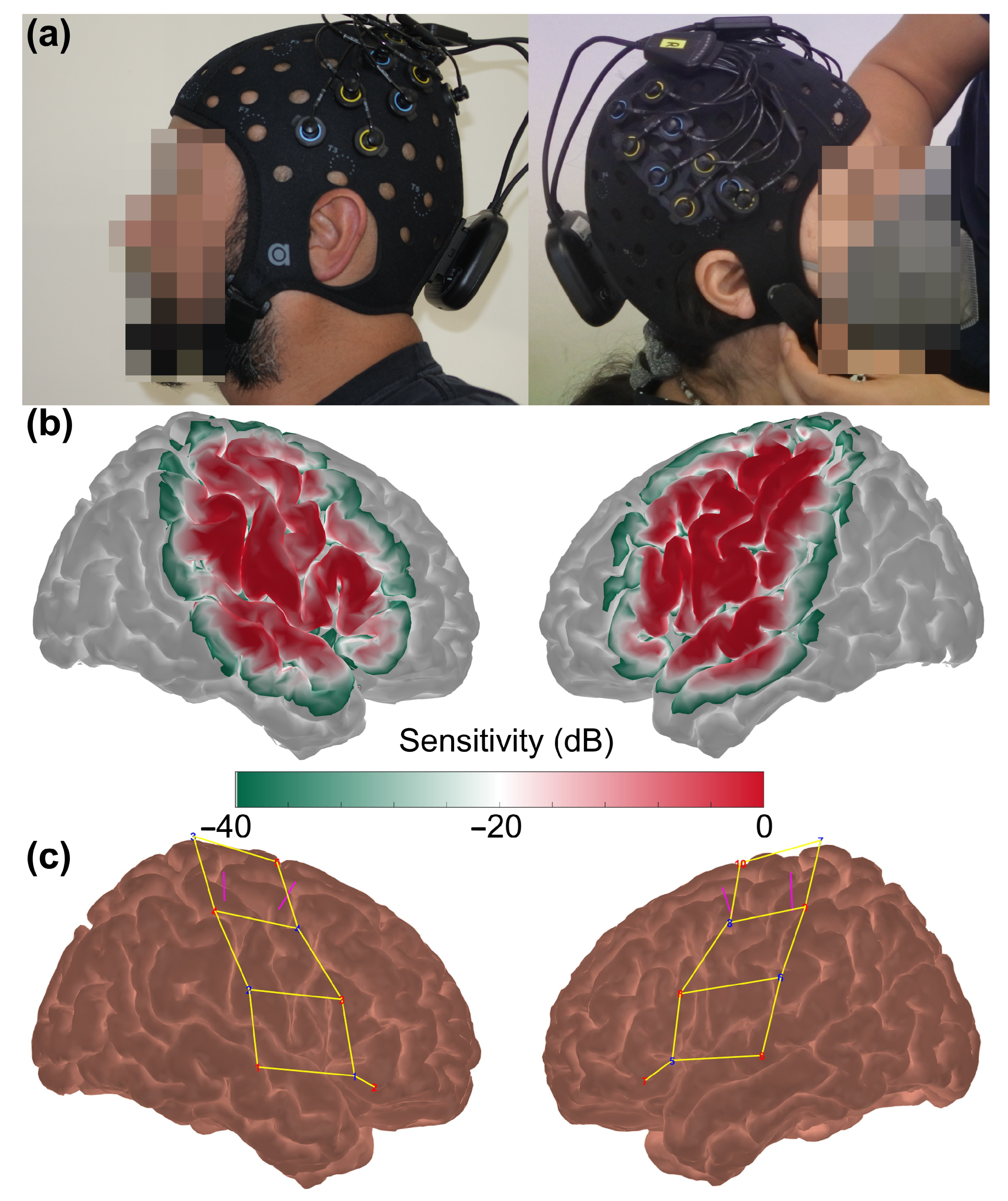
The portable fNIRS system Brite MKII (Artinis Medical Systems BV, Elst, The Netherlands) was used to measure cortical brain activity across bilateral motor regions, employing ten dual-wavelength LEDs centered at 757 and 843 nm, as well as eight detectors, with a sampling frequency of 25 Hz, sufficient to avoid aliasing effects from blood pressure, respiratory, and cardiac frequencies. Participants were fitted with a black neoprene head cap (54–60 cm) with the optodes arranged 3 cm apart for 20 long channels and 1.5 cm apart for two short channels to optimize hemodynamic measurement [], as depicted in Figure 1a. The cap was placed on participants’ heads with sources and detectors manually adjusted for efficient optode-scalp coupling, as determined in real time by the manufacturer’s software, to ensure sufficient sensitivity in the areas of interest (Figure 1b). The long channels measured hemoglobin changes in bilateral motor regions, as shown in Figure 1c, while short channels minimized superficial hemodynamic effects [,].
Figure 1.
(a) Images displaying the fNIRS probe placed on the head of the participants. (b) A logarithmic plot that represents the sensitivity of the optodes array to detect hemodynamic changes and (c) registered probe geometry covering the motor cortices: channels are indicated by yellow lines, the eight detectors are depicted in blue numbers while the 10 sources are indicated by red numbers; magenta lines delimit the activation area of fingertapping.
A finger-tapping task was used to activate the primary motor cortex (M1), and its associated hemodynamic activity was recorded. Participants were then instructed to tap their thumbs in sequence with the other fingers of the same hand (e.g., thumb and index finger, thumb and middle finger, etc.). The movements were carried out in short bursts of 10 s, with rest intervals between 20 and 24 s. Video cues guided the start and stop of each movement. The variable rest periods aimed to prevent synchronization with physiological hemodynamic oscillations and minimize anticipatory responses [,].
2.3. fNIRS Data Pre-Processing
Data were processed and analyzed in MATLAB R2017b using Homer3 [] and custom-made scripts. Initial quality control excluded readings with high/low optical density changes, low signal-to-noise ratio (SNR), or long source-detector separation. Raw data were transformed into optical density (OD) units, and motion artifacts were identified and corrected using cubic spline interpolation (p = 0.99) [,] and wavelet correction (interquartile range of 1.5) []. Residual motion artifacts were rejected within a −2 to 10 s window. OD data were bandpass filtered (0.01–3 Hz) in order to suppress slow drifts and baseline fluctuations while preserving the cardiac frequency component as a physiological marker to visually assess and ensure adequate optode-scalp coupling [], then converted to oxy- and deoxygenated hemoglobin (HbO, HbR) using the modified Bouguer–Beer–Lambert law with a partial pathlength factor of one to avoid assuming subject-specific pathlengths given their variability [,], thus yielding units of M-mm []. Hemodynamic response functions (HRF) were estimated via a General Linear Model (GLM) with short separation channels regressing superficial hemodynamics [,]. Short-channel regression was applied to attenuate superficial hemodynamic contributions arising from scalp and extracerebral tissues. By regressing out these short-distance signals, the analysis preferentially preserved deeper components of the hemodynamic response, thereby increasing the likelihood that the remaining signal reflects cortical activity of neural origin [,]. We prioritized HbO over HbR because HbO typically exhibits a higher signal-to-noise ratio, making it more robust in detecting functional changes [].
2.4. Feature Extraction and Classification
Given the structure of our fNIRS dataset, we utilized all available time points from each patient, generating a robust dataset composed of 29,737 labeled samples. These samples were derived from the time-series recordings of each subject, capturing 132 measurement variables corresponding to oxy-hemoglobin (HbO) signals recorded from 20 channels strategically positioned across the motor cortex, as detailed in Section 2.3. Each sample was labeled according to patient condition: 0 for healthy controls (23,655 samples) and 1 for postCOVID patients (6084 samples).
For each sample, we retained the original 132 measurement variables without further summarization, thus preserving the full temporal resolution of the hemodynamic response. To investigate how different representations of fNIRS data influence model performance, four distinct datasets were constructed based on alternative feature extraction strategies. These included the original time-series data preserving the full temporal resolution of the hemodynamic signal; a PCA-transformed version in which the number of components was selected to retain at least 95% of the explained variance; a dataset composed of global statistical descriptors extracted from each channel (mean, standard deviation, minimum, and maximum); and a hybrid representation that combines both time-series features and statistical descriptors. No sliding-window segmentation was used; each sample corresponds to a native fNIRS time point, and subject-aware cross-validation ensured that all samples from a participant were confined to either the training or test fold in each split.
For each dataset, six supervised machine learning algorithms were implemented: Random Forest, K-Nearest Neighbors (KNNs), Support Vector Machine (SVM), XGBoost, Logistic Regression, and Multi-Layer Perceptron (MLP). Model performance was rigorously evaluated using a stratified subject-aware five-fold cross-validation (CV5) scheme, in which all samples from each subject were assigned exclusively to either the training or testing set within each fold. In each fold of cross-validation, models were independently trained using the training subset and subsequently evaluated on the corresponding test subset, ensuring strict separation between training and testing phases and thereby preventing data leakage. Age and sex were similar across groups (Table 1), and all cross-validation folds were stratified by group to avoid biases. Performance was assessed through a comprehensive set of metrics: , , (), , , and the Area Under the Receiver Operating Characteristic Curve (). These metrics were computed for each fold and then averaged across folds to derive a reliable estimation of each model’s predictive capability. This rigorous methodological framework allowed us to objectively determine the effectiveness of each algorithm and to evaluate the impact on classification performance. To ensure methodological transparency, all classifiers were implemented using standard, widely adopted hyperparameters as recommended in their respective libraries (e.g., scikit-learn, XGBoost). This choice was deliberate, as the study’s primary objective was to compare feature representations rather than perform model-specific optimization. Additionally, a feature-level interpretation was performed based on subject-level summaries (mean, standard deviation, minimum, and maximum values for each fNIRS channel and hemoglobin signal [HbO, HbR, HbT]), allowing the identification of informative features at the individual level. These four statistical descriptors were selected because they capture complementary aspects of the hemodynamic response: the overall level (mean), variability (standard deviation), and amplitude of extrema (minimum and maximum), providing a compact yet physiologically meaningful representation of each subject’s signal profile. This interpretability analysis was carried out using two complementary approaches: supervised feature importance ranking through Random Forests and unsupervised loading inspection via Principal Component Analysis (PCA), both applied under stratified subject-level cross-validation.
Table 1.
Demographic and clinical data of postCOVID patients and controls. Unless otherwise indicated, the median values are given, with interquartile range in brackets. Reporting: p-values are shown with four decimals. p-values < 0.05 denoted in bold. 1 Wilcoxon–Mann–Whitney p-value. 2 Fisher’s exact test p-value. Sample sizes were and for all comparisons; no missing data were present.
2.5. Metrics for Evaluation
The models were evaluated using various metrics to ensure a comprehensive assessment of their performance. The performance metrics included , , , Positive Predictive Value (), and Negative Predictive Value (). The formulas used to calculate the metrics were as follows:
is given by
is given by
Positive Predictive Value () is given by
Negative Predictive Value () is given by
The is given by
where (True Positive) represents the number of correctly identified positive cases, (False Negative) represents the number of positive cases incorrectly identified as negative, (True Negative) represents the number of correctly identified negative cases, and (False Positive) represents the number of negative cases incorrectly identified as positive.
Given the 1:3 class imbalance (postCOVID vs. controls), we additionally report imbalance-robust metrics: (average for the positive class = postCOVID), class-wise s, , and Matthews correlation coefficient (). All metrics were computed on the held-out fold in a subject-aware five-fold CV and then averaged across folds.
The , which balances and , is defined as
where is equivalent to the Positive Predictive Value () and is equivalent to .
The Matthews correlation coefficient (), which captures overall classification quality in the presence of class imbalance, is given by
3. Results
The demographic and clinical data of postCOVID patients and the control group are summarized in Table 1. Significant differences were observed in MoCA scores (p = 0.0297) between the groups, as determined by the Wilcoxon–Mann–Whitney test. Other parameters did not show statistically significant differences.
3.1. Results Using Hybrid Features
In this section, the classification performance of a hybrid feature representation combining raw time series with global statistical descriptors (mean, standard deviation, minimum, and maximum) extracted from each fNIRS channel was evaluated. This strategy aims to capture both the local temporal dynamics and global patterns of cortical hemodynamics. The models were trained and evaluated on a robust dataset consisting of 29,737 labeled samples derived from 37 participants (9 postCOVID and 28 controls).
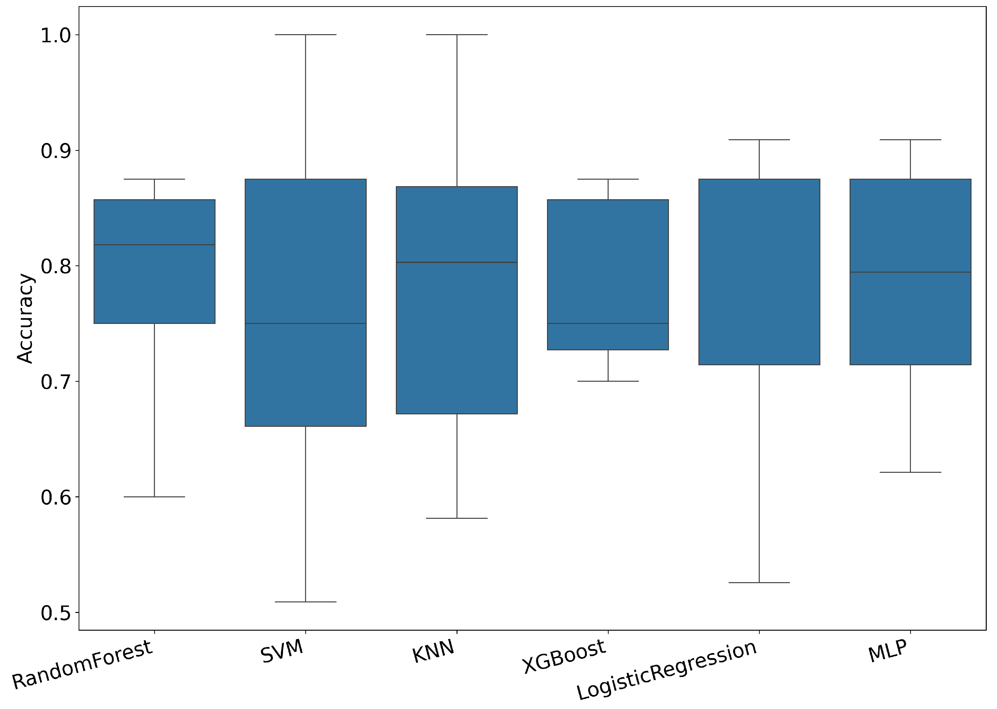
The performance of six machine learning models (Random Forest, Support Vector Machine (SVM), K-Nearest Neighbors (KNNs), XGBoost, Logistic Regression, and Multi-Layer Perceptron (MLP)) was evaluated to classify postCOVID-19 patients. Each model underwent rigorous evaluation through stratified subject-aware cross-validation with a CV5, ensuring that data from each participant were included exclusively in either the training or test folds. Figure 2 shows the distribution across folds for each model. While performance varies across classifiers and participants, most models achieved median accuracies above 75%, demonstrating the overall viability of the hybrid feature approach when generalizing to unseen subjects.
Figure 2.
distribution across models using hybrid features under subject-aware CV5.
Table 2 summarizes the average clinical performance metrics obtained for each machine learning model using hybrid features under subject-aware CV5, including , , , Positive Predictive Value (), negative predictive value (), and the Area Under the ROC Curve ().
Table 2.
Average (%) performance metrics using hybrid features for each model in CV5. Bold values indicate the highest average performance achieved in their respective column.
The results demonstrate variability in classification performance across all models. While accuracies ranged between 75.9% and 78.5%, significant variability was observed in and metrics. KNNs achieved the highest average (78.5%), although the highest (90.9%) was attained by SVM, indicating a strong discriminative capability despite lower . Logistic Regression and MLP models also presented balanced metrics, with Logistic Regression achieving notably higher (50.0%) and (63.5%). These results underscore the value of the hybrid feature approach, highlighting its potential clinical relevance and robust generalization when applied to previously unseen subjects.
Figure 3 displays the average ROC curves for each of the evaluated machine learning models using the hybrid features and subject-aware cross-validation. The SVM model achieved the highest (0.909), reflecting a stronger overall discriminative capability, followed by Logistic Regression (0.851) and Random Forest (0.839). These ROC curves further support the robustness and clinical potential of the hybrid approach when classifying postCOVID-19 patients based on fNIRS-derived hemodynamic signals.
Figure 3.
Average curves across models using hybrid features under subject-aware CV5.
3.2. Comparative Analysis of Feature Extraction Methods
In this subsection, a systematic comparison of four different approaches to classify postCOVID-19 patients based on fNIRS signals was conducted. The evaluated methods include (1) the proposed hybrid features, combining original time-series and statistical descriptors, previously detailed in Section 3.1; (2) the full raw time-series data (original data), consisting of all 132 measured variables; (3) PCA-based dimensionality reduction, in which Principal Component Analysis (PCA) was implemented using a subject-aware pipeline (StandardScaler → PCA) within each cross-validation fold. In each fold, the scaler and PCA were fit exclusively on the training data and then applied to the corresponding test data, thereby preventing any information leakage. The number of components was determined to retain 95% of the explained variance of the training subset in each fold, resulting in a variable number of principal components across folds. This approach ensures that dimensionality reduction is performed in a leakage-free manner, strictly adhering to best practices for cross-validation; and (4) statistical features, created by flattening the temporal dynamics into summary statistics (mean, standard deviation, minimum, and maximum) calculated for each of the 132 measurement variables, yielding 528 statistical features per participant. Each feature representation was evaluated using six machine learning models under a rigorous subject-aware CV5 strategy to ensure realistic generalization to unseen subjects.
Table 3 provides a comprehensive summary of the average classification performance achieved by each machine learning model across the four evaluated feature representation approaches (hybrid, original time-series, PCA-reduced, and statistical features). The best-performing models within each feature representation are highlighted in bold.
Table 3.
Average comprehensive comparison of classification models across different feature representation approaches. Values are presented as percentages (%) except for . Bold values indicate the highest average performance metric achieved within each feature representation approach.
The results in Table 3 clearly show distinct performance patterns across the different feature representation strategies. The hybrid approach achieved the highest discriminative performance under class imbalance, attaining the best (90.9%) and (81.2%), along with a competitive (0.429), indicating strong calibration and robustness to class imbalance. In contrast, statistical features, while producing the highest (81.1%), exhibited limited – performance ( = 43.1%), reducing their clinical utility. PCA-based and original time-series methods delivered intermediate yet consistent performance, with values around 84.8% and 82.1%, respectively, and scores near 63%. These findings underscore that the hybrid representation yields not only balanced but also superior robustness to imbalanced conditions, as reflected by higher and values. Overall, these results highlight the benefit of combining temporal dynamics with global statistical descriptors, yielding a more robust and generalizable representation of fNIRS signals for subject-level classification.
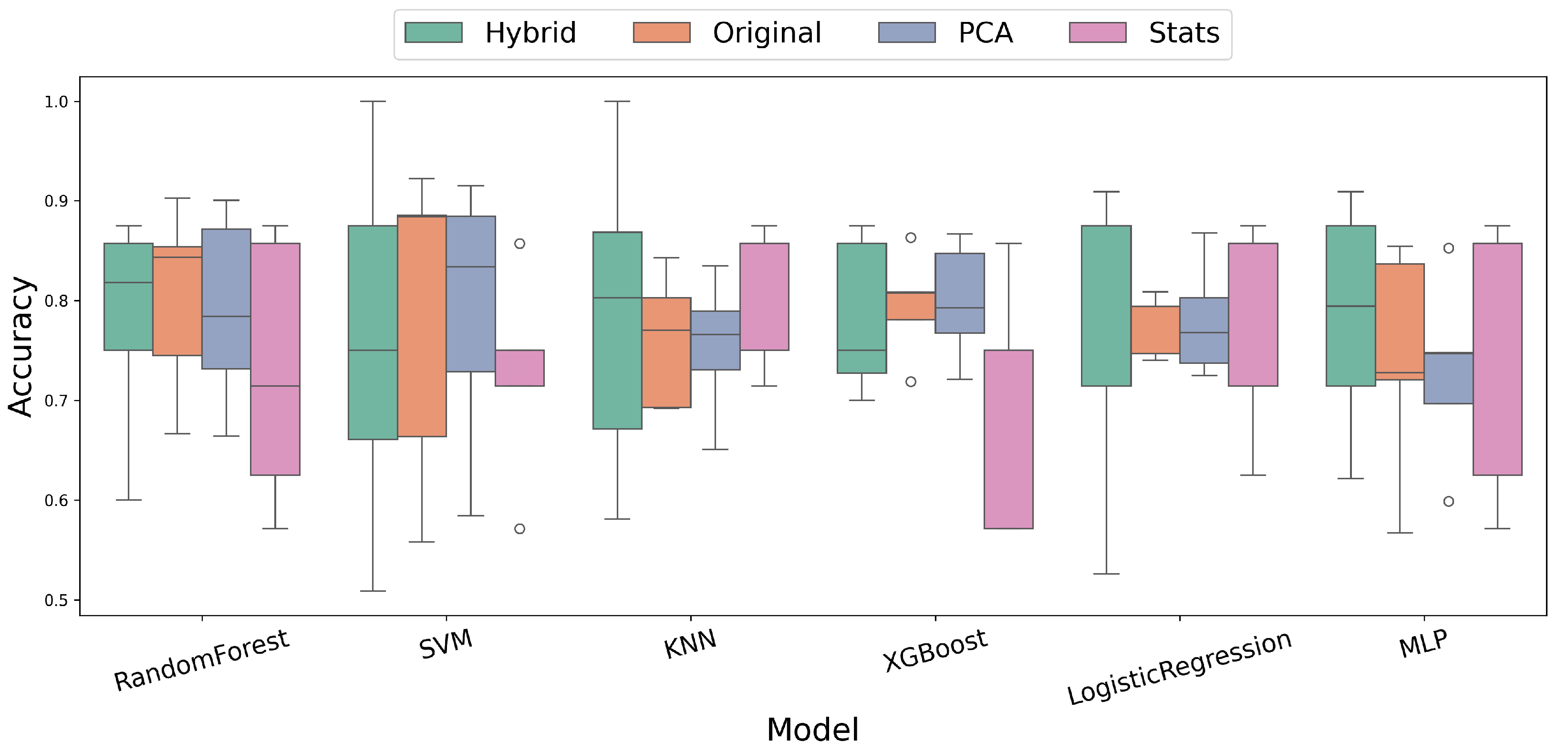
Figure 4 provides a comprehensive visualization of distributions across the six evaluated machine learning models for each feature representation approach. Notably, the hybrid approach consistently yielded balanced distributions across most classifiers, demonstrating robustness and generalizability. In contrast, statistical features exhibited high variability, particularly with models such as XGBoost and MLP, indicating limited stability and clinical applicability.
Figure 4.
distributions for each machine learning model across different feature representation approaches under subject-aware CV5.
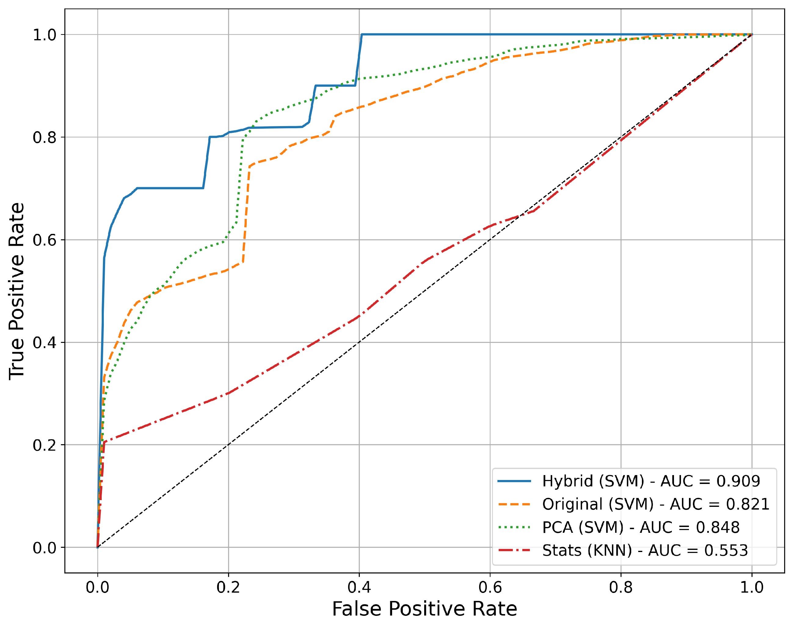
Further comparison of discriminative performance among the best models from each feature representation strategy is depicted in Figure 5. The hybrid feature representation (SVM) outperformed all other approaches, achieving the highest (0.909). The original time-series and PCA-based approaches showed intermediate performance, with values of approximately 0.821 and 0.848, respectively. The statistical feature representation (KNN) demonstrated the lowest discriminative capability ( = 0.553), emphasizing the limited utility of purely statistical descriptors in this context.
Figure 5.
curves comparing the best performing models from each feature representation approach. The hybrid approach (SVM) achieved the highest , highlighting its superior discriminative capability.
3.3. Feature-Level Interpretation and Informative Channels
To move beyond model benchmarking and provide interpretable insight from the fNIRS data, we conducted a two-step feature-level analysis: (i) a supervised ranking of informative features using Random Forest (RF) feature importance computed under stratified five-fold cross-validation at the subject level and (ii) an unsupervised inspection of PCA loadings (weighted by explained variance ratio) also performed within stratified five-fold CV to avoid leakage.
3.3.1. Random Forest Feature Importance (Supervised)
We first built one row per subject using summary statistics (mean, standard deviation, minimum, and maximum) for each channel and hemoglobin signal (HbO, HbR, HbT). A Random Forest classifier (1000 trees, balanced-subsample) was trained and evaluated under stratified five-fold CV at the subject level. For each fold, we extracted feature_importances_ and averaged them across folds to obtain a stable ranking. The top-ranked features concentrated on extrema and dispersion of deoxygenated hemoglobin and total hemoglobin (e.g., min_2HRF HbR_3_4, min_2HRF HbR_6_5, std_2HRF HbT_5_3), together with a subset of mean HbO descriptors (e.g., mean_2HRF HbO_1_1, mean_2HRF HbO_3_1). A compact summary of the Top 10 RF features is provided in Table 4.
Table 4.
Top ranked informative features obtained from supervised RF and unsupervised PCA (subject-level CV). Feature names follow the original notation.
3.3.2. PCA Loadings (Unsupervised)
In parallel, we fitted PCA within each CV fold (StandardScaler → PCA, retaining 95% variance) and computed per-feature contributions as the sum over components of the absolute loading multiplied by the component’s explained-variance ratio. The highest PCA contributions arose primarily from HbT and HbO descriptors involving maxima/minima and variability (e.g., max_2HRF HbT_4_3, max_2HRF HbO_4_3, min_3HRF HbT_1_2). The Top 10 PCA contributions are listed side by side in Table 4.
3.3.3. Convergence of Supervised and Unsupervised Evidence
Both analyses point to a coherent signature dominated by extrema and dispersion of HbR/HbT, with supportive contributions from mean HbO features. This convergence indicates that the discriminative performance observed in our classifiers is not driven by arbitrary statistical artifacts but by consistent hemodynamic patterns captured across methods.
RF highlights minima and dispersion in HbR/HbT together with a subset of mean HbO features; independently, PCA emphasizes maxima/minima and variability in HbT/HbO and partially overlaps with RF (e.g., std_2HRF HbT_4_3). This agreement across methods supports the presence of consistent hemodynamic differences between postCOVID-19 and controls and indicates that the observed discrimination is not driven by model-specific artifacts (Table 4).
4. Discussion
The machine learning models selected for this study were chosen based on their complementary methodological strengths and established effectiveness for analyzing complex, high-dimensional datasets, thus enabling a robust evaluation of different feature representation approaches. Random Forest was included due to its ability to manage large feature spaces and reduce overfitting through ensemble methods. K-Nearest Neighbors (KNNs) provided a nonparametric, distance-based classifier suitable for capturing nonlinear relationships in the data. Support Vector Machine (SVM) was selected for its strong generalization capabilities in high-dimensional settings through margin maximization. XGBoost was utilized given its proven gradient-boosting performance and robustness against class imbalance. Logistic Regression contributed with probabilistic interpretability and clearly defined model parameters. Finally, Multi-Layer Perceptron (MLP) was chosen to leverage its capacity for modeling hierarchical, nonlinear relationships through deep learning architectures. Collectively, these models provided a comprehensive methodological framework to reliably explore the neural correlates of postCOVID-19 through fNIRS, with a particular emphasis on ensuring robust generalization across unseen subjects.
The classification results obtained in this study demonstrate clear and clinically relevant differences among the evaluated feature representation approaches. The hybrid representation, combining temporal dynamics with global statistical descriptors, provided the highest discriminative performance, achieving an average of 0.909 using the Support Vector Machine (SVM) classifier. This result highlights that integrating both local and global hemodynamic information yields a robust biomarker for identifying postCOVID-19 patients. In contrast, models trained solely on the original time-series or PCA-reduced data showed moderate yet consistent performance, with values around 82% and 85%, respectively, reflecting stable but less discriminative capacity compared to the hybrid representation. Meanwhile, the statistical feature representation, despite yielding the highest overall (81.1%), demonstrated markedly lower robustness to class imbalance, with limited – performance ( = 43.1%) and moderate calibration ( = 0.26), reducing its clinical interpretability. These findings underscore the critical importance of feature representation choice, emphasizing that capturing both temporal patterns and global statistical characteristics significantly improves the robustness and clinical applicability of fNIRS-based machine learning classifiers.
The classification results obtained through the subject-aware cross-validation strategy enhance the credibility and clinical relevance of the findings. By strictly ensuring that data from individual participants were exclusively assigned to either training or testing sets, the potential risk of data leakage and overly optimistic performance estimation was effectively mitigated. The hybrid feature representation demonstrated superior discriminative performance, which can be attributed to its capacity to simultaneously capture both the subtle temporal fluctuations and the overarching statistical characteristics of fNIRS-derived hemodynamic responses. The results indicate that the temporal dynamics of cortical hemodynamic activity encode valuable diagnostic information that complements global statistical patterns, thus providing a robust and clinically relevant biomarker for identifying postCOVID-19 neural correlates.
Beyond performance metrics, Table 4 summarizes a coherent feature-level signature. Supervised RF ranks extrema and dispersion in HbR/HbT among the most informative descriptors, with complementary mean HbO features. Independently, PCA loadings weighted by explained variance emphasize related HbT/HbO variables and overlap with RF (e.g., std_2HRF HbT_4_3). This convergence indicates stable hemodynamic differences between postCOVID-19 and controls and provides a concise, data-driven set of candidate features to guide future biomarker refinement and external validation. We consider the two top-ranked features identified by both the supervised and unsupervised analyses (Table 4). The features are named as follows: [statistical operation]_[finger-tapping: 2(left), 3(right)] [hemoglobin type]_[source]_[detector]. The fNIRS channel comprising source 3 and detector 4 (Figure 6a) measures activity in the upper-lateral part of the right precentral gyrus; this area of the motor cortex controls voluntary movements of the left hand and fingers. Figure 6b shows that the ipsilateral premotor areas also represent highly ranked features. Similarly, the channel consisting of Source 4 and Detector 3 (Figure 6c,d) corresponds to the region responsible for movements of the left thumb and facial muscles. It is natural that these specific cortical sites are activated during fine motor tasks performed with the contralateral (left) hand. Since all participants identified their left hand as non-dominant, performing the finger-tapping task with that hand was slightly more challenging than using their dominant (right) hand. In participants with postCOVID-19 symptoms, this task demands even greater effort, resulting in a more pronounced hemodynamic response, specifically higher oxygenated hemoglobin (HbO) and total hemoglobin (HbT) signals with lower deoxygenated hemoglobin (HbR) amplitudes. This exacerbated brain activity is consistent with recent neuroimaging findings showing that long COVID patients experiencing “brain fog” exhibit abnormally increased brain activation during tasks [,]. Such hyperactivation likely reflects a compensatory mechanism, wherein additional neural resources are recruited to maintain normal performance []. Finding top ranked features in areas of ipsilateral activation can be explained by the fact that ipsilateral areas may be recruited to support task performance, acting as a compensatory backup network []. Further research involving larger patient cohorts is recommended to fully validate and expand upon these promising findings.
Figure 6.
Hemodynamic response function (HRF) time course where the top ranked features were extracted: (a) min_2HRF HbR_3_4, (b) min_2HRF HbR_6_5, (c) max_2HRF HbT_4_3, and (d) max_2HRF HbO_4_3. All HRF time courses are shown as mean ± standard deviation and correspond to finger tapping of the left hand (non-dominant).
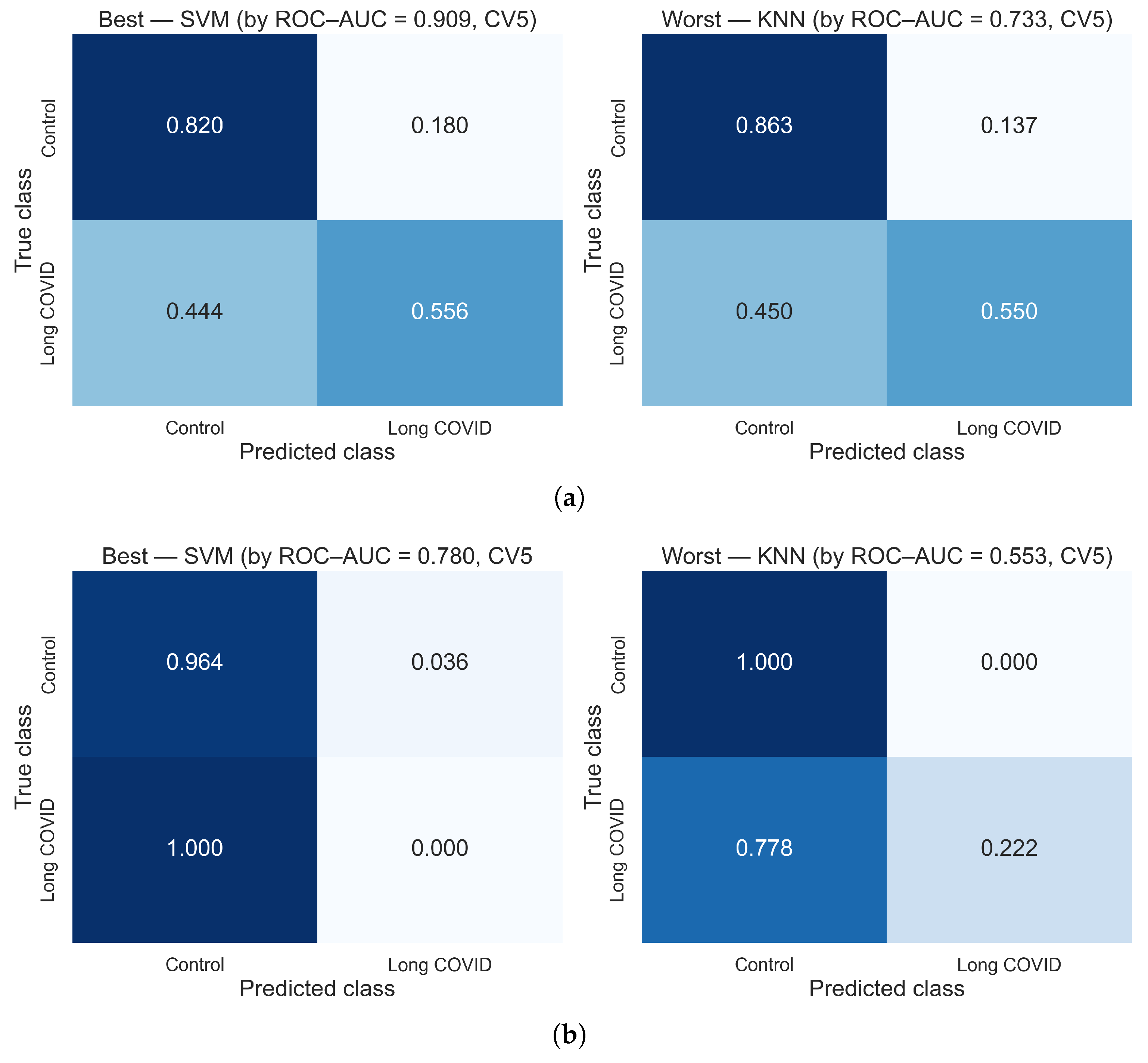
To further illustrate the differences in classification performance between feature representation approaches, normalized confusion matrices for the best and worst performing models using hybrid and statistical features were generated (Figure 7). The hybrid approach yielded balanced performance (Figure 7a), with the best model being SVM ( = 0.909), yielding an approximately 0.556 true positive rate for postCOVID and a 0.820 true negative rate for controls; the worst was KNN ( = 0.733), with approximately 0.550 and 0.863, respectively. In the statistical feature representation (Figure 7b), the best model by was SVM (0.780), which at the default threshold showed near-zero for postCOVID (0.000) despite high for controls (0.964); the worst was KNN ( = 0.553), with a postCOVID of approximately 0.222. These results visualize why hybrid features provide more clinically meaningful balance, whereas purely statistical descriptors tend to collapse .
Figure 7.
Normalized confusion matrices comparing the best and worst performing models for (a) hybrid and (b) statistical feature representations. The hybrid approach showed a more balanced – profile, while the statistical approach exhibited very low .
The findings collectively highlight that the choice of fNIRS feature representation significantly impacts classification performance. The hybrid representation, integrating temporal dynamics with global statistical descriptors, emerged clearly as the most effective approach, demonstrating balanced and , as well as superior discriminative capability. In contrast, purely statistical features, despite achieving high , exhibited very low , severely limiting their clinical applicability. Among the classifiers evaluated, models such as SVM and Logistic Regression showed strong discriminative power (high ), suggesting suitability for clinical contexts where robust generalization is essential. Nevertheless, the moderate observed across all models underscores the need for larger patient cohorts to further optimize and validate the clinical utility of these approaches. Thus, careful selection and optimization of both feature representations and classification algorithms remain crucial for achieving reliable identification of postCOVID-19 neural correlates. We verified that no demographic variable alone could explain the classification and that the ML models rely on fNIRS signal patterns.
This study represents, to the best of our knowledge, one of the first applications of functional near-infrared spectroscopy (fNIRS) technology in the analysis of postCOVID-19 patients, specifically targeting the motor cortex, although fNIRS has traditionally been used in the prefrontal cortex []. The implementation of this novel approach highlights the potential of fNIRS to provide valuable insights into the neural correlates of postCOVID-19 symptoms, which are often challenging to diagnose using traditional clinical methods. Beyond the immediate classification performance, the successful application of this methodology opens a promising avenue for developing tangible clinical tools. First, the hybrid fNIRS-based classification approach offers potential as an objective biomarker for postCOVID-19 neurological impairment, complementing current diagnostic methods that frequently rely on subjective symptom reporting. An accessible, non-invasive test could provide clinicians with quantitative, physiologically based evidence to support diagnostic decisions. Second, the portability and ease of use associated with fNIRS technology make it an ideal tool for longitudinal monitoring. Clinicians could employ this method to objectively track patients’ responses to rehabilitation therapies or pharmacological interventions over time, facilitating more personalized and data-driven treatment strategies. Ultimately, this research constitutes a foundational step toward integrating portable neuroimaging into the standard clinical management of postCOVID-19, potentially transforming how the condition is diagnosed, monitored, and understood.
However, it is important to acknowledge several limitations of this study. Although the current analysis included a robust dataset (29,737 total samples from 37 participants: 9 postCOVID and 28 controls), external validation with larger and independent patient cohorts remains necessary to confirm the generalizability and robustness of these findings. We acknowledge that the relatively small number of participants may limit statistical power; future work will involve recruiting additional subjects to validate and extend these preliminary findings. Additionally, the moderate observed across all classifiers, likely influenced by the limited number of postCOVID-19 cases, highlights the need for larger clinical samples to enhance the models’ diagnostic and reliability. Furthermore, clinical correlations are still required to better understand the relationship between fNIRS-derived neural signatures and the diverse spectrum of postCOVID-19 symptoms. Additional research incorporating larger, clinically diverse, and multicenter datasets is essential to further validate, refine, and improve the proposed models’ diagnostic . The complexity and subtlety of neurological symptoms related to postCOVID-19 underscore the continued need for rigorous, clinically informed studies.
Moreover, recent guidelines for the rigorous application of machine learning in fNIRS research emphasize the importance of strict validation protocols, transparent reporting of preprocessing and analytical steps, and caution when interpreting results, particularly in studies with clinical diagnostic implications []. Therefore, despite the analytical approach and the extensive dataset employed, these findings should still be interpreted cautiously, with validation from future studies involving larger, diverse, and independently collected samples strongly recommended.
The performance variability observed across different classifiers within the hybrid feature representation underscores the critical importance of careful model selection and parameter optimization in machine learning analyses of high-dimensional fNIRS data. Although classifiers such as SVM achieved the highest discriminative performance ( = 0.909), the moderate observed across models highlights the need for further optimization to enhance their suitability for clinical applications requiring higher . Future research should prioritize exploring advanced feature selection techniques, parameter tuning, and ensemble approaches to optimize classifier performance, aiming to pinpoint a minimal set of fNIRS channels or metrics as a diagnostic signature, particularly given the subtle and heterogeneous neurological presentations associated with postCOVID-19 syndrome.
In summary, the findings demonstrate that machine learning analysis of fNIRS-derived hemodynamic signals can effectively discriminate postCOVID-19 patients from healthy controls, with the hybrid feature representation showing superior discriminative performance ( = 0.909) and, at the default threshold, high and moderate . Nevertheless, the moderate observed underscores the importance of external validation with larger and clinically diverse cohorts. This study highlights both the potential and the critical methodological considerations involved in applying portable neuroimaging and machine learning tools to clinical diagnosis and longitudinal monitoring of complex neurological conditions such as postCOVID-19 syndrome.
5. Conclusions
In this study, the integration of functional near-infrared spectroscopy (fNIRS) and machine learning was investigated as a strategy to identify neural correlates associated with postCOVID-19 syndrome. A total of six machine learning classifiers—Random Forest, Support Vector Machine (SVM), K-Nearest Neighbors (KNNs), XGBoost, Logistic Regression, and Multi-Layer Perceptron (MLP)—were evaluated using a subject-aware CV5 strategy on a dataset comprising 29,737 labeled samples from 37 participants (9 postCOVID and 28 controls). Among the four tested feature representation approaches, a hybrid strategy that combines raw time-series data with statistical descriptors achieved the best overall performance. In particular, the SVM model trained with hybrid features reached an of 0.909, demonstrating superior discriminative ability with high and moderate at the default threshold.
To ensure methodological rigor and minimize the risk of overfitting or data leakage, a stratified subject-aware CV5 scheme was employed, in which all data from each participant were assigned exclusively to either the training or testing sets within each fold. This approach preserved class balance while simulating real-world clinical conditions by evaluating model generalization to entirely unseen subjects. For dimensionality reduction, PCA was applied within each training fold and subsequently transferred to the corresponding test fold, ensuring a leakage-free and reproducible transformation process. These methodological considerations enhance the reliability and clinical relevance of the reported results.
While all evaluated models demonstrated robust overall performance, remained moderate across classifiers, particularly in comparison to their consistently high . This performance profile suggests that models such as SVM and Logistic Regression, despite their lower , may still be valuable in clinical contexts where minimizing false positives is critical. Nonetheless, the observed variability emphasizes the importance of tailoring model selection and tuning to specific diagnostic priorities. Threshold calibration for specific clinical targets (e.g., high- screening) will be addressed in future work using independent validation cohorts, once larger datasets become available. Future research should focus on validating these approaches in larger and more clinically diverse populations, and on exploring the relationship between fNIRS-derived neural signatures and the heterogeneous neurological manifestations of postCOVID-19.
In conclusion, the integration of portable neuroimaging techniques such as fNIRS with machine learning represents a promising avenue for the non-invasive identification of neural alterations associated with postCOVID-19. The findings from this study suggest that feature representations capturing the temporal dynamics of the hemodynamic signal provide superior discriminative power compared to purely statistical descriptors. This underscores the relevance of temporal information in differentiating postCOVID-19 patients from healthy controls. Although further validation is required, this approach holds significant potential to improve diagnostic , support individualized interventions, and contribute to the clinical management of patients experiencing persistent neurological symptoms following COVID-19.
Author Contributions
Conceptualization, B.N.Z.-M. and E.G.; methodology, A.M.-C., B.N.Z.-M., R.F.-R. and E.G.; software, A.M.-C. and V.H.; validation, V.H. and A.A.L.-C.; formal analysis, A.M.-C.; investigation, B.N.Z.-M. and R.F.-R.; resources, R.F.-R.; data curation, A.A.L.-C.; writing—original draft preparation, A.M.-C.; writing—review and editing, B.N.Z.-M., R.F.-R. and E.G.; supervision, R.F.-R. and E.G.; funding acquisition, E.G. All authors have read and agreed to the published version of the manuscript.
Funding
This research received no external funding.
Institutional Review Board Statement
The study was conducted in accordance with the Declaration of Helsinki, and approved by the State Research Ethics Committee in Health of San Luis Potosí, Mexico (protocol code SLP/08-2020 and date of approval: 4 July 2022).
Informed Consent Statement
Informed consent was obtained from all subjects involved in the study.
Data Availability Statement
The data and code supporting this study’s findings are publicly available in Zenodo at the following DOI: https://doi.org/10.5281/zenodo.16429864.
Acknowledgments
We express our gratitude to “La Secretaría de Ciencia, Humanidades, Tecnología e Innovación” (SECIHTI) for the postdoctoral grant and SEDEAM for providing the equipment and workplace (Antony Morales-Cervantes).
Conflicts of Interest
On behalf of all authors, the corresponding author states that there are no conflicts of interest.
References
- World Health Organization. Post COVID-19 Condition. 2023. Available online: https://www.paho.org/es/temas/coronavirus/brote-enfermedad-por-coronavirus-covid-19/condicion-post-covid-19 (accessed on 28 September 2024).
- Davis, H.E.; McCorkell, L.; Vogel, J.M.; Topol, E.J. Long COVID: Major findings, mechanisms and recommendations. Nat. Rev. Microbiol. 2023, 21, 133–146. [Google Scholar] [CrossRef]
- Centers for Disease Control and Prevention (CDC). Long COVID Statistics. 2024. Available online: https://www.cdc.gov/covid/?CDC_AAref_Val=https://www.cdc.gov/coronavirus/2019-ncov/long-term-effects.html (accessed on 28 September 2024).
- Seo, J.W.; Kim, S.E.; Kim, Y.; Kim, E.J.; Kim, T.; Kim, T.; Lee, S.H.; Lee, E.; Lee, J.; Seo, Y.B.; et al. Updated Clinical Practice Guidelines for the Diagnosis and Management of Long COVID. Infect. Chemother. 2024, 56, 122–157. [Google Scholar] [CrossRef] [PubMed]
- Mina, Y.; Enose-Akahata, Y.; Hammoud, D.A.; Videckis, A.J.; Narpala, S.R.; O’Connell, S.E.; Carroll, R.; Lin, B.C.; McMahan, C.C.; Nair, G.; et al. Deep phenotyping of neurologic postacute sequelae of SARS-CoV-2 infection. Neurol. Neuroimmunol. Neuroinflamm. 2023, 10, e200097. [Google Scholar] [CrossRef] [PubMed]
- Serrano del Pueblo, V.M.; Serrano-Heras, G.; Romero Sánchez, C.M.; Piqueras Landete, P.; Rojas-Bartolome, L.; Feria, I.; Morris, R.G.M.; Strange, B.; Mansilla, F.; Zhang, L.; et al. Brain and cognitive changes in patients with long COVID compared with infection-recovered control subjects. Brain 2024, 147, 3611–3623. [Google Scholar] [CrossRef]
- Greene, C.; Connolly, R.; Brennan, D.; Laffan, A.; O’Keeffe, E.; Zaporojan, L.; O’Callaghan, J.; Thomson, B.; Connolly, E.; Argue, R.; et al. Blood–brain barrier disruption and sustained systemic inflammation in individuals with long COVID-associated cognitive impairment. Nat. Neurosci. 2024, 27, 421–432. [Google Scholar] [CrossRef] [PubMed]
- Kim, M.; Sim, S.; Yang, J.; Kim, M. Multivariate prediction of long COVID headache in adolescents using gray matter structural MRI features. Front. Hum. Neurosci. 2023, 17, 1202103. [Google Scholar] [CrossRef]
- Bhattacharya, B.S.; Mitchell, E.G.; Shetty, G.; Delong, G.; Munia, T.T.K.; Tariq, A.A. A Long COVID Risk Predictor Focused on Clinical Workflow Integration. medRxiv 2023. [Google Scholar] [CrossRef]
- Epsi, N.J.; Lindholm, D.A.; Ganesan, A.; Lalani, T.; Smith, A.G.; Mody, R.; Jones, M.; Bazan, S.; Colombo, R.E.; Colombo, C. Precision phenotyping of “long COVID” through machine learning. Open Forum Infect. Dis. 2022, 9, ofac492.907. [Google Scholar] [CrossRef]
- Chen, C.; Li, R.; Shen, H.; Xia, L. Long Short-Term Memory Based Framework for Longitudinal Assessment of COVID-19 Using CT Imaging and Laboratory Data. IEEE Access 2022, 10, 55533–55545. [Google Scholar] [CrossRef]
- Barnden, L.; Thapaliya, K.; Eaton-Fitch, N.; Barth, M.; Marshall-Gradisnik, S. Altered brain connectivity in long COVID during cognitive exertion: A pilot study. Front. Neurosci. 2023, 17, 1182607. [Google Scholar] [CrossRef]
- García-González, L.; Marti-Sarrias, A.; Puertas, M.C.; Bayón-Gil, Á.; Resa-Infante, P.; Martinez-Picado, J.; Navarro, A.; Acosta, S. Understanding the neurological implications of acute and long COVID using brain organoids. Dis. Model. Mech. 2023, 16, dmm049902. [Google Scholar] [CrossRef]
- Matias-Guiu, J.A.; Díez-Cirarda, M. Are there cognitive and neuroimaging signatures in long COVID? Brain Commun. 2023, 5, fcad189. [Google Scholar] [CrossRef]
- Kim, J.; Young, G.S. Neuroimaging of COVID-19. Semin. Neurol. 2023, 43, 205–218. [Google Scholar] [CrossRef]
- Ayaz, H.; Baker, W.B.; Blaney, G.; Boas, D.A.; Bortfeld, H.; Brady, K.; Brake, J.; Brigadoi, S.; Buckley, E.M.; Carp, S.A.; et al. Optical imaging and spectroscopy for the study of the human brain: Status report. Neurophotonics 2022, 9, S24001. [Google Scholar] [CrossRef]
- Lodha, N.; McCarthy, S. Do fine motor impairments constitute a key feature of mild cognitive impairment? A systematic review and meta-analysis. Alzheimer’s Dement. 2021, 17, e056433. [Google Scholar] [CrossRef]
- de Paula, J.J.; Albuquerque, M.R.; Lage, G.M.; Bicalho, M.A.; Romano-Silva, M.A.; Malloy-Diniz, L.F. Impairment of fine motor dexterity in mild cognitive impairment and Alzheimer’s disease dementia: Association with activities of daily living. Rev. Bras. Psiquiatr. 2016, 38, 235–238. [Google Scholar] [CrossRef]
- Hayward, W.; Buch, E.R.; Norato, G.; Iwane, F.; Dash, D.; Salamanca-Girón, R.F.; Bartrum, E.; Walitt, B.; Nath, A.; Cohen, L.G. Procedural motor memory deficits in patients with long-COVID. Neurology 2024, 102, e208073. [Google Scholar] [CrossRef]
- Sanal-Hayes, N.E.M.; Hayes, L.D.; Mclaughlin, M.; Berry, E.C.J.; Sculthorpe, N.F. People with long COVID and ME/CFS exhibit similarly impaired dexterity and bimanual coordination: A case-case-control study. Am. J. Med. 2025, 138, 893–900. [Google Scholar] [CrossRef] [PubMed]
- Lee, C.K.; Sun, C.W.; Lee, P.L.; Lee, H.C.; Yang, C.; Jiang, C.P.; Tong, Y.P.; Yeh, T.C.; Hsieh, J.C. Study of photon migration with various source-detector separations in near-infrared spectroscopic brain imaging based on three-dimensional Monte Carlo modeling. Opt. Express 2005, 13, 8339–8348. [Google Scholar] [CrossRef] [PubMed]
- Brigadoi, S.; Cooper, R.J. How short is short? Optimum source–detector distance for short-separation channels in functional near-infrared spectroscopy. Neurophotonics 2015, 2, 025005. [Google Scholar] [CrossRef]
- Tachtsidis, I.; Scholkmann, F. False positives and false negatives in functional near-infrared spectroscopy: Issues, challenges, and the way forward. Neurophotonics 2016, 3, 031405. [Google Scholar] [CrossRef]
- Yeung, M.K. An optical window into brain function in children and adolescents: A systematic review of functional near-infrared spectroscopy studies. Neuroimage 2021, 227, 117672. [Google Scholar] [CrossRef] [PubMed]
- Lloyd-Fox, S.; Blasi, A.; Elwell, C.E. Illuminating the developing brain: The past, present and future of functional near infrared spectroscopy. Neurosci. Biobehav. Rev. 2010, 34, 269–284. [Google Scholar] [CrossRef] [PubMed]
- Huppert, T.J.; Diamond, S.G.; Franceschini, M.A.; Boas, D.A. HomER: A review of time-series analysis methods for near-infrared spectroscopy of the brain. Appl. Opt. 2009, 48, D280–D298. [Google Scholar] [CrossRef]
- Scholkmann, F.; Spichtig, S.; Muehlemann, T.; Wolf, M. How to detect and reduce movement artifacts in near-infrared imaging using moving standard deviation and spline interpolation. Physiol. Meas. 2010, 31, 649–662. [Google Scholar] [CrossRef]
- Selb, J.J.; Yücel, M.A.; Phillip, D.; Schytz, H.W.; Iversen, H.K.; Vangel, M.; Ashina, M.; Boas, D.A. Effect of motion artifacts and their correction on near-infrared spectroscopy oscillation data: A study in healthy subjects and stroke patients. J. Biomed. Opt. 2015, 20, 056011. [Google Scholar] [CrossRef] [PubMed]
- Novi, S.L.; Roberts, E.; Spagnuolo, D.; Spilsbury, B.M.; Price, D.C.; Imbalzano, C.A.; Forero, E.; Tellis, G.M.; Tellis, C.M.; Mesquita, R.C. Functional near-infrared spectroscopy for speech protocols: Characterization of motion artifacts and guidelines for improving data analysis. Neurophotonics 2020, 7, 015001. [Google Scholar] [CrossRef]
- Di Rosa, E.; Brigadoi, S.; Cutini, S.; Tarantino, V.; Dell’Acqua, R.; Mapelli, D.; Braver, T.S.; Vallesi, A. Reward motivation and neurostimulation interact to improve working memory performance in healthy older adults: A simultaneous tDCS-fNIRS study. Neuroimage 2019, 202, 116062. [Google Scholar] [CrossRef]
- Pereira, J.; Direito, B.; Lührs, M.; Castelo-Branco, M.; Sousa, T. Multimodal assessment of the spatial correspondence between fNIRS and fMRI hemodynamic responses in motor tasks. Sci. Rep. 2023, 13, 2244. [Google Scholar] [CrossRef]
- Yücel, M.A.; Lühmann, A.V.; Scholkmann, F.; Gervain, J.; Dan, I.; Ayaz, H.; Boas, D.; Cooper, R.J.; Culver, J.; Elwell, C.E.; et al. Best practices for fNIRS publications. Neurophotonics 2021, 8, 012101. [Google Scholar] [CrossRef]
- Delpy, D.T.; Cope, M.; van der Zee, P.; Arridge, S.; Wray, S.; Wyatt, J. Estimation of optical pathlength through tissue from direct time of flight measurement. Phys. Med. Biol. 1988, 33, 1433–1442. [Google Scholar] [CrossRef]
- Gagnon, L.; Cooper, R.J.; Yücel, M.A.; Perdue, K.L.; Greve, D.N.; Boas, D.A. Short separation channel location impacts the performance of short channel regression in NIRS. Neuroimage 2012, 59, 2518–2528. [Google Scholar] [CrossRef]
- Saager, R.B.; Berger, A.J. Direct characterization and removal of interfering absorption trends in two-layer turbid media. J. Opt. Soc. Am. A 2005, 22, 1874–1882. [Google Scholar] [CrossRef] [PubMed]
- Li, Y.; Zhang, H.; Yu, M.; Yu, W.; Frederick, B.d.; Tong, Y. Systemic low-frequency oscillations observed in the periphery of healthy human subjects. J. Biomed. Opt. 2018, 23, 057001. [Google Scholar] [CrossRef]
- Chang, L.; Ryan, M.C.; Liang, H.; Zhang, X.; Cunningham, E.; Wang, J.; Wilson, E.; Herskovits, E.H.; Kottilil, S.; Ernst, T.M. Changes in brain activation patterns during working memory tasks in people with post-COVID condition and persistent neuropsychiatric symptoms. Neurology 2023, 100, e2409–e2423. [Google Scholar] [CrossRef]
- Tanashyan, M.M.; Kuznetsova, P.I.; Morozova, S.N.; Annushkin, V.A.; Raskurazhev, A.A. Neuroimaging correlates of post-COVID-19 symptoms: A functional MRI approach. Diagnostics 2024, 14, 2180. [Google Scholar] [CrossRef]
- Strens, L.H.A.; Fogelson, N.; Shanahan, P.; Rothwell, J.C.; Brown, P. The ipsilateral human motor cortex can functionally compensate for acute contralateral motor cortex dysfunction. Curr. Biol. 2003, 13, 1201–1205. [Google Scholar] [CrossRef] [PubMed]
- Clemente, L.; La Rocca, M.; Quaranta, N.; Iannuzzi, L.; Vecchio, E.; Brunetti, A.; Gentile, E.; Dibattista, M.; Lobasso, S.; Bevilacqua, V.; et al. Prefrontal dysfunction in post-COVID-19 hyposmia: An EEG/fNIRS study. Front. Hum. Neurosci. 2023, 17, 1240831. [Google Scholar] [CrossRef] [PubMed]
- Benerradi, J.; Clos, J.; Landowska, A.; Valstar, M.F.; Wilson, M.L. Benchmarking framework for machine learning classification from fNIRS data. Front. Neuroergonom. 2023, 4, 994969. [Google Scholar] [CrossRef]
Disclaimer/Publisher’s Note: The statements, opinions and data contained in all publications are solely those of the individual author(s) and contributor(s) and not of MDPI and/or the editor(s). MDPI and/or the editor(s) disclaim responsibility for any injury to people or property resulting from any ideas, methods, instructions or products referred to in the content. |
© 2025 by the authors. Licensee MDPI, Basel, Switzerland. This article is an open access article distributed under the terms and conditions of the Creative Commons Attribution (CC BY) license (https://creativecommons.org/licenses/by/4.0/).