Abstract
In this work, we present a wearable sensor patch for the early detection of extravasation by using a simple, direct printing process. Interdigitated electrodes are printed on a flexible film, which can be attached to skin. The electrodes are integrated with a top electrode to form a flexible pressure-sensing device utilizing an electrical contact resistance (ECR) variation mechanism. The detector possesses good sensitivity and a low detection limit for pressure variation. By adjusting the printing parameters, sensors of millimeter size can be fabricated and allow the potential for multiple detection points in a large area. In addition, by using silver nanowire inks, the sensor becomes nearly transparent to prevent patients’ panic. The possibility and feasibility of this device for early extravasation detection is also evaluated.
1. Introduction
Extravasation is the leakage of intravenously (IV) infused medication into the extravascular tissue surrounding the site of infusion. Depending on the drug’s properties and the amount extravasated, this can result in a reaction ranging from local irritation to tissue necrosis. Studies in extravasation injuries in pediatric hospitals have quoted rates of 11–70% in children receiving IV medications [1]. Extravasation is frequently detected late when a large subcutaneous “bump” or swelling is noticed at the IV cannula site. At this stage, a large amount of infused fluid has extravasated. Previous methods for early detection of extravasation include a wearable sensor patch incorporated with a strain sensor [2] and an electrode patch that can attach to the skin to sense electrical information [3]. However, these fabrication methods usually involve complicated fabrication processes. Moreover, these sensors are made of metals and thus obscure the site of cannulation, which prevents direct observation by medical professionals or patients.
To resolve these problems, in this study, we present a wearable transparent sensor patch for the early detection of extravasation by using a simple, direct printing process. The fabricated sensors can detect pressure variation using an electrical contact resistance (ECR) variation mechanism as proposed in our previous study [4]. Combined with the use of a conductive polymer, poly(3,4-ethylenedioxythiophene) (PEDOT), or silver nanowires (AgNWs), our sensing device not only preserves the advantage of resistive-type sensors in low-cost and easy fabrication, but also possesses good sensitivity.
2. Materials and Methods
A total of 10% polyethylene glycol (PEG) and 0.5% Triton X-100 (surfactant) were added to the poly(3,4-ethylenedioxythiophene) polystyrene sulfonate (PEDOT:PSS) dispersion (Clevios PH 1000, Heraeus, Germany). A quantity of 15% diethylene glycol (DEG) was added to the AgNW dispersion (Cambrios Technologies Corp., Sunnyvale, CA, USA). These two inks were printed using a dispensing system (DT-200F, Dispenser Tech Co., Ltd., Taipei City, Taiwan). A nozzle with an inner diameter of 0.06 mm (34G) with a 5 mL syringe was used to print the inks over 3M TegadermTM transparent film dressings.
3. Results and Discussion
The pressure-sensing device consists of two layers: An indium tin oxide coated polyethylene terephthalate (ITO/PET) conductive film is used as the top electrode, whereas the bottom layer is an interdigitated electrode printed on a 3M TegadermTM transparent film dressing. By using the dispensing system depicted in Figure 1a, the bottom electrodes are printed using either PEDOT:PSS or AgNW solutions and dried on a hot plate at 70 °C. Results of the printed sensing device with the PEDOT:PSS solution are shown in Figure 1b. To achieve better conductivities, one can increase the number of printing passes. The sensor has a dimension of 6 × 8 mm2, and can be made smaller with printing adjustments. After assembling the two electrodes on the Tegaderm film dressing, the sensor can be attached to human skin. The device printed with AgNW solution is nearly transparent (Figure 1c), and thus can be used for detection without notice. The resistance of the device is measured between the two contact pads on the interdigitated electrode using a multimeter. The relationship between the measured resistance and pressure is shown in Figure 1d. When the applied pressure is small, the two electrodes are compressed and the ECR rapidly decays. As the pressure increases, the ECR value changes at a slower rate with pressure. Under pressure larger than 10 kPa, the ECR value reaches an asymptotic value of 0.2 MΩ. More tests are still undergoing to evaluate the feasibility of using this device for early extravasation detection.
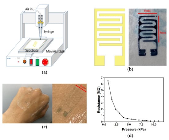
Figure 1.
(a) Schematic of the setup used to print the detector, (b) results of the printed sensing device, printed with poly(3,4-ethylenedioxythiophene) polystyrene sulfonate (PEDOT:PSS), (c) transparent silver nanowire (AgNW) detector attached to skin, and (d) characterization of the PEDOT:PSS detector.
Funding
The authors are grateful to the Ministry of Science and Technology (MOST) of Taiwan for supporting this research.
Conflicts of Interest
The authors declare no conflict of interest.
References
- Murphy, A.D.; Robert, F.G.; Coombs, C.J. Extravasation injury in a paediatric population. ANZ J. Surg. 2019, 89, E122–E126. [Google Scholar] [CrossRef] [PubMed]
- Cheng, M.-Y.; Damalerio, R.; Lim, R.; Chen, W.; Tan, K.L.; Bong, C.L.; Tan, S.K. Wearable Sensor Patch for Early Extravasation Detection. In Proceedings of the 2016 IEEE 66th Electronic Components and Technology Conference (ECTC), Las Vegas, NV, USA, 31 May–3 June 2016. [Google Scholar]
- Goodman, J.; Zimmet, A. Extravasation Detection Electrode Patch. U.S. Patent No. 5,964,703, 12 October 1999. [Google Scholar]
- Shiau, C.-C.; Liao, Y.; Kao, Z.; Yeh, Y.; Lu, Y. Based Flexible Taxel Device Using Electrical Contact Resistance Variation for Elasticity Measurement on Biological Objects. IEEE Sens. J. 2013, 13, 4038–4044. [Google Scholar] [CrossRef]
Publisher’s Note: MDPI stays neutral with regard to jurisdictional claims in published maps and institutional affiliations. |
© 2020 by the authors. Licensee MDPI, Basel, Switzerland. This article is an open access article distributed under the terms and conditions of the Creative Commons Attribution (CC BY) license (https://creativecommons.org/licenses/by/4.0/).