Describing Clinical and Histological Outcome of Oral Cancer Patients with Recurrent Malignant or Premalignant Oral Lesions: A Retrospective Series with a Follow-Up of 15 Years †

1. Introduction
2. Methods
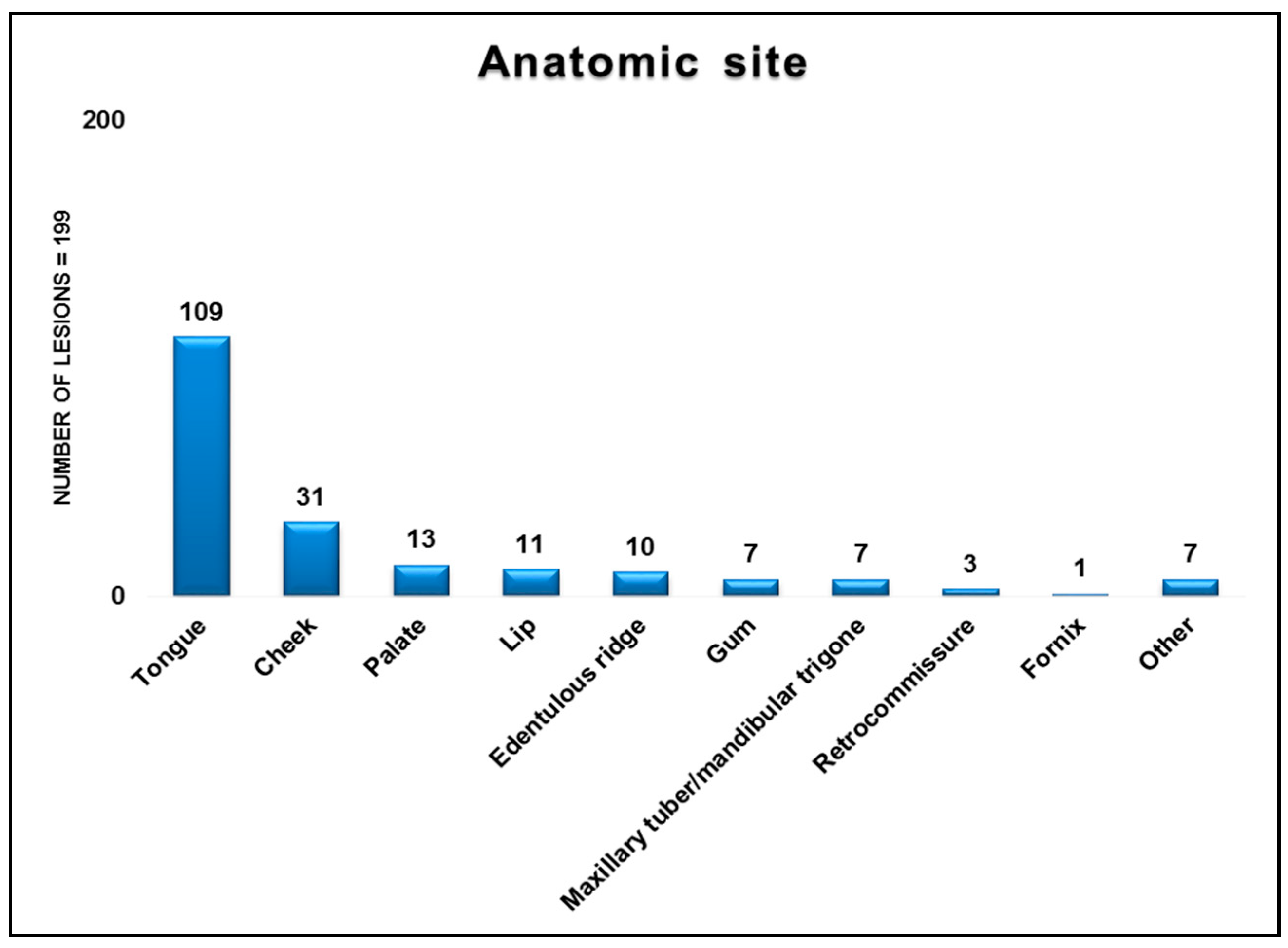
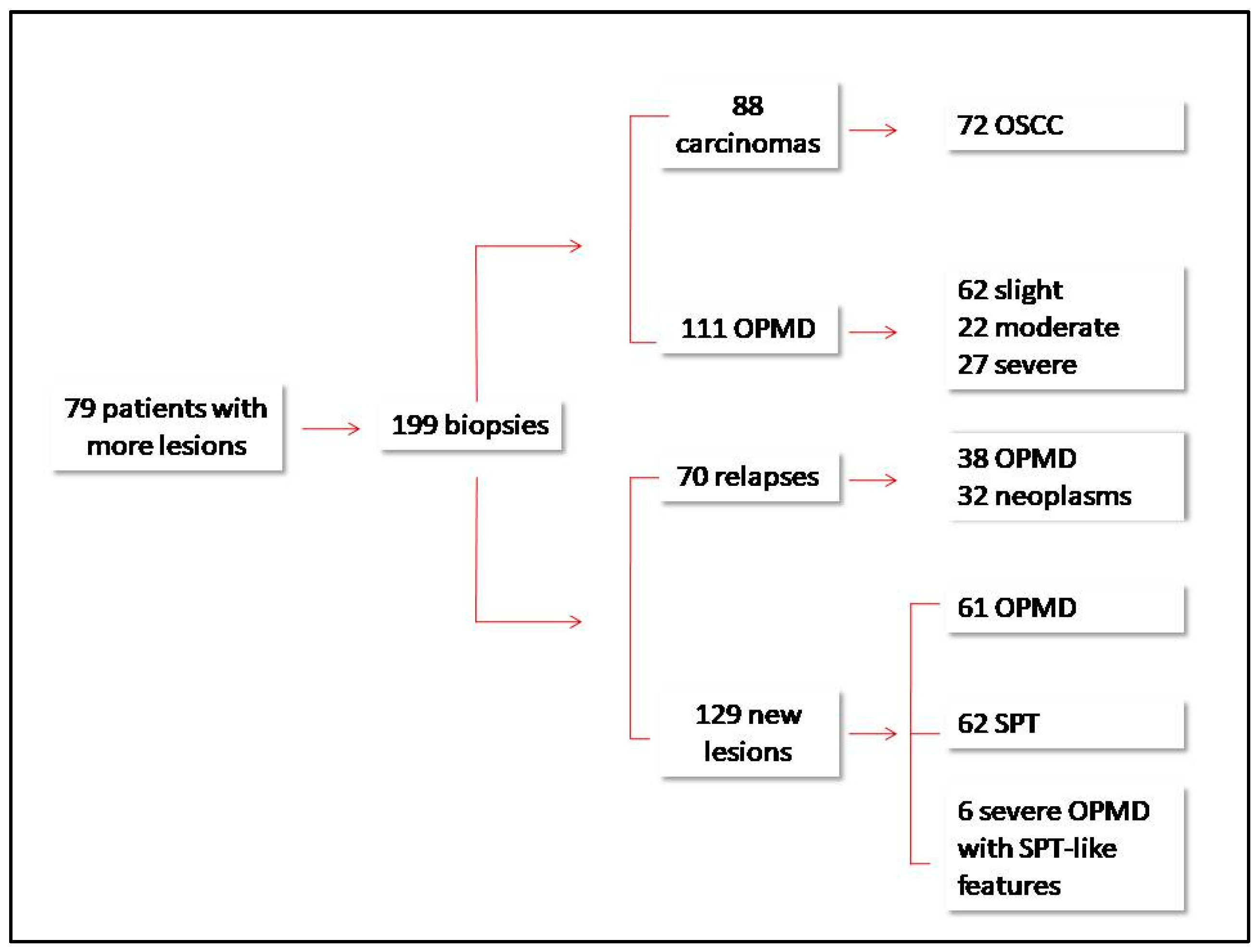
3. Results
4. Discussion
References
- Brands, M.T.; Smeekens, E.A.J.; Takes, R.P.; Kaanders, J.H.A.M.; Verbeek, A.L.M.; Merkx, M.A.W.; Geurts, S.M.E. Time patterns of recurrence and second primary tumors in a large cohort of patients treated for oral cavity cancer. Cancer Med. 2019, 8, 5810–5819. [Google Scholar] [CrossRef] [PubMed]
- Gleber-Netto, F.O.; Braakhuis, B.J.; Triantafyllou, A.; Takes, R.P.; Kelner, N.; Rodrigo, J.P.; Strojan, P.; Vander Poorten, V.; Rapidis, A.D.; Rinaldo, A.; et al. Molecular events in relapsed oral squamous cell carcinoma: Recurrence vs. secondary primary tumor. Oral Oncol. 2015, 51, 738–744. [Google Scholar] [CrossRef] [PubMed]

© 2019 by the authors. Licensee MDPI, Basel, Switzerland. This article is an open access article distributed under the terms and conditions of the Creative Commons Attribution (CC BY) license (http://creativecommons.org/licenses/by/4.0/).
Share and Cite
Cafaro, A.; Cabras, M.; Gambino, A.; Garrone, M.; Arduino, P.G.; Broccoletti, R. Describing Clinical and Histological Outcome of Oral Cancer Patients with Recurrent Malignant or Premalignant Oral Lesions: A Retrospective Series with a Follow-Up of 15 Years. Proceedings 2019, 35, 49. https://doi.org/10.3390/proceedings2019035049
Cafaro A, Cabras M, Gambino A, Garrone M, Arduino PG, Broccoletti R. Describing Clinical and Histological Outcome of Oral Cancer Patients with Recurrent Malignant or Premalignant Oral Lesions: A Retrospective Series with a Follow-Up of 15 Years. Proceedings. 2019; 35(1):49. https://doi.org/10.3390/proceedings2019035049
Chicago/Turabian StyleCafaro, Adriana, Marco Cabras, Alessio Gambino, Marco Garrone, Paolo Giacomo Arduino, and Roberto Broccoletti. 2019. "Describing Clinical and Histological Outcome of Oral Cancer Patients with Recurrent Malignant or Premalignant Oral Lesions: A Retrospective Series with a Follow-Up of 15 Years" Proceedings 35, no. 1: 49. https://doi.org/10.3390/proceedings2019035049
APA StyleCafaro, A., Cabras, M., Gambino, A., Garrone, M., Arduino, P. G., & Broccoletti, R. (2019). Describing Clinical and Histological Outcome of Oral Cancer Patients with Recurrent Malignant or Premalignant Oral Lesions: A Retrospective Series with a Follow-Up of 15 Years. Proceedings, 35(1), 49. https://doi.org/10.3390/proceedings2019035049