


Vision, Volume 9, Issue 1 (March 2025) – 24 articles
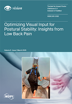
Cover Story (view full-size image):
Chronic low back pain (LBP) is a leading cause of disability, often disrupting balance control due to deficits in sensory integration. While visual input plays a crucial role in postural stability, limited research has explored its effects on dynamics in individuals with LBP. This study investigates the influence of visual conditions on postural control, examining stability indices, sway excursions, and compensatory strategies during repeated one-leg standing tasks. By identifying key sensory deficits in populations with LBP, this study underscores the need for targeted balance interventions that optimize visual and proprioceptive function to reduce fall risk. The findings pave the way for innovative rehabilitation strategies focusing on sensory reweighting and neuromuscular adaptation, improving postural control in individuals with LBP. View this paper
- Issues are regarded as officially published after their release is announced to the table of contents alert mailing list.
- You may sign up for e-mail alerts to receive table of contents of newly released issues.
- PDF is the official format for papers published in both, html and pdf forms. To view the papers in pdf format, click on the "PDF Full-text" link, and use the free Adobe Reader to open them.
Previous Issue
Next Issue