


Abstract
Aquarium fish are increasingly being recognized as reservoirs of zoonotic pathogens, with Aeromonas species posing a notable risk because of their environmental resilience and opportunistic pathogenicity. This study presents the most comprehensive genome-based investigation to date of Aeromonas diversity in aquarium fish, aiming to characterize their taxonomic distribution, population structure, and genomic features. A total of 64 Aeromonas isolates were collected from various aquarium fish species. Wholegenome sequencing was conducted on all isolates to facilitate comparative genomic analyses. Key approaches included multilocus sequence typing (MLST), pairwise Digital DNA-DNA hybridization (dDDH), and the construction of a phylogenomic tree for species-level classification. Furthermore, a population structure analysis was performed to explore genomic diversity and evolutionary trends among the isolates. The results identified 14 distinct Aeromonas species, with A. veronii, A. caviae, and A. hydrophila being the most common. Importantly, several isolates exhibited taxonomic ambiguity, indicating the possible presence of new species or subspecies lineages. Furthermore, antimicrobial resistance gene profiles and virulence factor distributions varied significantly across clades, indicating genomic plasticity. This study highlights the underappreciated genomic complexity of Aeromonas populations in aquarium environments and raises concerns about the public health implications of pathogen reservoirs in ornamental fish.
Key Contribution:
Population structure analysis clustered the isolates into four major genomic clades, partially aligning with species demarcation and highlighting potential host-specific adaptations.
1. Introduction
The genus Aeromonas, which has been the subject of extensive research and remains persistently relevant, continues to be enigmatic due to its pathogenic species and expanding taxonomy [1]. Aeromonas are Gram-negative, facultatively anaerobic, oxidase-positive, motile rods that are ubiquitous in aquatic environments and frequently behave as opportunistic pathogens. In fish, Aeromonas infections are commonly associated with ulcerative dermatitis, fin erosion, hemorrhages, and septicemia, particularly under stressors such as crowding, transport, and suboptimal water quality. In humans, Aeromonas can cause gastroenteritis, wounds and soft-tissue infections, and, in vulnerable individuals, invasive disease. These features make ornamental fish systems a relevant interface for One Health-oriented surveillance. In humans, numerous terrestrial animal species, aquatic animals, and across aquatic ecosystems, it is widely distributed and implicated in many infections; in fish, a large number of pathogenic species have been reported, including Aeromonas hydrophila, A. sobria, A. caviae, A. salmonicida, A. veronii, A. dhakensis, and A. bestiarum [1,2,3,4]. While studies report Aeromonas as a ubiquitous microorganism, the ecological distribution and routes of transmission of these agents have not been fully elucidated [5,6,7]. Because it possesses a complex genus structure, despite advances in whole-genome sequencing technology, it has yielded relatively few newly described species; as of 2025, the number of child taxa with a validly published and correct name is 34 (https://lpsn.dsmz.de/genus/Aeromonas, accessed 22 December 2025). The complex structure of the genus and the relatively lower rate of discovery of new species compared with other bacterial genera have led to more detailed investigation of known species, and it has been reported that species such as A. hydrophila, A. sobria, and A. caviae have broad host ranges and high tolerance to temperature and other factors [7,8]. Aeromonas numbers are higher in lotic than in lentic systems and are higher in thermal gradients ranging from +4 °C to 42 °C, in addition to conductivities, pHs, and turbidities, with only those habitats with extreme ranges of these parameters (extremely saline environments, thermal springs, and highly polluted waters) failing to yield aeromonads [1,9,10,11,12].
Especially in climatic aquatic conditions and high adaptability to warm temperatures, such as approximately 37 °C (the same as mammalian and human body temperatures), these factors play an accelerating role in the spread of Aeromonas species and in rapid bacterial growth [7,12,13]. In contexts where water temperatures align with climatic conditions and where there is a high global capacity for fish trade, aquarium fish farming serves as “hidden transfers” (such as inadvertent cross-border dissemination of bacteria via the aquarium fish trade) for the dissemination of Aeromonas species [6,14,15]. The trade at the retail level is worth more than USD 10 billion with an average annual growth of over 10%, while the entire industry, including plants, accessories, aquariums, feed, and drugs, is estimated to be worth more than USD 18–20 billion [16]. Ornamental fish production is projected to increase from USD 6.36 billion in 2023 to USD 11.30 billion in 2030. The expected near 100% growth in market size in the coming years underscores the increasing significance of aquarium fish farming [16]. A trade volume of such high economic capacity entails the inter-country transfer of bacteria/pathogens; however, in recent years, the practice of municipal aquaria that house large numbers of fish, obtaining fish from their natural habitats for transfer, has also increased the inter-country distribution of Aeromonas species. Individually, home aquarium keepers contaminate themselves and the environment with tap water during water changes and handling/manipulation [17]. In municipal aquaria, the distribution of Aeromonas species also increases due to the discharge of waters in which fish are kept into the environment, often through non-treated wastewater treatment practices [18]. For this reason, in situations that are not immediately visible and appear to concern only aquarium fish trade or inter-country aquarium fish transfer, it is foreseen that many bacterial agents are in fact transferred between countries, and, owing to the faster spread in the aquatic ecosystem, the dissemination of the genus Aeromonas constitutes silent risks [6].
This study aims to conduct a detailed investigation of genome-based Aeromonas diversity in aquarium fish, while also seeking to reveal inter-country similarities of species through genome-based comparison analysis of Aeromonas disseminated via so-called hidden transfers, namely the international movement of aquarium fish. In addition, our study aims to further clarify the taxonomy of the genus Aeromonas by characterizing strains identified by genomic analysis as potential new Aeromonas species.
2. Materials and Methods
2.1. Bacterial Isolation
This study examined ornamental fish collected from three high-capacity producers in Turkey that distribute nationwide (located in Istanbul, Bursa, and Eskişehir), as well as from wholesalers in provinces such as Istanbul, İzmir, Antalya, Bursa, and Eskişehir where imported ornamental fish are aggregated and sold, each with an average monthly sales capacity of approximately 50,000 fish. In addition, samples were obtained from different fish species ranging from 100 g to 10 kg, supplied by Istanbul Aquarium—a large municipal aquarium described as the world’s largest thematic aquarium, housing more than 17,000 organisms and comprising 17 distinct geographic themes. The sampled fish included species inhabiting freshwater, brackish, and marine salinities, as well as species maintained in artificial environments such as aquaria.
Sampling was conducted between June and November 2021 in two major urban centers in Turkey: Istanbul and Bursa. A total of 1184 ornamental fish were collected from eight different commercial aquariums, with more than one aquarium sampled within the same locality in several districts. Sampling included fish of various species, sizes, and health conditions. Sampling targeted clinically affected fish and acute mortality events; some fish appeared externally normal at the time of sampling.
A total of 60 different fish species were examined. The most frequently sampled species were Poecilia reticulata (guppy, n = 242), Pangasius sanitswongi (shark catfish, n = 123), Albino pangasius (Albino Pangasius sutchi, n = 110) Poecilia sphenops (molly, n = 103), Paracheirodon innesi (neon tetra, n = 136), Pseudotropheus demasoni (Demasoni, n = 75), and Carassius auratus (goldfish, n = 62). Other species were represented in smaller numbers, some with only a single individual. This was a surveillance-style sampling aiming to capture the breadth of host diversity rather than statistical inference by species.
Lesions were observed in several species, primarily involving the eyes, fins, skin, and internal organs. General lesions were as follows: hemorrhages in the eyes, ulcerations of the caudal fin, and ulcerations or discolorations of the skin and underlying muscle tissue. Ocular hemorrhage was noted in several species, including Carassius auratus, Maylandia lombardoi, Astronotus ocellatus, Hyphessobrycon rosaceus and Heros efasciatus. Caudal fin ulceration was commonly detected in Poecilia reticulata, Paracheirodon innesi, Xiphophorus maculatus, Hyphessobrycon rosaceus, Colisa unicolor, and Betta splendens. Skin ulceration and discoloration were seen in species such as Astronotus ocellatus, Poecilia sphenops, Sparisoma cretense, Barbonymus schwanenfeldii, Symphysodon discus, and Metriaclima estherae. In a smaller number of cases, hyperemia, hemorrhagic lesions of the internal organs, abscesses, and exophthalmia were also recorded, indicating a variety of possible underlying causes, including infectious and traumatic processes. For bacteriological isolation, swab/tissue samples were streaked onto tryptic soy agar (TSA), blood agar (BA), and marine agar (MA) (for marine species) and incubated at 28 °C for 24–48 h. Dominant colony morphotypes were subcultured to purity prior to MALDI-TOF MS identification and downstream analyses.
The samples were collected aseptically from the smaller fish, with a total length of up to 4 cm (≤4 cm), using a sterile loop from the internal cavity or lesioned tissue. In the case of fish larger than 4–5 cm (>4–5 cm), each individual microbiological sampling was aseptically performed from internal organs such as the liver, spleen, and kidney. Samples were plated on tryptic soy agar (105458, TSA, Merck KGaA, Darmstadt, Germany), blood agar (BA; with 5% sheep blood), tryptic soy agar added 1.5% NaCl, and Marine broth (76448, Merck KGaA, Darmstadt, Germany).
According to the health surveillance program, fish samples were also used to investigate the presence of other bacterial pathogens. Sampling procedures were conducted in accordance with guidelines for diagnosing fish diseases and in compliance with international standards for animal welfare and aquatic animal health surveillance [9]. All isolates were cultured in tryptic soy broth (TSB) (Merck, 105,459) at 28 °C for 24–48 h, and pure cultures were supplemented with 20% glycerol and stored at −80 °C for long-term preservation. The fish species, numbers, and lesions/symptoms used in this study are listed in Table 1.
Table 1.
Fish species, numbers, and observed lesions sampled in this study.
2.2. Microbiological Characterization and Primer Identification by MALDI TOF MS and qPCR
The biochemical characteristics of the isolates were determined using conventional microbial tests, including assessment of colony morphology, Gram staining, oxidase and catalase activities, glucose fermentation, and other differentiational biochemical tests [11,19]. Bacterial motility was observed by the hanging drop technique and checked on sulfide-indole-motility (SIM) medium (1054700500, Merck KGaA, Darmstadt, Germany) [19].
Bacterial isolates obtained from fish samples were identified using matrix-assisted laser desorption/ionization–time of flight mass spectrometry (MALDI-TOF MS). Pure bacterial colonies were first subcultured onto tryptic soy agar (TSA) and incubated at 28 °C for 24–48 h. Following incubation, a small amount of fresh bacterial biomass was directly applied onto a MALDI target plate. Each spot was overlaid with 1 μL of a saturated solution of α-cyano-4-hydroxycinnamic acid (HCCA) matrix, prepared in a solution of 50% acetonitrile and 2.5% trifluoroacetic acid, and then air-dried at room temperature [20].
Spectra acquisition was performed using a MALDI Biotyper system (Bruker Daltonics, Bremen, Germany) in linear positive mode, within a mass range of 2000–20,000 Da. Calibration was carried out using a bacterial test standard (Bruker Bacterial Test Standard, BTS) according to the manufacturer’s instructions. The obtained spectra were compared against the Bruker reference database using Biotyper software (version 2.0, Bruker Daltonics GmbH & Co. KG, Bremen, Germany). Identification scores were interpreted based on the manufacturer’s criteria: a score ≥2.000 was considered reliable for species-level identification, 1.700–1.999 for genus-level identification, and <1.700 as unreliable [20]. Isolates with insufficient scores were retested, and those still producing low-quality spectra were subjected to 70% formic acid extraction prior to analysis to improve identification reliability. After genus-based identification by MALDI TOF, further validation of the strains was performed using Real-Time PCR (qPCR) at the genus level, as described by Duman et al. [21].
2.3. Genomic Characterization
Genomic DNA was extracted using the Nucleogene DNA Extraction Kit (Nucleogene, Istanbul, Turkey) and quantified with a Qubit fluorometer. Whole-genome sequencing was carried out on the Oxford Nanopore PromethION (The Oxford Science Park, Littlemore, Oxford, UK) platform using the ligation sequencing kit (SQK-NBD114-24). DNA was extracted from fresh overnight cultures grown on TSA at 28 °C, using the manufacturer’s protocol (including cell lysis, binding, wash steps, and elution in 100 µL). DNA quantity and quality were assessed using a Qubit at an A260/280 ratio. Raw reads were basecalled with Guppy (v6.1.7) in high-accuracy mode and processed for quality filtering and adapter trimming. De novo assembly was performed with Flye (v2.9.1), followed by iterative polishing with Racon, Minimap2, and Pilon. Only contigs ≥1000 bp were retained, and assembly quality was assessed with QUAST (V. 5.3). Species-level identification was obtained with the Type Strain Genome Server (TYGS) using digital DNA–DNA hybridization (dDDH) values [22]. In general, dDDH values of approximately ≥70% support assignment to the same species; values below this threshold indicate increasing genomic divergence from the closest type strain. Final annotation was completed using the NCBI Prokaryotic Genome Annotation Pipeline (PGAP), and draft genomes were deposited in GenBank [23].
2.4. MLST and MLPA
MLST was performed for Aeromonas isolates using PubMLST, adopting the six-locus scheme proposed by Martino et al. [24] based on gltA, groL, gyrB, metG, ppsA, and recA. In total, 65 isolates were available from this study. One isolate (43P) displayed low genome coverage and was therefore excluded from whole-genome characterization; nevertheless, it was included in MLST/MLPA because the MLST loci could be obtained for typing purposes [25]. Allele and ST assignment were initially attempted through PubMLST. As reported in Table S2, eight isolates did not receive allele numbers (and thus ST designations) from PubMLST at this stage.
Because rpoD provides high discriminatory power within Aeromonas [26,27], we additionally retrieved rpoD sequences and used them to strengthen isolate characterization and to support downstream sequence-based analyses. This approach provided sufficient data for four of the eight initially unresolved isolates to be retained in MLST/MLPA analyses, whereas four isolates (127P, 132P, 135P, and 80P) still lacked allele/ST assignments and were excluded from MLST/MLPA downstream analyses. Therefore, all subsequent MLST/MLPA analyses were conducted on 61 isolates.
Clonal relationships and deep population structure were first explored using the goeBURST algorithm (Global Optimal eBURST) implemented in Phyloviz (V. 2.0) [25]. For population genetics and evolutionary analyses, we compiled a genome dataset comprising 94 genomes: 33 type strains representing all validly published Aeromonas species and 61 isolates from this study. In addition to the six MLST loci, rpoD was included in the concatenated sequence dataset. Alignment statistics were computed with AMAS [28], and maximum-likelihood phylogenetic inference was performed with IQ-TREE2 [29]. Genetic variation was quantified in DnaSP v6 [30] using the number of segregating sites (S) and nucleotide diversity (π). Demographic history and deviations from neutrality were tested using Tajima’s D, Fu and Li’s D* and F*, and Fu’s Fs. Recombination was summarized by the minimum number of recombination events (Rmin) estimated via the four-gamete test [31]. Evidence for episodic diversifying selection was examined using BUSTED (Branch-site Unrestricted Statistical Test for Episodic Diversification) implemented in HyPhy [32], which evaluates whether a proportion of sites experienced positive selection (ω > 1) on at least one branch while accounting for phylogenetic structure. Finally, Bayesian clustering was performed with STRUCTURE [33] by exploring K = 2–9, applying a burn-in of 50,000 iterations followed by 50,000 MCMC iterations. The optimal K (Evanno method) and bar plot visualizations were generated using pophelper [34], and final annotations were visualized with iTOL v6 [35].
2.5. Genome-Based Phylogenetic Analysis
To investigate the phylogenomic relationships of the genus Aeromonas, a genome-based analysis was performed using the Bacterial Genome Tree Service on the BV-BRC platform (https://www.bv-brc.org/app/PhylogeneticTree, accessed on 24 December 2025) [36]. The dataset comprised 99 genomes in total, including 34 type strains representing all validly published Aeromonas species and 65 additional strains generated in this study. Core-genome phylogenetic reconstruction was based on 1000 single-copy orthologous protein-coding genes following the methodology [37,38]. The final tree, inferred from 339,023 aligned amino acid positions, provided a high-resolution comparative framework spanning both reference and novel Aeromonas strains. Tree visualization and annotation were performed using iTOL (Interactive Tree of Life, https://itol.embl.de/) [38], which integrates metadata such as host association, environmental source, and geographic origin. Branch lengths were scaled to represent substitutions per site, with a tree scale of 0.01 substitutions per site.
2.6. AMR and Virulence Gene Detection
Antimicrobial resistance (AMR) genes were identified using AMRFinderPlus (v3.10.40) via the command-line interface, employing both nucleotide and protein FASTA files alongside annotated GFF files for each assembled Aeromonas genome, as determined by whole-genome characterization [39]. Virulence factors were detected by conducting BLASTp searches against the Virulence Factors Database (VFDB) (core dataset, Set A). Protein sequences from each assembled genome were queried locally against the VFDB protein database. Only hits with ≥90% identity and ≥60% coverage were retained for annotation [40].
2.7. Antibiotic Resistance
Antimicrobial susceptibility was assessed by the Kirby–Bauer disk diffusion method, following the CLSI M42-A guidelines for bacteria from aquatic animals [41]. The test panel comprised oxolinic acid (OA); tetracyclines [doxycycline (DO), TE, oxytetracycline (OT)]; a sulfonamide combination [trimethoprim/sulfamethoxazole (SXT, 1/19)]; aminopenicillins [amoxicillin (AML), amoxicillin–clavulanate (AMC)]; a fluoroquinolone [enrofloxacin (ENR)]; and a phenicol [florfenicol (FFC)]. Disks were applied to Mueller–Hinton agar seeded with pure cultures and incubated at 28 °C for 24–28 h. Zone diameters (mm) were measured and interpreted using EUCAST breakpoints (https://www.eucast.org/bacteria/clinical-breakpoints-and-interpretation/clinical-breakpoint-tables/, accessed on 22 December 2025). Escherichia coli ATCC 25922 served as the quality-control strain, with CLSI performance ranges verified for each run [42]. Where species- or host-specific clinical breakpoints were not available or not applicable to ornamental fish isolates, we report raw zone diameters and highlight fully resistant phenotypes as 0 mm inhibition zones.
2.8. Ethical Statements
This research was supported by the Scientific and Technological Research Council of Turkey and approved by the Local Ethics Commission (report 2021-07-07).
3. Results
3.1. Isolation and Primer Identification
Across the 1184 fish, including 60 different species, isolates of Aeromonas species were recovered from a broad host spectrum dominated by cyprinids and popular ornamental taxa. Positives were confirmed in goldfish (Carassius auratus), pangasius (commercial shark catfish lines), neon tetra (Paracheirodon innesi), swordtail (Xiphophorus hellerii), dwarf gourami (Trichogaster lalius), molly (Poecilia sphenops), Dalmatian/black sailfin molly (Poecilia latipinna), discus (Symphysodon discus), pufferfish (Tetraodon lineatus), bichir (Polypterus senegalus), mosquitofish (Gambusia holbrooki), and several African cichlids (like Maylandia estherae, Chindongo socolofi, Aequidens pulcher), among others (Table 2). Clinical signs in Aeromonas-positive hosts mirrored the dominant syndromes in the survey: caudal-fin and dermal ulceration, ocular hemorrhage, and peracute mortality/septicemia. Other bacteria were occasionally recovered but were outside the scope of this Aeromonas-focused genomic study.
Table 2.
Summary of the comparison of fish species and observed lesions.
Where the denominator of sampled fish matched the isolation dataset, crude isolation proportions illustrate the host–lesion links. In goldfish (n = 62; lesions mainly caudal-fin ulceration and occasional ocular hemorrhage), Aeromonas sp. was isolated from 10 individuals (16.1%). In Pangasius spp. (n = 233 across commercial lines; frequent acute losses and occasional exophthalmia), Aeromonas sp. was cultured from nine fish (3.9%). In neon tetras (n = 136; fin ulceration and acute mortality), four were positive (2.9%). Dwarf gourami (n = 11; acute mortality) yielded three positives (27.3%). Swordtail (n = 5; acute mortality) yielded five positives (100%), reflecting intense case clustering within a single vendor/batch rather than a population rate, and should be interpreted cautiously. Additional Aeromonas recoveries were documented from molly (P. sphenops; n = 103; fin/skin ulceration) and pufferfish (T. lineatus; n = 14; acute mortality), with one positive (7.1%), and discus (n = 3; skin ulceration), with one positive (33.3%). For several other hosts (such as G. holbrooki, bichir, selected cichlids), total numbers sampled were not systematically enumerated in the clinical dataset; for these, we report the presence of isolation without a denominator (Table 3).
Table 3.
The Aeromonas sp. isolation rates based on fish species.
Lesion patterns within Aeromonas-positive fish were consistent across taxonomic groups. Ulcerative disease predominated in cyprinids and poeciliids (goldfish, mollies, swordtail), while small characins (neon tetra) and labyrinth fish (dwarf gourami) more often presented with peracute mortality, occasionally accompanied by caudal-fin necrosis. Ocular hemorrhage co-occurred with isolation in species where this sign was prevalent (like goldfish, oscar), and rare septicemic presentations (such as rays, pufferfish, discus) aligned with Aeromonas recovery in representative cases (Table 2 and Table 3). Aggregating the unresolved lineages with named strains did not materially alter these lesion associations; most genomically novel candidates (labeled by “closest-to” references) came from fish manifesting the same ulcerative or peracute syndromes (discus with skin ulceration; neon tetra with acute mortality/fin ulceration; pufferfish with acute mortality; pangasius and goldfish with ulcerative/hemorrhagic signs), suggesting clinical equivalence at the syndrome level within the Aeromonas complex.
All Aeromonas isolates collected from diseased fish in this study were obtained from dominant colonies grown on TSA, BA, or MA after 24 h of incubation. Other colony morphotypes were occasionally observed and/or recovered; however, they were not characterized further because they were outside the scope of this Aeromonas-focused genomic study. For each sample, a selection was made from colonies that appeared as dominant colonies and were considered likely to represent potential pathogens in fish tissues. Particular attention was given to selecting morphologically dominant colonies rather than randomly picking isolates, ensuring that colonies most likely to represent the dominant bacteria present in the sampled tissues were included in further genomic analyses.
Initial phenotypic characterization revealed that all isolates were Gram-negative, motile, and tested positive for both oxidase and catalase activities—traits consistent with the general features of the genus Aeromonas. To avoid clonal redundancy and account for possible intrasample species diversity, colonies with similar morphology were not repeatedly selected from the same plate. Instead, where present, morphologically distinct colonies were preferentially isolated to increase the likelihood of detecting multiple Aeromonas species from the same sample. This approach allowed for the identification of different Aeromonas species from the same anatomical site or individual fish, reflecting the ecological complexity and co-occurrence potential of closely related taxa within aquatic environments. The isolated strains were confirmed as Aeromonas species by qPCR analysis, and species identification was confirmed by MALDI-TOF MS analysis. A total of 64 bacterial isolates were identified to the species level by MALDI-TOF MS, and these identifications were supported by additional phenotypic and genotypic methods. Although 65 Aeromonas isolates were initially collected, strain 43P was excluded from the genomic analyses due to low coverage and limited discriminatory power (but it was retained in the MLST analyses). The MALDI-TOF system most frequently identified A. hydrophila, A. sobria, and A. caviae, likely reflecting the limited representation of aquatic Aeromonas species diversity in the database.
3.2. Genomic Characterization
Of the 64 Aeromonas isolates analyzed, 43 could be assigned to recognized species based on genome-based taxonomy, whereas the remaining 21 isolates did not meet species-level thresholds against available type strains and were therefore reported as putative novel lineages according to their closest phylogenomic relatives by molecular identification: A. veronii (13 strains; 30.2%), A. hydrophila (11; 25.6%)), A. caviae (8; 18.6%), A. dhakensis (8; 18.6%), A. enteropelogenes (1; 2.3%), A. ichthiosmia (1; 2.3%), and A. jandaei (1; 2.3%). The highest strain diversity was observed in Istanbul, where six Aeromonas species were detected, whereas Bursa yielded five species (Table 4).
Table 4.
Aeromonas sp. was detected in aquariums and isolated in counts.
Temporal distribution of isolations showed that most were obtained in 2021, with peak activity in June, July, and September. Aeromonas species were recovered from 20 ornamental fish species, with Carassius auratus (Goldfish), Pangasius pangasius (Shark catfish), Poecilia latipinna (Dalmatian molly), and Xiphophorus hellerii (Koi Kohaku Wag) as the most common hosts. Notably, A. dhakensis was isolated exclusively from Carassius auratus and Pangasius pangasius, whereas A. veronii exhibited the broadest host range. In terms of aquarium distribution, Aquarium 1 in Istanbul harbored the greatest species richness, with five Aeromonas species detected.
In addition to the strains identified at the species level, 21 isolates could not be confidently assigned to a definitive taxonomic identity using MALDI-TOF MS and subsequent genome-based comparisons. Building on the baseline species distribution above, comparative genomics of the aquarium isolates revealed a set of Aeromonas lineages that did not meet the accepted digital DNA–DNA hybridization (dDDH) threshold for species assignment (≈70%) to any type strain. Because these lineages have not been formally identified, we provisionally labeled them by their nearest-neighbor similarity (such as “closest to A. veronii CECT 4257” or “closest to A. ichthiosmia CCM 7244) to indicate affinity rather than taxonomic status. In total, 21 isolates clustered into 14 putative novel taxa (groups 1–14, excluding group 6), with dDDH values ranging from 49.0% to 69.9%. Most candidates were affiliated with the A. veronii complex (20/21 isolates across 14 groups), while one strain was closest to A. ichthiosmia (1/21 isolates across group 6). Several groups formed a tight complex just below the species boundary, particularly the group 2 variants (2–2a/2–2b/2–2c; 6 isolates, dDDH 69.1–69.8% to A. veronii CECT 4257)—suggesting a cohesive but heterogeneous lineage within the A. veronii clade. Group 6 consisted of a single isolate with 68.7% dDDH to Aeromonas ichthiosmia CCM 7244—just below the 70% species threshold—consistent with a distinct yet closely related lineage. The most divergent candidate was group 1 (dDDH 49% to A. veronii), indicating deep separation from recognized species (Table 5). The genome accession numbers are presented in Supplementary Table S1.
Table 5.
The species diversity and genome-based identification results of Aeromonas strains based on isolation date, fish species, and aquariums.
These candidate taxa were unevenly distributed by geography, aquariums, host fish, and time. Fifteen of the twenty-one candidates originated from Istanbul and six from Bursa. Candidates were concentrated in a few aquariums: Istanbul aquaria 1, 5, and 6 together accounted for fifteen isolates (with aquaria 5 and 6 yielding 9/21), whereas Bursa aquaria 7 and 8 contributed six. Host range was broad and overlapped with the confirmed species set; recurrent hosts included Pangasius pangasius (three candidates in groups 2–2b, 9, and 13), Paracheirodon innesi (two candidates in group 10), and Poecilia latipinna (two candidates in groups 2–2c and 8); Carassius auratus occurred twice (groups 2–2a and 6). The temporal pattern was 2021-centric, with clusters in late June and July and additional peaks in early October and early November. Together with the culture-based results, where the A. veronii complex predominated, the genomic screening indicates that ornamental fish in urban retail settings harbor both recognized Aeromonas species and multiple closely related, potentially novel lineages, particularly within the A. veronii complex, with signatures of local clustering by vendor and host.
3.3. Genome-Based Phylogenetic Tree
The composite phylogenomic analysis resolves eight placement groups for our Aeromonas isolates, and our assignments align with these clades. Isolates assigned to the A. veronii complex—41P, 130A, 68P, 93A, 22P, 69P, 132P, 131P, 112P, 40P, 8P, 123P, 31A, CECT 4257ᵀ, 132A, and 106A—cluster within the A. veronii clade (Figure 1). Additional isolates (102P, 84A, 23P, 96A, 11P, 107A, 80P, 61P, 6P, 3P, 95A, 30P, 55A, 82P, 97P, 31P, 94A, 75A, 74A) form a sister subclade within the A. veronii complex and are best described as Aeromonas sp. closely related to A. veronii. The A. caviae group (98P, 95P, 99P, 57P, 55P, 56P, 96P, 97P) clusters tightly with the type strain A. caviae NCTC 12244ᵀ and forms a distinct subclade. Isolates identified as A. dhakensis (11A, 117P, 107P, 108P, 111P, 115P, 88P, 89P) group with A. dhakensis CIP 107500ᵀ. The A. hydrophila isolates (141A, 71A, 102A, 24P, 129P, 127P, 154P, 72A, 146A, 29P, 135P) cluster within the A. hydrophila group, together with the type strain A. hydrophila subsp. hydrophila ATCC 7966ᵀ (Figure 1). To the best resolution, the circular genome tree is also supplied in Figure S1. Most study isolates cluster near recognized type strains; however, strain 164P occupies a distinct single branch that is nearest to A. jandaei (CECT 4228ᵀ) and appears proximate to A. jacus (AE122ᵀ). Finally, A. enteropelogenes isolate 81A falls in a discrete subclade with A. enteropelogenes CECT 4487ᵀ.
Figure 1.
Genome-based phylogenetic tree of the strains isolated in this study and Aeromonas type strains.
3.4. MLST and MLPA Analysis
The new Aeromonas isolates were classified into 43 different STs: 71, 709, 1764, 1824, 2007, 2212, 2560, 2616, 2963, and 3646 to 3680 (Table S2). The most abundant ST was 3650, which grouped six A. dhakensis isolates (107P, 108P, 111P, 115P, 117P and 11A) belonging to this scheme, whereas the majority of ST (n = 36) only grouped one strain. This genetic difference is reflected in the goeBURST analysis, where most ST (n = 36) represent singletons connected exclusively to the ST 3672 considered by the algorithm to be the founder (Figure 2).
Figure 2.
goeBURST all MLST network. Each ST is linked to other STs based on similarities of the studied genes. Bigger labels correspond to ST with a larger number of isolates. All STs have blue labels, except for the founders of each group, which are represented by green labels.
After assessing the clonality and deep population structure within the Aeromonas strains, we analyzed evolutionary pressures using population genetics metrics (Table 6). A high degree of genetic diversity was observed across all housekeeping genes, as evidenced by the number of unique sequences (haplotypes) and the proportion of variable sites. For instance, nearly every strain was unique, with the number of different sequences ranging narrowly from 73 (rpoD) to 77 (gyrB) out of 94 total strains tested. This uniqueness was further reflected in the high density of variation, where the proportion of variable sites ranged from 0.381 to 0.489 across the genes. We then used these diversity metrics to assess deviations from the neutral evolution model, finding that while the overall Tajima’s D statistics were negative but close to zero (ranging from −1.37 to −0.39), the Fu’s FS statistic identified strong signals of recent expansion and (Fs > 0) for ppsA, recA, and gltA alignments. However, the Fs statistic of the concatenated set of housekeeping genes was significantly different from zero and positive, meaning that the population could be experiencing a bottleneck. In fact, population analysis with STRUCTURE confirmed that the current population evolved from four large genetic populations (Figure 3), although phylogenomic analysis revealed eight established groups at present. It should be noted that several strains that belong to the “type species” phylogenetic group, such as A. enteropelogenes or A. taiwanensis, are chimeric results of potential interspecies recombination.
Table 6.
General metrics of the alignment genes tested for MLST analysis and the concatenation of all genes.
Figure 3.
MLST tree and population structure of Aeromonas strains. Maximum likelihood tree of nucleotide sequences obtained with IQTREE2. Numbers in branches represent bootstrap support based on 1000 replicates. Each color represents the proportion of each population group calculated with STRUCTURE for an optimum K value of 4.
Regarding episodic diversifying selection (EDS) with BUSTED analysis, we could not find evidence for this phenomenon for the gyrB and rpoD genes with a level of significance of p < 0.05, although we found six codons with potential EDS in the former. On the contrary, metG exhibited the strongest signal with 36 codons potentially under EDS. When concatenated, the overall study of the genes suggests that EDS is a selective force that accelerates the evolution of the genus, potentially enabling adaptation to new niches.
Finally, we assessed the influence of recombination in the seven Aeromonas housekeeping genes. The minimum number of recombination events, Rmin, was exceptionally high across all loci, ranging from 17 in ppsA to 216 gyrB, and reached a cumulative total of 958 events in the concatenated alignment (Table 6).
3.5. Antibiotic Susceptibility and Resistance Genes
Because no clinical breakpoints or epidemiological cut-offs are available for these Aeromonas species antibiotic combinations in CLSI or EUCAST, no “susceptible/intermediate/resistant” interpretation was applied. Only isolates with a zone diameter of 0 mm were treated as fully resistant (explicitly, 0 mm zones were evaluated as resistant). Among isolates that produced any inhibition zone (>0 mm), we summarized the distribution by lower (≤20 mm) versus higher (≥21 mm) zone diameters (Table 7).
Table 7.
Zone-diameter distribution (count of isolates).
Oxolinic acid (OA): 47/64 (73.4%) showed 0 mm. Of the 17 with zones, 11/17 (64.7%) were ≤20 mm and 6/17 (35.3%) ≥ 21 mm. Oxytetracycline (OT): 39/64 (60.9%) showed 0 mm. Of 25 with zones, 17/25 (68.0%) were ≤20 mm and 8/25 (32.0%) ≥ 21 mm. Amoxicillin (AML): 63/64 (98.4%) showed 0 mm. The single zone-forming isolate had ≥21 mm (0/1 ≤ 20 mm; 1/1 ≥ 21 mm). Enrofloxacin (ENR): 23/64 (35.9%) showed 0 mm. Among the 41 with zones, 8/41 (19.5%) were ≤20 mm and 33/41 (80.5%) ≥ 21 mm. Florfenicol (FFC): 12/64 (18.8%) showed 0 mm. Of the 52 with zones, 12/52 (23.1%) were ≤20 mm and 40/52 (76.9%) ≥ 21 mm. Doxycycline (DO): 21/64 (32.8%) showed 0 mm. Of the 43 with zones, 35/43 (81.4%) were ≤20 mm and 8/43 (18.6%) ≥ 21 mm. Trimethoprim–sulfamethoxazole (SXT): 37/64 (57.8%) showed 0 mm. Of the 27 with zones, 10/27 (37.0%) were ≤20 mm and 17/27 (63.0%) ≥ 21 mm. Amoxicillin–clavulanate (AMC): 42/64 (65.6%) showed 0 mm. Of the 22 with zones, 20/22 (90.9%) were ≤20 mm and 2/22 (9.1%) ≥ 21 mm.
Overall, 0 mm zones (treated as fully resistant) were most frequent for AML, AMC, OT, and OA. Among zone-forming isolates, ENR and FFC most often produced higher diameters (≥21 mm), whereas DO and AMC were dominated by lower diameters (≤20 mm).
Under the Aeromonas strains analyzed, every clinical and reference strain carried at least one acquired or intrinsic antimicrobial resistance determinant, spanning β-lactams, tetracyclines, quinolones, sulfonamides, aminoglycosides, macrolides, rifamycins, trimethoprim, phenicols, quaternary ammonium compounds, and, in a subset, colistin. β-lactam-associated loci (including OXA-type oxacillinases, MOX/AQU/FOX/TRU families, cphA metallo-β-lactamase, and cephalosporinases such as cepS/cepH or ampC-Asob) were ubiquitous across isolates and type strains. Among clinical isolates, β-lactam genes frequently co-occurred with tetracycline determinants [either tet(A) or tet(E)] and with sulfonamide [sul1/sul2] and quaternary ammonium compound genes [qacE/qacEΔ1/qacG2]; aminoglycoside [aadA/aph/aac families], macrolide [mph(A)], trimethoprim [dfrA variants], and phenicol [floR/cmlA5] genes were observed in multi-drug profiles of several urine and pus isolates. Quinolone resistance was represented by plasmid-mediated qnrS/qnrS2 and by the hybrid aac(6′)-Ib-cr variants. The colistin resistance gene mcr-3 was detected in one clinical isolate and multiple Aeromonas type/reference strains within the panel.
At the isolate level, β-lactam genes were present in essentially all numbered isolates (including 3P, 6P, 8P, 11A/11P, 22P–24P, 29P–31A/31P, 40P, 54A–57P, 61P, 68P–69P, 71A–75A, 80P–84A, 88P–89P, 93A–99P, 102A/102P–164P). Tetracycline genes were present in most of these, typically alongside β-lactam genes; sulfonamide and QAC genes co-occurred in many of the same isolates, especially 22P, 23P, 24P, 55P–56P, 61P, 69P, 72P, 80P–81A, 84A, and 93A–97P. Quinolone resistance loci were found in 6P, 61P, 82P, 93A, 107P, 108P, 111P–112P, 117P, and 130A. Colistin resistance was observed in reference/type strains A. media ATCC 33907, A. piscicola LMG 24783, A. salmonicida subsp. salmonicida CIP 103209, and, in addition, Aeromonas strains labeled 74A and 75A. Reference/type strains also mirrored the clinical gene-class spectrum, with frequent OXA-type β-lactamases and cphA and occasional MOX/AQU/FOX/TRU families; several carried additional phenicol, aminoglycoside, sulfonamide, tetracycline, and QAC genes (Table S3).
A per-strain summary table is provided in Supplementary Table S1, with total AMR gene count and the presence/absence matrix by antibiotic class (β-lactam, tetracycline, quinolone, sulfonamide, aminoglycoside, macrolide, rifamycin, trimethoprim, phenicol, QAC, colistin) for each isolated strain.
3.6. Virulence Genes
Overall, the Aeromonas collection showed pronounced heterogeneity in VFDB-annotated content. A subset of isolates carried a very broad virulence repertoire spanning the large Aeromonas hydrophila-like cluster in the matrix (the dense block of VFG0383xx–VFG0389xx entries). This “high-burden” profile was typical of many waterborne/planktonic isolates such as 24P, 29P, 30P, 40P, 68P, 74P, 75P, 80P, 88P, 89P, 102A/102P, 107P, 108P, 111P, 115P, 117P, 123P, 127P, 129P, 132A/132P, 135P, 141A, 146A, 154P, and several others: these genomes showed long tracts of contiguous “1”s across the core block, indicating co-occurrence of dozens to hundreds of virulence entries per isolate. By contrast, a smaller “low-burden” group (such as 3P, 6P, 8P, 31A/31P, 41P, 54P, 82P, 84P, 93P, 94A, 95A/95P, 96A/96P, 97A/97P, 98P, 99P, 106A, 107A, 130P, 141P, 164P) showed sparse or punctate signals (short runs of “1”s separated by long stretches of “0”s). For many A/P pairs taken from the same source (such as 11A/11P, 55A/55P, 95A/95P, 96A/96P, 97A/97P, 102A/102P, 132A/132P), the virulence gene patterns were nearly indistinguishable, consistent with clonal relatedness at the virulence locus level (Table S4).
Across isolates with rich virulence content, the most consistently represented region was the mid-matrix Aeromonas block (VFG038317 onward), which in Aeromonas encompasses multiple secretion-associated and cell-surface/iron-related entries in VFDB. Within this block, presence was typically “all-or-none” at the scale of tens of adjacent entries—suggesting these loci are maintained as operon- or island-like segments rather than as isolated genes. Several isolates (such as 127P, 129P, 141A, 146A, 154P) carried the essentially entire block, whereas low-burden genomes retained only scattered fragments at their margins. In brief, virulence gene carriage in this Aeromonas panel ranges from minimal to extensive. Most isolates fall into one of two modes: (i) a dense, near-complete Aeromonas virulence block indicating a large accessory repertoire versus (ii) a sparse profile with only a few VFDB hits. Paired isolates from the same sample generally share the same virulence configuration, and high-burden genomes frequently harbor long, contiguous runs of virulence entries rather than singletons.
4. Discussion
To the best of our knowledge, this study is one of the most comprehensive investigations of Aeromonas prevalence in aquarium fish to date, encompassing 60 host species sourced from seven wholesale distributors and one municipal aquarium and a total of 1,184 individuals examined. In addition to culture-based detection, whole-genome analyses were performed on 64 Aeromonas isolates, thereby providing a high-resolution perspective on species composition and strain diversity across a wide ornamental trade network. The combination of host coverage, multi-vendor sampling frame, and integration of genomic data positions this work to refine estimates of Aeromonas burden in aquarium systems and to contextualize clinical presentations with species-level and lineage-level resolution.
Previous studies offer helpful context for understanding our findings. In a relatively large survey, 126 aquarium fish were examined, and 112 (88.8%) were found to have bacterial infections. Among the identified pathogens, Aeromonas veronii (26.3%) and A. hydrophila (16.2%) were common [14]. Earlier reports often identified A. hydrophila as the main species [43], while a study analyzing aquarium waters found an Aeromonas prevalence of only 2% [44]. Our data, based on whole-genome sequencing, shows that A. veronii was the most frequently detected species in culture-positive cases (30.2%), followed by A. hydrophila (25.6%), A. caviae (18.6%), and A. dhakensis (18.6%). A. enteropelogenes, A. ichthiosmia, and A. jandaei each appeared in one isolate (2.3%). Therefore, while A. hydrophila was among the frequently detected species in our culture-positive cases, genome-level identification shifts the apparent species order and emphasizes the importance of A. veronii in aquarium fish. Another key finding is the high level of uncharacterized diversity. About one-third of the isolates did not meet accepted standards for species assignment and likely represent potential new taxa. Most of these unresolved isolates clustered within the A. veronii species complex (20 out of 21 isolates across 14 groups), with one isolate most closely related to A. ichthiosmia. These findings suggest that studies relying solely on phenotypic tests or partial genotyping may have underestimated the contribution of A. veronii complex lineages and the overall diversity of undescribed Aeromonas in ornamental fishes. Reassessing reported isolates with whole-genome sequencing could improve species identification and, in some cases, alter conclusions about the dominant Aeromonas species involved in aquarium disease settings.
Aeromonas species are frequently described as opportunistic or secondary pathogens; nevertheless, several species have been implicated as primary agents of disease in ornamental fishes and even in rainbow trout, where fin and skin ulceration have been reported as leading clinical presentations [6]. In a comparatively large case series, septicemia alone or in combination with ocular lesions dominated the clinical spectrum, followed by isolated skin lesions, and mixed lesions were also common. Only about one-third of cases involved a single bacterial species, with the remainder representing polymicrobial infections [14]. Beyond aquatic hosts, Aeromonas is recognized as a cause of soft-tissue and invasive infections in humans, with severity ranging from self-limited diarrhea to necrotizing fasciitis, septicemia, meningitis, cholera-like illness, and hemolytic uremic syndrome—underscoring the occupational risk to those handling diseased fish [45,46]. From a One Health perspective, exposure may occur during aquarium maintenance (such as water changes, filter cleaning, handling of fish and equipment) through direct contact with contaminated water, aerosols, or minor skin injuries. This is particularly relevant for hobbyists, aquarium enthusiasts, and individuals with immunocompromised conditions. Therefore, our findings support improved hygiene and basic biosecurity practices (like gloves for wound-prone handling, hand washing, avoiding exposure of open cuts) and highlight the value of genomic surveillance in ornamental systems. The species most consistently associated with disease across fish and humans are A. hydrophila and A. veronii [47,48]. Notably, recent clinical reports highlight the diagnostic drawback posed by phenotypic overlap, including cases in which A. caviae mimicked as V. cholerae and would likely have been misreported as absent in genome-based confirmation, with clear public health implications in cholera-endemic settings [49]. In our surveillance, clinical patterns were consistent with those reported in the existing cases. Ulcerative disease of the caudal fin and skin predominated in cyprinids and poeciliids. Ocular hemorrhage occurred mainly in taxa where this sign was historically common (e.g., cyprinids), and peracute mortality/septicemia was more typical of small characins and labyrinth fishes. Where denominators were available, Aeromonas isolation among clinically affected fish ranged from low single digits to double digits across hosts—for example, approximately 1/6 goldfish, 1/25 pangasius, and 1/35 neon tetras were culture-positive—while certain small clusters (like swordtails from a single batch) yielded high within-batch positivity and should be interpreted as outbreak signals rather than population rates. Taken together, these data indicate that the lesion profile in ornamental fishes with Aeromonas infection is dominated by ulcerative presentations, with a smaller contribution from ocular and septicemic manifestations, consistent with prior reports. This also highlights the potential for batch-level clustering and mixed infections to shape case severity and detection rates.
In line with the literature reporting Aeromonas as pathogens of humans and multiple animal hosts, the pathogenic effects we observed in aquarium fish are concordant with prior findings; moreover, the detection of a high-burden VFDB Aeromonas virulence block (VFG0383xx–VFG0389xx)—comprising long, contiguous tracts of secretion-associated, surface/adhesion, and iron-acquisition loci and present near-completely in isolates such as 127P, 129P, 141A, 146A, and 154P—provides genetic evidence that these strains possess bona fide pathogenic potential.
To contextualize our findings, we compared the observed virulence/AMR patterns with published datasets and linked phenotypic resistance to the detected resistance determinants. Our VFDB survey identified a bimodal distribution of virulence factors, aligning with the species-level patterns described by Guerra et al. [2]. Isolates containing the dense “A. hydrophila-like” virulence block (VFG0383xx–VFG0389xx) exhibited widespread presence of aer/act/fla homologues and comprised most of our high-burden group, similar to the >50% gene positivity and the 31 fully positive A. hydrophila strains reported by Guerra et al. [2]. When species data were available, genomes labeled as A. veronii tended to be enriched for aer, act, and fla, whereas A. caviae–labeled genomes generally showed sparser, “low-burden” profiles with only some markers (for example, hlyA ± fla) retained, with a small number lacking all surveyed markers—consistent with the literature noting that several A. veronii and A. caviae strains are gene-negative [2]. Overall, our findings and previous studies suggest a species-specific virulence architecture—A. hydrophila and A. veronii usually possess broader virulence repertoires than A. caviae and support the idea that Aeromonas species adopt different infection strategies depending on their lineage, as proposed by de Oliveira et al. [15].
In a previous work by Fernández-Bravo and Figueras [46], the molecular analysis of a total of 1,852 human clinical Aeromonas isolates revealed that 95.4% of these isolates belonged to four distinct species: A. caviae (37.26%), A. veronii (23.49%), A. dhakensis (21.54%), and A. hydrophila (13.07%). This comprehensive study established these species as the most prevalent taxa across a range of infection sites, thereby providing a valuable foundation for further research in this field [14,15,43,44,47,48]. In the dataset under consideration, species designation derived from whole-genome comparisons likewise resolved the majority of aquarium-fish isolates to these same four species, indicating close concordance between genome-based identifications in aquatic sources and the dominant human-associated taxa. This agreement underscores the necessity for genome-resolved taxonomy in Aeromonas, considering the documented limitations of phenotypic systems and the enhanced precision of genomic indices, such as ANI/dDDH. Moreover, it emphasizes the zoonotic interface, underscoring the potential for ornamental fish to harbor lineages that are also prevalent in human disease.
Focusing on phylogeny, our core-genome tree recapitulates a topology that is broadly congruent with current Aeromonas taxonomy, while also refining species boundaries within taxonomically complex groups. This pattern aligns with recent phylogenomic studies, which demonstrate that genome-scale analyses—particularly those based on concatenated core gene alignments and average nucleotide identity (ANI)—enable the robust circumscription of Aeromonas species that were previously unresolved through single-locus or phenotypic approaches [46,50]. In our dataset, the A. caviae, A. dhakensis, and A. hydrophila lineages each formed well-supported, monophyletic clades anchored by their respective type strains (NCTC 12244ᵀ, CIP 107500ᵀ, and ATCC 7966ᵀ), indicating taxonomic stability under genome-resolved conditions. This aligns with previous findings that position these taxa as distinct and consistently recovered entities within the genus (LPSN, 2025). By contrast, the A. veronii complex exhibited a deeper internal structure. Specifically, we recovered two adjacent partitions: a canonical A. veronii clade comprising isolates tightly coalescing with the type strain, and a sister subclade composed of Aeromonas isolates that are closest to—but clearly distinct from—A. veronii. Similar patterns have been observed in recent comparative genomic studies, which identified cryptic divergence within A. veronii lineages across diverse hosts and geographical contexts [51,52]. The consistent recovery of this sister lineage across multiple ornamental fish hosts argues against sampling artifacts and supports the hypothesis of a genomically distinct, yet previously unrecognized, taxon within the A. veronii complex.
Importantly, the observed phylogenetic structure undermines the resolution provided by conventional identification schemes, which have historically relied on 16S rRNA, gyrB, or phenotypic traits. These markers often fail to discriminate closely related Aeromonas species or overlook cryptic lineages entirely [50]. Our genome-scale analysis, when integrated with ANI, AAI, and isDDH metrics, provides improved taxonomic resolution that supports the delineation of novel or under-described taxa within the genus. This is especially evident in the placement of isolate 164P, which forms a long, isolated branch nearest to A. jandaei and adjacent to A. jacus. This branching pattern suggests either an early-diverging A. jandaei lineage or the presence of a previously undescribed species—an interpretation that warrants further investigation using recombination-aware phylogenomic frameworks and genomic similarity indices [53].
A similar case is observed for the A. enteropelogenes clade, where our isolate clusters tightly with the type strain CECT 4487ᵀ, reinforcing the validity of this infrequently sampled species. Notably, the recovery of this lineage from ornamental systems implies that aquaria may serve as underrecognized reservoirs for Aeromonas diversity. This observation builds upon prior work that highlights aquatic systems, including aquaculture and retail tanks, as hotspots for microbial diversification and the emergence of opportunistic pathogens [46].
Strikingly, host of origin does not impose a clear phylogenetic structure within major clades. Isolates from cyprinids, poeciliids, and catfishes are interspersed across both the A. veronii complex and the A. hydrophila clade, suggesting that cross-host transmission is frequent and that ecological plasticity, rather than host specificity, drives population structure. This pattern is consistent with Aeromonas’s recognized ability to colonize a broad spectrum of aquatic vertebrates and invertebrates, often functioning as both a commensal and opportunistic pathogen [46].
Taken together, our findings reinforce the need for genome-scale phylogenetic frameworks—supplemented with quantitative relatedness metrics (ANI/isDDH) and recombination-aware models—to resolve species boundaries within the genus Aeromonas. Such an approach not only stabilizes taxonomic assignments for isolates from ornamental fish but also facilitates recognition of cryptic diversity with potential epidemiological relevance. Given the growing use of Aeromonas as a model for aquatic microbial ecology and host–pathogen interactions, these refined phylogenomic tools provide a critical baseline for future studies on strain virulence, environmental distribution, and the evolution of antimicrobial resistance.
The wide host range and frequent transmission align with the MLST population genetics results, where the individual Aeromonas lineages show strong evidence of pervasive interspecies recombination (Rmin = 958) and a strong population structure that exchange genetic information (see Figure 3). This recombination would potentially give rise to new variants that are further selected through positive selection, as evidenced by the BUSTED test, and would help the recombinant individuals to adapt to new ecological niches [54]. HGT among sympatric populations is frequent in aquatic bacteria and provides the community with plasticity that helps the adaptation to an environment whose physicochemical properties are complex and can change through time [55]. Previous studies on Aeromonas species have revealed that the phylogeny of the genus is influenced by genes present in the core genome, which could have facilitated its rapid evolution [56]. As a result, we can observe an expanding population based on several pieces of information: (i) several different MLST profiles, (ii) low clonality for the goeBURST analysis, (iii) high number of different haplotypes, and (iv) negative values for Tajima’s D test. However, our data suggests that this selective pressure seems to be different in the analyzed housekeeping genes. These results, along with the STRUCTURE population analysis, suggest that Horizontal Gene Transfer (HGT) events are frequent within the genus, giving rise to new variants that would putatively help the species to adapt to different environments. For instance, genes involved in core metabolism, such as gltA and ppsA, which encode citrate synthase and phosphoenolpyruvate synthase, are subjected to EDS, while genes involved in the replication and transcription machinery, such as gyrB and rpoD, are not. One possible explanation could be the radiation and colonization of new niches, although further investigation is needed.
Another important factor that could affect the expansion of Aeromonas could be climate change. Temperature can influence several aspects of the biology of these species such as bacterial growth or biofilm formation [57]. Since rising water temperatures facilitate the proliferation of Aeromonas species, this intrinsic genetic capacity for rapid expansion poses a risk to public health for human and animal populations [58], a threat shared with other worrying bacteria such as Vibrio spp. [58,59].
In ornamental aquaculture, there is no standardized antimicrobial regimen, largely because small body size, interspecies variability, and mixed-species husbandry complicate dose, duration, and even mechanism-of-action considerations. Routine microbiological diagnostics are seldom performed, so antibiotics are rarely used in a systematic manner. Despite this context, our Aeromonas panel exhibited substantial resistance. The most striking finding was the near-universal absence of inhibition to amoxicillin, with 63/64 isolates (98.4%) showing 0 mm zones. High rates of 0 mm zones were also observed for oxolinic acid (47/64, 73.4%), amoxicillin–clavulanate (42/64, 65.6%), oxytetracycline (39/64, 60.9%), and trimethoprim–sulfamethoxazole (37/64, 57.8%). By contrast, among zone-forming isolates, enrofloxacin and florfenicol more often yielded larger diameters (≥21 mm; 33/41, 80.5% for enrofloxacin; 40/52, 76.9% for florfenicol), although fully resistant subsets persisted (0 mm: 35.9% and 18.8%, respectively). Doxycycline showed an intermediate profile, with 21/64 (32.8%) yielding 0 mm and the majority of zone-formers at ≤20 mm (35/43, 81.4%), suggesting limited activity in our collection.
These patterns align with and extend findings from aquatic systems. Enrofloxacin is widely used in aquaculture, and resistance to this antibiotic in Aeromonas has been increasingly documented [60]. Our 35.9% fully resistant fraction aligns with the 34.6% resistance reported by Zhu et al. [60] and supports concerns that environmental or indirect exposures can select for non-wild-type populations, even where formal licensing is limited [61]. Florfenicol resistance has also been rising with increased and sometimes improper use in aquatic environments [62]; while most of our zone-forming isolates exhibited large diameters, the 18.8% 0 mm fraction highlights the same trend [62]. Tetracyclines remain key in fish farming in several countries [63], and susceptibility has been described in some Aeromonas groups [64], yet our oxytetracycline results indicate mostly low inhibition, closer to the high resistance levels (>70%) reported in zebrafish systems by Hossain et al. [65]. Overall, the near-complete lack of inhibition to aminopenicillins (with or without clavulanate) and the high 0 mm rates for older quinolones and folate inhibitors suggest that many aquarium-associated Aeromonas may originate from or be influenced by environments under antibiotic pressure, consistent with selection driven by antimicrobial residues and resistant bacteria spreading through wastewater and aquatic networks. This interpretation, along with evidence from the literature of broad susceptibility differences across aminoglycosides, tetracyclines, amphenicols, quinolones, and β-lactams in Aeromonas [3,61], underscores the need for targeted surveillance and careful stewardship tailored to ornamental systems, rather than extrapolating from food–fish or terrestrial models.
The phenotypic data not only align with the literature but also match the detected genotypic profiles. The widespread presence of β-lactamase determinants (OXA-type, AmpC/cepS/cepH, and cphA), along with tetracycline [tet(A)/tet(E)], sulfonamide (sul1/sul2), quinolone (qnrS and aac(6′)-Ib-cr), and phenicol (floR/cmlA5) resistance genes, provides a clear mechanistic basis for the antimicrobial resistance observed in our Aeromonas isolates.
Limitations of this study include the fact that retail and wholesale ornamental fish systems are commonly influenced by transport- and handling-related stress, crowding, and fluctuating water quality parameters. In the present study, water parameters were not systematically recorded, and no experimental challenge/re-isolation was performed. Additionally, clinical presentations in ornamental fish can be multifactorial and often involve polymicrobial infections. Therefore, our findings should be interpreted as a genome-based characterization of Aeromonas lineages recovered from clinical/diagnostic cases and as evidence of potential carriage of strains with virulence and antimicrobial resistance determinants, rather than proof that Aeromonas was the primary cause of lesions or mortality. In further research, we suggest that each bacterial species needs to be illuminated for pathogenicity for ornamental fish species.
5. Conclusions
Aeromonas remains a genus of ongoing research interest due to its wide host range, the probable existence of undescribed species, and an incompletely understood pathogenesis. Despite extensive studies in humans and various animals, ornamental aquarium fish have mostly been overlooked as “hidden carriers” in transmission studies. Our research fills this gap by comprehensively analyzing 64 Aeromonas isolates from aquarium fish using whole-genome sequencing, revealing significant taxonomic diversity and identifying 21 isolates with genomic traits suggestive of potential new species. The alignment between phenotypic susceptibility tests and resistome data, along with a broad array of VFDB-annotated virulence factors, highlights that aquarium fish can harbor strains with both antimicrobial resistance and notable virulence. Despite the high number of distinct sequence types, population structure consistently partitioned the isolates into major genetic clusters, highlighting a structured yet dynamic Aeromonas population. The strong recombination signal supports horizontal gene flow as a key driver of diversification and rapid adaptation in aquarium-associated lineages. Overall, these findings indicate that Aeromonas diversity in aquarium-associated settings is high and that virulence- and resistance-associated determinants are widespread among the recovered lineages. However, because water quality parameters were not systematically assessed and no challenge experiments were performed, our study does not establish Aeromonas as the primary cause of the observed clinical signs or mortality. Instead, the data support the view that ornamental fish and their associated environments may act as potential reservoirs of Aeromonas lineages with pathogenic potential and antimicrobial resistance, which is relevant from a One Health perspective. These findings underscore the importance of targeted surveillance, basic biosecurity practices in ornamental systems, and genomic monitoring in enhancing risk assessment and detecting taxonomic novelties.
Supplementary Materials
The following supporting information can be downloaded at: https://www.mdpi.com/article/10.3390/fishes11020074/s1, Figure S1: Circular genome tree; Table S1: Aeromonas-Isoloate No and Accession Numbers; Table S2: Aeromonas alleles and ST; Table S3: AMR_merged_presence_matrix; Table S4: vfdb_gene.
Author Contributions
Conceptualization, M.D., I.B.S. and J.L.R.; methodology, M.D., N.A., G.T., H.C.-S., I.B.S. and J.L.R.; software, M.D., N.A., G.T., H.C.-S., I.B.S. and J.L.R.; validation, M.D., N.A., G.T., H.C.-S., I.B.S. and J.L.R.; formal analysis, M.D., N.A., G.T., H.C.-S., I.B.S. and J.L.R.; investigation, M.D., N.A., G.T., A.Y. and I.B.S.; resources, M.D., N.A., A.Y. and I.B.S.; data curation, M.D., N.A., G.T., I.B.S. and J.L.R.; writing—original draft preparation, M.D.; writing—review and editing, M.D., H.C.-S., I.B.S., J.L.R., N.A. and G.T.; visualization, M.D. and H.C.-S.; supervision, I.B.S. and J.L.R.; project administration, M.D. and I.B.S.; funding acquisition, S.A. and J.L.R. contributed to genomic analyses, software/database use, and manuscript preparation/revision. All authors have read and agreed to the published version of the manuscript.
Funding
This research was supported by the Research Fund of Bursa Uludag University (Project Number: TGA-2024-1716).
Institutional Review Board Statement
This research was supported by the Scientific and Technological Research Council of Turkey and approved by the Local Ethics Commission (Approval Code: 2021-07/07; Approval Date: 1 June 2021).
Data Availability Statement
The data presented in this study are available in the article.
Acknowledgments
The genomic sequence of Aeromonas strains has been deposited in the NCBI GenBank database, and accession numbers are presented in Supplementary Table S1. The authors have completely declared to have used Generative AI software in any section of this manuscript.
Conflicts of Interest
The authors declare that they have no conflicts of interest.
References
- Pessoa, R.B.G.; de Oliveira, W.F.; Marques, D.S.C.; dos Santos Correia, M.T.; de Carvalho, E.V.M.M.; Coelho, L.C.B.B. The genus Aeromonas: A general approach. Microb. Pathog. 2019, 130, 81–94. [Google Scholar] [CrossRef] [Scilit]
- Guerra, I.M.; Fadanelli, R.; Figueiró, M.; Schreiner, F.; Delamare, A.P.L.; Wollheim, C.; Costa, S.O.P.; Echeverrigaray, S. Aeromonas associated diarrhoeal disease in south Brazil: Prevalence, virulence factors and antimicrobial resistance. Braz. J. Microbiol. 2007, 38, 638–643. [Google Scholar] [CrossRef] [Scilit]
- Batra, P.; Mathur, P.; Misra, M.C. Aeromonas spp.: An emerging nosocomial pathogen. J. Lab. Physicians 2016, 8, 1–4. [Google Scholar] [CrossRef] [Scilit]
- Janda, J.M.; Abbott, S.L. The genus Aeromonas: Taxonomy, pathogenicity, and infection. Clin. Microbiol. Rev. 2010, 23, 35–73. [Google Scholar] [CrossRef] [Scilit]
- Piotrowska, M.; Popowska, M. The prevalence of antibiotic resistance genes among Aeromonas species in aquatic environments. Ann. Microbiol. 2014, 64, 921–934. [Google Scholar] [CrossRef] [Scilit]
- Duman, M.; Saticioglu, I.B.; Janda, J.M.; Altun, S. The determination of the infectious status and prevalence of motile Aeromonas species isolated from disease cases in rainbow trout (Oncorhynchus mykiss) and aquarium fish. J. Fish Dis. 2018, 41, 1843–1857. [Google Scholar] [CrossRef] [Scilit]
- Abdella, B.; Shokrak, N.M.; Abozahra, N.A.; Elshamy, Y.M.; Kadira, H.I.; Mohamed, R.A. Aquaculture and Aeromonas hydrophila: A complex interplay of environmental factors and virulence. Aquac. Int. 2024, 32, 7671–7681. [Google Scholar] [CrossRef] [Scilit]
- Harikrishnan, R.; Balasundaram, C. Modern trends in Aeromonas hydrophila disease management with fish. Rev. Fish. Sci. 2005, 13, 281–320. [Google Scholar] [CrossRef] [Scilit]
- Austin, B.; Newaj-Fyzul, A. (Eds.) Diagnosis and Control of Diseases of Fish and Shellfish; Wiley: Chichester, UK, 2017. [Google Scholar]
- Abbott, S.L.; Cheung, W.K.; Janda, J.M. The genus Aeromonas: Biochemical characteristics, atypical reactions, and phenotypic identification schemes. J. Clin. Microbiol. 2003, 41, 2348–2357. [Google Scholar] [CrossRef] [Scilit] [PubMed]
- Martin-Carnahan, A.; Joseph, S.W. Aeromonas. In Bergey’s Manual of Systematics of Archaea and Bacteria; Whitman, W.B., Ed.; Wiley: Hoboken, NJ, USA, 2015; pp. 1–44. [Google Scholar] [CrossRef] [Scilit]
- Talagrand-Reboul, E.; Jumas-Bilak, E.; Lamy, B. The social life of Aeromonas through biofilm and quorum sensing systems. Front. Microbiol. 2017, 8, 37. [Google Scholar] [CrossRef] [Scilit]
- Chen, Y.; Wang, J.; Cai, H.; Lin, M.; Zhang, Y.; Huang, L. Enhanced hemolytic activity of mesophilic Aeromonas salmonicida SRW-OG1 is brought about by elevated temperatures. Microorganisms 2022, 10, 2033. [Google Scholar] [CrossRef] [Scilit]
- Cardoso, P.H.; Moreno, L.Z.; De Oliveira, C.H.; Gomes, V.T.; Silva, A.P.S.; Barbosa, M.R.; Sato, M.I.Z.; Balian, S.C.; Moreno, A.M. Main bacterial species causing clinical disease in ornamental freshwater fish in Brazil. Folia Microbiol. 2021, 66, 231–239. [Google Scholar] [CrossRef] [Scilit]
- de Oliveira, C.H.; Moreno, L.Z.; Cardoso, P.H.; Silva, A.P.S.; Gomes, V.T.; Barbosa, M.R.; Balian, S.C.; Moreno, A.M. Characterization of Aeromonas isolates from ornamental fish: Species, virulence genes, and antimicrobial susceptibility. Microorganisms 2024, 12, 176. [Google Scholar] [CrossRef] [Scilit]
- Duman, M.; Satıcıoğlu, I.B.; Janda, J.M. A review of the industrial importance, common bacterial diseases, and zoonotic risks of freshwater aquarium fish. Vector-Borne Zoonotic Dis. 2024, 24, 69–85. [Google Scholar] [CrossRef] [Scilit] [PubMed]
- Gołaś, I.; Szmyt, M.; Glińska-Lewczuk, K. Water as a source of indoor air contamination with potentially pathogenic Aeromonas hydrophila in aquaculture. Int. J. Environ. Res. Public Health 2022, 19, 2379. [Google Scholar] [CrossRef] [Scilit]
- Figueira, V.; Vaz-Moreira, I.; Silva, M.; Manaia, C.M. Diversity and antibiotic resistance of Aeromonas spp. in drinking and waste water treatment plants. Water Res. 2011, 45, 5599–5611. [Google Scholar] [CrossRef] [Scilit] [PubMed]
- Austin, B.; Austin, D.A. Aeromonadaceae representatives (motile aeromonads). In Bacterial Fish Pathogens: Disease of Farmed and Wild Fish; Springer International Publishing: Cham, Switzerland, 2016; pp. 161–214. [Google Scholar] [CrossRef] [Scilit]
- Benagli, C.; Demarta, A.; Caminada, A.; Ziegler, D.; Petrini, O.; Tonolla, M. A rapid MALDI-TOF MS identification database at genospecies level for clinical and environmental Aeromonas strains. PLoS ONE 2012, 7, e48441. [Google Scholar] [CrossRef] [Scilit]
- Duman, M.; Saticioglu, I.B.; Ajmi, N.; Tasci, G.; Altun, S.; Standish, I. A genus-based qPCR probe design for rapid and accurate detection of Aeromonas spp., Pseudomonas spp., Vibrio spp. and Mycobacterium spp. Aquaculture 2025, 602, 742315. [Google Scholar] [CrossRef] [Scilit]
- Meier-Kolthoff, J.P.; Carbasse, J.S.; Peinado-Olarte, R.L.; Göker, M. TYGS and LPSN: A database tandem for fast and reliable genome-based classification and nomenclature of prokaryotes. Nucleic Acids Res. 2022, 50, D801–D807. [Google Scholar] [CrossRef] [Scilit]
- Richter, M.; Rosselló-Móra, R.; Oliver Glöckner, F.; Peplies, J. JSpeciesWS: A web server for prokaryotic species circumscription based on pairwise genome comparison. Bioinformatics 2016, 32, 929–931. [Google Scholar] [CrossRef] [Scilit] [PubMed]
- Martino, M.E.; Fasolato, L.; Montemurro, F.; Rosteghin, M.; Manfrin, A.; Patarnello, T.; Novelli, E.; Cardazzo, B. Determination of microbial diversity of Aeromonas strains on the basis of multilocus sequence typing, phenotype, and presence of putative virulence genes. Appl. Environ. Microbiol. 2011, 77, 4986–5000. [Google Scholar] [CrossRef] [Scilit]
- Nascimento, M.; Sousa, A.; Ramirez, M.; Francisco, A.P.; Carriço, J.A.; Vaz, C. PHYLOViZ 2.0: Providing scalable data integration and visualization for multiple phylogenetic inference methods. Bioinformatics 2017, 33, 128–129. [Google Scholar] [CrossRef] [Scilit]
- Jolley, K.A.; Bray, J.E.; Maiden, M.C. Open-access bacterial population genomics: BIGSdb software, the PubMLST. org website and their applications. Wellcome Open Res. 2018, 3, 124. [Google Scholar] [CrossRef] [Scilit]
- Puthucheary, S.D.; Puah, S.M.; Chua, K.H. Molecular characterization of clinical isolates of Aeromonas species from Malaysia. PLoS ONE 2012, 7, e30205. [Google Scholar] [CrossRef] [Scilit]
- Borowiec, M.L. AMAS: A fast tool for alignment manipulation and computing of summary statistics. PeerJ 2016, 4, e1660. [Google Scholar] [CrossRef] [Scilit]
- Minh, B.Q.; Schmidt, H.A.; Chernomor, O.; Schrempf, D.; Woodhams, M.D.; Von Haeseler, A.; Lanfear, R. IQ-TREE 2: New models and efficient methods for phylogenetic inference in the genomic era. Mol. Biol. Evol. 2020, 37, 1530–1534. [Google Scholar] [CrossRef] [Scilit]
- Rozas, J.; Ferrer-Mata, A.; Sánchez-DelBarrio, J.C.; Guirao-Rico, S.; Librado, P.; Ramos-Onsins, S.E.; Sánchez-Gracia, A. DnaSP 6: DNA sequence polymorphism analysis of large data sets. Mol. Biol. Evol. 2017, 34, 3299–3302. [Google Scholar] [CrossRef] [Scilit]
- Hudson, R.R.; Kaplan, N.L. Statistical properties of the number of recombination events in the history of a sample of DNA sequences. Genetics 1985, 111, 147–164. [Google Scholar] [CrossRef] [Scilit]
- Weaver, S.; Shank, S.D.; Spielman, S.J.; Li, M.; Muse, S.V.; Kosakovsky Pond, S.L. Datamonkey 2.0: A modern web application for characterizing selective and other evolutionary processes. Mol. Biol. Evol. 2018, 35, 773–777. [Google Scholar] [CrossRef] [Scilit]
- Pritchard, J.K.; Stephens, M.; Donnelly, P. Inference of population structure using multilocus genotype data. Genetics 2000, 155, 945–959. [Google Scholar] [CrossRef] [Scilit]
- Francis, R. pophelper: An R package and web app to analyse and visualize population structure. Mol. Ecol. Resour. 2017, 17, 27–32. [Google Scholar] [CrossRef] [Scilit]
- Letunic, I.; Bork, P. Interactive Tree of Life (iTOL) v6: Recent updates to the phylogenetic tree display and annotation tool. Nucleic Acids Res. 2024, 52, W78–W82. [Google Scholar] [CrossRef] [Scilit]
- Wick, R.R.; Judd, L.M.; Gorrie, C.L.; Holt, K.E. Unicycler: Resolving bacterial genome assemblies from short and long sequencing reads. PLoS Comput. Biol. 2017, 13, e1005595. [Google Scholar] [CrossRef] [Scilit]
- Alperi, A.; Figueras, M.J.; Inza, I.; Martínez-Murcia, A.J. Analysis of 16S rRNA gene mutations in a subset of Aeromonas strains and their impact in species delineation. Int. Microbiol. 2008, 11, 185–194. [Google Scholar] [CrossRef] [Scilit]
- Olson, R.D.; Assaf, R.; Brettin, T.; Conrad, N.; Cucinell, C.; Davis, J.J.; Dempsey, D.M.; Dickerman, A.; Dietrich, E.M.; Kenyon, R.W.; et al. Introducing the bacterial and viral bioinformatics resource center (BV-BRC): A resource combining PATRIC, IRD and ViPR. Nucleic Acids Res. 2023, 51, D678–D689. [Google Scholar] [CrossRef] [Scilit]
- Feldgarden, M.; Brover, V.; Gonzalez-Escalona, N.; Frye, J.G.; Haendiges, J.; Haft, D.H.; Hoffmann, M.; Pettengill, J.B.; Prasad, A.B.; Tillman, G.E.; et al. AMRFinderPlus and the Reference Gene Catalog facilitate examination of the genomic links among antimicrobial resistance, stress response, and virulence. Sci. Rep. 2021, 11, 12728. [Google Scholar] [CrossRef] [Scilit]
- Liu, B.; Zheng, D.; Jin, Q.; Chen, L.; Yang, J. VFDB 2019: A comparative pathogenomic platform with an interactive web interface. Nucleic Acids Res. 2019, 47, D687–D692. [Google Scholar] [CrossRef] [Scilit]
- Clinical and Laboratory Standards Institute (CLSI). Method for Antimicrobial Disk Susceptibility Testing of Bacteria İsolated from Aquatic Animals; Approved Guideline M42-A; Clinical and Laboratory Standards Institute: Wayne, PA, USA, 2006. [Google Scholar]
- Clinical and Laboratory Standards Institute (CLSI). Methods for Broth Dilution Susceptibility Testing of Bacteria İsolated from Aquatic Animals: Approved Guideline, CLSI Document VET04-A2, 2nd ed.; Clinical and Laboratory Standards Institute: Wayne, PA, USA, 2014. [Google Scholar]
- Musa, N.; Wei, L.S.; Shaharom, F.; Wee, W. Surveillance of bacteria species in diseased freshwater ornamental fish from aquarium shop. World Appl. Sci. J. 2008, 3, 903–905. [Google Scholar]
- Smith, K.F.; Schmidt, V.; Rosen, G.E.; Amaral-Zettler, L. Microbial diversity and potential pathogens in ornamental fish aquarium water. PLoS ONE 2012, 7, e39971. [Google Scholar] [CrossRef] [Scilit]
- Parker, J.L.; Shaw, J.G. Aeromonas spp. clinical microbiology and disease. J. Infect. 2011, 62, 109–118. [Google Scholar] [CrossRef] [Scilit]
- Fernández-Bravo, A.; Figueras, M.J. An update on the genus Aeromonas: Taxonomy, epidemiology, and pathogenicity. Microorganisms 2020, 8, 129. [Google Scholar] [CrossRef] [Scilit]
- Honein, K.; Jagoda, S.D.S.; Arulkanthan, A.; Ushio, H.; Asakawa, S. Draft genome sequence of Aeromonas hydrophila strain Ae25, isolated from a septicemic moribund koi carp (Cyprinus carpio) in Sri Lanka. Genome Announc. 2018, 6, e01523-17. [Google Scholar] [CrossRef] [Scilit]
- Wu, C.J.; Ko, W.C.; Lee, N.Y.; Su, S.L.; Li, C.W.; Li, M.C.; Chen, Y.W.; Su, Y.C.; Shu, C.Y.; Lin, Y.T.; et al. Aeromonas isolates from fish and patients in Tainan City, Taiwan: Genotypic and phenotypic characteristics. Appl. Environ. Microbiol. 2019, 85, e01360-19. [Google Scholar] [CrossRef] [Scilit]
- van Zwetselaar, M.; Nyombi, B.; Sonda, T.; Kumburu, H.; Chamba, N.; Dekker, M.C.; Kilonzo, K.G.; Urasa, S.J.; Mmbaga, B.T. Aeromonas caviae mimicking Vibrio cholerae infectious enteropathy in a cholera-endemic region with possible public health consequences: Two case reports. J. Med. Case Rep. 2018, 12, 71. [Google Scholar] [CrossRef] [Scilit]
- Bertran, X.; Figueras, M.J.; Alperi, A. Taxonomic identification of different species of the genus Aeromonas by whole-genome sequencing and species-specific β-lactamases. Antibiotics 2021, 10, 354. [Google Scholar] [CrossRef] [Scilit]
- Tekedar, H.C.; Kumru, S.; Blom, J.; Perkins, A.D.; Griffin, M.J.; Abdelhamed, H.; Karsi, A.; Lawrence, M.L. Comparative genomics of Aeromonas veronii: Identification of a pathotype impacting aquaculture globally. PLoS ONE 2019, 14, e0221018. [Google Scholar] [CrossRef] [Scilit]
- Lawal, O.U.; Bryan, N.; Parreira, V.R.; Anderson, R.; Chen, Y.; Precious, M.; Goodridge, L. Phylogenomics of novel clones of Aeromonas veronii recovered from a freshwater lake reveals unique biosynthetic gene clusters. Microbiol. Spectr. 2024, 12, e01171-24. [Google Scholar] [CrossRef] [Scilit]
- Page, A.J.; Cummins, C.A.; Hunt, M.; Wong, V.K.; Reuter, S.; Holden, M.T.; Fookes, M.; Falush, D.; Keane, J.A.; Parkhill, J. Roary: Rapid large-scale prokaryote pan genome analysis. Bioinformatics 2015, 31, 3691–3693. [Google Scholar] [CrossRef] [Scilit]
- Didelot, X.; Maiden, M.C. Impact of recombination on bacterial evolution. Trends Microbiol. 2010, 18, 315–322. [Google Scholar] [CrossRef] [Scilit]
- Pernthaler, J. Competition and niche separation of pelagic bacteria in freshwater habitats. Environ. Microbiol. 2017, 19, 2133–2150. [Google Scholar] [CrossRef] [Scilit]
- Zhong, C.; Han, M.; Yang, P.; Chen, C.; Yu, H.; Wang, L.; Ning, K. Comprehensive analysis reveals the evolution and pathogenicity of Aeromonas, viewed from both single isolated species and microbial communities. Msystems 2019, 4, e00252-19. [Google Scholar] [CrossRef] [Scilit] [PubMed]
- Grilo, M.L.; Pereira, A.; Sousa-Santos, C.; Robalo, J.I.; Oliveira, M. Climatic alterations influence bacterial growth, biofilm production and antimicrobial resistance profiles in Aeromonas spp. Antibiotics 2021, 10, 1008. [Google Scholar] [CrossRef] [Scilit]
- LeChevallier, M.W.; Prosser, T.; Stevens, M. Opportunistic pathogens in drinking water distribution systems—A review. Microorganisms 2024, 12, 916. [Google Scholar] [CrossRef] [Scilit]
- EFSA Panel on Biological Hazards (BIOHAZ); Koutsoumanis, K.; Allende, A.; Alvarez-Ordóñez, A.; Bolton, D.; Bover-Cid, S.; Chemaly, M.; De Cesare, A.; Herman, L.; Hilbert, F.; et al. Public health aspects of Vibrio spp. related to the consumption of seafood in the EU. EFSA J. 2024, 22, e8896. [Google Scholar] [CrossRef] [Scilit]
- Zhu, F.; Yang, Z.; Zhang, Y.; Hu, K.; Fang, W. Transcriptome differences between enrofloxacin-resistant and enrofloxacin-susceptible strains of Aeromonas hydrophila. PLoS ONE 2017, 12, e0179549. [Google Scholar] [CrossRef] [Scilit]
- Baron, S.; Granier, S.A.; Larvor, E.; Jouy, E.; Cineux, M.; Wilhelm, A.; Gassilloud, B.; Le Bouquin, S.; Kempf, I.; Chauvin, C. Aeromonas diversity and antimicrobial susceptibility in freshwater—An attempt to set generic epidemiological cut-off values. Front. Microbiol. 2017, 8, 503. [Google Scholar] [CrossRef] [Scilit]
- Zhao, X.L.; Chen, H.; Zhong, K.K.; Li, L.; Kong, X.H. Myo-inositol as an adjuvant to florfenicol against Aeromonas hydrophila infection in common carp Cyprinus carpio. FEMS Microbiol. Lett. 2018, 365, fny212. [Google Scholar] [CrossRef] [Scilit]
- Hossain, S.; Heo, G.J. Ornamental fish: A potential source of pathogenic and multidrug-resistant motile Aeromonas spp. Lett. Appl. Microbiol. 2021, 72, 2–12. [Google Scholar] [CrossRef] [Scilit]
- Sharma, A.; Chanu, T.I.; Nayak, S.K.; Jahageerdar, S.; Krishna, G. Pathogenesis of Aeromonas caviae in Clarias magur. Microb. Pathog. 2022, 169, 105662. [Google Scholar] [CrossRef] [Scilit]
- Hossain, S.; Dahanayake, P.S.; De Silva, B.C.J.; Wickramanayake, M.V.K.S.; Wimalasena, S.H.M.P.; Heo, G.J. Multidrug resistant Aeromonas spp. isolated from zebrafish (Danio rerio): Antibiogram, antimicrobial resistance genes and class 1 integron gene cassettes. Lett. Appl. Microbiol. 2019, 68, 370–377. [Google Scholar] [CrossRef] [Scilit] [PubMed]
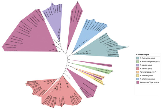
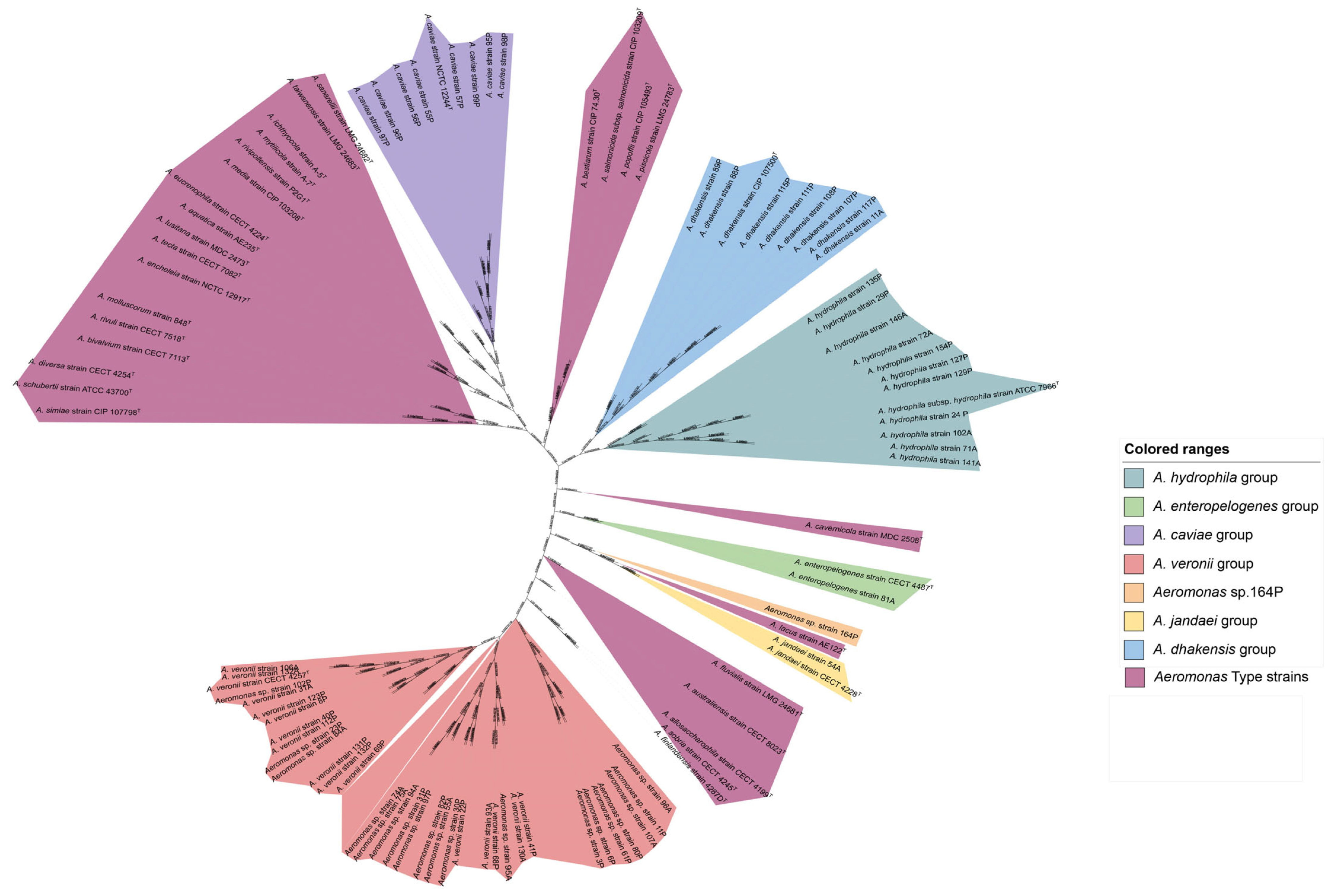
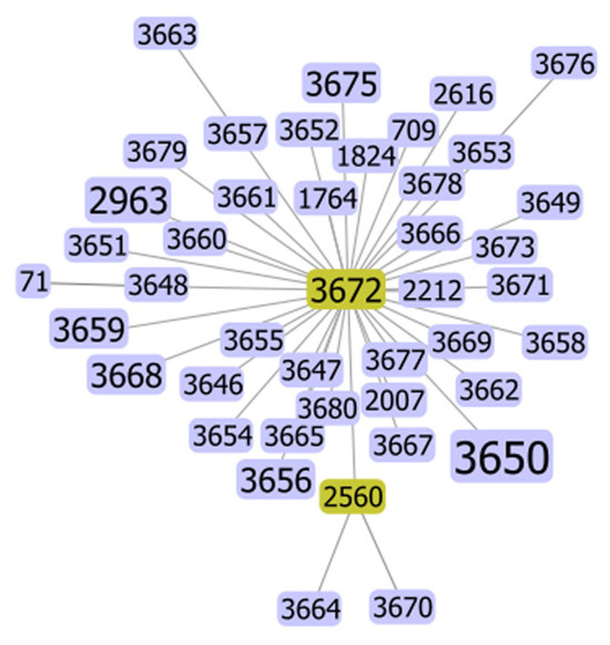
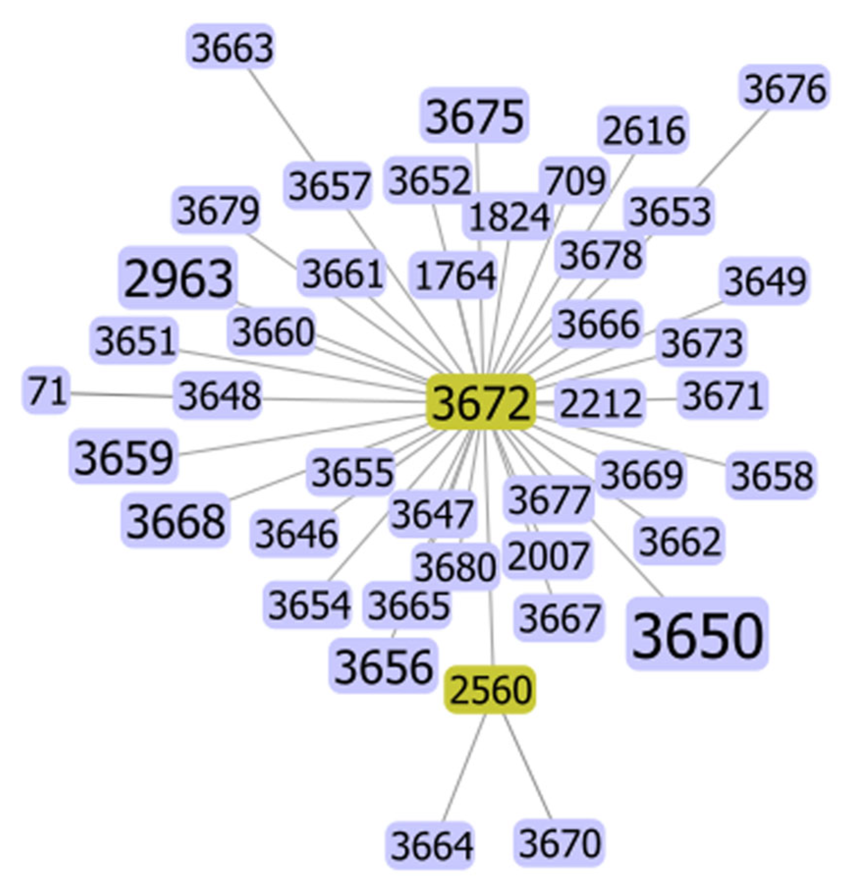
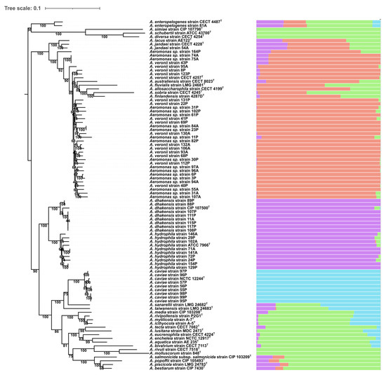
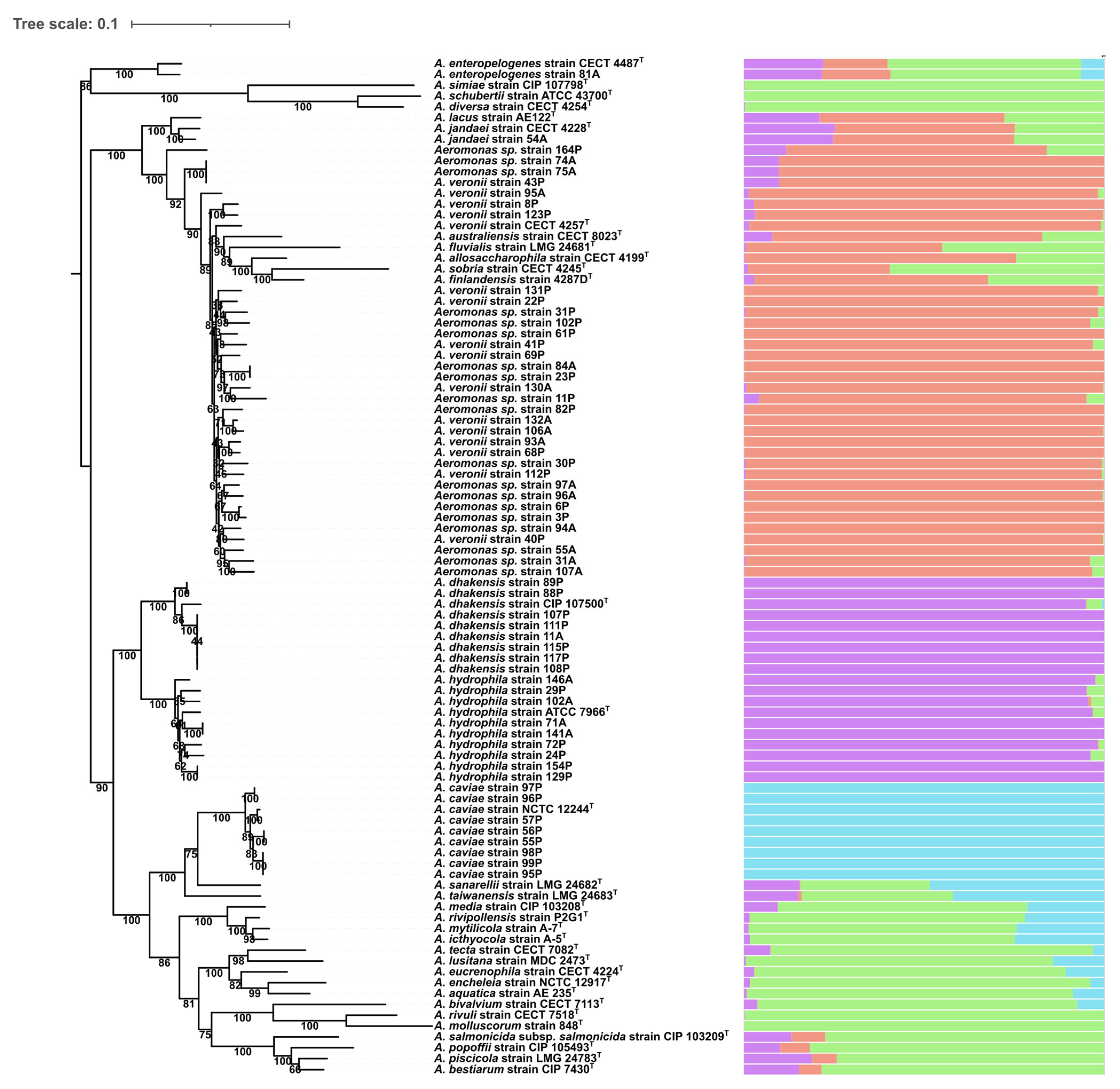

Disclaimer/Publisher’s Note: The statements, opinions and data contained in all publications are solely those of the individual author(s) and contributor(s) and not of MDPI and/or the editor(s). MDPI and/or the editor(s) disclaim responsibility for any injury to people or property resulting from any ideas, methods, instructions or products referred to in the content. |
© 2026 by the authors. Licensee MDPI, Basel, Switzerland. This article is an open access article distributed under the terms and conditions of the Creative Commons Attribution (CC BY) license.