
Velopharyngeal Insufficiency Treatment in Cleft Palate Patients: Umbrella Review

 ,
,  ,
,  ,
,  ,
,  , and
, and 
Abstract
:1. Introduction
2. Materials and Methods
2.1. Protocol Registration
2.2. PICO Question
2.3. Search Strategy
2.4. Eligibility Criteria
2.5. Study Selection and Data Collection
2.6. Quality Assessment
2.7. Analysis of the Degree of Overlap in Studies
3. Results
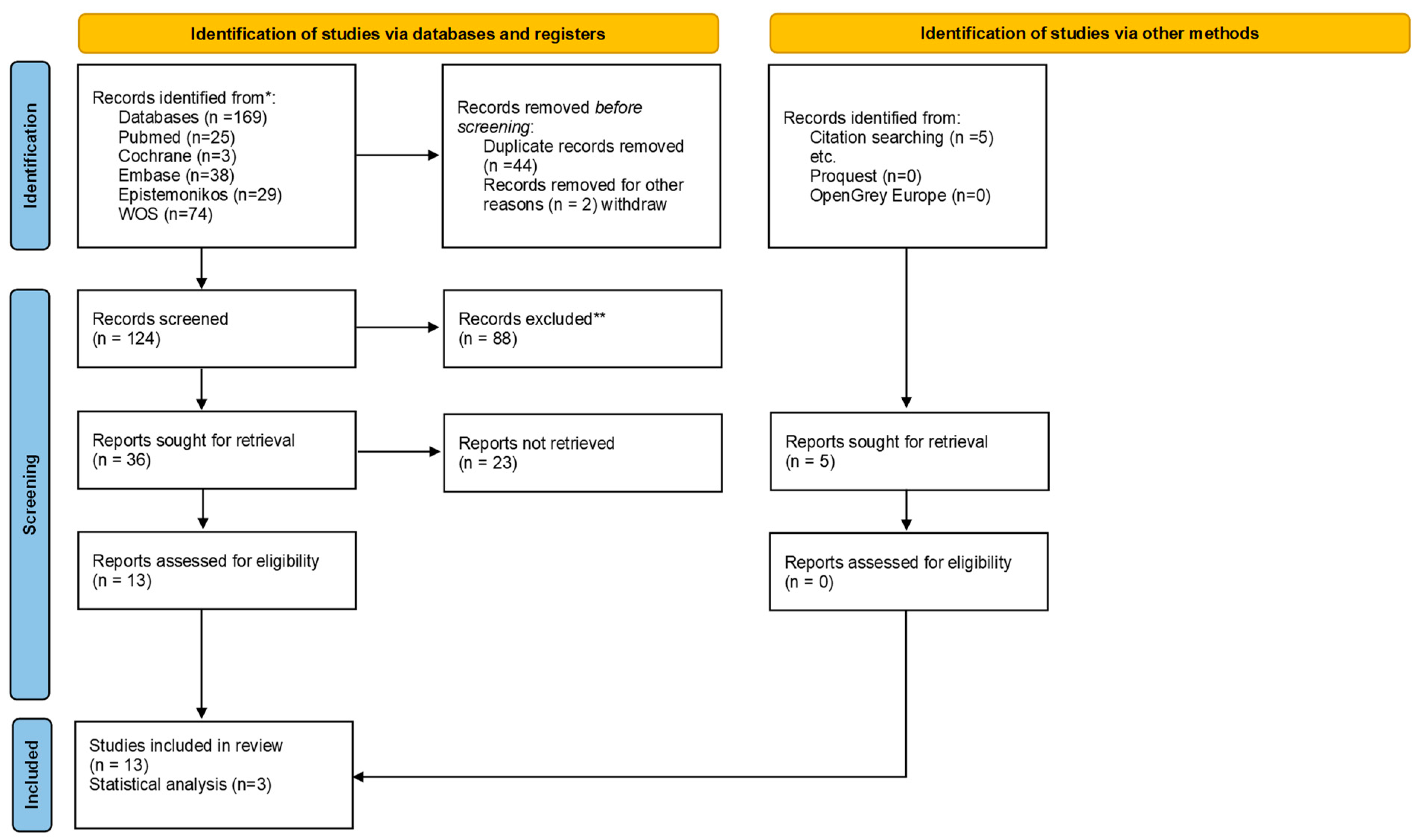
3.1. Study Selection
3.2. Description of the Included Reviews
3.3. Quantitative Synthesis of the Results
3.4. Quality of Included Reviews
3.5. Analysis of the Degree of Overlap in Studies
4. Discussion
5. Conclusions
Author Contributions
Funding
Institutional Review Board Statement
Informed Consent Statement
Data Availability Statement
Conflicts of Interest
References
- Crockett, D.J.; Goudy, S.L. Cleft lip and palate. Facial Plast. Surg. Clin. N. Am. 2014, 22, 573–586. [Google Scholar] [CrossRef] [PubMed]
- Kloukos, D.; Fudalej, P.; Sequeira-Byron, P.; Katsaros, C. Maxillary distraction osteogenesis versus orthognathic surgery for cleft lip and palate patients. Cochrane Database Syst. Rev. 2016, 9, CD010403. [Google Scholar] [CrossRef] [PubMed]
- Gilleard, O.; Sell, D.; Ghanem, A.M.; Tavsanoglu, Y.; Birch, M.; Sommerlad, B. Submucous cleft palate: A systematic review of surgical management based on perceptual and instrumental analysis. Cleft Palate Craniofac. J. 2014, 51, 686–695. [Google Scholar] [CrossRef] [PubMed]
- Kurnik, N.M.; Weidler, E.M.; Lien, K.M.; Cordero, K.N.; Williams, J.L.; Temkit, M.H.; Beals, S.P.; Singh, D.J.; Sitzman, T.J. The Effectiveness of Palate Re-Repair for Treating Velopharyngeal Insufficiency: A Systematic Review and Meta-Analysis. Cleft Palate Craniofac. J. 2020, 57, 860–871. [Google Scholar] [CrossRef] [PubMed]
- Sainsbury, D.; Williams, C.; de Blacam, C.; Mullen, J.; Chadha, A.; Wren, Y.; Hodgkinson, P. Non-Interventional Factors Influencing Velopharyngeal Function for Speech in Initial Cleft Palate Repair: A Systematic Review Protocol. Syst. Rev. 2019, 8, 261. [Google Scholar] [CrossRef] [PubMed]
- Sales, P.H.H.; Costa, F.W.G.; Cetira Filho, E.L.; Silva, P.G.B.; Albuquerque, A.F.M.; Leão, J.C. Effect of maxillary advancement on speech and velopharyngeal function of patients with cleft palate: Systematic Review and Meta-Analysis. Int. J. Oral Maxillofac. Surg. 2021, 50, 64–74. [Google Scholar] [CrossRef] [PubMed]
- Bell, R.; Cowan, K.; Marston, A.P. Metanalysis of alloplastic materials versus autologous fat for injection augmentation pharyngoplasty treatment of velopharyngeal insufficiency. Int. J. Pediatr. Otorhinolaryngol. 2021, 146, 10738. [Google Scholar] [CrossRef] [PubMed]
- Paniagua, L.M.; Signorini, A.V.; da Costa, S.S.; Martins Collares, M.V.; Dornelles, S. Velopharyngeal dysfunction: A systematic review of major instrumental and auditory-perceptual assessments. Int. Arch. Otorhinolaryngol. 2013, 17, 251–256. [Google Scholar] [PubMed]
- de Blacam, C.; Smith, S.; Orr, D. Surgery for velopharyngeal dysfunction: A systematic review of interventions and outcomes. Cleft Palate Craniofac. J. 2018, 55, 405–422. [Google Scholar] [CrossRef] [PubMed]
- Boyce, J.O.; Kilpatrick, N.; Morgan, A.T. Speech and language characteristics in individuals with nonsyndromic submucous cleft palate—A systematic review. Child Care Health Dev. 2018, 44, 818–831. [Google Scholar] [CrossRef] [PubMed]
- Nigh, E.; Rubio, G.A.; Hillam, J.; Armstrong, M.; Debs, L.; Thaller, S.R. Autologous fat injection for treatment of velopharyngeal insufficiency. J. Craniofac. Surg. 2017, 28, 1248–1254. [Google Scholar] [CrossRef] [PubMed]
- Nasser, M.; Fedorowicz, Z.; Newton, T.; Nouri, M. Interventions for the management of submucous cleft palate. Cochrane Database Syst. Rev. 2008, 1, CD006703. [Google Scholar]
- Neumann, S.; Romonath, R. Effectiveness of nasopharyngoscopic biofeedback in clients with cleft palate speech: A systematic review. Logop. Phoniatr. Vocol. 2012, 37, 95–106. [Google Scholar] [CrossRef] [PubMed]
- Timbang, M.R.; Gharb, B.B.; Rampazzo, A.; Papay, F.; Zins, J.; Doumit, G. A systematic review comparing furlow double-opposing Z-plasty and straight-line intravelar veloplasty methods of Cleft palate repair. Plast Reconstr. Surg. 2014, 134, 1014–1022. [Google Scholar] [CrossRef] [PubMed]
- Rossell-Perry, P.; Romero-Narvaez, C.; Olivencia-Flores, C.; Marca-Ticona, R.; Anaya, M.H.; Cordova, J.P.; Luque-Tipula, M. Effect of Nonradical Intravelar Veloplasty in Patients with Unilateral Cleft Lip and Palate: A Comparative Study and Systematic Review. J. Craniofac. Surg. 2021, 32, 1999–2004. [Google Scholar] [CrossRef] [PubMed]
- Haenssler, A.; Perry, J. The Effectiveness of the Buccal Myomucosal Flap on Speech and Surgical Outcomes in Cleft Palate: A Systematic Review. Int. J. Speech Lang. Pathol. Audiol. 2020, 8, 50–54. [Google Scholar]
- Salna, I.; Jervis-Bardy, J.; Wabnitz, D.; Rees, G.; Psaltis, A.; Johnson, A. Partial adenoidectomy in patients with palatal abnormalities. J. Craniofac. Surg. 2019, 30, E454–E460. [Google Scholar] [CrossRef] [PubMed]
- Collins, J.; Cheung, K.; Farrokhyar, F.; Strumas, N. Pharyngeal flap versus sphincter pharyngoplasty for the treatment of velopharyngeal insufficiency: A meta-analysis. J. Plast. Reconstr. Aesthetic Surg. 2012, 65, 864–868. [Google Scholar] [CrossRef] [PubMed]
- Téblick, S.; Ruymaekers, M.; van de Casteele, E.; Nadjmi, N. Effect of Cleft Palate Closure Technique on Speech and Middle Ear Outcome: A Systematic Review. J. Oral Maxillofac. Surg. 2018, 77, 405.e1–405.e15. [Google Scholar] [CrossRef] [PubMed]
- Spruijt, N.E.; ReijmanHinze, J.; Hens, G.; Poorten, V.V.; Van Der Molen, A.B.M. In search of the optimal surgical treatment for velopharyngeal dysfunction in 22q11.2 deletion syndrome: A systematic review. PLoS ONE 2012, 7, e34332. [Google Scholar] [CrossRef] [PubMed] [Green Version]

| PICO Question | |
|---|---|
| Population | Cleft palate patients (unilateral or bilateral) |
| Intervention | Invasive (surgical or other medical procedures) and non-invasive (prosthetic devices, physical therapy, speech therapy) methods |
| Comparison | Different available interventions |
| Outcome | Resolution of velopharyngeal dysfunction |
| Databases | Search Keys |
|---|---|
| Pubmed via Medline | (“cleft palate” [MeSH] OR “cleft palate” OR “oral cleft*” OR “orofacial cleft*”) AND (“Velopharyngeal Insufficiency” [Mesh] OR velopharyngeal OR VPI OR Palatopharyngeal Filters: systematic reviews |
| Web of Science | TS = (“cleft Palate*“ OR “oral cleft*” OR “orofacial cleft*” OR “Palate*, Cleft”) AND TS = (velopharyngeal OR VPI OR Palatopharyngeal) TS = (“cleft Palate*” OR “oral cleft*” OR “orofacial cleft*” OR “Palate*, Cleft”) AND TS = (velopharyngeal OR VPI OR Palatopharyngeal) AND (TS = ”Systematic review*”) |
| Cochrane Library | #1 MeSH descriptor: [Cleft Palate] explode all trees #2 Cleft palate #3 oral cleft* #4 orofacial cleft* #5 MeSH descriptor: [Velopharyngeal Insufficiency] explode all trees #6 velopharyngeal #7 VPI #8 Palatopharyngeal |
| EMBASE | (‘cleft palate’/exp OR ‘cleft palate’ OR ‘oral cleft*’ OR ‘orofacial cleft*’) AND (‘palatopharyngeal incompetence’/exp OR velopharyngeal OR palatopharyngeal OR vpi) AND ‘review’/it |
| Author/Year | Design | Registration | No. of Trials and Design | Bias Analysis | Quality of Evidence | Age of Participants | Intervention | Comparison Unit | Primary Outcome | Results |
|---|---|---|---|---|---|---|---|---|---|---|
| Neumann et al., 2012 [13] | SR | NR | RCT (1) Single-case studies or case-series studies (5) | R | Very low (level 4) | 7–50 years | (n = 83) Nasopharyngoscopy biofeedback (NPB) | NR | - Activation of lateral pharyngeal wall and velopharyngeal closure in articulation - Reduction of hypernasality - Nasal emission or nasal turbulence - Improvement of articulation or intelligibility in connected speech | Preliminary results show effectiveness of visual feedback by flexible nasopharyngoscopy in helping older children and adults improve their VPC during articulation, but only in combination with conventional speech therapy. No studies published measuring the effectiveness of NPB without additional treatments, such as secondary surgery or speech-language therapy. Thus, no conclusive evidence of effectiveness of NPB as a unique therapeutic method. |
| Author/Year | Design | Registration | No. of Trials and Design | Bias Analysis | Quality of Evidence | Age of Participants | Intervention | Comparison Unit | Primary Outcome | Results |
|---|---|---|---|---|---|---|---|---|---|---|
| Timbang et al., 2014 [14] | SR | NR | RS (11) RCT P (1) | NR | NR | 9–18 months (age at palate repair); >4 years (estimated age at speech assessment) | (n = 927) Repair of the cleft palate with Furlow double-opposing Z-plasty | (n = 1205) Repair of the cleft palate with straight-line intravelar veloplasty | - Speech (need for secondary procedures and hypernasality) - Oronasal fistula | Furlow group: - Need for secondary procedures to correct VPI: 0–11.4% in isolated cleft palate (ICP) and 0–6.7% in unilateral cleft lip and cleft palate (UCLP); - Hypernasality: 13–14.3% in ICP and 8.9–18.5% in UCLP; - Oronasal fistula rate: 7.87% (p = 0.14). Straight-line intravelar group: - Need for secondary procedures to correct VPI: 9.1–29.2% in ICP and 6.7–19.4% in UCLP; - Hypernasality: 11.1–20.0% in ICP and 29.1–33.3% in UCLP; - Oronasal fistula rate: 9.81% (p = 0.14). |
| Nigh et al., 2017 [11] | SR | NR | 15 | NR | NR | 2–56 years | (n = 251) Autologous fat injection (combined with surgery; augmentation of soft palate alone; posterior pharyngeal wall augmentation; combined soft palate, posterior, and lateral wall augmentation) | Traditional VPI surgical treatments | - Speech quality - Rate of velopharyngeal insufficiency (RVPI) - Velopharyngeal distance by magnetic resonance imaging (MRI) - Nasometry - Nasendoscopy | In general, AFI should be reserved for patients with mild to moderate VPI (usually <50% closure gap defect or a closure defect between 0.5 and 2 cm2 with adequate velar mobility). Majority of studies, with one exception, required a trial of speech therapy to maximize mobility of the velum prior to AFI. Studies that included patients with VPI secondary to velocardiofacial syndrome reported satisfactory results with no major complications. Major complications were rare. Only one patient with graft hypertrophy reported obstructive sleep apnea. |
| Kurnik et al., 2020 [4] | SR/MA | Prospero | Retrospective cohort (10) Prospective cohort (5) Cohort (3) | R | NR | Any age | Palate re-repair: Furlow double-opposing Z-plasty, radical intravelar veloplasty (IVVP), and radical IVVP with mucosal lengthening. | NR | - Hypernasality - Nasal air emission - Additional velopharyngeal surgery - Obstructive sleep apnea | The overall incidence of achieving no consistent hypernasality following palate re-repair was 61% (95% CI: 44–75%). The incidence of achieving no hypernasality, a more stringent outcome, was 53% (95% CI: 40–65%). The incidence of less than mild hypernasality, a less stringent outcome, was 65% (95% CI: 54–75%). The incidence of no consistent nasal air emission was 78% (95% CI: 60–89%). The incidence of additional velopharyngeal surgery for persistent VPI symptoms was 21% (95% CI: 12–33%). The overall incidence of OSA following re-repair was 28% (95% CI: 13–49%). The incidence of OSA following re-repair (86%) was substantially lower than the incidence of OSA following pharyngeal flap (95%; CI: 63–96%; p = 0.0007). Radical IVVP had a higher incidence of achieving no consistent nasal air emission compared with Furlow DOZ (p = 0.0081). For the remainder of the speech outcomes there was no significant difference among techniques (p > 0.10). The indication for performing re-repair was not associated with the incidence of achieving no consistent hypernasality (p = 0.6572) |
| Rossell-Perry et al., 2021 [15] | SR | Prospero | 10 | Oxford CEBM and GRADE | Low | NR | (n = 503) Radical intravelar veloplasty (IVVP) | (n = 864) Nonradical IVV (preserving the attachment of the tendon of the tensor veli palatini muscle) | - Evaluation of speech development - Middle ear function | Definitive conclusions could not be drawn regardingthe effectiveness of radical IVV on velopharyngeal and middle ear function. |
| Bell et al., 2021 [7] | SR/MA | NR | 29 | NR | Level 3 evidence | 3–75 years | (n = 116) Injection pharyngoplasty with (n = 5) GAX collagen, (n = 36) calcium hydroxyapatite, (n = 72) dextranomer and hyaluronic acid, (n = 3) acellular dermal matrix (Alloderm ®) | (n = 471) Injection pharyngoplasty with autologous fat | - Changes in resonance (reduction in hypernasality) - Degree of velopharyngeal closure | Functional improvements in nasality were recorded in a large proportion of patients (0.79, 95% CI: 0.75–0.82). When stratified for injection material, the proportion of patients with reduced or resolved hypernasality among those receiving synthetic injections was 0.88 (95% CI: 0.82–0.95) and 0.75 (95% CI: 0.71–0.80) for those receiving autologous fat injections (χ2 = 7.035, p = 0.008). Complete velopharyngeal gap closure post-injection was achieved at a higher frequency with injection of synthetic materials compared with autologous fat (χ2 = 11.270; 88% of n = 61/69 vs. 64% of n = 58/91; p = 0.001). |
| Gilleard et al., 2014 [3] | SR | NR | OS (11) CS (14) RCT (1) | NR | Methodological quality score of 6/12; Cohen kappa coefficient 0.63 (range 0.27 to 0.81) | NR | Surgery for VPI in SMCP | Z-palatoplasty; pharyngeal flap; radical velar muscle correction; island flap pushback (Millard); and pharyngeal flap | - Assessing speech outcome following surgery in SMCP - In n = 2, speech was evaluated from previously taken audio/video recordings (Ysunza et al., 2001; Sommerlad et al., 2004), whereas for the others, it was evaluated live | Furlow Z-plasty = 67– 97% (Chen et al., 1996; Sullivan et al., 2011), muscle correction/retropositioning = 30–33% (Sommerlad et al., 2004; Reiter et al., 2011; Sullivan et al., 2011), pharyngeal flap surgery = 32–100% (Crikelair et al., 1970; Porterfield et al., 1976; Peat et al., 1994; Isotalo et al., 2007; Sullivan et al., 2011), and sphincter pharyngoplasty 50–72% (Seagle et al., 1999; Pryor et al., 2006). |
| Blacam et al., 2018 [9] | SR | Pros | RCT(2) Case-control studies (3) Cross sectional studies (2) Retrospective case series (76/83, 91.5%) | Cochrane guidelines | Level IV evidence (According to the 2011 Oxford centre for evidence-based medicine criteria) | 9.64 years (range 1–69.1 years) | Surgery for VPD | Pharyngeal flap; sphincter pharyngoplasty; palatoplasty; and posterior pharyngeal wall augmentation | - Speech assessment, need for further surgery, and occurrence of OSA were the outcomes of interest | Pharyngeal flap surgery was the most common procedure (64% of patients). Overall, 70.7% of patients attained normal resonance and 65.3% attained normal nasal emission. There was no notable difference in speech outcomes, need for further surgery, or occurrence of OSA across the four categories of surgery examined. |
| Haenssler et al., 2020 [16] | SR | NR | Retrospective reviews (11) | Risk bias was not performed | NR | NR | Buccal myomucosal flap surgical approach used for primary palatoplasty and secondary surgery for velopharyngeal insufficiency (VPI) in individuals with cleft palate | NR | - Speech and velopharyngeal competence outcomes following the buccal myomucosal flap surgical approach used for primary palatoplasty and secondary surgery for velopharyngeal insufficiency (VPI) in individuals with cleft palate | Post-surgery, normal resonance was achieved in 77.4% of patients and no nasal air emission was reported in 54.7% of patients. An improvement in velopharyngeal closure was reported in 81.8% of patients. A variety of perceptual speech assessment scales and methods for assessing velopharyngeal competence were used in the studies. |
| Salna et al., 2019 [17] | SR | NR | RCT (7) PS (1) | NR | low-level evidence | 5.5 years | Adenoidectomy | NR | VPI following adenoid surgery | Nearly all patients showed improvement in nasal airway obstruction and snoring. The pooled risk for velopharyngeal insufficiency across all studies was 2 out of 122, which approximates to 1.6% of patients. There were very few complications. |
| Collins et al., 2012 [18] | SR/MA | NR | RCT (2) | p value of 0.10 and an I2 value of 64% | Detsky and MINORS scales The intra-class coalition coefficient was 0.977 (95% CI: 9.0–99.0%). | NR | Operative procedures for the treatment of velopharyngeal insufficiency | Pharyngeal flap or sphincter pharyngoplasty | Velopharyngeal insufficiency resolution | The forest plot of this data was produced through a random effects model analysis. The odds ratio was found to be 2.95 (95% CI: 0.66–13.23) in favour of the pharyngeal flap. |
| Téblick et al., 2018 [19] | SR | NR | RCT (19) prospective cohort studies (4) | NR | For level of evidence, all studies were level 2 (n = 3) or 3 (n = 20) | 2 to 28 years | Cleft palate repair surgical technique | Furlow double-opposing Z-plasty; intravelar veloplasty;von Langenbeck palatoplasty; VWK, Veau-Wardill-Kilner 2-flap palatoplasty | Otitis media with effusion and disturbed speech after cleft palate repair | Four out of five studies concluded that the Furlow palatoplasty, von Langenbeck palatoplasty, VWK palatoplasty, and Sommerlad IVVP had no relevant effect on OME prevalence. Only one study reported a lower incidence of OME after the Kriens IVVP compared with the VWK palatoplasty. |
| Spruijt et al., 2012 [20] | SR | No | Cochrane Collaboration’s tool | Levels 2c or 4 evidence | 2.4–31 years | Surgical procedure | Fat injection, Furlow, intravelar veloplasty (IVP), PF, Honig, SP, or Hynes | Determined whether a particular surgical procedure results in a greater percentage of postoperative normal resonance in patients with 22qDS and VPD | None of the interventions in current use were completely successful in correcting VPD. The low rate of normal resonance may be attributed to the short postoperative follow-up after which the full effect of speech therapy has not yet been achieved. |
| Author/Year | PICO | Protocol | Inclusion Criteria | Comprehensive Search | Duplicate in Selection | Duplicate in Data Extraction | List of Excluded Studies | Description of Included Studies | Assessing Risk of Bias | Funding of Included Studies | Results of Statistical Combination | ROB Effect on the Statistical Combination | ROB in the Discussion | Discussion for the Heterogeneity | Publication Bias | Author’s Funding and COF Reporting | Overall Quality |
|---|---|---|---|---|---|---|---|---|---|---|---|---|---|---|---|---|---|
| Bell et al., 2021 [7] | No | No | No | Partial Yes | Yes | No | No | Partial Yes | No | No | Yes | No | No | Yes | No | Yes | Low |
| Blacam et al., 2018 [9] | No | Partial Yes | No | Partial Yes | No | No | No | Partial Yes | Partial Yes | No | No | No | Yes | Yes | No | Yes | Low |
| Collins et al., 2012 [18] | Yes | Yes | No | Partial Yes | Yes | Yes | No | Partial Yes | Partial Yes | No | Yes | Yes | No | Yes | No | Yes | Low |
| Gilleard et al., 2014 [3] | No | Partial Yes | No | Partial Yes | No | Yes | No | No | Partial Yes | No | No | No | No | No | No | No | Low |
| Haenssler et al., 2020 [16] | No | No | No | Partial Yes | No | No | No | No | No | No | No | No | No | Yes | No | No | Low |
| Kurnik et al., 2020 [4] | No | Partial Yes | No | Partial Yes | Yes | Yes | No | Partial Yes | No | No | Yes | No | No | Yes | No | Yes | Low |
| Neumann et al., 2012 [13] | No | Partial Yes | No | Partial Yes | Yes | Yes | No | Yes | Partial Yes | No | No | No | Yes | Yes | No | No | Low |
| Nigh et al., 2017 [11] | No | No | No | Partial Yes | No | Yes | No | No | No | No | No | No | No | No | No | No | Low |
| Rossell-Perry et al., 2021 [15] | Yes | Partial Yes | No | Partial Yes | Yes | Yes | No | No | Partial Yes | No | No | No | No | Yes | No | Yes | Low |
| Salna et al., 2019 [17] | No | No | No | Partial Yes | Yes | Yes | No | Partial Yes | No | No | No | No | No | Yes | No | No | Low |
| Spruijt et al., 2012 [20] | No | No | Yes | Partial Yes | No | No | No | Partial Yes | Yes | No | Yes | No | Yes | Yes | No | Yes | Low |
| Téblick et al., 2018 [19] | No | No | No | Partial Yes | Yes | Yes | No | Partial Yes | Yes | No | No | No | No | No | No | No | Low |
| Timbang et al., 2014 [14] | No | No | No | Partial Yes | Yes | No | No | Partial Yes | No | No | Yes | No | No | Yes | No | No | Low |
| Systematic Reviews | |||||||||||||
|---|---|---|---|---|---|---|---|---|---|---|---|---|---|
| Primary Studies | Neumann et al., 2012 | Timbang et al., 2014 | Nigh et al., 2017 | Kurnik et al., 2020 | Rossell-Perry et al., 2021 | Bell et al., 2021 | Gilleard et al., 2014 | Blacam et al., 2018 | Haenssler et al., 2020 | Salna et al., 2019 | Collins et al., 2012 (n = 2) | Téblick et al., 2018 | Spruijt et al., 2012 |
| Abdel-Aziz et al., 2007 | x | x | |||||||||||
| Antonelli et al., 2011 | x | x | |||||||||||
| Argamaso et al., 1994 | x | x | |||||||||||
| Arneja et al., 2008 | x | x | |||||||||||
| Boneti et al., 2015 | x | x | x | ||||||||||
| Brandao et al., 2011 | x | x | |||||||||||
| Brigger et al., 2010 | x | x | |||||||||||
| Cantarella et al., 2011 | x | x | x | ||||||||||
| Cao et al., 2013 | x | x | |||||||||||
| Chen et al., 1994 | x | x | |||||||||||
| Chen et al., 1996 | x | x | |||||||||||
| DÁndrea et al., 2018 | x | x | |||||||||||
| Dejonckere and van Wijngaarden et al., 2001 | x | x | |||||||||||
| Deren et al., 2005 | x | x | |||||||||||
| Doucet et al., 2013 | x | x | |||||||||||
| Filip et al., 2013 | x | x | x | ||||||||||
| Filip et al., 2011 | x | x | |||||||||||
| Guerrerosantos et al., 2004 | x | x | |||||||||||
| Klotz et al., 2001 | x | x | |||||||||||
| Lau et al., 2013 | x | x | x | ||||||||||
| Leboulanger et al., 2011 | x | x | x | ||||||||||
| Leuchter et al., 2010 | x | x | x | ||||||||||
| Logjes et al., 2017 | x | x | |||||||||||
| Mazzola et al., 2015 | x | x | |||||||||||
| Mehendale et al., 2004 | x | x | |||||||||||
| Milczuk et al., 2007 | x | x | |||||||||||
| Nakamura et al., 2003 | x | x | |||||||||||
| Park et al., 2000 | x | x | |||||||||||
| Pensler et al., 1988 | x | x | |||||||||||
| Piotet et al., 2015 | x | x | x | ||||||||||
| Robertson et al., 2008 | x | x | |||||||||||
| Rouillon et al., 2009 | x | x | |||||||||||
| Sie et al., 1998 | x | x | |||||||||||
| Spruijt et al., 2011 | x | x | |||||||||||
| Widdershoven et al., 2008 | x | x | |||||||||||
| Yu et al., 2014 | x | x | |||||||||||
Publisher’s Note: MDPI stays neutral with regard to jurisdictional claims in published maps and institutional affiliations. |
© 2022 by the authors. Licensee MDPI, Basel, Switzerland. This article is an open access article distributed under the terms and conditions of the Creative Commons Attribution (CC BY) license (https://creativecommons.org/licenses/by/4.0/).
Share and Cite
Vale, F.; Paula, A.B.; Travassos, R.; Nunes, C.; Ribeiro, M.P.; Marques, F.; Pereira, F.; Carrilho, E.; Marto, C.M.; Francisco, I. Velopharyngeal Insufficiency Treatment in Cleft Palate Patients: Umbrella Review. Biomimetics 2022, 7, 118. https://doi.org/10.3390/biomimetics7030118
Vale F, Paula AB, Travassos R, Nunes C, Ribeiro MP, Marques F, Pereira F, Carrilho E, Marto CM, Francisco I. Velopharyngeal Insufficiency Treatment in Cleft Palate Patients: Umbrella Review. Biomimetics. 2022; 7(3):118. https://doi.org/10.3390/biomimetics7030118
Chicago/Turabian StyleVale, Francisco, Anabela Baptista Paula, Raquel Travassos, Catarina Nunes, Madalena Prata Ribeiro, Filipa Marques, Flávia Pereira, Eunice Carrilho, Carlos Miguel Marto, and Inês Francisco. 2022. "Velopharyngeal Insufficiency Treatment in Cleft Palate Patients: Umbrella Review" Biomimetics 7, no. 3: 118. https://doi.org/10.3390/biomimetics7030118
APA StyleVale, F., Paula, A. B., Travassos, R., Nunes, C., Ribeiro, M. P., Marques, F., Pereira, F., Carrilho, E., Marto, C. M., & Francisco, I. (2022). Velopharyngeal Insufficiency Treatment in Cleft Palate Patients: Umbrella Review. Biomimetics, 7(3), 118. https://doi.org/10.3390/biomimetics7030118