Review

Benign Cutaneous Neoplasms with Syndromic Associations

1
Island Medical Program, University of British Columbia, Victoria, BC V8W 2Y2, Canada
2
Department of Pathology and Laboratory Medicine, University of Calgary, Calgary, AB T2N 1N4, Canada
3
Alberta Precision Laboratories, Diagnostic and Scientific Centre, 9-3535 Research Road NW, Calgary, AB T2L 2K8, Canada
*
Author to whom correspondence should be addressed.
These authors contributed equally to this work.
Dermatopathology 2025, 12(4), 34; https://doi.org/10.3390/dermatopathology12040034
Submission received: 6 August 2025 / Revised: 17 September 2025 / Accepted: 25 September 2025 / Published: 8 October 2025

Abstract

There are many benign skin neoplasms encountered in dermatopathology practice that can be associated with underlying genetic disorders. Although benign themselves, these lesions can offer insight into the potential for development of internal malignancies in patients with these hereditary syndromes. An astute dermatopathologist will recognize clues that suggest a syndromic association of these lesions, such as the presence of multiple lesions, distinct histologic growth patterns, and the results of ancillary immunohistochemical testing. The dermatopathologist can then guide the referring clinician to obtain additional clinical and family history and, if appropriate, pursue further screening and genetic testing. This review article will provide an overview of the clinical and histologic features associated with select common and uncommon benign skin neoplasms with syndromic associations.

1. Introduction

Benign skin tumors are among the most frequently encountered lesions in dermatologic practice [1]. While they often arise sporadically and may be regarded as clinically insignificant, there are some instances in which the presence of these lesions may serve as important cutaneous indicators of underlying genetic syndromes, some of which are associated with an elevated risk of internal malignancy or other systemic complications [2]. In many cases, the skin provides the first manifestation of these conditions, with these benign skin neoplasms sometimes appearing years before internal disease becomes clinically apparent. Using cutaneous findings to identify an underlying hereditary condition presents a critical opportunity for early recognition and intervention in these genetic disorders [2,3].
This review highlights the clinical and histopathological features of select benign cutaneous neoplasms of adnexal, mesenchymal, and melanocytic origin with the aims of helping dermatopathologists become more aware of the possible syndromic associations of these lesions and recognize the clinical and histological clues that may prompt further recommendations to the referring provider to consider an underlying genetic syndrome in a patient.

2. Hair Follicle Tumors

2.1. Fibrofolliculoma/Trichodiscoma

Fibrofolliculomas and trichodiscomas are benign follicular hamartomas that may occur sporadically or, more notably, in association with Birt–Hogg–Dubé syndrome (OMIM# 135150). Clinically, these lesions present as small, dome-shaped, white-to-skin-colored papules most commonly distributed on the face, neck, and upper torso. In patients with BHD, they typically appear after puberty and may number in the dozens or even hundreds, sometimes coalescing into plaques [4]. Histologically, fibrofolliculomas are characterized by distorted or dilated central hair follicles embedded in a fibromucinous stroma with thin, anastomosing epithelial strands radiating from the follicular wall (Figure 1A). Trichodiscomas exhibit a more prominent stromal component and may include peripheral sebaceous lobules (Figure 1B). Both lesions are now considered histologic variants of the same process [5]. Immunohistochemical studies demonstrate diffuse expression of CD34 and scattered expression of factor XIIIa in the stromal component, with nestin and c-kit occasionally marking spindle-shaped stromal cells. The follicular epithelium may show aberrant CK15 expression. These findings support a shared origin in the differentiation of hair follicle bulge stem cells [5,6]. Markers of neural differentiation, such as S100, are negative in the stromal component [6].
Fibrofolliculomas and trichodiscomas are considered hallmark cutaneous findings of Birt–Hogg–Dubé syndrome, an autosomal dominant disorder caused by pathogenic variants in the FLCN gene on chromosome 17p11.2 [4]. BHD is associated with a spectrum of internal tumors, most notably renal neoplasms, including hybrid oncocytic tumors (most frequent), chromophobe renal cell carcinoma, renal oncocytomas, and less commonly, clear cell renal carcinoma [4,7]. Pulmonary manifestations are also common and include bilateral basilar lung cysts and recurrent spontaneous pneumothoraces [6]. While acrochordons may also be observed, they are not specific to BHD given their prevalence in the general population. Recognition of multiple fibrofolliculomas or trichodiscomas, especially in patients with a suggestive personal or family history, should prompt consideration of BHD and initiate appropriate genetic testing and systemic screening.

2.2. Tricholemmoma

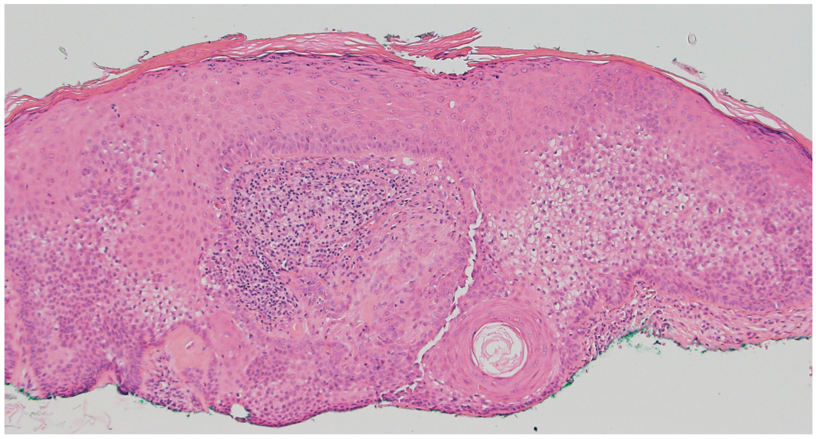
Tricholemmoma is a benign follicular neoplasm that arises from the outer root sheath of the hair follicle and most commonly presents as a small, smooth, skin-colored or slightly pigmented papule on the face, particularly around the nose and upper lip [8]. These lesions are often solitary, but the presence of multiple facial tricholemmomas is a hallmark of Cowden syndrome and should prompt consideration of a syndromic association [9]. Histologically, tricholemmomas are well-circumscribed lobular proliferations composed of pale or clear glycogen-rich keratinocytes with peripheral palisading and are surrounded by a thickened basement membrane, mimicking the vitreous layer of the outer root sheath (Figure 2) [8]. Desmoplastic tricholemmoma demonstrates epithelial cords embedded in a dense sclerotic stroma, sometimes resembling an invasive carcinoma, such as basal cell carcinoma [10]. Periodic acid–Schiff (PAS) positivity, which is diastase-labile, highlights the glycogen content. Immunohistochemically, tricholemmomas typically express CD34 in diffuse or peripheral patterns, as well as CK17 and calretinin, supporting their outer root sheath origin [11]. A lack of expression for BerEP4 can help differentiate these lesions from basal cell carcinoma [12].
The presence of multiple tricholemmomas is strongly associated with PTEN hamartoma tumor syndrome (PHTS), a spectrum of autosomal dominant conditions caused by germline mutations in the tumor suppressor gene PTEN located on chromosome 10q23.31 [9,13]. PHTS includes Cowden syndrome (OMIM #158350), Bannayan–Riley–Ruvalcaba syndrome (OMIM #158350), Proteus syndrome (OMIM #176920), and Proteus-like syndrome. Among these, Cowden syndrome is the most well-defined and is associated with a significantly increased lifetime risk of malignancies including breast cancer (25–50%), follicular thyroid cancer (up to 10%), and endometrial cancer (5–10%), as well as possible increased risks of renal cell carcinoma, melanoma, and gliomas [9]. Other clinical features may include macrocephaly, mucocutaneous papillomas, acral keratoses, oral cobblestoning, multinodular goiter, and lipomas [9]. The dermatologic manifestations often precede the development of internal malignancies and serve as valuable diagnostic clues. Loss of PTEN protein expression on immunohistochemistry may be observed in these lesions and, if present, can support the diagnosis of PHTS, although its absence does not rule it out [14,15]. In a clinical context suggestive of PHTS, recognition of multiple tricholemmomas should prompt referral for genetic counseling and PTEN mutation testing, as early diagnosis enables appropriate cancer screening and risk reduction strategies.

2.3. Pilomatricoma

Pilomatricoma, also known as calcifying epithelioma of Malherbe, is a benign adnexal tumor derived from immature hair matrix cells. It typically presents in children and young adults as a solitary, firm, slow-growing, subcutaneous nodule with overlying skin that may appear skin-toned, reddish, or bluish [16]. Lesions are most often located on the head, neck, and upper limbs, and commonly measure under 3 cm in diameter [16]. A useful clinical clue is the “tent sign”, where stretching the skin over the lesion reveals angular protrusions due to underlying calcification [17]. Histologically, pilomatricomas are sharply circumscribed, lobulated tumors of the dermis or subcutis, composed of peripheral basaloid cells that transition abruptly into central anucleate eosinophilic shadow (ghost) cells (Figure 3). Dystrophic calcification and foreign-body giant cell reaction are common features [18,19]. Immunohistochemically, nuclear and cytoplasmic accumulation of β-catenin is frequently observed in basaloid cells, consistent with dysregulated Wnt signaling [20]. This can result from activating mutations in CTNNB1 or from upstream disruption of APC regulation in syndromic cases [19].
The presence of multiple pilomatricomas, especially six or more, warrants evaluation for syndromic associations, particularly Gardner syndrome, a subtype of familial adenomatous polyposis (FAP; OMIM #175100). Gardner syndrome is an autosomal dominant disorder caused by germline mutations in the APC gene on chromosome 5q21–q22 and is associated with colorectal adenomatous polyps (nearly 100% lifetime cancer risk) and extracolonic features such as osteomas, desmoid tumors, epidermoid cysts, supernumerary teeth, and retinal pigment epithelium hypertrophy [21,22]. Pilomatricomas in this context may occur as multiple independent lesions or as pilomatricoma-like changes within epidermoid cysts, showing histologic features such as shadow cells and basaloid matrical cells and nuclear β-catenin positivity [23,24]. Multiple familial pilomatricomas have been reported as early dermatologic signs of Gardner syndrome, and their presence, particularly in conjunction with a relevant family history, should prompt genetic counseling, colonoscopic screening, and APC gene testing [21].

2.4. Trichoepithelioma

Multiple trichoepitheliomas may be found in Brooke–Spiegler Syndrome (OMIM #605041) and multiple familial trichoepitheliomas (OMIM #601606). These autosomal dominant disorders are characterized by germline inactivating mutations in the tumor suppressor gene CYLD, located on chromosome 16q12-13. These syndromes are phenotypic variations in CYLD cutaneous syndrome.
Trichoepitheliomas present as skin-colored, non-ulcerated papules or nodules on the face, most commonly on the nose and nasolabial folds [25]. Syndrome associated tumors typically appear during puberty and progressively accumulate during adulthood. These tumors can number in the hundreds, with the potential to cause disfigurement [25,26]. Visual impairment and hearing loss can result from extensive involvement of the eyelids and external auditory canal [27].
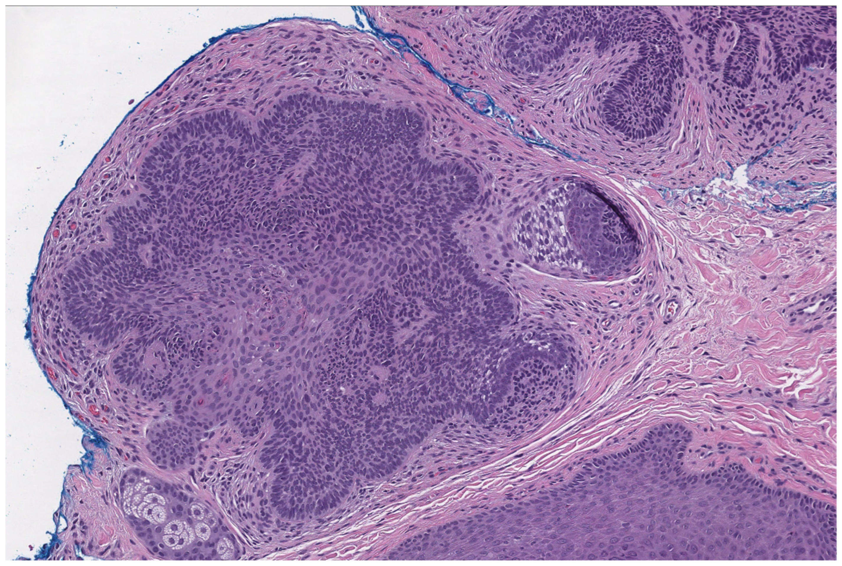
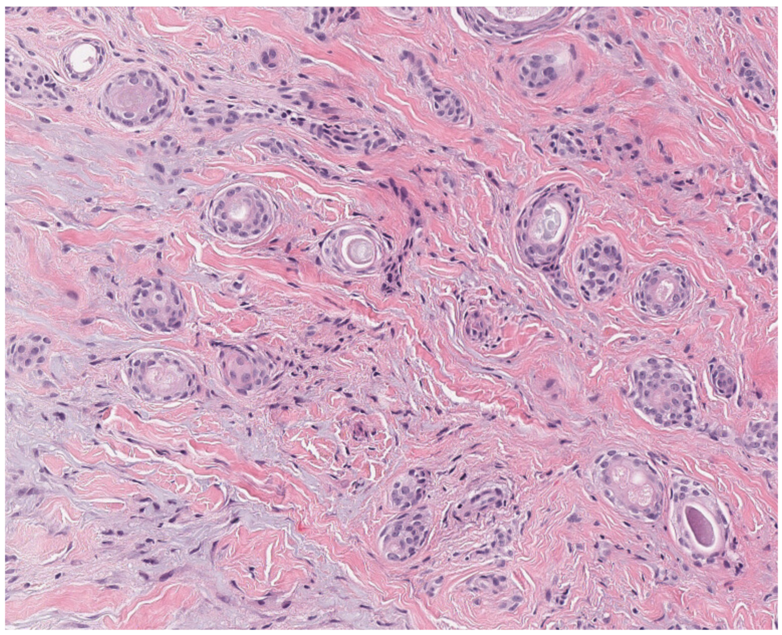
Histological examination of trichoepitheliomas demonstrates nests of bland basaloid cells with peripheral palisading, associated papillary mesenchymal bodies and horn cysts, and a fibrous stroma (Figure 4). Desmoplastic trichoepitheliomas are characterized by a prominent desmoplastic stroma surrounding small cords and islands of basaloid cells. Histologically, these lesions may mimic basal cell carcinoma [28]. The presence of numerous CK20-positive Merkel cells favors a diagnosis of trichoepithelioma [29]. BerEP4 is not helpful in distinguishing these lesions from basal cell carcinoma [30].
Brooke–Spiegler Syndrome is associated with the presence of multiple cylindromas, spiradenomas, spiradenocylindromas, and trichoepitheliomas [26]. Multiple familial trichoepitheliomas is associated with numerous trichoepitheliomas only. Malignant transformation of pre-existing skin neoplasms may develop in 5–10% of these patients [27]. CYLD cutaneous syndrome can also be associated with salivary gland tumors and rarely pulmonary and mammary cylindromas [27,31]. A CYLD cutaneous syndrome should be suspected in patients with multiple trichoepitheliomas with or without other skin adnexal neoplasms from a young age and with a family history of similar multiple tumors. Genetic testing can help facilitate early diagnosis and surveillance.

2.5. Basaloid Follicular Hamartoma

Basaloid follicular hamartomas may arise sporadically or be associated with nevoid basal cell carcinoma syndrome (NBCC)/Gorlin syndrome (OMIM #109400). This is an autosomal dominant disorder caused by germline mutations of the Sonic Hedgehog (SHH) pathway. Inactivation of genes, particularly PTCH1 (chromosome 9q22.32) or SUFU (chromosome 10q24.31), leads to activation of the SHH pathway. This syndrome is characterized by an early age of onset of multiple basal cell carcinomas (the most common neoplasm in this syndrome), odontogenic keratocysts of the jaw, palmar or plantar pits, skeletal abnormalities, lamellar calcification of the falx cerebri, and characteristic coarse facial features (e.g., frontal bossing and macrocephaly) [32]. SUFU mutations are less prevalent and are associated with an increased risk of early-onset medulloblastoma [32]. Basaloid follicular hamartomas may represent up to 24% of skin tumors in NBCC [32]. Meningiomas, craniopharyngiomas, cardiac fibromas, and bilateral calcified ovarian fibromas may also be found in this syndrome [33]. Multiple basaloid follicular hamartomas in association with hypotrichosis may also develop in generalized basaloid follicular hamartoma syndrome (OMIM #605827), an autosomal dominant disorder of unknown molecular basis [33].
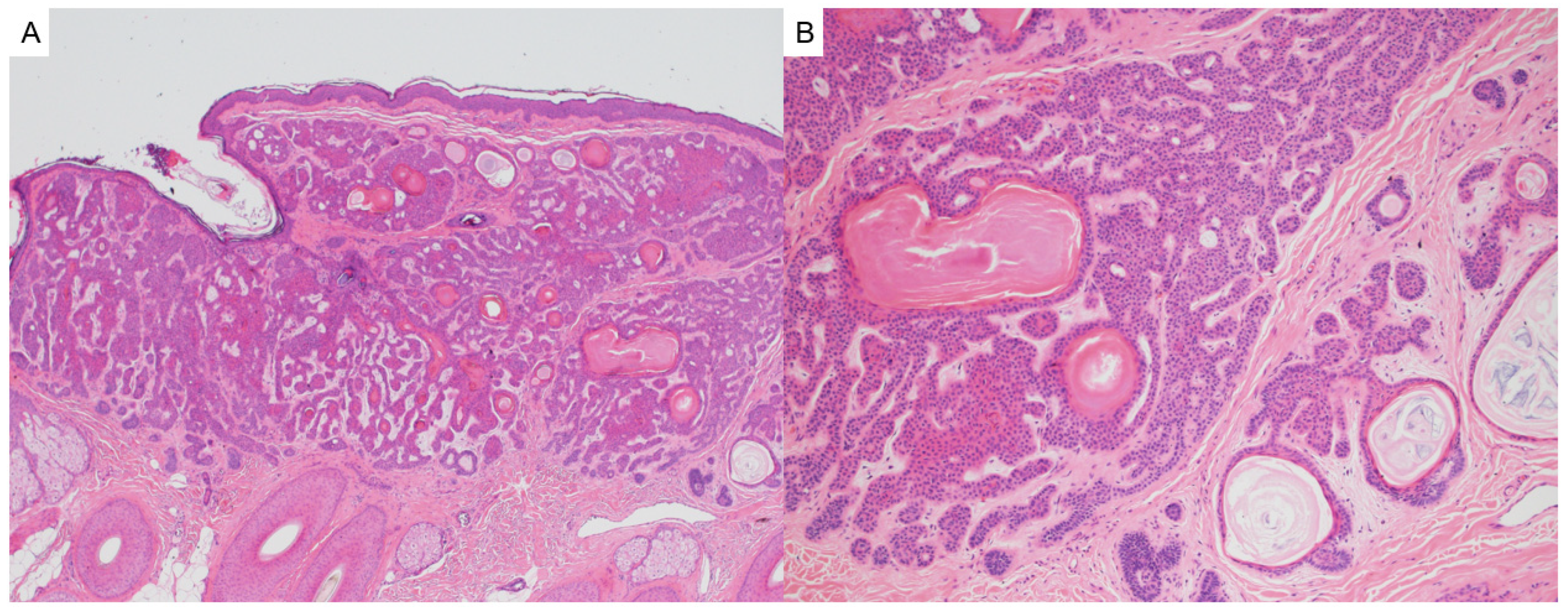
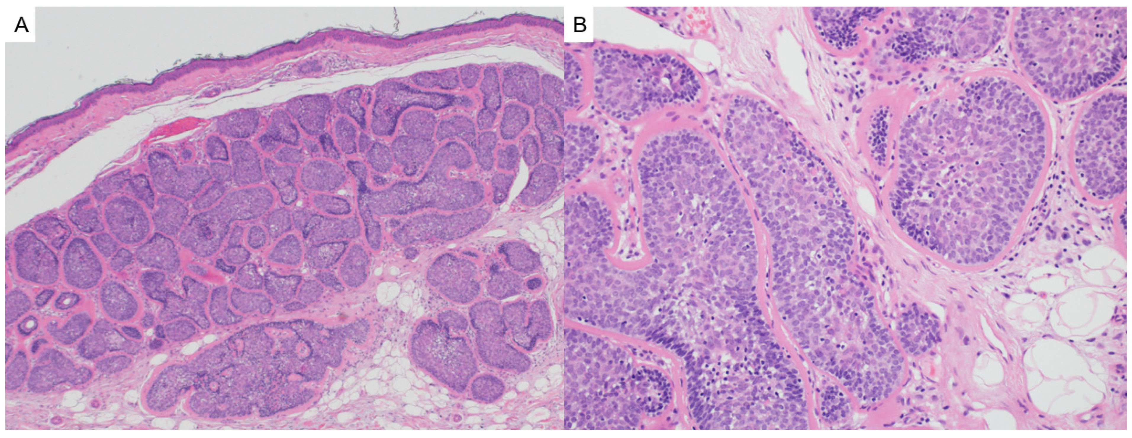
Clinically, basaloid follicular hamartomas are small, stable, slightly raised, skin-colored lesions on the face, scalp, and upper trunk [34,35]. On histology, basaloid follicular hamartomas may mimic infundibulocystic basal cell carcinoma and are characterized as symmetrical, well-circumscribed, superficial perifollicular neoplasms composed of radiating anastomosing cords and strands of basaloid and squamoid cells with associated horn cysts in a loose fibrous stroma (Figure 5). They demonstrate stromal-dermal clefting (but not tumor-stroma clefting), bland cytology, minimal mitotic activity, minimal apoptosis, and lack the peripheral palisading and inflammation that may be associated with basal cell carcinoma [32,34]. Immunohistochemistry can be helpful in distinguishing these lesions from basal cell carcinoma. Basaloid follicular hamartomas demonstrate the presence of CK20 positive Merkel cells (usually not present in basal cell carcinomas), focal EpCAM expression (diffuse expression in basal cell carcinoma), low Ki67 proliferative index (higher in basal cell carcinoma), and wild-type p53 expression (can be overexpressed in basal cell carcinoma) [32,35,36,37]. It is suggested that basaloid follicular hamartomas may be closely related or precursors to basal cell carcinoma [32].
Given the potential syndromic association and the risk for development of other associated lesions, the presence of multiple basaloid follicular hamartomas should prompt consideration for genetic evaluation.

3. Sebaceous Tumors

3.1. Sebaceous Adenoma/Sebaceous Epithelioma

Sebaceous adenomas and sebaceous epitheliomas are cutaneous adnexal tumors that can serve as clinical hallmarks of Muir–Torre syndrome (MTS), a subtype of Lynch syndrome [38]. Clinically, these neoplasms appear as solitary or multiple, small, yellow-to-pink papules or nodules, most commonly found on the face, scalp, and trunk [39].
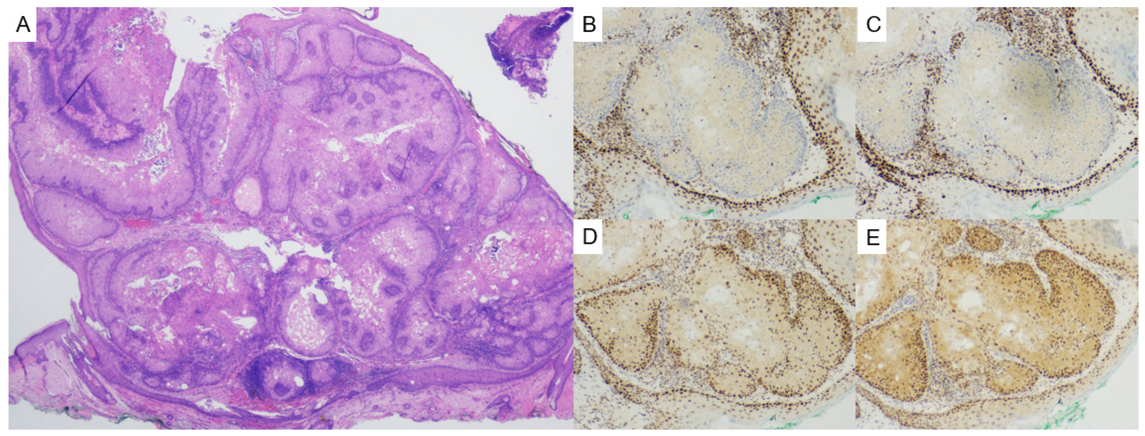
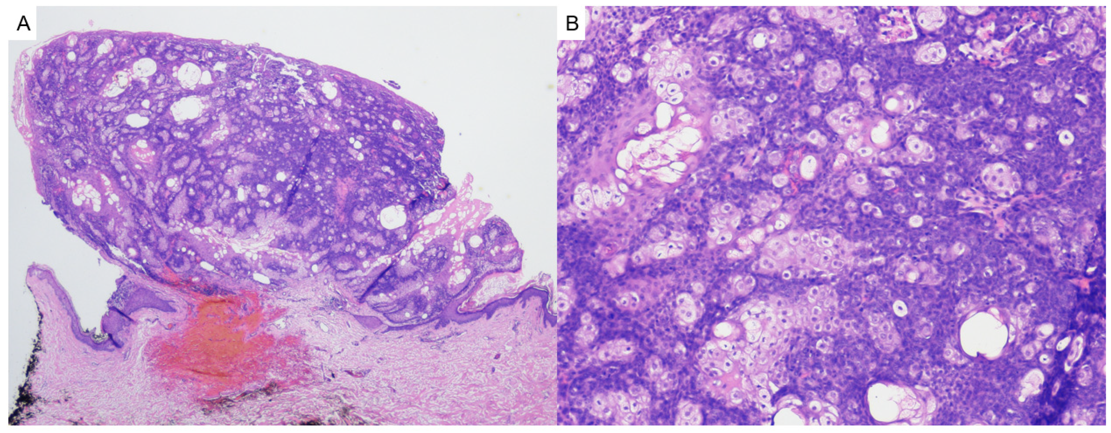
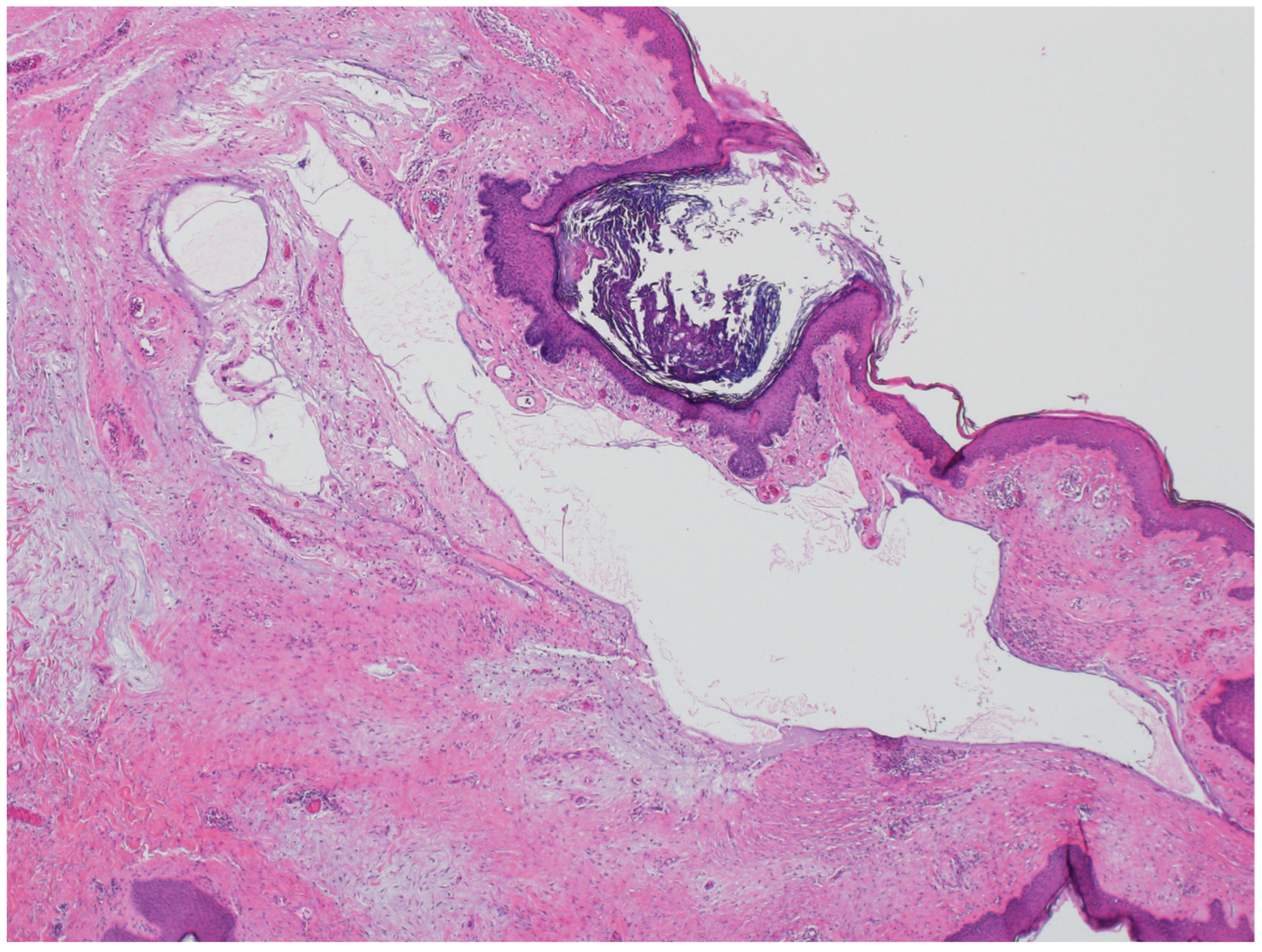
Histologically, sebaceous adenomas show well-circumscribed lobules with a predominant population (i.e., more than 50% of the tumor) of central mature sebocytes with a less predominant population of basaloid cells peripherally (Figure 6). Sebaceous epitheliomas (sebaceomas) are histologically similar but contain a higher proportion of basaloid cells (i.e., more than 50% of the tumor) (Figure 7) [38]. Sebaceous neoplasms in MTS may exhibit cystic or folliculocentric architecture [40]. Immunohistochemically, evaluation of DNA mismatch repair (MMR) proteins MLH1, MSH2, MSH6, and PMS2 is critical for identifying MTS-associated lesions. Loss of nuclear expression on immunohistochemistry for one or more of these proteins in tumor cells suggests mismatch repair deficiency and should prompt further investigation. Currently, a consensus screening strategy for MMR proteins in sebaceous tumors is not available [41]. The most recent American Society of Dermatopathology appropriate use criteria suggest a targeted screening approach to MMR immunohistochemistry, with a four antibody panel being appropriate in patients with multiple sebaceous tumors regardless of patient age, keratoacanthoma with sebaceous differentiation regardless of patient age, cystic sebaceous tumor regardless of patient age, MTS-associated neoplasm and/or visceral malignancy regardless of patient age, and sebaceous tumors arising in non-head and neck locations in individuals ≤60 years of age [42]. While immunohistochemistry is widely used, it does have a high false positive rate and low specificity [42], and germline sequencing remains the gold standard for diagnosing hereditary MTS [39].
Muir–Torre syndrome (OMIM# 158320) is an autosomal dominant condition caused by germline mutations in MSH2 (accounts for ~90% of cases), MLH1, MSH6, and PMS2, all of which are components of the mismatch repair pathway [43,44]. Sebaceous neoplasms can precede, coincide with, or follow the diagnosis of internal malignancy, most commonly colorectal carcinoma, followed by genitourinary, endometrial, gastric, pancreatic, and breast cancers [45]. The presence of even a single sebaceous neoplasm—especially sebaceous adenoma, epithelioma, or carcinoma—warrants evaluation for underlying malignancy, particularly in patients with personal or family histories of Lynch-related cancers [40]. Keratoacanthomas have also been associated with MTS and may occur in combination with sebaceous tumors [46].
Detection of MMR deficiency in a sebaceous tumor, particularly with concurrent or personal/family history of visceral malignancy, should prompt genetic counseling, colonoscopy, and MMR germline testing.

3.2. Steatocystoma

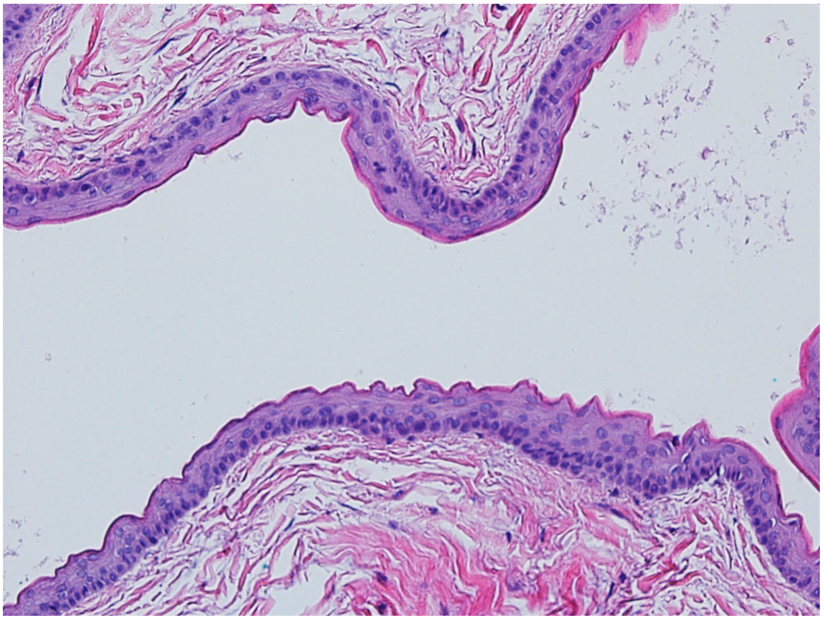
Although cystic lesions are not considered neoplasms, steatocystomas have been included in this review because of their potential syndromic associations. These lesions may be seen in association with steatocystoma multiplex (OMIM #184500), an autosomal dominant disorder resulting from a mutation of KRT17 on chromosome 17q21.2.1 [47,48]. Steatocystoma multiplex presents as an early onset of multiple translucent skin-colored to yellow dome-shaped papules or nodules on the trunk, head and neck, and limbs [49]. Histologically, steatocystomas are characterized as thin-walled dermal-based multiloculated cysts lined by stratified squamous epithelium lacking a granular layer and with an undulating, eosinophilic cuticle forming the inner cyst wall lining (Figure 8). Sebaceous lobules are usually identified within the cyst wall. Steatocystomas may become inflamed, infected, and scarred (steatocystoma multiplex suppurativum), causing cosmetic disfigurement [49]. Early recognition of a syndromic association in a patient with multiple steatocystomas, followed by appropriate counselling may alleviate some of the psychological issues associated with this syndrome [50].
Multiple steatocystomas may also be seen in pachyonychia congenita type 2 (OMIM #167210), which also results from mutations in keratin genes including KRT17. Affected patients may additionally present with severe nail abnormalities, painful palmoplantar keratoderma, oral leukokeratosis, vellus hair cysts, palmoplantar hyperhidrosis, natal or prenatal teeth, and follicular keratoses on the trunk and limbs [51].

4. Sweat Gland Tumors

4.1. Cylindroma/Spiradenoma

Cylindromas and spiradenomas are benign adnexal neoplasms most commonly affecting the scalp, face, and neck. Clinically, they present as firm, pink to reddish dome-shaped nodules. Cylindromas may coalesce into large masses on the scalp known as “turban tumors”, while spiradenomas are often painful and more likely to occur on the trunk or extremities. Lesions usually appear in adolescence or early adulthood and progressively increase in number and size [52,53].
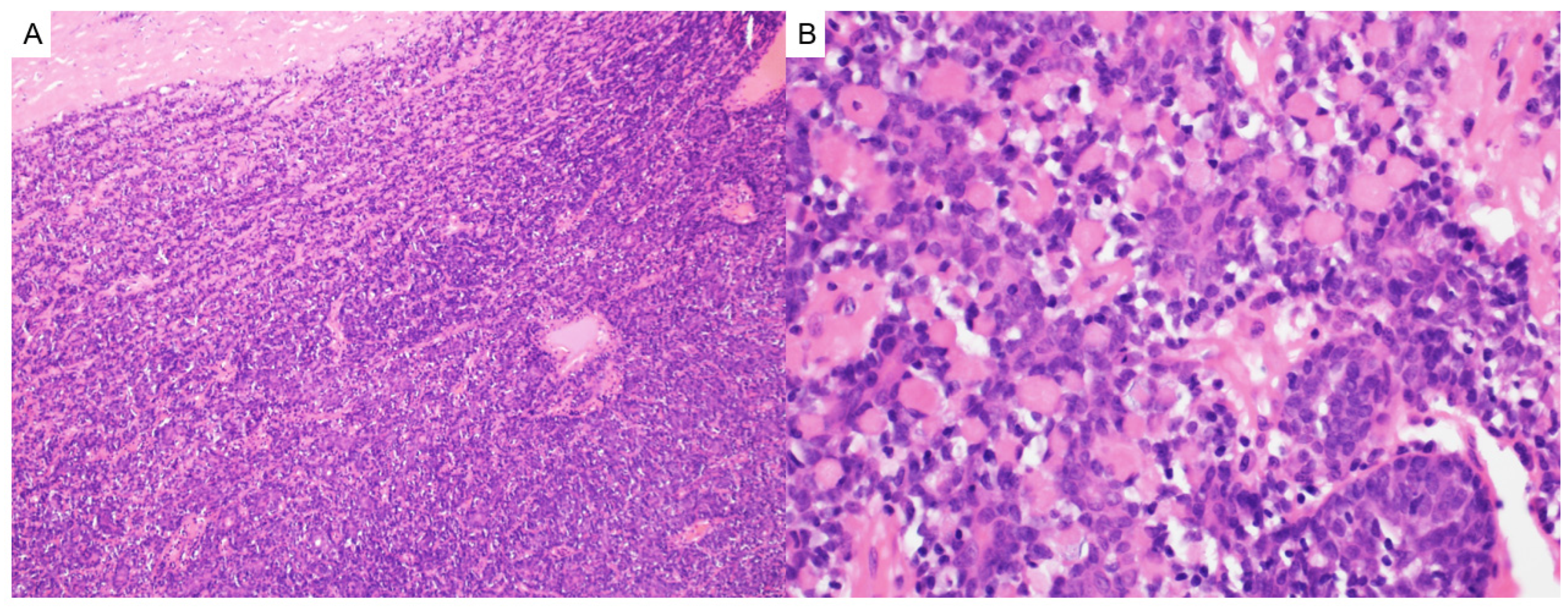
Histologically, cylindromas demonstrate basaloid cells arranged in a distinctive “jigsaw puzzle” pattern surrounded by thick PAS-positive eosinophilic basement membrane material (Figure 9) [53,54]. Spiradenomas, in contrast, show lobular architecture with dual populations of small dark peripheral and large pale central cells, surrounded by prominent lymphocytic infiltrates (Figure 10) [52]. Hybrid tumors, called spiradenocylindromas, containing features of both may also occur.
Immunohistochemically, both tumors exhibit cytokeratin and SOX10 positivity [55], with myoepithelial markers such as calponin, SMA, S100, and p63 expressed in surrounding cells. Ductal differentiation is highlighted by CEA and EMA. Spiradenomas uniquely contain CD3+ T-cells and CD1a+ Langerhans cells [56]. Malignant transformation (to spiradenocarcinoma or cylindrocarcinoma) is rare but marked by an elevated Ki67 proliferative index and loss of characteristic MYB expression in spiradenomas [56,57].
These tumors are central to the diagnosis of Brooke–Spiegler syndrome (OMIM# 605041), an autosomal dominant condition caused by inactivating mutations in the tumor suppressor gene CYLD, located on chromosome 16q12-13. Brooke–Spiegler syndrome presents with multiple cylindromas, spiradenomas, spiradenocylindromas, and trichoepitheliomas and occasionally basal cell carcinomas or salivary gland tumors. It exhibits variable penetrance and significant phenotypic heterogeneity even within the same family [58]. Multiple cylindromas can also be found in familial cylindromatosis (OMIM #132700). Brooke–Spiegler syndrome and familial cylindromatosis are phenotypic variants of CYLD cutaneous syndrome.
Early identification of multiple adnexal tumors, especially in young individuals or those with a family history, should prompt evaluation for CYLD cutaneous syndrome. Genetic confirmation via CYLD mutation analysis is diagnostic. Regular follow-up is essential to monitor for malignant transformation and associated internal neoplasms [56].

4.2. Syringoma

Syringomas are common tumors that typically present as small, skin-colored to yellow papules on the lower eyelids and periorbital areas. Eruptive syringomas present as multiple widespread scattered papules on the trunk and limbs, which may coalesce to form plaques [59,60]. Histologically, they are characterized as well-circumscribed proliferations of small ducts, nests, and cords of bland pale eosinophilic to clear cells in the superficial dermis (Figure 11). Ducts can have a tadpole-like appearance with comma-like tails. While syringomas usually occur sporadically, they have also been found to be associated with Down syndrome (OMIM #190685, characterised by trisomy 21) and rarely in other syndromes, such as Nicolau Balus syndrome, Brooke–Spiegler syndrome (OMIM# 605041), Costello syndrome (OMIM# 218040), and steatocystoma multiplex (OMIM #184500) [61,62,63,64,65]. In Down syndrome, syringomas are most commonly found around the eyes [66,67]. Other dermatologic manifestations that may be seen in Down syndrome include inflammatory dermatoses (such as atopic dermatitis), alopecia areata, and elastosis perforans serpiginosa [62]. Patients with Down syndrome have an increased risk of acute leukemias [68]. Nicolau Balus syndrome is a rare disorder characterized by syringomas, atrophorderma vermiculata, and milia [69].

5. Neural Tumors

Neurofibromas

Neurofibromas are benign peripheral nerve sheath tumors composed of a mixture of Schwann cells, fibroblasts, mast cells, and perineurial cells. While they can arise sporadically, the presence of multiple neurofibromas is a cardinal feature of Neurofibromatosis Type 1 (NF1, OMIM# 162200), an autosomal dominant neurocutaneous syndrome caused by mutations in the NF1 gene on chromosome 17q11.2. This gene encodes neurofibromin, a tumor suppressor that negatively regulates RAS signaling through its GTPase-activating protein function. Loss of neurofibromin leads to dysregulated cell proliferation, especially in neural crest–derived tissues [70,71].
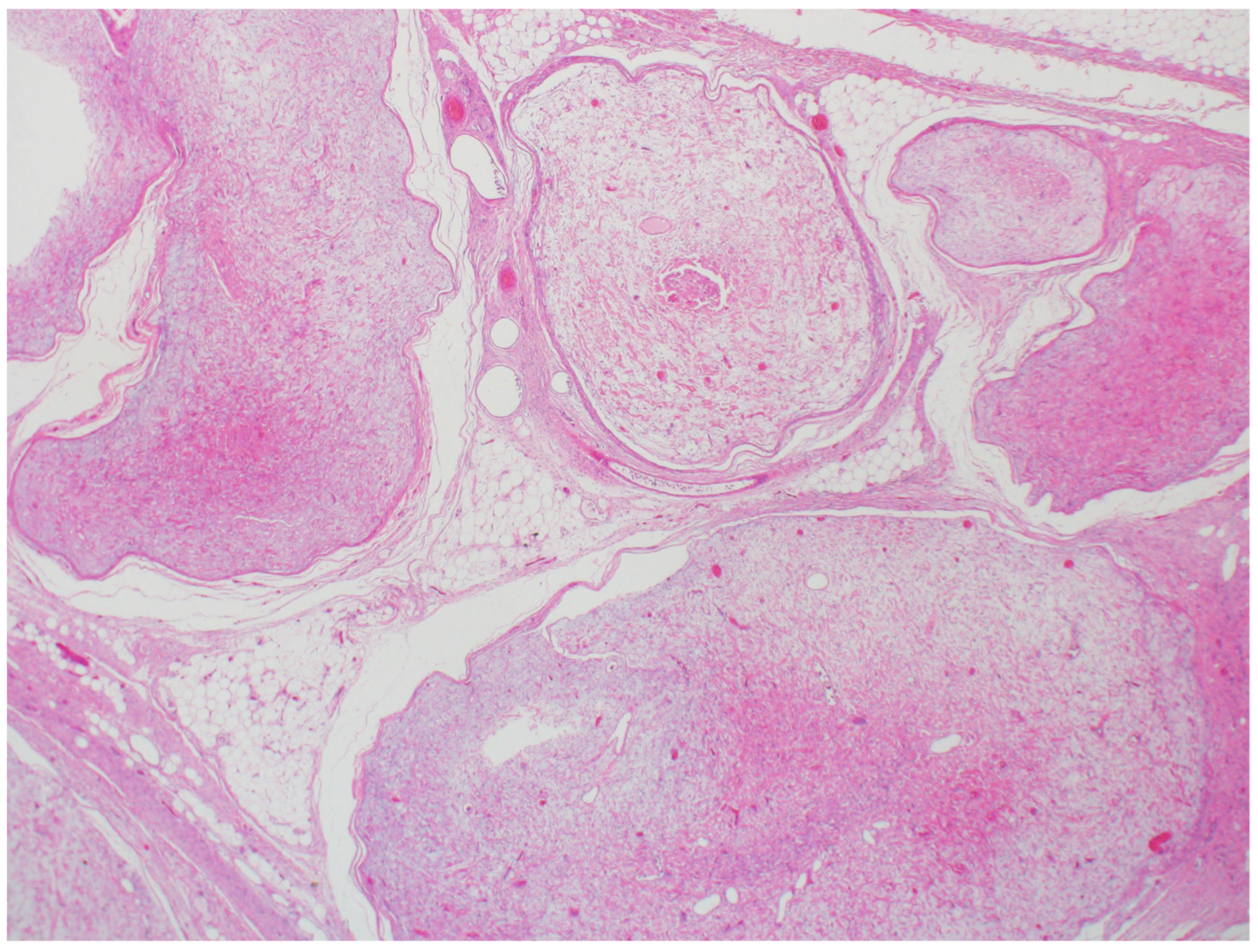
Clinically, neurofibromas typically begin to appear during adolescence and increase in number and size with age. Cutaneous neurofibromas are soft, skin-colored to violaceous papules or nodules with a characteristic “buttonhole” invagination upon palpation. Plexiform neurofibromas, which involve multiple nerve fascicles in a tortuous, infiltrative manner, are often congenital and considered pathognomonic for NF1 [72]. These lesions carry a 10% lifetime risk of malignant transformation into malignant peripheral nerve sheath tumors (MPNST), particularly when rapidly enlarging or associated with pain or neurologic changes. Histologically, neurofibromas are unencapsulated and composed of S100-positive Schwann cells and CD34-positive fibroblasts within a myxoid or collagenous stroma. Immunohistochemistry may also demonstrate CD117-positive mast cells and EMA-positive perineurial cells [73]. Plexiform subtypes show a more diffuse, infiltrative growth along large nerve trunks and may involve adjacent soft tissue and skin (Figure 12).
NF1 is a multisystem disorder with complete penetrance and highly variable expressivity. The diagnostic criteria include two or more of the following: six or more café-au-lait macules (>5 mm in prepubertal or >15 mm in postpubertal individuals), axillary or inguinal freckling, two or more neurofibromas of any type or one plexiform neurofibroma, optic pathway glioma, two or more iris Lisch nodules (iris hamartomas), distinctive osseous lesions such as sphenoid wing dysplasia or tibial pseudoarthrosis, and a first-degree relative with NF1 [74]. Lisch nodules are seen in over 90% of adults with NF1 and are considered a sensitive ophthalmologic finding [73]. Additional systemic features may include learning disabilities, skeletal dysplasia, vascular abnormalities (e.g., renal artery stenosis, hypertension), and increased risk of malignancies including MPNSTs, breast cancer (especially in women under 50), and gastrointestinal stromal tumors [73,75].
Cutaneous neurofibromas, while benign, are a major cause of disfigurement and quality-of-life burden in patients with NF1. Their number increases throughout life and may surge during puberty or pregnancy due to hormonal influences [76]. Ongoing surveillance and multidisciplinary management, including regular dermatologic, ophthalmologic, neurologic, and genetic evaluations, are essential for early detection and management of complications in this complex, lifelong condition. The presence of multiple neurofibromas or a plexiform neurofibromas should prompt consideration of NF1.

6. Smooth Muscle Tumors

Leiomyomas

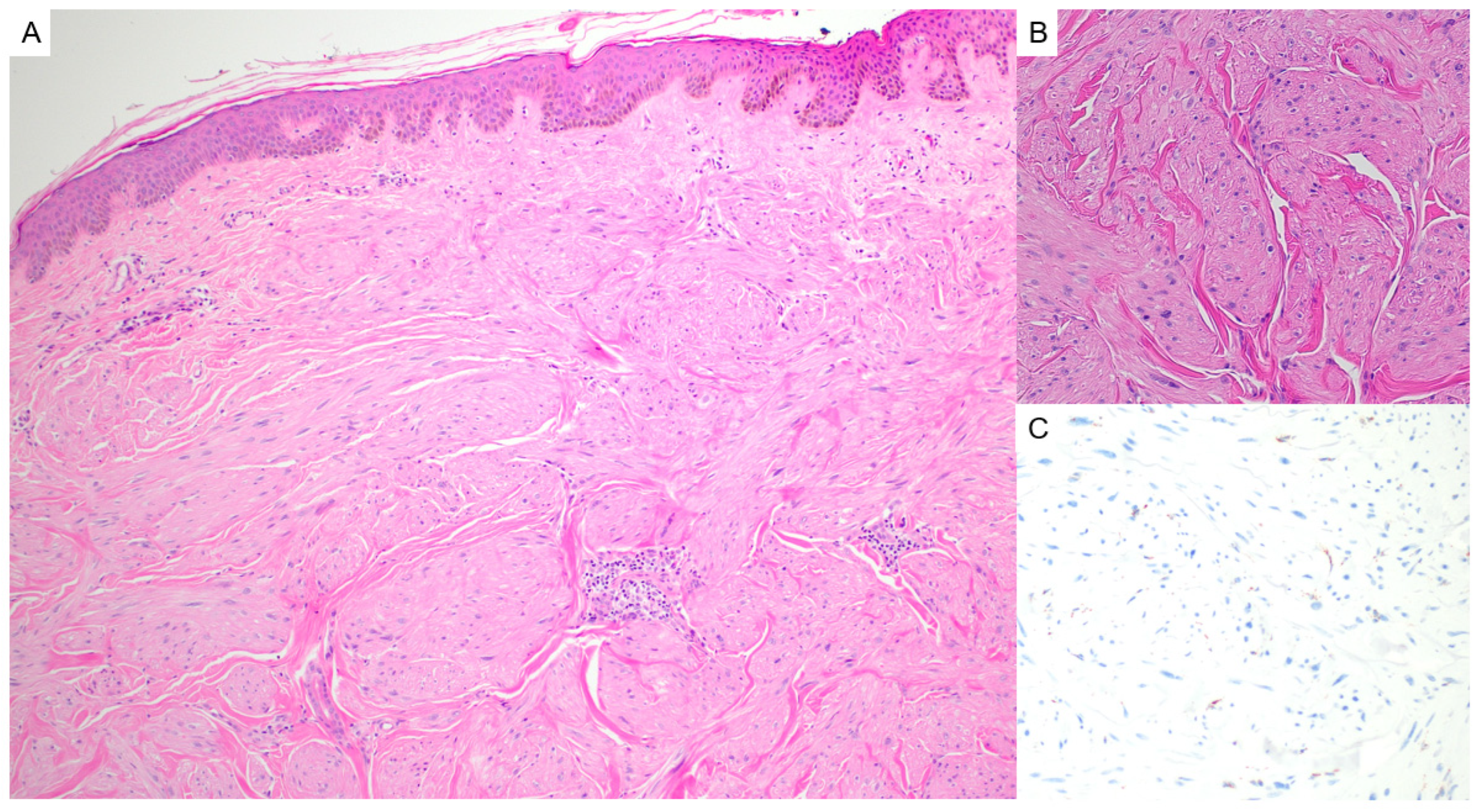
Leiomyomas are benign smooth muscle tumors that may occur sporadically or in association with genetic syndromes [77]. Clinically, cutaneous leiomyomas present as firm, skin-colored to erythematous papules or nodules, often painful to touch or with cold exposure [78]. Histologically, these tumors are composed of interlacing bundles of spindle cells with eosinophilic cytoplasm and blunt-ended nuclei, consistent with smooth muscle differentiation (Figure 13) [79]. Immunohistochemically, they are positive for smooth muscle markers such as desmin, caldesmon and smooth muscle actin (SMA) [80].
In the syndromic context, multiple cutaneous and uterine leiomyomas may indicate Hereditary Leiomyomatosis and Renal Cell Cancer (HLRCC, OMIM #150800), also known as Reed syndrome. This autosomal dominant condition is caused by germline mutations in the FH gene, located on chromosome 1q43, which encodes fumarate hydratase (FH), a key enzyme in the Krebs cycle. Loss of FH function leads to metabolic dysregulation and increased risk for malignancy, particularly type 2 papillary renal cell carcinoma [81]. Immunohistochemical staining for FH can demonstrate loss of cytoplasmic expression in lesional cells, supporting a syndromic diagnosis. Additionally, affected tumors may show positive staining for 2-succinocysteine (2SC), a surrogate marker for FH deficiency [82].
Given the potential for life-threatening, aggressive renal cancer, recognition of multiple cutaneous leiomyomas with loss of FH expression and a history of uterine leiomyomas should prompt evaluation for HLRCC, including genetic counseling, imaging surveillance, and screening of at-risk family members [83].

7. Vascular Tumors

7.1. Angiokeratoma Corporis Diffusum

Angiokeratoma corporis diffusum is a vascular lesion that presents in childhood as clusters of dark red to black papules, typically distributed over the lower trunk, buttocks, and thighs. These lesions represent dilated capillaries in the superficial dermis with overlying epidermal hyperkeratosis. Histologically, they reveal telangiectatic blood vessels in the papillary dermis, and can be accompanied by epidermal acanthosis and hyperkeratosis (Figure 14). Angiokeratoma corporis diffusum may demonstrate PAS-positive and Sudan black-positive lipid granules within endothelial cells, pericytes, and fibroblasts on frozen section examination.
Angiokeratoma corporis diffusum is a hallmark of Anderson–Fabry disease (OMIM #301500), a rare X-linked recessive lysosomal storage disorder caused by deficiency of the enzyme α-galactosidase A. The GLA gene, located on chromosome Xq22, is responsible for encoding this enzyme, and its deficiency leads to systemic accumulation of globotriaosylceramide in various tissues. Ultrastructural examination of tissue by electron microscopy, particularly kidney biopsies, reveals the pathognomonic presence of lamellar cytoplasmic inclusions, often described as “zebra bodies” [84,85].
In addition to cutaneous findings, Anderson–Fabry disease is associated with a range of systemic features, including renal failure, cardiomyopathy, acroparesthesias, and cerebrovascular disease. Rarely, angiokeratoma corporis diffusum may occur with deficiencies of other lysosomal enzymes such as α-L-fucosidase (fucosidosis, OMIM #230000) or β-mannosidase (β-mannosidosis, OMIM #248510), but Anderson–Fabry disease remains the most common association [86,87]. Recognition of a syndromic association and early diagnosis is critical, as enzyme replacement therapy may slow disease progression and improve quality of life [88].

7.2. Other Vascular Anomalies

The International Society for the Study of Vascular Anomalies classification system highlights an exhaustive list of other vascular tumors and malformations with syndromic associations [89]. These include vascular anomalies associated with Sturge–Weber syndrome (OMIM# 185300), Capillary Malformation-Arteriovenous Malformation syndromes (OMIM# 608354 and 618196), Hereditary Hemorrhagic Telangiectasia syndrome (OMIM# 175050), Klippel–Trenaunay syndrome (OMIM# 149000), and Proteus syndrome (OMIM# 158350) [89]. Further details on these vascular lesions and syndromes are outside the scope of this review.

8. Adipocytic Tumors

Lipomas

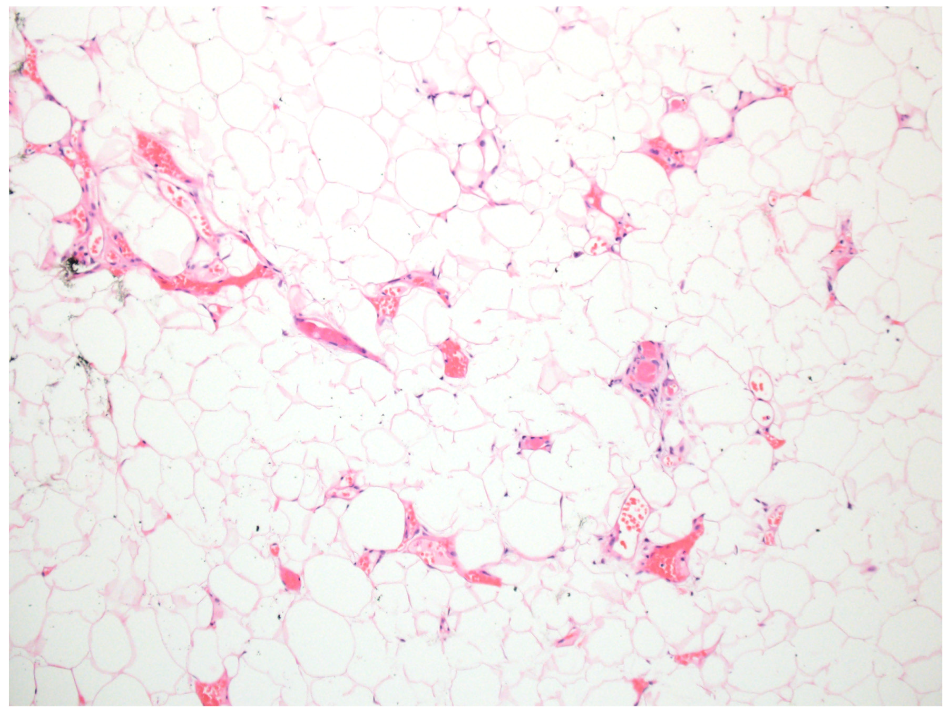
Lipomas are benign tumors composed of mature adipocytes and are the most common soft tissue neoplasms. Clinically, they appear as soft, mobile, subcutaneous nodules that are typically painless and slow-growing. Histological examination demonstrates mature adipose tissue (Figure 15). Multiple lipomas may arise sporadically or suggest an underlying genetic syndrome.
Several syndromes are associated with multiple lipomas, including Cowden syndrome (OMIM #158350), Proteus syndrome (OMIM #176920), and familial multiple lipomatosis (OMIM #151900). Cowden syndrome is an autosomal dominant disorder caused by mutations in the PTEN gene, located on chromosome 10q23.31, and is part of the PTEN hamartoma tumor syndrome spectrum. Affected individuals may have lipomas alongside trichilemmomas, oral papillomas, and an increased risk of breast, thyroid, and endometrial cancers [90]. Proteus syndrome is a rare, mosaic condition caused by somatic activating mutations in the AKT1 gene, located on chromosome 14q32.33. It is characterized by asymmetric overgrowth of bones, skin, and other tissues, including lipomas, connective tissue nevi, and vascular malformations [91]. Familial multiple lipomatosis is an autosomal dominant condition associated with variants in exon 5 of the HMGA2 gene, located on chromosome 12q14-15 [92]. It is characterized by the presence of multiple, slow-growing lipomas and angiolipomas on the trunk and limbs and increased body habitus.
Recognition of multiple lipomas in the context of the syndromic features mentioned should prompt genetic evaluation and multidisciplinary management.

9. Fibrohistiocytic/Fibrovascular Tumors

9.1. Superficial Angiomyxoma

Superficial angiomyxoma is a rare, benign cutaneous mesenchymal tumor. Clinically, these lesions present as polypoid or nodular masses, often on the trunk, head, or neck. Histologically, they are characterized by a well-circumscribed but non-encapsulated proliferation of spindle or stellate cells in a prominent myxoid matrix, interspersed with small, thin-walled blood vessels, neutrophils, and occasional entrapped benign epithelial elements (Figure 16). Immunohistochemistry typically shows positivity for CD34 [93].
Superficial angiomyxomas can occur sporadically or in association with Carney complex (OMIM #160980), an autosomal dominant multiple neoplasia syndrome. Carney complex is caused by mutations in the PRKAR1A gene, which encodes a regulatory subunit of protein kinase A and is located on chromosome 17q24. In this context, superficial angiomyxomas may be multiple and recurrent. Carney complex is also associated with cardiac myxomas, spotty skin pigmentation (lentigines), endocrine tumors (e.g., pituitary adenomas, adrenal tumors), and other myxoid neoplasms [94,95,96]. Recognition of this tumor, especially in younger individuals or in the presence of other stigmata, should prompt consideration of syndromic evaluation and genetic counselling [83].

9.2. Facial Angiofibromas/Acral Fibrokeratomas

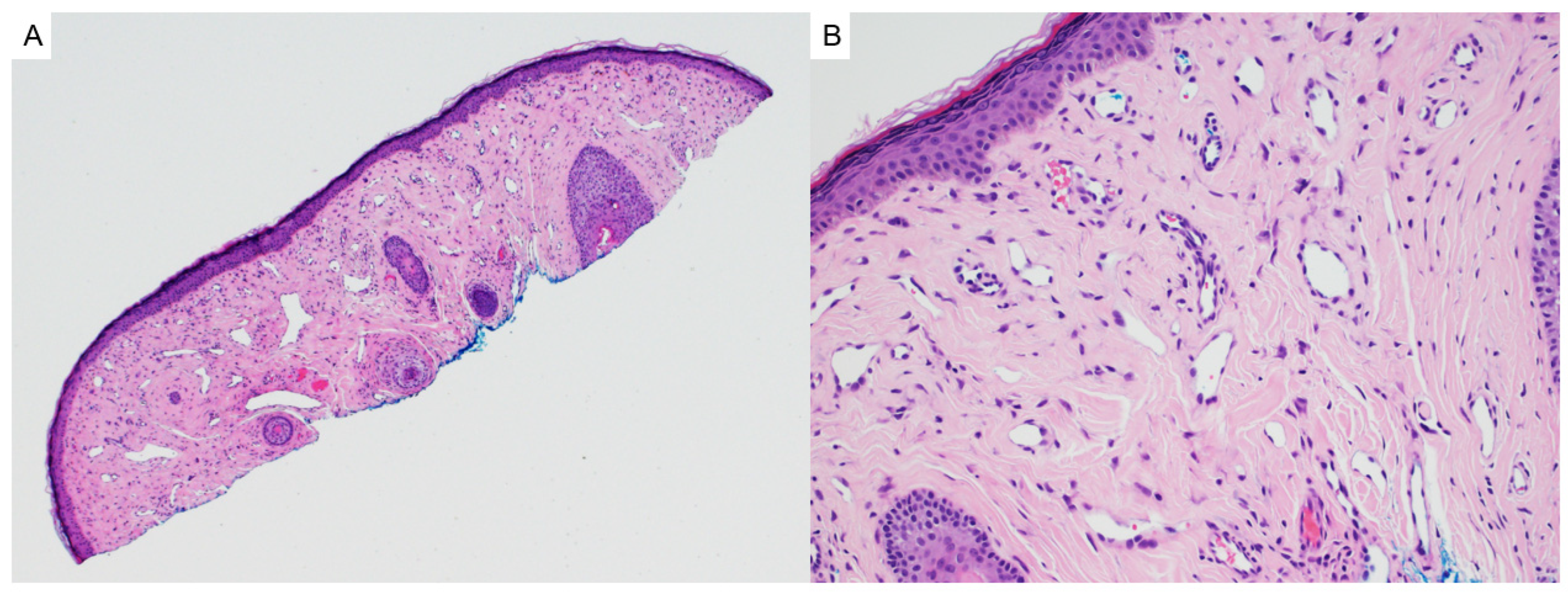
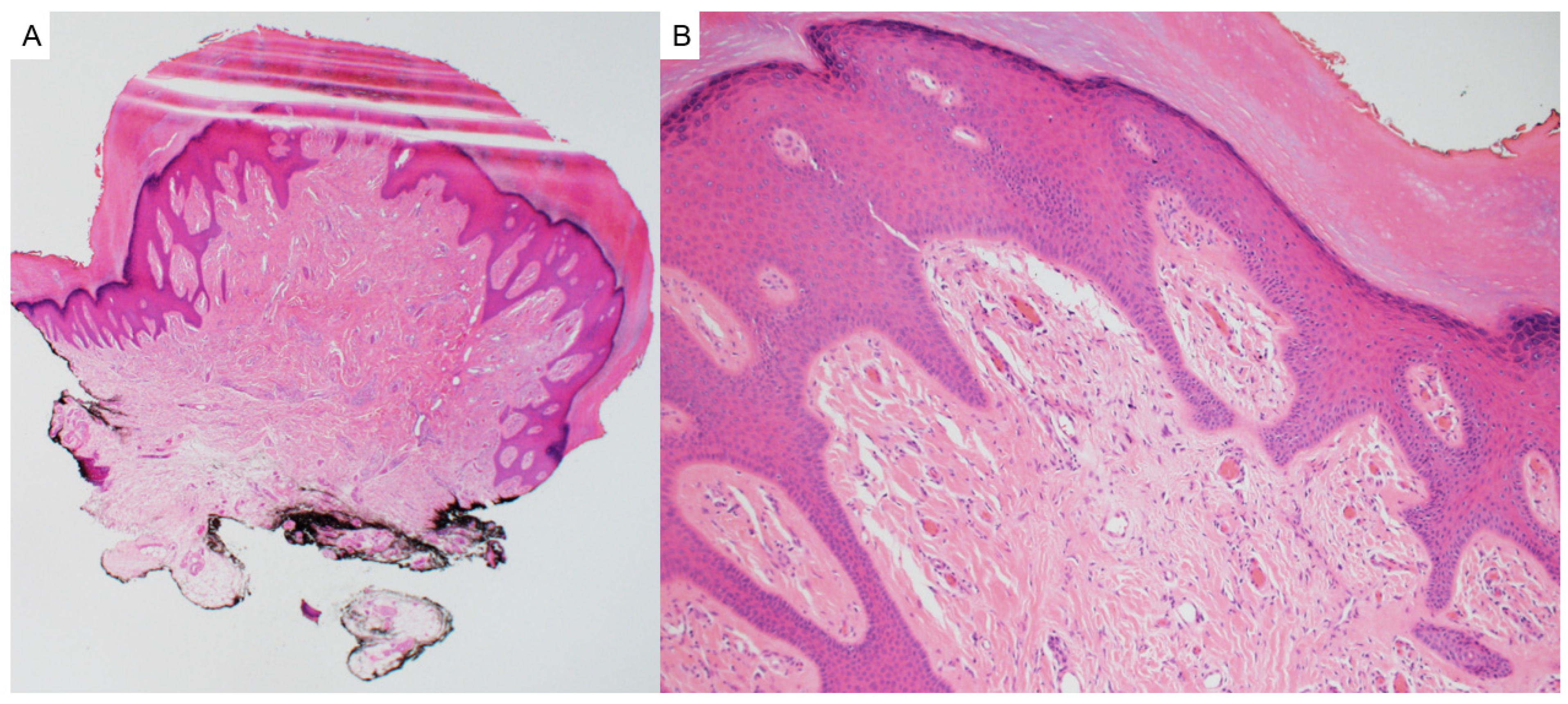
Facial angiofibromas (adenoma sebaceum) and acral fibrokeratomas can be associated with tuberous sclerosis complex (TSC), a multisystem genetic disorder. Angiofibromas associated with TSC typically manifest as multiple small, reddish papules distributed symmetrically across the central face, particularly the nasolabial folds and cheeks [97,98]. Acral fibrokeratomas, or “Koenen tumors”, are periungual or subungual fibromas that emerge in adolescence or adulthood and may be painful or disfiguring [99].
Histologically, angiofibromas lesions demonstrate scattered bland dermal fibroblasts and collagen bundles with prominent vasculature (Figure 17). Acral fibrokeratomas demonstrate a polypoid lesion with hyperkeratosis, acanthosis, dermal fibroblasts, thick collagen bundles, and scattered blood vessels (Figure 18). Immunohistochemistry findings are generally nonspecific but may aid in ruling out mimics [100].
Tuberous sclerosis complex is caused by mutations in either the TSC1 (hamartin, located on chromosome 9q34) (OMIM #191100) or TSC2 (tuberin, located on chromosome 16p13.3) (OMIM #613254) genes and follows an autosomal dominant inheritance pattern with variable expressivity. The syndrome is associated with a wide spectrum of manifestations, including cortical tubers, subependymal giant cell astrocytomas, renal angiomyolipomas, cardiac rhabdomyomas, and pulmonary lymphangioleiomyomatosis [101]. Multiple clustered angiofibromas and/or acral fibrokeratomas, especially in young patients, should prompt consideration of these syndromes.

10. Melanocytic Tumors

10.1. BAPomas

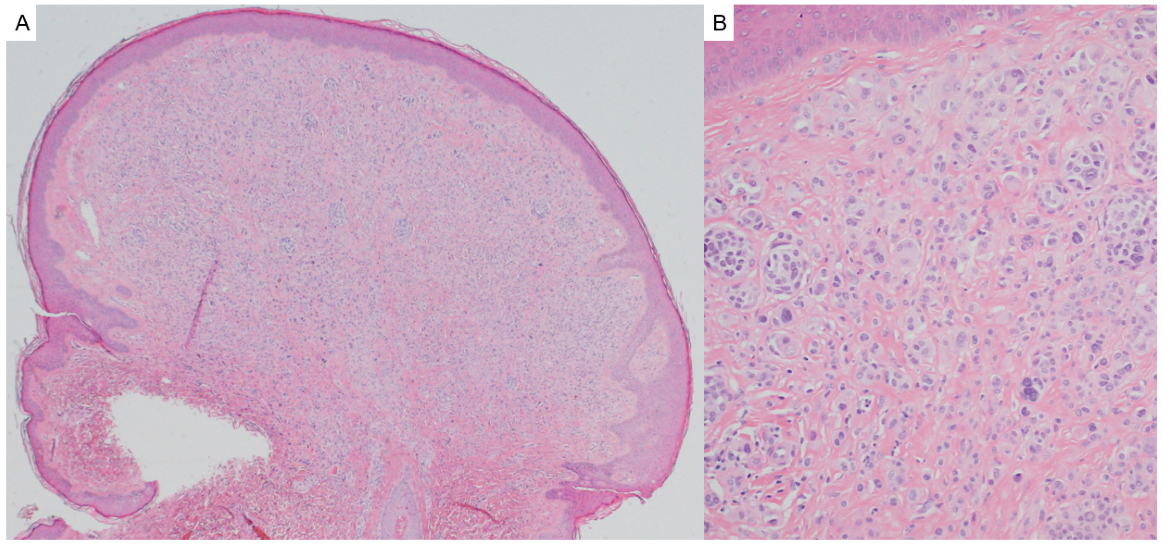
BAP-1 inactivated melanocytic tumors (BAPomas) are generally indolent melanocytic tumors associated with inactivating mutations in the BAP1 gene (BRCA1-associated protein-1), a tumor suppressor gene on chromosome 3p21.1 [102]. Clinically, BAPomas usually present as dome-shaped, skin-colored to lightly pigmented papules that may resemble banal nevi and may arise anywhere on the body. Histologically, BAPomas are distinguished by a predominantly intradermal proliferation of epithelioid melanocytes with abundant cytoplasm, well-defined cellular borders, vesicular nuclei, distinct nucleoli, and occasional multinucleation (Figure 19) [103,104]. They may arise in association with a conventional nevus. Loss of nuclear BAP1 expression on immunohistochemistry is helpful in confirming a diagnosis of BAPoma [105].
BAPomas may occur sporadically or may arise in association with BAP1 tumor predisposition syndrome (BAP1-TPDS, OMIM #614327). BAP1-TPDS is an autosomal dominant disorder caused by germline BAP1 mutations [102]. BAPomas are frequently multiple and appear in early adulthood in BAP1-TPDS. BAP1-TPDS is associated with an increased risk for uveal melanoma, mesothelioma, cutaneous melanoma, and renal cell carcinoma [106]. Identification of multiple BAPomas in a patient with a personal or family history suggestive of BAP1-TPDS should prompt genetic testing and surveillance for associated malignancies.

10.2. Pigmented Epithelioid Melanocytoma

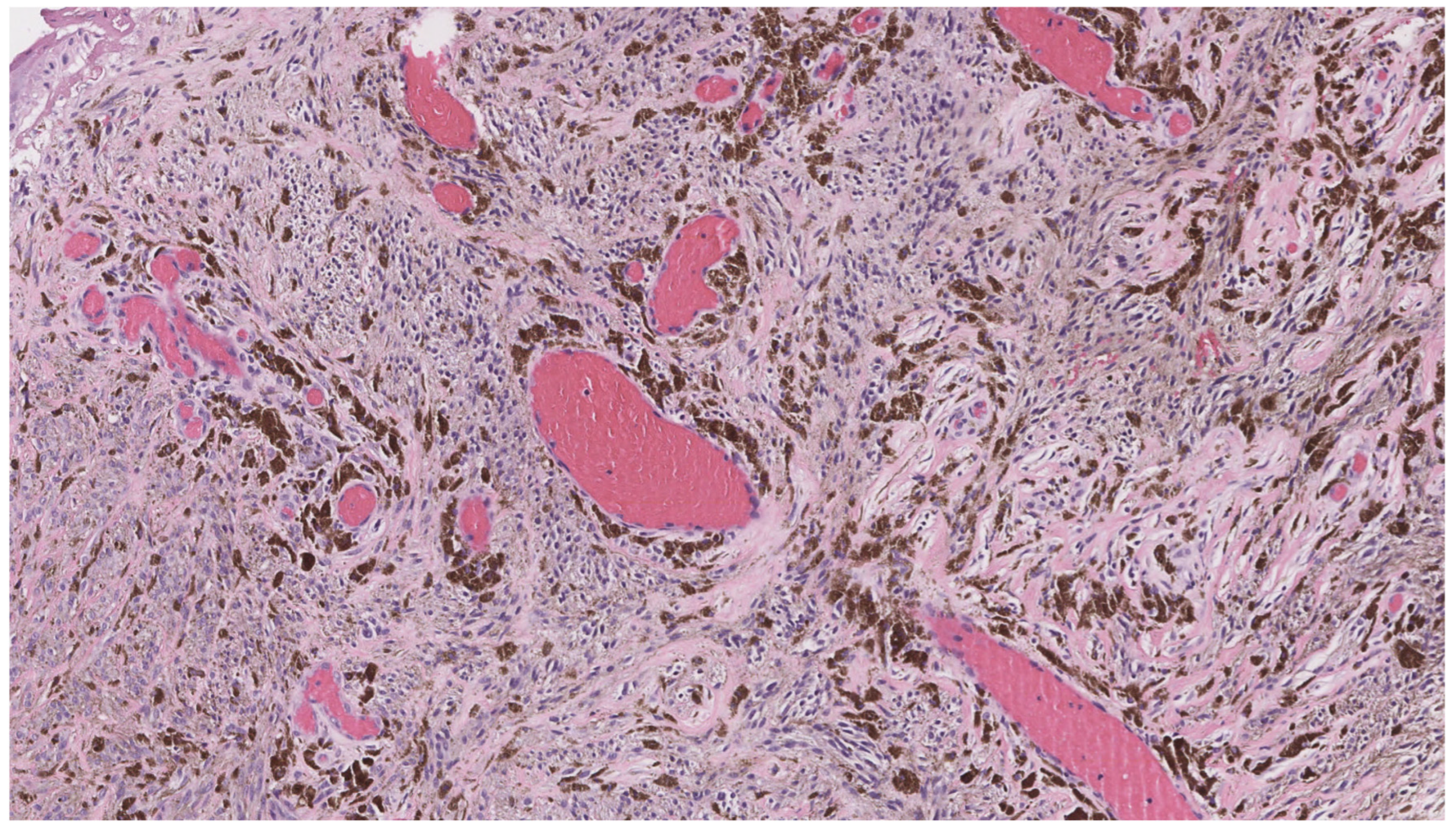
Pigmented epithelioid melanocytoma (PEM), also known as epithelioid blue nevus-like tumor, is a rare, deeply pigmented melanocytic neoplasm characterized by low to intermediate malignant potential and frequent sentinel lymph node involvement, though distant metastasis is rare [107,108]. Clinically, it presents as a darkly pigmented nodule, typically located on the extremities or trunk [108,109]. Histologically, PEM is composed of heavily pigmented epithelioid and spindle-shaped melanocytes arranged in nests or sheets within the dermis (Figure 20). Immunohistochemistry demonstrating loss of PRKAR1A expression can be useful for confirming a diagnosis of PEM [110].
PEM can occur sporadically or be associated with Carney complex (OMIM #160980), a multiple neoplasia syndrome caused by mutations in the PRKAR1A gene on chromosome 17q24 [111]. In Carney complex, PEMs may occur alongside cardiac and cutaneous myxomas, endocrine tumors, schwannomas, and lentigines. Carney complex is inherited in an autosomal dominant fashion [112]. The presence of multiple PEMs should prompt consideration for a syndromic association.

10.3. Dysplastic/Atypical Nevi

Dysplastic/atypical nevi, formerly known as “Clark’s nevi”, demonstrate clinical atypia compared to ordinary nevi, including larger size, irregular borders, and color variation [113]. On histology, they are characterized by the presence of architectural disorder (shoulder phenomenon, bridging of junctional nests between elongated rete ridges, pagetoid scatter, and lentiginous growth), cytologic atypia (enlarged and pleomorphic nuclei, hyperchromasia, and prominent nucleoli), and host response (lamellar and concentric fibroplasia of the papillary dermis and a patchy superficial lymphocytic infiltrate) [114]. Multiple dysplastic/atypical nevi may be associated with Familial Atypical Multiple Mole and Melanoma Syndrome (OMIM# 155600), a typically autosomal dominant disorder caused by inherited mutations in tumor suppressor genes associated with melanoma susceptibility, such as CDKN2A, CDK4, and ARF [115]. Individuals with these syndromes have a significantly increased risk for the development of melanoma and an increased risk for the development of internal malignancies, especially pancreatic carcinoma [115,116,117]. The presence of multiple dysplastic/atypical nevi in an individual should prompt regular dermatologic surveillance for the early detection and management of melanoma in these patients and their families and consideration for genetic testing.

11. Conclusions

Benign skin tumors with syndromic associations represent a valuable and often underrecognized opportunity for the early identification of genetic syndromes. Though often overlooked due to their innocuous appearance, these lesions can display subtle, yet diagnostically meaningful features on histopathologic evaluation, particularly when interpreted with clinical context [118]. When such findings present in multiples, demonstrate distinct morphological features, or occur in younger individuals or with a relevant family history, a syndromic etiology should be considered [2]. Timely recognition in these cases is critical, as it allows for risk stratification and surveillance for associated internal malignancies [3].
This review synthesizes the clinical and histological features of benign cutaneous neoplasms with known syndromic associations (Table 1). In this diagnostic process, dermatopathologists play a pivotal role. Their careful evaluation, supported by ancillary testing, is often essential for uncovering the systemic implications of these lesions even in the absence of overt clinical signs [119]. Once a syndromic diagnosis is established, patient management should include regular surveillance for associated malignancies and referral for genetic counselling for both the patient and at-risk family members [2]. Notably, in several syndromes, including Muir–Torre, Cowden, and Birt–Hogg–Dubé, the skin may be the earliest or only organ involved at presentation. Failure to recognize these early cutaneous signs within their syndromic context may delay critical interventions and allow for progression of occult malignancies [120,121]. It is thus paramount that dermatopathologists pay attention to clinical and histological clues in these lesions that may suggest a syndrome and guide the referring clinician to consider further screening and genetic counselling for these patients and their families.

Author Contributions

S.L. and C.M.: Writing—Original Draft Preparation. P.G.: Conceptualization & Design, Writing—Original Draft Preparation, Review & Editing, and Figure Preparation. All authors have read and agreed to the published version of the manuscript.

Funding

This research received no external funding.

Acknowledgments

ChatGPT (OpenAI, GPT-4.0) was used by the author CM for parts of Section 1, Section 2, Section 3, Section 4 and Section 5 in an iterative manner for language editing, which included improvements for grammar, clarity of phrasing, and readability. The software did not contribute to the research tasks of identifying, interpreting, or synthesizing literature. Suggested edits were checked by CM for accuracy before incorporation. Microsoft Copilot was used by the author SL during the manuscript preparation process for parts of Section 5, Section 6, Section 7, Section 8 and Section 9 to assist with grammar correction, sentence restructuring, and enhancing clarity. The tool was applied iteratively to refine phrasing and improve readability across these sections. All substantive content, including the selection, synthesis, and discussion/analysis of literature was conducted without the assistance of Copilot or other artificial intelligence (AI) tools. All AI-suggested edits were reviewed and approved by SL to ensure accuracy and appropriateness. The conceptualization and design of this manuscript and all aspects of this manuscript drafted by PG were done without the use of AI tools. Furthermore, the sections prepared by CM and SL were reviewed and revised by PG without the use of AI tools. The figures and table were created without the use of AI tools.

Conflicts of Interest

The authors declare no conflicts of interest.

References

  1. Gruber, V.; Hofmann-Wellenhof, R.; Wolf, P.; Hofmann-Wellenhof, E.L.; Schmidt, H.; Berghold, A.; Wedrich, A. Common Benign Melanocytic and Non-Melanocytic Skin Tumors among the Elderly: Results of the Graz Study on Health and Aging. Dermatology 2023, 239, 379–386. [Google Scholar] [CrossRef]
  2. Tidman, A.S. Be vigilant for skin manifestations of inherited cancer syndromes. Practitioner 2017, 261, 23–27. Available online: http://www.ncbi.nlm.nih.gov/pubmed/29023082 (accessed on 10 June 2025).
  3. Shen, Z.; Hoffman, J.D.; Hao, F.; Pier, E. More than just skin deep: Faciocutaneous clues to genetic syndromes with malignancies. Oncologist 2012, 17, 930–936. [Google Scholar] [CrossRef]
  4. Schmidt, L.S.; Linehan, W.M. Molecular genetics and clinical features of Birt-Hogg-Dubé syndrome. Nat. Rev. Urol. 2015, 12, 558–569. [Google Scholar] [CrossRef] [PubMed]
  5. Misago, N.; Kimura, T.; Narisawa, Y. Fibrofolliculoma/trichodiscoma and fibrous papule (perifollicular fibroma/angiofibroma): A revaluation of the histopathological and immunohistochemical features. J. Cutan. Pathol. 2009, 36, 943–951. [Google Scholar] [CrossRef]
  6. Kutzner, H.; Requena, L.; Rütten, A.; Mentzel, T. Spindle cell predominant trichodiscoma: A fibrofolliculoma/trichodiscoma variant considered formerly to be a neurofollicular hamartoma: A clinicopathological and immunohistochemical analysis of 17 cases. Am. J. Dermatopathol. 2006, 28, 1–8. [Google Scholar] [CrossRef] [PubMed]
  7. Scalvenzi, M.; Argenziano, G.; Sammarco, E.; Delfino, M. Hereditary multiple fibrofolliculomas, trichodiscomas and acrochordons: Syndrome of Birt-Hogg-Dubè. J. Eur. Acad. Dermatol. Venereol. 1998, 11, 45–47. Available online: http://www.ncbi.nlm.nih.gov/pubmed/9731965 (accessed on 23 May 2025). [CrossRef] [PubMed]
  8. Wong, T.Y.; Suster, S.; Cheek, R.F.; Mihm, M.C. Benign cutaneous adnexal tumors with combined folliculosebaceous, apocrine, and eccrine differentiation. Clinicopathologic and immunohistochemical study of eight cases. Am. J. Dermatopathol. 1996, 18, 124–136. [Google Scholar] [CrossRef]
  9. Gustafson, S.; Zbuk, K.M.; Scacheri, C.; Eng, C. Cowden syndrome. Semin. Oncol. 2007, 34, 428–434. [Google Scholar] [CrossRef]
  10. Kaptan, M.A.; Kattampallil, J.; Rosendahl, C. Trichilemmoma in continuity with pigmented basal cell carcinoma; with dermatoscopy and dermatopathology. Dermatol. Pract. Concept. 2015, 5, 57–59. [Google Scholar] [CrossRef]
  11. Tardío, J.C. CD34-reactive tumors of the skin. An updated review of an ever-growing list of lesions. J. Cutan. Pathol. 2009, 36, 89–102. [Google Scholar] [CrossRef]
  12. Ung, T.; Tan, J.H.; Mudhar, H. Three patients with desmoplastic tricholemmoma with an incidental histological surprise impacting on management. Br. J. Ophthalmol. 2012, 96, 461–462. [Google Scholar] [CrossRef]
  13. Romano, C.; Schepis, C. PTEN gene: A model for genetic diseases in dermatology. Sci. World J. 2012, 2012, 252457. [Google Scholar] [CrossRef]
  14. Al-Zaid, T.; Ditelberg, J.S.; Prieto, V.G.; Lev, D.; Luthra, R.; Davies, M.A.; Diwan, A.H.; Wang, W.; Lazar, A.J. Trichilemmomas show loss of PTEN in Cowden syndrome but only rarely in sporadic tumors. J. Cutan. Pathol. 2012, 39, 493–499. [Google Scholar] [CrossRef] [PubMed]
  15. Hobert, J.A.; Eng, C. PTEN hamartoma tumor syndrome: An overview. Genet. Med. 2009, 11, 687–694. [Google Scholar] [CrossRef] [PubMed]
  16. Hernández-Núñez, A.; Nájera Botello, L.; Romero Maté, A.; Martínez-Sánchez, C.; Busquets, M.U.; Komáromy, A.C.; Martínez, J.B. Retrospective study of pilomatricoma: 261 tumors in 239 patients. Actas Dermo-Sifiliográficas 2014, 105, 699–705. [Google Scholar] [CrossRef]
  17. Neema, S.; Kashif, A.W.; Vasudevan, B. Dermoscopy of Pilomatrixoma. Indian Dermatol. Online J. 2023, 14, 450–451. [Google Scholar] [CrossRef] [PubMed]
  18. Thompson, L.D. Pilomatricoma. Ear Nose Throat J. 2012, 91, 18–20. [Google Scholar] [CrossRef]
  19. Di Brizzi, E.V.; Piccolo, V.; Moscarella, E.; Pellerone, S.; Argenziano, G. Dermoscopy of pilomatricoma: Retrospective descriptive study on 35 paediatric patients. J. Eur. Acad. Dermatol. Venereol. 2025, 39, e141–e143. [Google Scholar] [CrossRef]
  20. Alnaqshanbandi, S.M.; McAfee, J.L.; Ko, J.S.; Billings, S.D.; Ronen, S. Role of Immunohistochemistry in the Diagnosis of Pilomatrical Tumors. Am. J. Surg. Pathol. 2024, 48, 1543–1550. [Google Scholar] [CrossRef]
  21. Pujol, R.M.; Casanova, J.M.; Egido, R.; Pujol, J.; de Moragas, J.M. Multiple familial pilomatricomas: A cutaneous marker for Gardner syndrome? Pediatr. Dermatol. 1995, 12, 331–335. [Google Scholar] [CrossRef]
  22. Ciriacks, K.; Knabel, D.; Waite, M.B. Syndromes associated with multiple pilomatricomas: When should clinicians be concerned? Pediatr. Dermatol. 2020, 37, 9–17. [Google Scholar] [CrossRef]
  23. Xin, T.Y.; Saniasiaya, J.; Kulasegarah, J.; Fan, C.S. Commonly Misdiagnosed Facial Lesion: Pilomatricoma. Acta Medica 2023, 66, 158–160. [Google Scholar] [CrossRef] [PubMed]
  24. Wang, Z.; Tan, X.; Yang, X.; Hu, H.; Lin, K.; Wang, C.; Fu, H.; Zhang, J. Retrosynthetic analysis via deep learning to improve pilomatricoma diagnoses. Comput. Biol. Med. 2024, 182, 109152. [Google Scholar] [CrossRef] [PubMed]
  25. Guerreiro, F.P.; Martins, A.I.; Costa e Silva, J.; Teixeira, N.; Ramos, J.N. Multiple Trichoepithelioma Syndrome: A Case Report. Cureus 2023, 15, e42930. [Google Scholar] [CrossRef] [PubMed]
  26. Nagy, N.; Dubois, A.; Szell, M.; Rajan, N. Genetic Testing in CYLD Cutaneous Syndrome: An Update. Appl. Clin. Genet. 2021, 14, 427–444. [Google Scholar] [CrossRef]
  27. Kazakov, D.V. Brooke-Spiegler Syndrome and Phenotypic Variants: An Update. Head Neck Pathol. 2016, 10, 125–130. [Google Scholar] [CrossRef]
  28. Mohammadi, A.A.; Seyed Jafari, S.M. Trichoepithelioma: A rare but crucial dermatologic issue. World J. Plast. Surg. 2014, 3, 142–145. Available online: http://www.ncbi.nlm.nih.gov/pubmed/25489539 (accessed on 5 August 2025).
  29. Rahman, J.; Tahir, M.; Arekemase, H.; Murtazaliev, S.; Sonawane, S. Desmoplastic Trichoepithelioma: Histopathologic and Immunohistochemical Criteria for Differentiation of a Rare Benign Hair Follicle Tumor From Other Cutaneous Adnexal Tumors. Cureus 2020, 12, e9703. [Google Scholar] [CrossRef]
  30. Gill, P.; Naugler, C.; Abi Daoud, M.S. Utility of Ber-EP4 and MOC-31 in Basaloid Skin Tumor Detection. Appl. Immunohistochem. Mol. Morphol. 2019, 27, 584–588. [Google Scholar] [CrossRef]
  31. Dubois, A.; Rajan, N. CYLD Cutaneous Syndrome; University of Washington: Seattle, WA, USA, 1993. Available online: https://pubmed.ncbi.nlm.nih.gov/24838222/ (accessed on 5 August 2025).
  32. Barbieux, S.; Jouenne, F.; Machet, M.-C.; Fraitag, S.; Macagno, N.; Battistella, M.; Cribier, B.; Sohier, P.; Laurent-Roussel, S.; Carlotti, A.; et al. Re-evaluation of the concept of basaloid follicular hamartoma associated with naevoid basal cell carcinoma syndrome: A morphological, immunohistochemical and molecular study. Pathology 2025, 57, 49–56. [Google Scholar] [CrossRef]
  33. Lee, D.A.; Grossman, M.E.; Schneiderman, P.; Celebi, J.T. Genetics of skin appendage neoplasms and related syndromes. J. Med. Genet. 2005, 42, 811–819. [Google Scholar] [CrossRef]
  34. Mills, O.; Thomas, L.B. Basaloid follicular hamartoma. Arch. Pathol. Lab. Med. 2010, 134, 1215–1219. [Google Scholar] [CrossRef]
  35. Jih, D.M.; Shapiro, M.; James, W.D.; Levin, M.; Gelfand, J.; Williams, P.T.; Oakey, R.J.; Fakharzadeh, S.; Seykora, J.T. Familial basaloid follicular hamartoma: Lesional characterization and review of the literature. Am. J. Dermatopathol. 2003, 25, 130–137. [Google Scholar] [CrossRef]
  36. Honarpisheh, H.; Glusac, E.J.; Ko, C.J. Cytokeratin 20 expression in basaloid follicular hamartoma and infundibulocystic basal cell carcinoma. J. Cutan. Pathol. 2014, 41, 916–921. [Google Scholar] [CrossRef]
  37. Gumaste, P.; Ortiz, A.E.; Patel, A.; Baron, J.; Harris, R.; Barr, R. Generalized basaloid follicular hamartoma syndrome: A case report and review of the literature. Am. J. Dermatopathol. 2015, 37, e37–e40. [Google Scholar] [CrossRef] [PubMed]
  38. Papadimitriou, I.; Vakirlis, E.; Sotiriou, E.; Bakirtzi, K.; Lallas, A.; Ioannides, D. Sebaceous Neoplasms. Diagnostics 2023, 13, 1676. [Google Scholar] [CrossRef]
  39. Eisen, D.B.; Michael, D.J. Sebaceous lesions and their associated syndromes: Part I. J. Am. Acad. Dermatol. 2009, 61, 549–560, quiz 561–562. [Google Scholar] [CrossRef]
  40. Cohen, P.R.; Kurzrock, R. Germline Testing of Mismatch Repair Genes Is Needed in the Initial Evaluation of Patients with Muir-Torre Syndrome-Associated Cutaneous Sebaceous Neoplasms: A Case Series. Cureus 2023, 15, e33975. [Google Scholar] [CrossRef] [PubMed]
  41. Gallon, R.; Holt, G.; Alfailakawi, W.; Husain, A.; Jones, C.; Sowter, P.; Santibanez-Koref, M.; Jackson, M.S.; Burn, J.; Cook, S.; et al. Novel microsatellite instability test of sebaceous tumours to facilitate low-cost universal screening for Lynch syndrome. Clin. Exp. Dermatol. 2025, 50, 1155–1162. [Google Scholar] [CrossRef] [PubMed]
  42. Task Force/Committee Members; Vidal, C.I.; Sutton, A.; Armbrect, E.A.; Lee, J.B.; Litzner, B.R.; Hurley, M.Y.; Panel, R.; Alam, M.; Duncan, L.M.; et al. Muir-Torre syndrome appropriate use criteria: Effect of patient age on appropriate use scores. J. Cutan. Pathol. 2019, 46, 484–489. [Google Scholar] [CrossRef]
  43. Mintsoulis, D.; Beecker, J. Muir-Torre syndrome. CMAJ 2016, 188, E95. [Google Scholar] [CrossRef] [PubMed]
  44. Jones, B.; Oh, C.; Mangold, E.; Egan, C.A. Muir-Torre syndrome: Diagnostic and screening guidelines. Australas. J. Dermatol. 2006, 47, 266–269. [Google Scholar] [CrossRef]
  45. Dores, G.M.; Curtis, R.E.; Toro, J.R.; Devesa, S.S.; Fraumeni, J.F. Incidence of cutaneous sebaceous carcinoma and risk of associated neoplasms: Insight into Muir-Torre syndrome. Cancer 2008, 113, 3372–3381. [Google Scholar] [CrossRef]
  46. Wang, J.; Qi, X.; Zhang, K.; Zhang, W. A case report of Muir-Torre syndrome (MTS) in a Chinese patient. BMC Ophthalmol. 2025, 25, 218. [Google Scholar] [CrossRef]
  47. Zhang, B.; Sun, L.; Fu, X.; Yu, G.; Liu, H.; Zhang, F. Mutation analysis of the KRT17 gene in steatocystoma multiplex and a brief literature review. Clin. Exp. Dermatol. 2020, 45, 132–134. [Google Scholar] [CrossRef]
  48. Brownstein, M.H. The genodermatopathology of adnexal tumors. J. Cutan. Pathol. 1984, 11, 457–465. [Google Scholar] [CrossRef] [PubMed]
  49. Georgakopoulos, J.R.; Ighani, A.; Yeung, J. Numerous asymptomatic dermal cysts: Diagnosis and treatment of steatocystoma multiplex. Can. Fam. Physician 2018, 64, 892–899. Available online: http://www.ncbi.nlm.nih.gov/pubmed/30541803 (accessed on 5 August 2025). [PubMed]
  50. Palaniappan, V.; Karthikeyan, K. Steatocystoma Multiplex. Indian Dermatol. Online J. 2024, 15, 105–112. [Google Scholar] [CrossRef]
  51. Smith, F.J.; Hansen, C.D.; Hull, P.R.; Kaspar, R.L.; McLean, W.H.I.; O’Toole, E.; Sprecher, E. Pachyonychia Congenita; University of Washington: Seattle, WA, USA, 1993. Available online: https://www.ncbi.nlm.nih.gov/books/NBK1280/ (accessed on 5 August 2025).
  52. Mohiuddin, W.; Laun, J.; Cruse, W. Brooke-Spiegler Syndrome. Eplasty 2018, 18, ic14. Available online: http://www.ncbi.nlm.nih.gov/pubmed/30093932 (accessed on 23 May 2025).
  53. Neema, S.; Sandhu, S.; Kashif, A.W. Dermoscopy of Cylindroma. Indian Dermatol. Online J. 2022, 13, 818–819. [Google Scholar] [CrossRef]
  54. Jordão, C.; de Magalhães, T.C.; Cuzzi, T.; Ramos-e-Silva, M. Cylindroma: An update. Int. J. Dermatol. 2015, 54, 275–278. [Google Scholar] [CrossRef] [PubMed]
  55. Cassarino, D.S.; Su, A.; Robbins, B.A.; Altree-Tacha, D.; Ra, S. SOX10 immunohistochemistry in sweat ductal/glandular neoplasms. J. Cutan. Pathol. 2017, 44, 544–547. [Google Scholar] [CrossRef]
  56. Macagno, N.; Sohier, P.; Kervarrec, T.; Pissaloux, D.; Jullie, M.-L.; Cribier, B.; Battistella, M. Recent Advances on Immunohistochemistry and Molecular Biology for the Diagnosis of Adnexal Sweat Gland Tumors. Cancers 2022, 14, 476. [Google Scholar] [CrossRef]
  57. Lobos, P.; Lobos, C.; Baksai, K. Reflectance confocal microscopy of spiradenoma. JAAD Case Rep. 2022, 24, 105–107. [Google Scholar] [CrossRef]
  58. Layegh, P.; Sharifi-Sistani, N.; Abadian, M.; Moghiman, T. Brooke-Spiegler syndrome. Indian J. Dermatol. Venereol. Leprol. 2008, 74, 632–634. [Google Scholar] [CrossRef]
  59. Lee, A.Y.; Kawashima, M.; Nakagawa, H.; Ishibashi, Y. Generalized eruptive syringoma. J. Am. Acad. Dermatol. 1991, 25, 570–571. [Google Scholar] [CrossRef]
  60. Lei, H.; Wang, Z.; Ma, X.; Zhang, Z.; Feng, Y.; Zheng, Y. Eruptive syringomas: Summary of ninety cases and a brief literature review. J. Cosmet. Dermatol. 2023, 22, 1128–1133. [Google Scholar] [CrossRef] [PubMed]
  61. Schepis, C.; Siragusa, M.; Palazzo, R.; Batolo, D.; Romano, C. Perforating milia-like idiopathic calcinosis cutis and periorbital syringomas in a girl with Down syndrome. Pediatr. Dermatol. 1994, 11, 258–260. [Google Scholar] [CrossRef]
  62. Lam, M.; Lu, J.D.; Elhadad, L.; Sibbald, C.; Alhusayen, R. Common Dermatologic Disorders in Down Syndrome: Systematic Review. JMIR Dermatol. 2022, 5, e33391. [Google Scholar] [CrossRef] [PubMed]
  63. Ranasinghe, A.; Rytina, E.; Batchelor, J.; Ha, T. Nicolau Balus syndrome with microcystic adnexal carcinoma. J. Am. Acad. Dermatol. 2013, 68, AB56. [Google Scholar] [CrossRef]
  64. Williams, K.; Shinkai, K. Evaluation and management of the patient with multiple syringomas: A systematic review of the literature. J. Am. Acad. Dermatol. 2016, 74, 1234–1240.e9. [Google Scholar] [CrossRef]
  65. Marzano, A.V.; Fiorani, R.; Girgenti, V.; Crosti, C.; Alessi, E. Familial syringoma: Report of two cases with a published work review and the unique association with steatocystoma multiplex. J. Dermatol. 2009, 36, 154–158. [Google Scholar] [CrossRef]
  66. Schepis, C.; Siragusa, M.; Palazzo, R.; Ragusa, R.M.; Massi, G.; Fabrizi, G. Palpebral syringomas and Down’s syndrome. Dermatology 1994, 189, 248–250. [Google Scholar] [CrossRef] [PubMed]
  67. Feingold, M. Syringomas in Down syndrome. Am. J. Dis. Child. 1991, 145, 966–967. [Google Scholar] [CrossRef]
  68. Rabin, K.R.; Whitlock, J.A. Malignancy in children with trisomy 21. Oncologist 2009, 14, 164–173. [Google Scholar] [CrossRef]
  69. Schaller, J.; Rytina, E.; Rütten, A.; Hendricks, C.; Ha, T.; Requena, L. Sweat duct proliferation associated with aggregates of elastic tissue and atrophodermia vermiculata: A simulator of microcystic adnexal carcinoma. Report of two cases. J. Cutan. Pathol. 2010, 37, 1002–1009. [Google Scholar] [CrossRef]
  70. Ly, I.; Romo, C.G.; Gottesman, S.; Kelly, K.M.; Kornacki, D.; York, Z.; Lee, S.Y.; Rhodes, S.D.; Staedtke, V.; Steensma, M.R.; et al. Target Product Profile for Cutaneous Neurofibromas: Clinical Trials to Prevent, Arrest, or Regress Cutaneous Neurofibromas. J. Invest. Dermatol. 2023, 143, 1388–1396. [Google Scholar] [CrossRef]
  71. Gerber, P.A.; Antal, A.S.; Neumann, N.J.; Homey, B.; Matuschek, C.; Peiper, M.; Budach, W.; Bölke, E. Neurofibromatosis. Eur. J. Med. Res. 2009, 14, 102–105. [Google Scholar] [CrossRef] [PubMed]
  72. Ortonne, N.; Wolkenstein, P.; Blakeley, J.O.; Korf, B.; Plotkin, S.R.; Riccardi, V.M.; Miller, D.C.; Huson, S.; Peltonen, J.; Rosenberg, A.; et al. Cutaneous neurofibromas: Current clinical and pathologic issues. Neurology. 2018, 91 (Suppl. 1), S5–S13. [Google Scholar] [CrossRef]
  73. Poplausky, D.; Young, J.N.; Tai, H.; Rivera-Oyola, R.; Gulati, N.; Brown, R.M. Dermatologic Manifestations of Neurofibromatosis Type 1 and Emerging Treatments. Cancers 2023, 15, 2770. [Google Scholar] [CrossRef] [PubMed]
  74. Duman, N.; Elmas, M. Dermoscopy of cutaneous neurofibromas associated with neurofibromatosis type 1. J. Am. Acad. Dermatol. 2015, 73, 529–531. [Google Scholar] [CrossRef] [PubMed]
  75. Ly, K.I.; Blakeley, J.O. The Diagnosis and Management of Neurofibromatosis Type 1. Med. Clin. N. Am. 2019, 103, 1035–1054. [Google Scholar] [CrossRef]
  76. Phillips, T.G.; Persia, O.R.; Jimenez Lopez, J.A. Neurofibromatosis type 1: More than skin deep. J. Fam. Pract. 2020, 69, 401–405. Available online: http://www.ncbi.nlm.nih.gov/pubmed/33175920 (accessed on 23 May 2025). [CrossRef]
  77. Marcoval, J.; Llobera-Ris, C.; Moreno-Vílchez, C.; Penín, R.M. Cutaneous Leiomyoma: A Clinical Study of 152 Patients. Dermatology 2022, 238, 587–593. [Google Scholar] [CrossRef]
  78. Malik, K.; Patel, P.; Chen, J.; Khachemoune, A. Leiomyoma cutis: A focused review on presentation, management, and association with malignancy. Am. J. Clin. Dermatol. 2015, 16, 35–46. [Google Scholar] [CrossRef]
  79. Dilek, N.; Yüksel, D.; Sehitoğlu, I.; Saral, Y. Cutaneous leiomyoma in a child: A case report. Oncol. Lett. 2013, 5, 1163–1164. [Google Scholar] [CrossRef]
  80. McCarthy, A.J.; Chetty, R. Benign Smooth Muscle Tumors (Leiomyomas) of Deep Somatic Soft Tissue. Sarcoma 2018, 2018, 2071394. [Google Scholar] [CrossRef] [PubMed]
  81. Valcarcel-Jimenez, L.; Frezza, C. Fumarate hydratase (FH) and cancer: A paradigm of oncometabolism. Br. J. Cancer 2023, 129, 1546–1557. [Google Scholar] [CrossRef]
  82. Buelow, B.; Cohen, J.; Nagymanyoki, Z.; Frizzell, N.; Joseph, N.M.; McCalmont, T.; Garg, K. Immunohistochemistry for 2-Succinocysteine (2SC) and Fumarate Hydratase (FH) in Cutaneous Leiomyomas May Aid in Identification of Patients with HLRCC (Hereditary Leiomyomatosis and Renal Cell Carcinoma Syndrome). Am. J. Surg. Pathol. 2016, 40, 982–988. [Google Scholar] [CrossRef]
  83. Hampel, H.; Bennett, R.L.; Buchanan, A.; Pearlman, R.; Wiesner, G.L.; Guideline Development Group of the American College of Medical Genetics and Genomics Professional Practice and Guidelines Committee and of the National Society of Genetic Counselors Practice Guidelines Committee. A practice guideline from the American College of Medical Genetics and Genomics and the National Society of Genetic Counselors: Referral indications for cancer predisposition assessment. Genet. Med. 2015, 17, 70–87. [Google Scholar] [CrossRef] [PubMed]
  84. Al-Chaer, R.N.; Folkmann, M.; Mårtensson, N.L.; Feldt-Rasmussen, U.; Mogensen, M. Cutaneous manifestations of Fabry disease: A systematic review. J. Dermatol. 2025, 52, 571–582. [Google Scholar] [CrossRef]
  85. Mehta, A.; Hughes, D.A. Fabry Disease; University of Washington: Seattle, WA, USA, 1993. Available online: https://pubmed.ncbi.nlm.nih.gov/20301469/ (accessed on 23 May 2025).
  86. Suzuki, N.; Konohana, I.; Fukushige, T.; Kanzaki, T. Beta-mannosidosis with angiokeratoma corporis diffusum. J. Dermatol. 2004, 31, 931–935. [Google Scholar] [CrossRef]
  87. Kanitakis, J.; Allombert, C.; Doebelin, B.; Deroo-Berger, M.; Grande, S.; Blanc, S.; Claudy, A. Fucosidosis with angiokeratoma. Immunohistochemical & electronmicroscopic study of a new case and literature review. J. Cutan. Pathol. 2005, 32, 506–511. [Google Scholar] [CrossRef]
  88. Harzer, K.; Beck-Wödl, S.; Haack, T.B. Angiokeratoma corporis diffusum with severe acroparesthesia, an endothelial abnormality, and inconspicuous genetic findings. J. Cutan. Pathol. 2022, 49, 293–298. [Google Scholar] [CrossRef]
  89. Goldenberg, D.C.; Vikkula, M.; Penington, A.; Blei, F.; Schultze-Kool, L.; Wassef, M.; Frieden, I.J.; the ISSVA Vascular Anomalies Classification Group. Updated Classification of Vascular Anomalies. A living document from the International Society for the Study of Vascular Anomalies Classification Group. J. Vasc. Anomalies 2025, 6, e113. [Google Scholar] [CrossRef]
  90. Yehia, L.; Eng, C. PTEN Hamartoma Tumor Syndrome; University of Washington: Seattle, WA, USA, 1993. Available online: https://pubmed.ncbi.nlm.nih.gov/20301661/ (accessed on 5 August 2025).
  91. Lindhurst, M.J.; Sapp, J.C.; Teer, J.K.; Johnston, J.J.; Finn, E.M.; Peters, K.; Turner, J.; Cannons, J.L.; Bick, D.; Blakemore, L.; et al. A mosaic activating mutation in AKT1 associated with the Proteus syndrome. N. Engl. J. Med. 2011, 365, 611–619. [Google Scholar] [CrossRef]
  92. Mejía Granados, D.M.; de Baptista, M.B.; Bonadia, L.C.; Bertuzzo, C.S.; Steiner, C.E. Clinical and Molecular Investigation of Familial Multiple Lipomatosis: Variants in the HMGA2 Gene. Clin. Cosmet. Investig. Dermatol. 2020, 13, 1–10. [Google Scholar] [CrossRef]
  93. Calonje, E.; Guerin, D.; McCormick, D.; Fletcher, C.D. Superficial angiomyxoma: Clinicopathologic analysis of a series of distinctive but poorly recognized cutaneous tumors with tendency for recurrence. Am. J. Surg. Pathol. 1999, 23, 910–917. [Google Scholar] [CrossRef] [PubMed]
  94. Neumann, N.M.; LeBoit, P.E.; Cohen, J.N. Superficial Angiomyxomas Frequently Demonstrate Loss of Protein Kinase A Regulatory Subunit 1 Alpha Expression: Immunohistochemical Analysis of 29 Cases and Cutaneous Myxoid Neoplasms with Histopathologic Overlap. Am. J. Surg. Pathol. 2022, 46, 226–232. [Google Scholar] [CrossRef] [PubMed]
  95. Hafeez, F.; Krakowski, A.C.; Lian, C.G.; Nazarian, R.M.; Maleszewski, J.J. Sporadic superficial angiomyxomas demonstrate loss of PRKAR1A expression. Histopathology 2022, 80, 1001–1003. [Google Scholar] [CrossRef]
  96. Wilkes, D.; McDermott, D.A.; Basson, C.T. Clinical phenotypes and molecular genetic mechanisms of Carney complex. Lancet Oncol. 2005, 6, 501–508. [Google Scholar] [CrossRef] [PubMed]
  97. Hasbani, D.M.; Crino, P.B. Tuberous sclerosis complex. Handb. Clin. Neurol. 2018, 148, 813–822. [Google Scholar] [CrossRef]
  98. Ebrahimi-Fakhari, D.; Meyer, S.; Vogt, T.; Pföhler, C.; Müller, C.S.L. Dermatological manifestations of tuberous sclerosis complex (TSC). J. Dtsch. Dermatol. Ges. 2017, 15, 695–700. [Google Scholar] [CrossRef]
  99. Liebman, J.J.; Nigro, L.C.; Matthews, M.S. Koenen tumors in tuberous sclerosis: A review and clinical considerations for treatment. Ann. Plast. Surg. 2014, 73, 721–722. [Google Scholar] [CrossRef]
  100. Göktay, F.; Altan, Z.M.; Haras, Z.B.; Güneş, P.; Yaşar, Ş.; Aytekin, S.; Haneke, E. Multibranched acquired periungual fibrokeratomas with confounding histopathologic findings resembling papillomavirus infection: A report of two cases. J. Cutan. Pathol. 2015, 42, 652–656. [Google Scholar] [CrossRef] [PubMed]
  101. Henske, E.P.; Jóźwiak, S.; Kingswood, J.C.; Sampson, J.R.; Thiele, E.A. Tuberous sclerosis complex. Nat. Rev. Dis. Prim. 2016, 2, 16035. [Google Scholar] [CrossRef] [PubMed]
  102. Wang, A.; Papneja, A.; Hyrcza, M.; Al-Habeeb, A.; Ghazarian, D. Gene of the month: BAP1. J. Clin. Pathol. 2016, 69, 750–753. [Google Scholar] [CrossRef]
  103. Wiesner, T.; Obenauf, A.C.; Murali, R.; Fried, I.; Griewank, K.G.; Ulz, P.; Windpassinger, C.; Wackernagel, W.; Loy, S.; Wolf, I.; et al. Germline mutations in BAP1 predispose to melanocytic tumors. Nat. Genet. 2011, 43, 1018–1021. [Google Scholar] [CrossRef]
  104. Garfield, E.M.; Walton, K.E.; Quan, V.L.; VandenBoom, T.; Zhang, B.; Kong, B.Y.; Isales, M.C.; Panah, E.; Kim, G.; Gerami, P. Histomorphologic spectrum of germline-related and sporadic BAP1-inactivated melanocytic tumors. J. Am. Acad. Dermatol. 2018, 79, 525–534. [Google Scholar] [CrossRef]
  105. Zhang, A.J.; Rush, P.S.; Tsao, H.; Duncan, L.M. BRCA1-associated protein (BAP1)-inactivated melanocytic tumors. J. Cutan. Pathol. 2019, 46, 965–972. [Google Scholar] [CrossRef]
  106. Walpole, S.; Pritchard, A.L.; Cebulla, C.M.; Pilarski, R.; Stautberg, M.; Davidorf, F.H.; De La Fouchardière, A.; Cabaret, O.; Golmard, L.; Stoppa-Lyonnet, D.; et al. Comprehensive Study of the Clinical Phenotype of Germline BAP1 Variant-Carrying Families Worldwide. J. Natl. Cancer Inst. 2018, 110, 1328–1341. [Google Scholar] [CrossRef]
  107. Isales, M.C.; Mohan, L.S.; Quan, V.L.; Garfield, E.M.; Zhang, B.; Shi, K.; Arva, N.; Beaubier, N.; Yazdan, P.; White, K.; et al. Distinct Genomic Patterns in Pigmented Epithelioid Melanocytoma: A Molecular and Histologic Analysis of 16 Cases. Am. J. Surg. Pathol. 2019, 43, 480–488. [Google Scholar] [CrossRef]
  108. Mandal, R.V.; Murali, R.; Lundquist, K.F.; Ragsdale, B.D.; Heenan, P.; McCarthy, S.W.; Mihm, M.C.; Scolyer, R.A.; Zembowicz, A. Pigmented epithelioid melanocytoma: Favorable outcome after 5-year follow-up. Am. J. Surg. Pathol. 2009, 33, 1778–1782. [Google Scholar] [CrossRef]
  109. Bax, M.J.; Brown, M.D.; Rothberg, P.G.; Laughlin, T.S.; Scott, G.A. Pigmented epithelioid melanocytoma (animal-type melanoma): An institutional experience. J. Am. Acad. Dermatol. 2017, 77, 328–332. [Google Scholar] [CrossRef] [PubMed]
  110. Zembowicz, A.; Knoepp, S.M.; Bei, T.; Stergiopoulos, S.; Eng, C.; Mihm, M.C.; Stratakis, C.A.M. Loss of expression of protein kinase a regulatory subunit 1alpha in pigmented epithelioid melanocytoma but not in melanoma or other melanocytic lesions. Am. J. Surg. Pathol. 2007, 31, 1764–1775. [Google Scholar] [CrossRef] [PubMed]
  111. Cohen, J.N.; Joseph, N.M.; North, J.P.; Onodera, C.; Zembowicz, A.; LeBoit, P.E. Genomic Analysis of Pigmented Epithelioid Melanocytomas Reveals Recurrent Alterations in PRKAR1A, and PRKCA Genes. Am. J. Surg. Pathol. 2017, 41, 1333–1346. [Google Scholar] [CrossRef]
  112. Bouys, L.; Bertherat, J. Management of Endocrine Disease: Carney complex: Clinical and genetic update 20 years after the identification of the CNC1 (PRKAR1A) gene. Eur. J. Endocrinol. 2021, 184, R99–R109. [Google Scholar] [CrossRef] [PubMed]
  113. Drozdowski, R.; Spaccarelli, N.; Peters, M.S.; Grant-Kels, J.M. Dysplastic nevus part I: Historical perspective, classification, and epidemiology. J. Am. Acad. Dermatol. 2023, 88, 1–10. [Google Scholar] [CrossRef]
  114. Shea, C.R.; Prieto, V.G.; Shachaf, C.M.; Florell, S.R. Grading Melanocytic Dysplasia: Updated Histopathologic Criteria. J. Cutan. Pathol. 2024. [Google Scholar] [CrossRef]
  115. Siddiqui, F.S.; Puckett, Y.; Dunn, C. Familial Atypical Multiple Mole and Melanoma Syndrome; StatPearls Publishing: Treasure Island, FL, USA, 2025. Available online: http://www.ncbi.nlm.nih.gov/pubmed/26892865 (accessed on 16 September 2025).
  116. Soura, E.; Eliades, P.J.; Shannon, K.; Stratigos, A.J.; Tsao, H. Hereditary melanoma: Update on syndromes and management: Genetics of familial atypical multiple mole melanoma syndrome. J. Am. Acad. Dermatol. 2016, 74, 395–407, quiz 408–410. [Google Scholar] [CrossRef] [PubMed]
  117. Lynch, H.T.; Fusaro, R.M.; Pester, J.; Lynch, J.F. Familial atypical multiple mole melanoma (FAMMM) syndrome: Genetic heterogeneity and malignant melanoma. Br. J. Cancer 1980, 42, 58–70. [Google Scholar] [CrossRef] [PubMed]
  118. Aslan Kayiran, M.; Karadağ, A.S.; Küçük, Y.; Çobanoğlu Şimşek, B.; Erdemir, V.A.; Akdeniz, N. Are clinicians successful in diagnosing cutaneous adnexal tumors? a retrospective, clinicopathological study. Turk. J. Med. Sci. 2020, 50, 832–843. [Google Scholar] [CrossRef]
  119. Brown, S.; Brennan, P.; Rajan, N. Inherited skin tumour syndromes. Clin. Med. 2017, 17, 562–567. [Google Scholar] [CrossRef]
  120. Cook, S.; Bajwa, D.; Hollestein, L.; Husain, A.; Rajan, N. A 5-year retrospective review of skin adnexal tumours received at a tertiary dermatopathology service: Implications for linked genetic diagnoses. Br. J. Dermatol. 2022, 186, 167–173. [Google Scholar] [CrossRef]
  121. Ponti, G.; Pellacani, G.; Seidenari, S.; Pollio, A.; Muscatello, U.; Tomasi, A. Cancer-associated genodermatoses: Skin neoplasms as clues to hereditary tumor syndromes. Crit. Rev. Oncol. Hematol. 2013, 85, 239–256. [Google Scholar] [CrossRef] [PubMed]
Figure 1. (A) Fibrofolliculomas are characterized by distorted or dilated central hair follicles embedded in a fibromucinous stroma (hematoxylin and eosin, 2×). (B) Trichodiscomas demonstrate a prominent fibromucinous stromal component with peripheral sebaceous lobules (hematoxylin and eosin, 2×).
Dermatopathology 12 00034 g001
Figure 2. Tricholemmomas are superficial well-circumscribed lobular proliferations composed of pale or clear glycogen-rich keratinocytes with peripheral palisading and a surrounding thick basement membrane (hematoxylin and eosin, 4×).
Dermatopathology 12 00034 g002
Figure 3. (A) Pilomatricoma may demonstrate rupture and are characterized as dermal or subcuticular tumors (hematoxylin and eosin, 2×) (B) composed of peripheral basaloid cells that transition abruptly into central anucleate eosinophilic shadow cells (hematoxylin and eosin, 20×).
Dermatopathology 12 00034 g003
Figure 4. Trichoepitheliomas demonstrate nests of bland basaloid cells with peripheral palisading, associated papillary mesenchymal bodies, and a fibrous stroma (hematoxylin and eosin, 10×).
Dermatopathology 12 00034 g004
Figure 5. (A) Basaloid follicular hamartomas are symmetrical, well-circumscribed, superficial neoplasms (hematoxylin and eosin, 4×) (B) composed of radiating anastomosing cords and strands of basaloid and squamoid cells with associated horn cysts in a loose fibrous stroma (hematoxylin and eosin, 10×).
Dermatopathology 12 00034 g005
Figure 6. (A) Sebaceous adenomas are characterized as well-circumscribed, multi-lobular proliferations with multiple epidermal attachments and are composed of an admixture of mature sebocytes and basaloid cells with more than 50% of tumor being composed of mature sebocytes (hematoxylin and eosin, 2×). Evaluation of mismatch repair proteins in this sebaceous adenoma shows (B) loss of nuclear MSH2 (MSH2, 10×), (C) loss of nuclear MSH6 (MSH6, 10×), (D) retained nuclear expression of MLH1 (MLH1, 10×), and (E) retained nuclear expression of PMS2 (PMS2, 10×).
Dermatopathology 12 00034 g006
Figure 7. (A) Sebaceomas are characterized as well-circumscribed, multi-lobular proliferations with multiple epidermal attachments (hematoxylin and eosin, 2×) and (B) are composed of an admixture of mature sebocytes and basaloid cells with more than 50% of tumor being composed of basaloid cells (hematoxylin and eosin, 10×).
Dermatopathology 12 00034 g007
Figure 8. Steatocystomas are characterized as multiloculated cysts lined by stratified squamous epithelium with an undulating, eosinophilic cuticle on the inner cyst wall lining (hematoxylin and eosin, 20×).
Dermatopathology 12 00034 g008
Figure 9. (A) Cylindromas show a jigsaw puzzle-like arrangement of nodules of basaloid cells containing or surrounded by eosinophilic basement membrane material (hematoxylin and eosin, 4×). (B) Peripheral cells can show palisading and are usually darker than the central cells of the nodules. There may be intraepithelial lymphocytes (hematoxylin and eosin, 20×).
Dermatopathology 12 00034 g009
Figure 10. (A) Spiradenomas can show large nodules composed of diffuse sheets (hematoxylin and eosin, 10×) of (B) small basaloid cells intermixed with paler cells and lymphocytes with associated basement membrane material (hematoxylin and eosin, 40×).
Dermatopathology 12 00034 g010
Figure 11. Syringomas show well-circumscribed superficial dermal proliferations of small nests, cords, and tadpole-like ducts composed of bland pale eosinophilic to clear cells (hematoxylin and eosin, 10×).
Dermatopathology 12 00034 g011
Figure 12. Plexiform neurofibroma shows an irregular, multinodular growth of expanded nerves (hematoxylin and eosin, 2×).
Dermatopathology 12 00034 g012
Figure 13. (A) Leiomyomas are composed of interlacing bundles of spindle cells (hematoxylin and eosin, 10×) with (B) eosinophilic cytoplasm and blunt-ended nuclei (hematoxylin and eosin, 20×). (C) Reed syndrome associated leiomyomas demonstrate loss of cytoplasmic expression with FH immunohistochemistry (FH, 20×).
Dermatopathology 12 00034 g013
Figure 14. Angiokeratomas are composed of dilated blood vessels in the papillary dermis the appear to herniate into the overlying hyperplastic epidermis (hematoxylin and eosin, 4×).
Dermatopathology 12 00034 g014
Figure 15. Lipomas are composed of mature adipose tissue with angiolipomas demonstrating a vascular component composed of clusters of small vessels with fibrin thrombi (hematoxylin and eosin, 10×).
Dermatopathology 12 00034 g015
Figure 16. Superficial angiomyxomas demonstrate a well-circumscribed, unencapsulated proliferation of spindle cells in a prominent myxoid matrix with interspersed blood vessels and entrapped epithelial elements (hematoxylin and eosin, 4×).
Dermatopathology 12 00034 g016
Figure 17. (A) Angiofibroma is a somewhat polypoid dermal lesion (hematoxylin and eosin, 2×) (B) composed of bland dermal fibroblasts and collagen bundles with prominent vasculature (hematoxylin and eosin, 10×).
Dermatopathology 12 00034 g017
Figure 18. (A) Acral fibrokeratoma is a polypoid dermal lesion (hematoxylin and eosin, 2×) (B) demonstrating hyperkeratosis, acanthosis, and a bland dermal proliferation of fibroblasts, collagen bundles, and prominent blood vessels (hematoxylin and eosin, 10×).
Dermatopathology 12 00034 g018
Figure 19. (A) BAP-1 inactivated melanocytic tumors can show a polypoid appearance (hematoxylin and eosin, 2×) and (B) are composed of a predominantly intradermal proliferation of epithelioid melanocytes with abundant cytoplasm, well-defined cellular borders, vesicular nuclei, distinct nucleoli, and occasional multinucleation (hematoxylin and eosin, 10×).
Dermatopathology 12 00034 g019
Figure 20. Pigmented epithelioid melanocytoma demonstrates heavily pigmented epithelioid and spindle-shaped melanocytes arranged in nests or sheets within the dermis (hematoxylin and eosin, 10×).
Dermatopathology 12 00034 g020
Table 1. Benign cutaneous neoplasms with syndromic associations and the genetic, dermatologic, and oncologic features associated with these syndromes.
EntityAssociated Syndrome(s)OMIM #Syndrome Associated Gene(s)Other Syndrome Associated Skin LesionsSyndrome Associated Malignancy
Fibrofolliculoma/TrichodiscomaBirt–Hogg–Dubé syndrome135150FLCNFacial angiofibromas, acrochordonsRenal cell carcinoma
TricholemmomaPTEN Hamartoma Tumor Syndrome (e.g., Cowden, Bannayan–Riley–Ruvalcaba, Proteus)153480 (Cowden)
158350 (Bannayan–Riley–Ruvalcaba), 176920 (Proteus)
PTENAcral keratoses, lipomas, milia, mucocutaneous papillomasBreast, thyroid, endometrial cancers
PilomatricomaGardner’s Syndrome175100APCEpidermoid cysts, fibromasColon cancer
Basaloid follicular hamartomaNevoid basal cell carcinoma syndrome/Gorlin syndrome109400PTCH1, SUFUBasal cell carcinoma, palmar or plantar pitsMedulloblastoma
Sebaceous adenoma/epitheliomaMuir–Torre syndrome158320MSH2, MLH1, MSH6, PMS2Sebaceous carcinoma, keratoacanthomaColorectal, genitourinary, endometrial, gastric, pancreatic, and breast carcinomas
SteatocystomaSteatocystoma multiplex and pachyonychia congenita type 2184500 (steatocystoma multiplex), 167210 (pachyonychia congenita type 2)KRT17Severe nail abnormalities, painful palmoplantar keratoderma, vellus hair cysts, follicular keratoses in pachyonychia congenita type 2-
Cylindroma/Spiradenoma/Spiradenocylindroma/TrichoepitheliomasCYLD cutaneous syndrome, including: Brooke–Spiegler syndrome, familial cylindromatosis, multiple familial trichoepitheliomas605041 (Brooke–Spiegler syndrome)
132700
(familial cylindromatosis)
601606
(multiple familial trichoepitheliomas)
CYLDBasal cell carcinomas and malignant neoplasms arising from spiradenoma, cylindroma, or spiradenocylindromaSalivary gland tumors
SyringomaDown syndrome, rare others190685Trisomy 21Inflammatory dermatoses (Down syndrome)Leukemia (Down syndrome)
NeurofibromaNeurofibromatosis Type 1162200NF1Café-au-lait macules, axillary/inguinal freckling, malignant peripheral nerve sheath tumorMalignant peripheral nerve sheath tumor, breast cancer
LeiomyomaHereditary leiomyomatosis and renal cell cancer (Reed syndrome)605839FH-Type 2 papillary renal cell carcinoma
Angiokeratoma corporis diffusumAnderson–Fabry disease301500GLA (α-galactosidase A)--
LipomaCowden and Proteus syndromes153480 (Cowden), 176920 (Proteus)PTEN (Cowden), AKT1 (Proteus)-Breast, thyroid, endometrial cancers (Cowden)
Superficial angiomyxomaCarney complex160980PRKAR1ASpotty skin pigmentationMelanocytic tumors, endocrine neoplasms
Facial angiofibromas/Acral fibrokeratomasTuberous sclerosis complex191100 (TSC1), 613254 (TSC2)TSC1, TSC2--
BAPomaBAP1 tumor predisposition syndrome614327BAP1Cutaneous melanomaUveal melanoma, mesothelioma, renal cell carcinoma
Pigmented epithelioid melanocytomaCarney complex (in some cases)160980PRKAR1ASpotty skin pigmentationMelanocytic tumors, endocrine neoplasms
Dysplastic/atypical melanocytic neviFamilial Atypical Multiple Mole and Melanoma Syndrome155600CDKN2A, CDK4, ARFMelanomaInternal malignancies, especially pancreatic carcinoma
Disclaimer/Publisher’s Note: The statements, opinions and data contained in all publications are solely those of the individual author(s) and contributor(s) and not of MDPI and/or the editor(s). MDPI and/or the editor(s) disclaim responsibility for any injury to people or property resulting from any ideas, methods, instructions or products referred to in the content.