
When Classic Signs Deceive: A Widespread Papulosquamous Eruption in Skin of Colour

 ,
, 
Abstract
1. Case Presentation
2. What Is the Diagnosis?
- Cutaneous lichen planus (LP);
- Plaque psoriasis;
- Atopic dermatitis;
- Pityriasis rosea;
- Tinea Corporis.
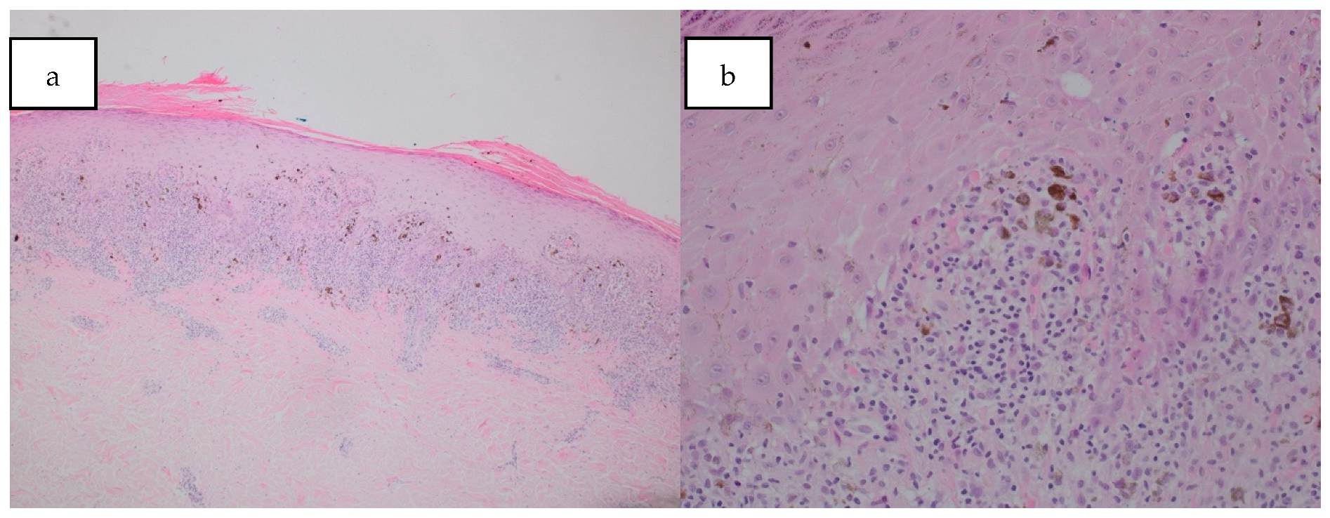
3. Diagnosis
4. Discussion
Author Contributions
Funding
Institutional Review Board Statement
Informed consent Statement
Data availability Statement
Conflicts of Interest
References
- Boehncke, W.H.; Schön, M.P. Psoriasis. Lancet 2015, 386, 983–994. [Google Scholar] [CrossRef] [PubMed]
- McAleer, I.A. Atopic Dermatitis. In Dermatology, 5th ed.; Bolognia, J., Schaffer, J., Cerroni, L., Eds.; Elsevier: Philadelphia, PA, USA, 2025; p. 219. [Google Scholar]
- Cribier, B.; Wood, G.; Reizner, G. Other Papulosquamous Disorders. In Dermatology, 5th ed.; Bolognia, J., Schaffer, J., Cerroni, L., Eds.; Elsevier: Philadelphia, PA, USA, 2025; pp. 172–173. [Google Scholar]
- Solimani, F.; Forchhammer, S.; Schloegl, A.; Ghoreschi, K.; Meier, K. Lichen planus—A clinical guide. J. Dtsch. Dermatol. Ges. 2021, 19, 864–882. [Google Scholar] [CrossRef] [PubMed]
- Tziotzios, C.; Lee, J.Y.W.; Brier, T.; Saito, R.; Hsu, C.-K.; Bhargava, K.; Stefanato, C.M.; Fenton, D.A.; McGrath, J.A. Lichen planus and lichenoid dermatoses: Clinical overview and molecular basis. J. Am. Acad. Dermatol. 2018, 79, 789–804. [Google Scholar] [CrossRef] [PubMed]
- Kusari, A.; Ahluwalia, J. Lichen Planus. N. Engl. J. Med. 2018, 379, 567. [Google Scholar] [CrossRef] [PubMed]
- Jensen, M.B.; Staun, P.W.; Salim, S.; Svendsen, M.T. Lichen planus-like rash in a patient with skin of colour. Clin. Exp. Dermatol. 2023, 48, 1074–1076. [Google Scholar] [CrossRef] [PubMed]
- Ioannides, D.; Vakirlis, E.; Kemeny, L.; Marinovic, B.; Massone, C.; Murphy, R.; Nast, A.; Ronnevig, J.; Ruzicka, T.; Cooper, S.M. European S1 guidelines on the management of lichen planus: A cooperation of the European Dermatology Forum with the European Academy of Dermatology and Venereology. J. Eur. Acad. Dermatol. Venereol. 2020, 34, 1403–1414. [Google Scholar] [CrossRef] [PubMed]
- Tekin, B.; Xie, F.; Lehman, J.S. Lichen Planus: What is New in Diagnosis and Treatment? Am. J. Clin. Dermatol. 2024, 25, 735–764. [Google Scholar] [CrossRef] [PubMed]

Disclaimer/Publisher’s Note: The statements, opinions and data contained in all publications are solely those of the individual author(s) and contributor(s) and not of MDPI and/or the editor(s). MDPI and/or the editor(s) disclaim responsibility for any injury to people or property resulting from any ideas, methods, instructions or products referred to in the content. |
© 2025 by the authors. Published by MDPI on behalf of the European Society of Dermatopathology. Licensee MDPI, Basel, Switzerland. This article is an open access article distributed under the terms and conditions of the Creative Commons Attribution (CC BY) license (https://creativecommons.org/licenses/by/4.0/).
Share and Cite
Yong, J.F.; Howard-James, C.; Crowther, S.; Tobin, A.-M.; Hackett, C. When Classic Signs Deceive: A Widespread Papulosquamous Eruption in Skin of Colour. Dermatopathology 2025, 12, 21. https://doi.org/10.3390/dermatopathology12030021
Yong JF, Howard-James C, Crowther S, Tobin A-M, Hackett C. When Classic Signs Deceive: A Widespread Papulosquamous Eruption in Skin of Colour. Dermatopathology. 2025; 12(3):21. https://doi.org/10.3390/dermatopathology12030021
Chicago/Turabian StyleYong, Ji Fung, Claudine Howard-James, Stephen Crowther, Anne-Marie Tobin, and Caitriona Hackett. 2025. "When Classic Signs Deceive: A Widespread Papulosquamous Eruption in Skin of Colour" Dermatopathology 12, no. 3: 21. https://doi.org/10.3390/dermatopathology12030021
APA StyleYong, J. F., Howard-James, C., Crowther, S., Tobin, A.-M., & Hackett, C. (2025). When Classic Signs Deceive: A Widespread Papulosquamous Eruption in Skin of Colour. Dermatopathology, 12(3), 21. https://doi.org/10.3390/dermatopathology12030021